overview of the radiographers’ practice in 65 healthcare
TRANSCRIPT

ORIGINAL ARTICLE
Overview of the radiographers’ practice in 65 healthcare centersusing digital mammography systems in Portugal
Cláudia Sá dos Reis1,2,3 & Ana Pascoal3,4 & Lucian Radu3&
Mário Fartaria de Oliveira3,5,6 & João Alves6,7
Received: 31 October 2016 /Revised: 24 January 2017 /Accepted: 22 February 2017 /Published online: 16 March 2017# The Author(s) 2017. This article is published with open access at Springerlink.com
AbstractPurpose To assess current practices in digital mammogra-phy (DM) in Portuguese healthcare providers using digitalsystems. To investigate compliance with European stan-dards regarding mean glandular dose and quality controlpractice and to identify optimisation needs.Methods Two questionnaires, targeted at breast radio-graphers and chief radiographers, were designed and ap-plied in 65 imaging departments offering DM. Questionsfielded were focused on the staff profile and technical/clinical practice.Results Prior to starting their activity in DM, 70% (82 outof 118) of the respondents received training in DM. Thepractice in 29 out of 59 providers was established by the
manufacturers’ recommendations for image acquisition.Variations were observed between radiographers who be-long to the same provider namely the selection of expo-sure parameters such as the target-filter combination andautomatic mode. The use of the manual exposure modewas reported for imaging breast implants (44%) and sur-gical specimens (22%). The main causes of repeat exam-inations were skin folding (21%) and absence of pectoralmuscle (PM) (20%).Conclusions The study revealed opportunities to optimiseradiographers’ practice in DM regarding the selection ofexposure parameters. A robust and consistent trainingprogramme in DM and established local protocols canhelp to reduce the variations observed and improve clin-ical practice.
Main Messages• Radiographers adopted different practices selecting AECmodes and T/F combinations.
• Radiographer practice is more consistent using DR thanusing CR systems.
• The main causes for rejecting images were the visibility ofskin folding and PM absence.
• Radiographers were partly unaware of the dose indicator.• Radiographers’ training needs: QC, interventional proce-dures and breast dose optimisation.
Keywords Radiographer . Digital mammography . Practice .
Technique . Positioning
Introduction
Digital mammography (DM) is in use worldwide and ismost commonly used in America, Europe, Australia and
* Cláudia Sá dos [email protected]
1 Department of Medical Radiation Sciences, Curtin University,Perth, Western, Australia
2 Escola Superior de Tecnologia da Saúde de Lisboa/InstitutoPolitécnico de Lisboa (ESTeSL/IPL), Lisboa, Portugal
3 Faculdade de Engenharia, Universidade Católica Portuguesa, EstradaOctávio Pato, 2635-631 Rio de Mouro, Portugal
4 King’s College Hospital NHS Foundation Trust, Department ofMedical Engineering and Physics, London, UK
5 Department of Radiology, Centre Hospitalier Universitaire Vaudois(CHUV) and University of Lausanne (UNIL), Lausanne, Switzerland
6 Instituto Superior Técnico (IST), Laboratório de Proteção eSegurança Radiológica (LPSR), Universidade de Lisboa (UL),Estrada Nacional 10 (ao km 139,7), 2986-066 Bobadela, Portugal
7 Centro de Ciências e Tecnologias Nucleares (C2TN) do IST, UL, EN10 (ao km 139,7), 2986-066 Bobadela, Portugal
Insights Imaging (2017) 8:345–355DOI 10.1007/s13244-017-0550-9

Japan. The International Cancer Screening Network(ICSN) cited in its 2008 report that 15 out of 27European countries implemented DM in their breastscreening programmes [1]. In the US, the Food andDrug Administration approved DM in 2000 but its adop-tion in screening mammography programmes has beenslow. In 2006 less than 10% of mammography systemswere digital [1]. In the UK, mammographic screening iscoordinated by the National Health Service BreastScreening Programme (NHSBSP), which has implement-ed DM at their centres. As of July 2011, 85% of breastscreening units had at least one DM set [2].
Obstacles to the introduction of DM include the highcapital cost of equipment including archiving facilities,integration with existing X-ray systems, staff trainingand workflow reengineering [3, 4]. The successful transi-tion from screen film to DM and the cost-effective use ofDM require a clear understanding of the potential andlimitations of DM systems as well as their potential im-pacts on the established routines [5–7]. Fully integratedDM systems offer opportunities to streamline workflowand increase workload [7–9]. Prior to clinical use ofDM, specialised training for radiographers and radiolo-gists is essential as well as continuous refresher trainingto update knowledge and promote competent and safe useof the technology. The establishment of clinical protocolsand multidisciplinary meetings and feedback is importantto audit practice and identify opportunities for optimisa-tion. These strategies are essential to promote high-qualitystandards whilst minimising the radiation dose to the pa-tient [7, 10–12].
Various organisations [e.g. the European Commission,International Atomic Energy Agency (IAEA), Institute ofPhysics and Engineering in Medicine (IPEM) and NationalHealth Service Breast Screening Programme (NHSBSP)] re-lease guidelines aimed at promoting quality in mammographyand high-quality breast care. The currently available guide-lines provide advice on organisational, technical and clinicalmatters in DM [13–15].
In Portugal, DM was introduced in clinical practice in2000 and it is currently in use by various healthcare pro-viders for screening, diagnosis, intervention and follow-up. No clinical audit data have been found to assess theimplementation of DM in Portugal or the impact of ar-rangements put in place to promote cost-effective use ofthe imaging modality in the national health system. In factstudies combining all areas regarding radiographer prac-tice in DM are scarce worldwide. Usually, the studies arelimited in scope and focus only on specific subjects suchas communication with patients, patient positioning, qual-ity control practice, image evaluation and perception,breast compression or other breast imaging techniques[16–24].
The objectives of this study were to survey the academicand professional profile of radiography staff performingmam-mography and to characterise their routine practice in the useof the digital technology. It also aimed at providing recom-mendations to optimise the quality of DM performed inPortugal.
Methods
Development, testing and application of questionnaires
Two original questionnaires were designed to implementthe survey. One was targeted to radiographers performingmammography and the other was targeted to chiefradiographers who are frequently involved with the man-agement of DM in Portuguese hospitals. Before sendingthem, nine radiographers piloted the questionnaires andsuggestions were incorporated to improve the quality ofthe tool.
The questions were designed to capture data on thefollowing themes: demographic profile such as age andgender, experience in radiography and DM, specialisededucation and training in DM and self-assessed trainingneeds (Table 1).
Data on the type of mammography system available atthe facility, e.g., computed radiography (CR), direct digi-tal mammography (DDM), most frequently used tech-nique, e.g., manual vs. AEC, selected target-filter (T/F)combination, and the use of the breast dose (or exposure)indicator (Tables 2, 3 and 4), were also collected.Additionally, the variability in practice amongstradiographers working at the same centre regarding theprotocol selected (T/F combination and AEC mode) wasalso investigated.
The questionnaire was also designed to capture infor-mation on the use of guidelines to support the practice inplace. It collected data on staff preferences and viewsabout the impact of DM on established practice, namelyin radiographic technique, workflow and workload(Tables 3 and 4).
The questionnaires were posted or sent by e-mail to allproviders of mammography services using DM technolo-gy (CR or DR) in continental Portugal and the Azores andMadeira Islands. The healthcare providers invited to par-ticipate in the study included large public hospitals, uni-versity hospitals, private hospitals and diagnostic clinics.The national coordination centre for breast screening wasalso invited to take part in the study.
A cover letter informing about the context of thestudy and its objectives accompanied the question-naires. Contacts were established with the hospital ad-ministration and with the local radiology department.
346 Insights Imaging (2017) 8:345–355

The opportunity to take part in an independent assess-ment of compliance with the best practice in mammog-raphy was highlighted as a benefit to the healthcareprovider.
The data collected were screened for quality and the de-scriptive statistical analysis was performed using the softwarepackages MS Excel (version 2007, Microsoft ©) and SPSS(version 19, IBM).
Table 2 Summary of the questionnaire section to capture data on the specifications of the mammographic equipment available at the healthcareprovider questioned (target: breast radiographer and chief radiographer)
Table 1 Summary of the questionnaires targeted to radiographers illustrating the questions to capture data regarding education and training in digitalmammography
Insights Imaging (2017) 8:345–355 347

Table 3 Summary of the questionnaire targeted to the chief radiographer: questions to capture data to characterise the type of healthcare provider,activity and workload
Table 4 Summary of the questionnaire targeted to radiographers to capture data regarding the radiographic technique used and the reference guidanceused to support usual practice
348 Insights Imaging (2017) 8:345–355

Results and Discussion
The questionnaires were sent out to 270 institutions and 118responses were received from 65 centres representing a re-sponse rate of 24.1%.
Radiographers’ profiles
Age and gender
The majority of radiographers (98%; n = 118) were femalewithin the age range 20 to 59 years old. Among these were asignificant number of young professionals (46%) aged 20–29years. Since mammography is a diagnostic modality usedmost commonly for imaging female breasts, it seems naturalthat the healthcare staff should bemainly composed of womenas this may also contribute to the patient’s acceptance and self-assurance. Published studies [25, 26] indicate that some wom-en undergoing mammography may feel embarrassed whenassisted by a male radiographer. Fitzpatrick et al. [25] reportedthat overall, 17.5% of women agreed (or strongly agreed) withthe statement BIf there were male radiographers I would notreturn to BreastCheck (Irish screening program) for anotherscreening appointment and a further 18.3% were unsure^.
Specific education and training in digital mammography
Many responding breast radiographers (88%; 96/118) hadgraduated in radiography. The average work experience in
radiography was 10 years ranging between 1–39 years. Theaverage work experience in DM was 4.7 years (range: 1–15years).
Prior to enrolling as DM radiographers, the majority (70%;n = 118) of participating radiographers had received trainingin DM. A few (3%) (4) received training by attending courseson general radiology techniques. Participants stated that theyreceived training from the manufacturer’s study days (50),through workshops (39) and/or other training courses (21).The reported duration of the training varied with a predomi-nance of short-term (1–2 days) sessions (73%). Longer train-ing periods (≥1 month) were less frequent (27%). Topics cov-ered in the training included advantages and limitations ofdigital mammography, optimisation of exposure parameters,technological developments in mammography and artefactsrecognition.
More than one third of the respondents (38%) reportedself-assessed the need to refresh their knowledge on DM.Key areas of training needs that were highlighted were onquality control (QC), interventional procedures and breastdose optimisation. A previous study [10] about trainingon medical imaging concluded that when compared toCT and MRI, mammography was a less valued imagingmodality, which has also been highlighted in otherEuropean studies [11, 27]. As per current practice, quali-fied radiographers graduating in Portugal are entitled tostart performing DM promptly following graduation.Anecdotal evidence suggests that experienced staff usual-ly informally supervise radiographers starting to practicemammography. Currently there is no established comple-mentary training programme in place nationwide or a for-mal continuous professional development (CPD) trainingof 40 h/year as recommended by the EUREF guidelines.
Usual radiographic practice with digital mammographysystems
Support guidance for the technical protocol in use
The majority (63%) of centres followed the recommendationsprovided by the manufacturer to select exposure parameterson the DM systems. Some centres (29%) reported using pro-tocols developed locally and a small percentage (6%) stated
Departmentprotocol
29%
National Guideline
2%
Manufacturer
International Guideline
63%
6%
Fig. 1 Guidance to support mammography practice in the participatingcentres
Small Breast
17.1%
8.6%
Post-therapy
43.7%
Breast implants
1.6%
Equipment
22.0%
Surgical
specimens
6.9%
Other
Fig. 2 Justification for the use ofmanual exposure mode inmammography
Insights Imaging (2017) 8:345–355 349
using international guidelines [American College ofRadiology (ACR), European Protocol (EUREF)] to supportquality assurance in mammography (Fig. 1).
Exposure mode (AEC vs. manual)
Mammography imaging units incorporate an AutomaticExposure Control (AEC) system. This provides a means ofachieving adequate and consistent image quality (IQ), inde-pendent of the breast characteristics and the radiographer’sexperience in the selection of the X-ray tube exposure param-eters [28–30]. AEC systems can operate in various modes thatare manufacturer-specific. For each system usually threemodes are provided offering a range of IQ options (lower,standard and higher) selected by the radiographer according
to the clinical task (diagnostic or screening). The use of themanual mode or manual selection of the settings by theoperator is not recommended for standard mammographywith few justified exceptions, like in the cases of breastimplants, surgical specimens and breasts that have under-gone surgery or radiotherapy [31–33]. This was observedin our sample: manual mode selection was mainly usedwhen imaging breast implants (44%), surgical specimens(22%) collected during biopsies, small breasts and post-radiotherapy breasts (Fig. 2).
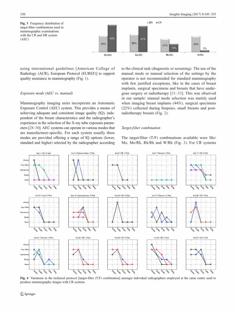
Target-filter combination
The target/filter (T/F) combinations available were Mo/Mo, Mo/Rh, Rh/Rh and W/Rh (Fig. 3). For CR systems
26%
43%
Mo/Mo
228%
41%
Mo/Rh
D
21%
R
DR CR
13%
Rh/Rh
25%
W/Rh
3%
Fig. 3 Frequency distribution oftarget-filter combinations used inmammographic examinationswith the CR and DR system(AEC)
Fig. 4 Variations in the technical protocol [target-filter (T/F) combination] amongst individual radiographers employed at the same centre used toproduce mammography images with CR systems
350 Insights Imaging (2017) 8:345–355

Mo/Mo and Mo/Rh were the most frequently used, Rh/Rhwas reported in 13% of the cases and W/Rh was rarelyused. For DR systems Mo/Rh seemed to be the most fre-quent T/F combination but Mo/Mo, Rh/Rh and W/Rhwere also frequently used.
Inter-radiographer variability (use of technical protocol)
The analysis of Figs. 4, 5, 6 and 7 shows that in some centressome radiographers adopted different practices in the selectionof T/F compared to their colleagues. This was observed for
Fig. 5 Variations in the technical protocol [target-filter (T/F) combination] amongst individual radiographers employed at the same centre used toproduce mammography images with DR systems
Fig. 6 Variability in the use of AEC mode in mammography (CR system) for various members of staff (radiographers) and various institutions (inst.)and manufacturers
Insights Imaging (2017) 8:345–355 351

CR systems at centres 1, 2, 11, 14, 17, 21, 26 and 27 (Fig. 4).The radiographers from institution 11 (Fig. 4) reported usingthe W/Ag combination, which was not available on the mam-mography equipment they were using. This may represent alack of clarity from staff regarding the feature of the equip-ment in use.
For DR systems the overall practice adopted radiographerswas more consistent. However, variations in the selection ofT/F were noticed for centres 4, 10, 15 and 23.
Regarding the selection of AEC mode, a spread of scoreswas observed for both CR andDR systems showing variationsbetween the centres (Figs. 6 and 7). The practice of individualradiographers appears to be consistent. A higher variabilitywas shown for CR namely in institutions 1, 2, 7, 11, 14, 21,
26 and 27. The available AEC modes in mammography de-vices are dependent of each manufacturer, and according toradiographers they followed the manufacturer recommenda-tions to choose the exposure mode. However, the answerswere not consistent with the options that are available in theequipment for the majority (75%) of the institutions that wereconsidered. Radiographers’ training in DM is expected tohave a significant impact on their practice. A clear understand-ing of the differences between the AEC modes provided bythe equipment (as well as the rationale for selection) is impor-tant to select the technical protocol in mammography.
As discussed above, 33% of radiographers have not hadspecific education and training in mammography and reportedlittle experience in using the technology. Also, the use of local
Fig. 7 Variability in the use of AEC mode in mammography (DR system) for various members of staff (radiographers) and various institutions (inst.)and manufacturers
12.7%
Technicalparameters
8.6%
Imageprocessing
18.2%
Artifacts
17.8%
Patientmotion
21.0%
Skin folders
20.4%
Absence ofMP
1.3%
Other
Fig. 8 Causes for rejection andrepeat of mammography imagesresported by breast radiographers
352 Insights Imaging (2017) 8:345–355

protocols and guidelines for good practice was not consistent-ly adopted. These factors could contribute to the observedvariations in practice amongst radiographers in the selectionof exposure settings.
Analysis of rejected/repeated examinations
About 88% of radiographers stated that they rarely needed toreject and/or repeat mammography examinations. When ex-aminations needed to be rejected and repeated, they reportedthat the main causes for this were the visibility of skin foldingon the image (21%), absence of pectoral muscle (PM) (20%),blur caused by patient motion (18%) and/or presence of othertypes of artefacts in the image (18%) (Fig. 8). Less frequentcauses for rejection were inappropriate image processing and/or technical parameter selection. However, the majority (65%)of radiographers do not perform analysis of rejected/repeatedmammography examinations in a systematic way as recom-mended by European guidelines (and ACR guidelines) affect-ing the opportunity to identify opportunities for optimisation.
Assessment of rejected/repeated images to identify thecauses of rejection is a valuable quality assurance practice alsorecommended by ACR and IAEA guidelines [14, 34–36].Implementing corrective and preventive measures to reducethe number of repeated mammography examinations is im-portant to ensure that mammographic images are producedat high quality/standards [14, 35] and comply with theALARA/ALARP principles. Additionally, financial gains
are expected because of the most efficient use of the equip-ment and radiographers’ time.
Use of the dose (or exposure) indicator
Monitoring and optimising the dose to the patient in mam-mography is a recommended quality control procedure byall international guidelines to ensure that the risk to the patientis kept low.
For DR systems the mean glandular dose (MGD) to thebreast can be monitored promptly following the exposureusing the dose indicator incorporated in the majority of DMsystems. For mammography with CR systems an MGD valueis usually not promptly available to the user. An indication ofexposure on the image receptor is used as it has an effect onpatient dose (and also on image quality). The name of theindex varies depending on the manufacturer. The responsesto the questionnaire showed that half (50%) of theradiographers were aware of the existence of a dose indicatoron the equipment and used its displayed value to monitor thedose to the patient following the mammography procedure.About 11% referred to being aware of the indicator but notusing it. A substantial percentage (21%) of the respondentswere not aware of the indicator’s existence at all.
The analysis of the exposure indicator system is an impor-tant QC practice that alerts the radiographer to the occurrenceof sub-optimal exposures; these may have a direct impact onthe IQ and patient dose. Considering the results of the survey
Num
ber o
f R
adio
graphers (
N)
0
20
40
60
80
100
Exp
para
posure
ameters Tar
ch
67
22
rget-filter
hanges
20
45
29
5
Positioning
8
89
3
Yes
No
Unknow
21
Fig. 9 Areas where changeswere observed following theintroduction of digitaltechnologies for mammography(radiographers’ personal view)
Num
ber
of R
adio
grap
hers
(N
)
0
20
40
60
Exam timeWorkflow
26
39
53
W
28
35
3 55
Workload
29
42
47
Unknow
No
Yes
Fig. 10 Impact of digitalmammography on workload,workflow and examination time(radiographers’ point of view)
Insights Imaging (2017) 8:345–355 353

it may be appropriate to provide radiographers with refreshertraining about the dose in mammography. The training shouldhighlight the QA tools provided by the equipment such as thedose indicator. Additionally, mechanisms to assess the impactof training, such as auditing, should be put in place.Appropriate communication and feedback mechanismsamong all staff members involved in mammography will alsobe important to promote consistent practice.
Personnel views on the impact of digital mammography
More than half (57%) of the total number of radiographersreported having noticed changes in practice following the in-troduction of DM, particularly changes in the exposure factorsused. Their perception was that digital mammography de-creased the patient dose compared to screen-film mammogra-phy because of the reduction of exposure time and increase ofbeam energy. They also reported that the introduction of AECmodes caused reduction of the number of repeated images.Variations in the T/F combination in use were reported by17%, mainly referring to a decrease in use of Mo/Mo andincreased use of W/Rh (Fig. 9).
The majority of radiographers (75%) considered that theintroduction of DM did not require changes to the positioningof the patient. A few radiographers (7%) reported that posi-tioning small breasts is now more difficult compared to ana-logue systems because of the larger platform size. Other au-thors also reported challenges with positioning of smallbreasts on large platforms and increased challenges to fullyfulfil the recommended criteria of good radiographic position-ing practice [13, 35, 37]. When positioning small breasts thereis a risk of including part of the arm in the image and some-times also part of the abdominal wall, which is not desirable.
Approximately half of the radiographers considered thatthe introduction of DM caused impacts on workload,workflow and examination time (Fig. 10). Image acquisitionwas faster and the number of mammography procedures per-formed per shift had increased. It was reported that time be-tween consecutive examinations also increased allowingradiographers to dedicate more attention to patients.
Conclusions
This study collected evidence and provided an overview ofradiographers’ profile and practices in use in digital mammog-raphy in Portugal.
The majority of radiographers were young females withlittle experience inDM. Specialised training inmammographyis not mandatory and radiographers were trained on the joband worked under supervision. The majority of radiographersidentified self-assessed need for training on digital mammog-raphy with focus on artefact recognition, dosimetry and
quality control. Limited evidence of compliance with the rec-ommended international standards of good for mammographypractice (EUREF) was found as the radiographers do not per-form 40 h/year of CPD in mammography and quality controltests are not performed following the main recommendationsprovided by the EUREF guidelines.
The study revealed opportunities for optimisation of radi-ographer practice in digital mammography in Portugal. A ro-bust and consistent training programme in digital mammog-raphy for radiography staff can help reduce the observed var-iations in practice. The training should take into considerationthe activities of the radiographer and include practice with theequipment. It should be developed by a multidisciplinary teamwith the input of the relevant stakeholders (radiographers,radiologists and medical physicists). The establishment of aninternational training network for mammography is likely toprovide a valuable contribution to improve and disseminatebest practice in digital mammography.
Open Access This article is distributed under the terms of the CreativeCommons At t r ibut ion 4 .0 In te rna t ional License (h t tp : / /creativecommons.org/licenses/by/4.0/), which permits unrestricted use,distribution, and reproduction in any medium, provided you give appro-priate credit to the original author(s) and the source, provide a link to theCreative Commons license, and indicate if changes were made.
References
1. Institute of Medicine-National Research Council (2001)Mammography and beyond: developing technologies for the earlydetection of breast cancer, 1st edn. National Cancer Policy Board-Institute of Medicine, Washington
2. Public Health England (2013) NHS Breast Cancer ScreeningProgramme, digital mammography. [Online]. Available: http://www.cancerscreening.nhs.uk/breastscreen/digital-mammography.html
3. Morin RL, Maidment ADA (2005) Digital mammography: comingof age. J Am Coll Radiol 2(9):798–801
4. Vinnicombe S, Pinto Pereira SM, McCormack VA, Shiel S, PerryN, Dos Santos Silva IM (2009) Full-field digital versus screen-filmmammography: comparison within the UK breast screening pro-gram and systematic review of published data. Radiology 251(2):347–358
5. Pisano ED, Gatsonis C, Hendrick E, Yaffe M, Baum JK, AcharyyaS, Conant EF, Fajardo LL, Bassett L, D’Orsi C, Jong R, Rebner M(2005) Diagnostic performance of digital versus film mammogra-phy for breast-cancer screening. N Engl J Med 353(17):1773–1783
6. Kuzmiak CM, Cole E, Zeng D, Kim E, KoomenM, LeeY, Pavic D,Pisano ED (2010) Comparison of image acquisition and radiologistinterpretation times in a diagnostic mammography center. AcadRadiol 17(9):1168–1174
7. Pisano ED, Zuley M, Baum JK, Marques HS (2007) Issues toconsider in converting to digital mammography. Radiol Clin NAm 45(5):813–830, vi
8. Mas N, Seinfeld J (2008) Is managed care restraining the adoptionof technology by hospitals? J Health Econ 27(4):1026–1045
354 Insights Imaging (2017) 8:345–355

9. Lettieri E, Masella C (2009) Priority setting for technology adop-tion at a hospital level: relevant issues from the literature. HealthPolicy 90(1):81–88
10. Leal J, Andrade AS, Ribeiro R (2012) Continuous professionaldevelopment : the perspective of radiographers in private and publicinstitutions of Lisbon region. Eur Soc Radiol C-1815:1–17
11. Cataliotti L, De Wolf C, Holland R, Marotti L, Perry N, RedmondK, Rosselli Del TurcoM, Rijken H, Kearney N, Ellis IO, Di Leo A,Orecchia R, Noel A, Andersson M, Audretsch W, Bjurstam N,Blamey RW, Blichert-Toft M, Bosmans H, Burch A, Bussolati G,Christiaens MR, Colleoni M, Cserni G, Cufer T, Cush S, DamilakisJ, Drijkoningen M, Ellis P, Foubert J, Gambaccini M, Gentile E,Guedea F, Hendriks J, Jakesz R, Jassem J, Jereczek-Fossa BA,Laird O, Lartigau E, Mattheiem W, O’higgins N, Pennery E,Rainsbury D, Rutgers E, Smola M, Van Limbergen E, vonSmitten K, Wells C, Wilson R (2007) Guidelines on the standardsfor the training of specialised health professionals dealing withbreast cancer. Eur J Cancer 43(4):660–675
12. Nodine CF, Kundel HL, Mello-Thoms C, Weinstein SP, Orel SG,Sullivan DC, Conant EF (1999) How experience and training in-fluence mammography expertise. Acad Radiol 6:575–585
13. European Communities/EUREF (2006) European guidelines forquality assurance in breast cancer screening and diagnosis, vol 19,4th edn. European Communities, Luxembourg, no. 4
14. International Atomic Energy Agency (2011) Quality assurance pro-gramme for digital mammography. International Atomic EnergyAgency, Vienna
15. National Health Care Breast Screening Programme (2009)Commissioning and routine testing of full field digital mammogra-phy systems - NHSBSP equipment report 0604,^ London
16. Spuur K, Hung WT, Poulos A, Rickard M (2011) Mammographyimage quality: model for predicting compliance with posterior nip-ple line criterion. Eur J Radiol 80(3):713–718
17. Poulos A, Llewellyn G (2005) Mammography discomfort: a holis-tic perspective derived from women’s experiences. Radiography11(1):17–25
18. Li Y, Poulos A, Mclean D, Rickard M (2010) A review of methodsof clinical image quality evaluation in mammography. Eur J Radiol74:122–131
19. D. O’ Leary, A. Teape, J. Hammond, L. Rainford, and T. Grant(2011) Compression force recommendations in mammographymust be linked to image quality. In: European Congress ofRadiology 2011 pp. 1–19
20. Ooms EA, Zonderland HM, Eijkemans MJC, Kriege M,Mahdavian Delavary B, Burger CW, Ansink AC (2007)Mammography: interobserver variability in breast density assess-ment. Breast 16(6):568–576
21. Toroi P, Zanca F, Young KC, van Ongeval C, Marchal G, BosmansH (2007) Experimental investigation on the choice of the tungsten/rhodium anode/filter combination for an amorphous selenium-based digital mammography system. Eur Radiol 17(9):2368–2375
22. Samei E (2005) AAPM/RSNA physics tutorial for residents: tech-nological and psychophysical considerations for digital mammo-graphic displays. Radiographics 25(2):491–501
23. C. Lança, C. Reis, and L. Lança (2012) Perceção visual naavaliação diagnóstica em mamografia: uma revisão sistemática.SAÚDE Tecnol. ed. online, vol. Temático 1, pp. 31–40
24. Reis C, Pascoal A, Sakellaris T, Koutalonis M (2013) Quality as-surance and quality control in mammography: a review of availableguidance worldwide. Insights Imaging 4(5):539–553
25. Fitzpatrick P, Winston A, Mooney T (2008) Radiographer genderand breast-screening uptake. Br J Cancer 98(11):1759–1761
26. Warren-Forward HM, Mackie B, Alchin M, Mooney T, FitzpatrickP (2016) Perceptions of Australian clients towards maleradiographers working in breast imaging: quantitative results froma pilot study. Radiography 23(1):3–8
27. Marshall G, Punys V, Sykes A (2008) The continuous professionaldevelopment (CPD) requirements of radiographers in Europe: aninitial survey. Radiography 14(4):332–342
28. Andolina V, Lyllé S (2011) Mammographic imaging—a practicalguide, 3rd edn. Wolters Kluwer Health-Lippincott Williams &Wilkins, Baltimore
29. National Healthcare System Breast Screening Programme(NHSBSP) (2007) Guidance on beam quality selection in theNHS Breast screening programme NHSBSP Equipment Report0704. Sheffield
30. Oduko J, Young K, Burch A (2012) Breast imaging—a survey ofpatient doses from digital mammography systems in the UK in2007 to 2009, vol 7361. Berlin, Springer Berlin Heidelberg
31. A. Reis, C. Sakellaris, T. Carrasqueiro, S. Pascoal (2012) Digitalmammography in Portugal: a national survey on technology andpractices. In: Symposium Mammographicum 2012 MeetingAbstracts pp. 1–15
32. Reis C (2013) Digital mammography: characterisation of practiceand equipment performance in portuguese healthcare providers.Universidade Católica Portuguesa, Lisboa
33. Reis C, Pascoal A (2010) Mamografia digital em portugal:caracterização da tecnologia instalada. Acta Radiol Port 22(145):21–23
34. American College of Radiology (2011) ACR-SPR practice guide-line for the performance of chest radiography. pp. 1–7
35. American College of Radiology (1999) Mammography qualitycontrol manual. American College of Radiology, Reston
36. International Atomic Energy Agency (2009) Quality assurance pro-gramme for screen film mammography, Internatio., no. 2.International Atomic Energy Agency, Vienna
37. The National Cancer Screening Service (2008) Guidelines for qual-ity assurance in mammography screening, 3rd edn. Members of theQuality Assurance Committee/The National Cancer ScreeningService Board, Dublin
Insights Imaging (2017) 8:345–355 355