overview of peripheral vascular disease - api · overview of peripheral vascular disease 89...
TRANSCRIPT

overvieW of peripHeraL vascuLar Disease89
Peripheral Vascular Disease of the lower extremity is an important cause of morbidity and affects 10 million people in India.1 It is a common condition with variable morbidity affecting men and women over the age of 45 years. It is going to be a major health problem in our country as the Indian population is aging. Atherosclerosis is a generalized disorder and involves medium and large sized arteries. It is estimated that 74% patients of atherosclerotic coronary artery disease have involvement of some other vascular bed also. 40% patients of coronary artery disease have associated peripheral vascular disease, 14% have carotid artery stenosis and 17% have associated renal artery stenosis. Therefore it becomes very important for the physicians to know the pathology, clinical presentations and treatment of common vascular disorders.Increasingly, peripheral vascular disease is becoming a focus of involvement for primary care physicians and cardiovascular specialists who must work in partnership. Awareness and interest in peripheral vascular disease is growing in India because of the following reasons:• Advancing age of the general population, resulting in
increase prevalence of the peripheral vascular disease.• Unwillingness of patients to accept the limitations and
associated morbidity of vascular disease when therapeutic options are available.
• The realization that vascular disease in one system should prompt investigation of other areas for coexistent disease
cAuSeS of VASculAr diSeASeSee Table 1.
overview of peripheral vascular Diseasenn khannaconsultant interventional cardiologist with special interest in peripheral vascular interventions, escorts Heart institute, okhla road, new Delhi & incharge escorts Heart centre, pandu nagar, Kanpur 19
riSk fActorS for AtHeroSclerotic VASculAr diSeASe See Table 2 for risk factors.
renAl Artery StenoSiSThe most common causes of renal artery stenosis are atherosclerosis and fibrous dysplasia. Asymptomatic renal artery stenosis is present in 40% cases.The common presentations of renal artery stenosis are hypertension, deterioration of renal functions and acute pulmonary edema Atherosclerotic renal artery stenosis is common in males over the age of 55 years, in diabetics and patients who have coronary artery stenosis or carotid artery stenosis. It should be suspected when the hypertension is of recent onset, when it is poorly
table 1 : causes of Vascular disease
Arterial Venous lymphatic 1. Atherosclerosis 1. Venous thrombosis 1. Lymphoedema 2. Thromboangiitis obliterans 2. Varicose veins 2. Lipidemia 3. Arteriosclerosis obliterans 3. AV fistula 4. Raynaud’s disease 5. Acrocynosis 6. Erythromelalgia 7. Vasculitis 8. Takayasu Arteritis
table 2 : risk factors for Atherosclerotic Vascular disease
conventional : 1. Smoking 2. Diabetes Mellitus 3. Hyperlipidemia 4. Hypertension5. Obesity 6. Syndrome X (Hypertriglyceridemia + Insulin Resistance)
Secondary / Possible : 1. Angiotensin Converting Enzyme Polymorphism2. Chlamydial infection 3. Lp(a)4. Lipid Remnants 5. IDL 6. Apoprotein B 7. Elevated serum fibrinogen level 8. Elevated Serum Iron level 9. Elevated Serum Uric acid level 10. Low Serum Folate level11. Hyperhomocysteinemia12. Hypothyroidism 13. Elevated protein C, protein S14. Oxidative Stress (lipid peroxidation)15. Platelet-receptor polymorphism 16. von Willebrand disease

MeDicine upDate 200590
in the abdominal cavity. Diagnosis is confirmed by ultra sonography, computer tomography, magnetic resonance imaging and digital subtraction angiography. 10% of these aneurysms are accompanied by intense inflammatory reaction. These represent a threat of rupture. Aneurysms which are > 5 cm or are expanding > 1 cm / year are at high risk of rupture and mandate treatment in form of surgery or endovascular treatment. More and more studies suggest that endoluminal grafting is better and safer than surgery.
Aortic diSSectionSThis occurs because of longitudinal cleavage of the aortic media by a dissecting column of blood. The split in the media may extend through the entire length of aorta. There are 3 types of dissection (Debekey Classification). Type I involves ascending aorta, arch and descending aorta. Type II involves only the ascending aorta while Type III involves descending thoracic aorta distal to the subclavian artery. Death occurs due to external rupture of the saccular aneurysm in the outer wall of the aneurysms.Aortic dissections occur more commonly in hypertensive males. Bicuspid aortic valve, coarctation of aorta, Marfans syndrome and Turner syndrome are common associations.The mortality is 35% in first 24 hours,50% in 48 hours, 70% by I week and 80% by 2 weeks. 50% of the survivors die in the next 3 months.Treatment includes drastic lowering of blood pressure upto 100 mmHg by using Labetalol, nitroprusside or nitroglycerine. Surgical treatment should be initiated as early as possible. Surgical mortality is 15% in Type I, 5% in Type II and 12% in Type III. 10 year survival of these surgically treated patients is 35% in Type I dissection and 50% in Type II. Many of these patients have been successfully treated by endovascular stent grafting
cArotid Artery StenoSiS14% patients of coronary artery disease have high grade stenosis in the carotid arteries. In 1990 the NASCET and ACAS trial clearly revealed the superiority of carotid end arterectomy over medical therapy. However the morbidity and mortality was still higher in high risk patients. After the famous CAVATAS trial and the SAPHIRE trial the carotid stenting has become the preferred treatment for carotid artery stenosis. The indications for stenting are symptomatic stenosis > 50% and Asymptomatic stenosis >80%. Medical therapy with dual antiplatelet therapy, ACE inhibiters and statins is the cornerstone of treatment of atherosclerotic carotid artery stenosis (Fig. 1).Stroke occurs because of low flow and embolism from atherosclerotic carotid plaque. The diagnosis is made by duplex scan, digital subtraction angiography and magnetic resonance angiography. Many a times carotid bruit is present.
loWer liMB PeriPHerAl VASculAr diSeASe
unique features of Peripheral Vascular disease in indians1. Presentation at younger age (mean age 45 years)2. Increased association of diabetes and presence of typical
Diabetic Peripheral Vascular Disease. .3. Takayasu arteritis
Fig. 1 : Carotid artery stenosis before and after stenting
controlled on 3 or more drugs or when the patient presents with hypertensive crisis or malignant hypertension. Bilateral renal artery stenosis is suspected when there is rise of serum creatinine after initiation of ACE inhibiter therapy. Diagnostic tests include Ultrasonography for renal size, Duplex Scanning, MR Angiography, CT Angiography, Digital subtraction Angiography. Captopril Renography and Differential Renal Vein Renin levels establishes renal artery stenosis as the cause of renovascular hypertension. Stenting of the renal arteries is an accepted form of treatment. We at Escorts Heart Institute have performed 287 renal artery stents with 100% procedural success.
Aortic AneurySMSAneurysms are areas of focal or diffuse dilatation of the aorta. They usually develop at sites of congenital or acquired weakness of media. Hypertension which is frequently present probably accelerates degeneration of aortic wall. Once begun The aneurysm formation and expansion is governed by the Laplace law. Atherosclerosis is responsible for majority of the cases of aortic aneurysms.Thoracic Aortic Aneurysms are usually because of atherosclerosis. Those limited to Ascending Aorta rarely produce symptoms unless they e undergoing active expansion or rupture.Aortic Regurgitation is present if the aneurysms extends to the aortic sinuses. Descending Thoracic Aneurysms are usually symptomatic Compression of the tracheo bronchial tree produces cough, dyspnoea and tracheal tug. Hoarseness is because of the compression of recurrent Laryngeal nerve. Superior Vena Caval syndrome is also seen because of compression of SVC. Chest pain may occur due to the erosion of rib cage and vertebras.Sometimes the aneurysms may rupture into the adjacent structures like mediastinum, pleural space, esophagus, pericardial cavity, lungs or tracheobronchial tree. 5 year mortality of Thoracic Aneurysms is 75%. Risk factors for rupture are diameter > 6 cm,advanced age, hypertension and presence of cardiovascular disease.Treatment is Percutaneous Endovascular Stent Grafting or Surgical Repair. Fig 3.Abdominal Aortic Aneurysms are usually infrarenal or juxtarenal. They are detected by detecting an expansile mass

overvieW of peripHeraL vascuLar Disease91
4. Strongest correlation with presence of Coronary Artery Disease (CAD) and Cerebrovascular Disease (CVD).
5. Poor understanding of this disease among the physicians, primary healthcare providers and patients. Poor resources and treatment standards
Atherosclerosis, Takayasu Arteritis and Berger’s disease (Thrombo Angiitis Obliterans {TAO}) are common etiologies of peripheral vascular disease in India. In India, Aortoiliac disease is present in 23.2%, femoropopliteal disease is present in 40%, and below knee arteries are involved in 37% cases. Atherosclerosis and Takayasu Arteritis is the etiology in 96% cases of Aortoiliac disease. 2,3
Mean age of these patients is 45 (+/-11.6) years. Atherosclerosis is predominantly the cause in femoropopliteal disease and Berger’s disease & arteriosclerosis obliterans is the cause in below knee arteries. The mean age of presentation in these patients is 37 (+/- 9.8) years. Berger’s disease in below knee arteries is associated with involvement of distal arteries of upper limb in 20% cases. In a clinicopathological study of pattern of occlusive peripheral vascular disease in India published in Angiology, TAO was present in 27% cases, Atherosclerosis Obliterans (ASO) in 60% & Thrombo Embolism Obliterans (TEO) in 13% cases. 4 Most common risk factors for peripheral vascular disease are smoking, diabetes mellitus, dyslipidemia. Elevated CRP and increased CRP+TC / HDL level are the strongest risk factor.
Peripheral Vascular disease in diabetesDiabetes Mellitus is an important risk factor of lower extremity arterial disease (LEAD) in India. Smoking and insulin resistance are frequently present in patients with diabetes and contribute an additional risk for vascular disease. Peripheral Vascular Disease (PVD) in diabetes is complicated by peripheral neuropathy and susceptibility to infection, which leads to foot ulceration, gangrene and amputation of the affected extremity. Diabetes accounts for ~ 50% of all non traumatic amputations in India. Mortality is increased in diabetic patients with PVD. Three years survival after an amputation is < 50%. In population based and epidemiology based studies,5,6 it is estimated that 20-30% of diabetic patients over 65 years of age have peripheral arterial disease. Approximately 30% of these diabetic patients with peripheral vascular disease require surgical or percutaneous revascularization. 10% require an amputation of the affected limb within 5-10 years of diagnosis. Progression from intermittent claudication to critical limb ischemia occurs at the rate of 1.4% per year. Five year mortality of diabetic patients with PVD approaches 30%.
PathogenesisA cluster of factors present in a diabetic leads to the development of peripheral arterial disease (PAD). This is depicted in Fig. 2.
DIABETES MELLITUS
Hyperglycemia Resistance Increased Fatty Acids Insulin
Increased Oxidative stressProtein Kinase C activation
Endothelium
Decreased NOIncreased thromboxaneRBC deformityAngiotensin II AggregationEndothelin I
Decreased
Decreased Response to Vasodilators Platelet
Leucocyte Adhesion Increased
Fibrinogen
Increased Vasoconstrictors Decreased Vasodilators
Decreased Blood flowIncreased Thrombogenicity
Fig. 2 : Peripheral arterial disease in diabetes mellitus

MeDicine upDate 200592
Some abnormalities in the microcirculation in diabetics are not occlusive but can alter the biology of the foot. There is evidence for thickening of capillary basement membrane,3 which is key in the exchange of nutrients and metabolic products between the capillary lumen and the interstitium. The chemical structure of the membrane is altered by glycosylation, causing crosslinking of proteins and a decreased in the number of highly charged sulphur groups.7 This may explain why molecules such as albumin leak through the capillary membrane in diabetics. There is no impairment in oxygen diffusion. Infact diabetics with foot ulceration have higher levels of transcutaneous pO2 than non diabetics.8
Further evaluation of microanatomy reveals more tortuous capillaries in diabetics, appearing as tufts instead of the typical hair pin loops. With ischemia there is less recruitment of new capillaries into the circulation, although per gram there is no difference in capillary concentration. Atherosclerotic occlusion in diabetes commonly involves tibial and peroneal arteries and spares superficial femoral artery and the arteries of the foot, especially the dorsalis pedis artery.9-13
takayashu Arteritis (non-specific Aorto Arieritis)This disease was first described by Dr. R. Adams in 1827 as pulseless disease. Dr. Mikito Takayasu described typical wreath like appearance of retinal blood vessels with absent pulses in 1908. In India, the incidence of this disease is highest in the world. The exact etiopathogenesis of this disease is unknown. It is typically an arteritis of aorta and its main branches, predominantly present in young females in their 2nd and 3rd decades. In India, the disease commonly involves the abdominal aorta and aortoiliac vessels. Many excellent monographs on the epidemiology, pathology and treatment of Takayasu arteritis have been published by great Indian researchers like Panja et.al,14 Sen et.al,15 Chhetri et.al16 and Tyagi et.al.17 Judicious use of corticosteroids and other immunosuppressants during the active phase of disease and balloon angioplasty / stenting or surgery during the remissant phase has considerably brought down the morbidity and mortality (10%) in aortoarteritis (fig. 3).
Thrombo Angiitis obliterans (tAo) or Buerger’s diseaseIt is an obliterative disease characterized by inflammatory changes in small and medium-sized arteries and veins. It occurs in cigarette smokers, predominantly in men aged 20 to 40 years. Only about 5% of cases occur in women. The frequency of diagnosis has decreased drastically in recent years because of better understanding of clinical and angiographic characteristics of this disease vs. arteriosclerosis obliterans. Although the cause is unknown, thromboangiitis obliterans has not been documented in nonsmokers, implicating cigarette and bidi smoking as a primary etiologic factor, perhaps as a delayed type of hypersensitivity or toxic angiitis. It may be a reaction to tobacco in persons with a specific phenotype. There is greater prevalence of HLA-A9 and HLA-B5 in persons with this disease. Unlike atherosclerosis, thromboangiitis obliterans does not involve the coronary arteries. The disease involves small and medium-sized arteries and frequently, superficial veins of the extremities in a segmental pattern. The pathologic appearance is that of a nonsuppurative panarteritis and of panphlebitis with thrombosis of involved vessels. Proliferation of endothelial cells and infiltration of the intimal layer with lymphocytes occur in the acute lesion, but the internal elastic lamina is typically intact. The thrombus becomes organized and later on incompletely recanalized. The media is well preserved but may be infiltrated with fibroblasts. Older lesions show periarterial fibrosis, which may also involve the adjacent veins and nerves. The symptoms and signs are those of arterial ischemia and of superficial thrombophlebitis. History of migratory phlebitis, usually in the superficial veins of the foot or leg, is present in about 40% of cases. Onset is gradual, starting in the most distal vessels of the upper and lower extremities and progressing proximally, culminating in distal gangrene. The patient may complain of coldness, numbness, tingling or burning before there is objective evidence of disease. Intermittent claudication occurs in the involved extremities. Arteriogram show segmental occlusions of distal arteries of hands and feet.
Fig. 3 : Severe involvement of Abdominal Aorta,Iliac arteries and Bilateral Renal Arteries in a young patient of Takayasu Arteritis. Treated successfully by PTA of bilateral Renal arteries and stenting of Abdominal Aorta and Iliac arteries.

overvieW of peripHeraL vascuLar Disease93
The patient is advised to walk for at least 30 minutes twice a day when there is no gangrene, ulceration or rest pain. If these signs are present, bed rest may be necessary. Feet should be protected by bandages with heel pads or by foam rubber boots. The head of the bed should be elevated 6 to 8 inches to assist arterial filling by gravity.raynaud’s disease : Spasm of arterioles occurs, usually in the digits and occasionally in other acral parts (e.g. nose, tongue) with intermittent pallor or cyanosis. Raynaud’s disease, most common in young women. 60 to 90% of reported cases are idiopathic. erythromelalgia : A rare syndrome of paroxysmal vasodilatation with burning pain, increased skin temperature and redness of the feet and hands.
nAturAl HiStory of PeriPHerAl VASculAr diSeASe In 75% cases peripheral vascular disease is asymptomatic, (manifested by ABI < 0.9). In 25% cases peripheral vascular disease is symptomatic with intermittent claudication, coldness and numbness of feet, weakness of lower limb, dependent rubor, non healing ulcer and gangrene. In patients with intermittent claudication, after 5 years 50% still have stable symptoms, 16% experience worsening of symptoms, 16% require revascularization by surgery / intervention and 4% have major amputation (fig 3).14 In a study. of 1244 patients of intermittent claudication the cumulative 10 years risk of ischaemic ulcer and rest pain was found to be 23% and 30% respectively. Amputation rate and mortality is higher in diabetics (41.4% and 57.7% respectively) as compared to non diabetics (11.5% and 25.6% respectively).There is a strong correlation between the presence of Peripheral Vascular Disease (PVD), Coronary artery disease (CAD) and Cerebrovascular disease (CVD) and mortality. 5 years mortality is 30% of which 75% deaths are because of cardiovascular causes. 20% patients have non fatal MI or stroke at 5 years. The mortality from PVD and CVD and CAD is directly related to the severity of PVD.In CAPRIE trial, there was 10.2% increase in relative risk of event rate and mortality for every 0.1 decrease in ABI (P=0.04).
clinicAl PreSentAtion of loWer liMB VASculAr diSorderS
1. intermittent claudication It is characterized by pain or fatigue in the affected leg on
walking and relieved by rest. It occurs when the oxygen demand of the skeletal muscle exceeds the blood supply
during exercise and is due to activation of local receptors by accumulated lactic acid. Claudication has been graded using different classifications. The commonly used classification is given in Table 3.
2. rest Pain
3. non Healing ulcers The prevalence of foot ulcers is15% in all diabetic patients
with peripheral vascular disease in a study. The prevalence is higher in individuals diagnosed at age > 30 years, is slightly higher in men (16%) than in women (13%), and is greater in insulin treated diabetic patients (17%) than in patients not taking insulin (10%). The prevalence increases with age.
4. Gangrene Gangrene is focal or extensive necrosis of the skin and
underlying tissue. There are several etiologies for gangrene viz. Peripheral vascular disease, Infection, and Neuropathy. The incidence of gangrene in patients with PVD is 39.6 per 1000 person -years in men and 37.1 per 1000 person- years in women.
5. diabetic foot Three pathogenic mechanisms, i.e. neuropathy, infection,
and ischemia, account for the complex pathobiology and clinical presentation of the diabetic foot. Successful management of the ischemia and limb salvage requires a clinical care plan that addresses all aspects of the underlying pathology. Intrinsic muscle atrophy from motor neuropathy leads to characteristic foot deformities (claw), creating pressure points under the metatarsal heads, tips of the toes, etc. Sensory neuropathy diminishes awareness of pressure-related trauma or other injuries to the skin, establishing a portal of entry for bacteria. Autonomic neuropathy leads to loss of sweat and oil gland production, resulting in dryness and fissuring of skin. The alterations in autonomic function can impair distal perfusion by arterio-venous shunting of blood around the capillary bed, leading to an increase in nonnutritive blood flow. In addition, the neurogenic inflammatory response mediated by fine sensory C-fibers and neurokinins is blunted. Thus, the usual signs and symptoms of early infections are absent. Under these circumstances, profound deep tissue infections can occur before a diagnosis is made. 18
eVAluAtion of loWer liMB PeriPHerAl VASculAr diSeASe
A. clinical Methods 1. Palpation of peripheral pulses : Absence of peripheral
pulses is an important finding. Absent posterior tibial, popliteal or femoral pulses with / without bruits indicate significant occlusive peripheral vascular disease especially if associated with symptoms like claudication. Physical examination often reveals decreased pulsations. Capillary refilling, increased venous filling time (>20 seconds), atrophic changes, loss of hair, discoloration
table 3 : fontaine classification
Stage Symptoms I Asymptomatic II Intermittent claudication IIa Pain free, claudication on walking > 200 meters IIb Pain free, claudication on walking < 200 metersIII Rest and nocturnal painIV Necrosis, gangrene

MeDicine upDate 200594
of skin and decreased temperature are common clinical findings.
2. Ankle brachial index : It is the ratio of the systolic blood pressure measurement of the ankle to that of the brachial artery.
a. a normal ABI should be less or equal to 1.1 b. An index of < 0.9 is abnormal and indicates
occlusive PVD, especially in presence of absent peripheral pulses.
c. ABI less than or equal to 0.8 indicates PVD regardless of symptoms. Lower the ABI, the more significant is the occlusion. ABI less then or equal to 0.5 suggests multiple level arterial disease. A resting ankle pressure of less then 50 mmHg or ABI less then 0.26 indicates severe limb threatening ischemia.
d. ABI becomes more sensitive with exercise. Measurement of ABI after 5 minutes of exercise indicates significant occlusive disease and becomes
positive long before the resting ABI becomes abnormal.
e. Because of the presence of calcific medial sclerosis which prevents the compression of the calcified vessel, diabetics present a challenge to the sensitivity of this method
f. Another condition in which ABI is falsely negative is when there is stenosis in the brachial as well as the calf muscle arteries or when the blockage is present but good collateral vessels have developed.
3. toe pressure measurement is a useful clinical method, especially in diabetes because digital vessels are usually spared form medial sclerosis. It is especially useful in detecting individual at high risk of gangrene, ulceration and infection associated with occlusive arterial disease. (Arterial BP at toes is approximately 60% systolic BP at ankle) ABI and Toe Systolic Pressure Index (TSPI) (normal value >0.6) are the two tests used to screen and diagnose PAD.
B. diagnostic tests 1. X ray : Can show calcified arteries that are associated
with low ABI levels. 2. Pulse Volume recording : This is based on the principle
that the contour of the pulse wave changes distal to a stenosis. There is loss of dicrotic notch, a slow rise, a rounded peak and a slow descent. Plethysmographic instruments are used to measure volumetric changes in the limb when the transducer is placed at different segments of the limbs.
3. doppler Velocimetry : The Doppler wave form changes if the probe is placed distal to an arterial stenosis. Normal Doppler wave form has forward flow during systole, transient flow reversal during early diastole and slow forward flow during shows a late diastole. Abnormal Doppler wave forms – are deceleration of systolic flow, loss of early diastolic reversal and diminished peak velocities.
4. duplex imaging : Methods include gray scale imaging, Doppler pulse and continuous wave spectral imaging and Doppler color flow imaging. Gray scale and color flow imaging are useful in localizing the diseased segment while spectral imaging is used to assess the severity of the lesion. A two fold or greater increase in peak systolic velocity at the site of stenosis indicates 50% or more stenosis. 3 fold increase in peak systolic velocity indicates more than 75% stenosis. Doppler signals are absent if artery is totally occluded.
5. digital contrast Angiography : Is the gold standard for identifying PVD It is indicated to visualize the arterial anatomy prior to a revascularization. The ability to store information in standardized DICOM format (Digital Imaging Communication in medicine), allows remote reading and serial comparison, use of telemedicine applications and universal access in healthcare.19
6. Mr Angiography : Is a non invasive test to visualize the peripheral arteries. Gadolinium enhanced MRA has
Fig. 4 : Thoracic Aortic Aneurysm before and after endoluminal stent grafting

overvieW of peripHeraL vascuLar Disease95
shown to have sensitivity and a specificity of 96-100%. It is done to evaluate symptomatic patients and helps in decision making prior to endovascular intervention. It is also of special use in evaluating arterial dissection and haematoma.
7. intra vascular ultrasound : Widely used as an adjunct to peripheral vascular intervention. Avoids drawbacks of transcutaneous duplex e.g. shadowing artifacts, wall calcification, acoustic impedance from interposed tissue.
MAnAGeMent of loWer liMB PeriPHerAl VASculAr diSeASeManagement of Peripheral Vascular Disease can be divided into two categories : I. Medical Therapy II. Lower limb revascularization
i Medical Therapy exercise : Supervised exercise therapy has been shown to
improve claudication in patients with PAD. In a meta-analysis of exercise programs, supervised exercise therapy increased the average distance walked to the onset of claudication by 179% and the maximal distance walked by 122%. The exercise schedule should be of at least 30 minutes duration at least twice a week. Supervised exercise therapy
benefits by development of collateral vessels, expression of angiogenic factors, improvement in the endothelial functions and cardiovascular fitness. Control of risk factors (e.g. Diabetes Mellitus, Dyslipidemia, Hypertension and others) and total abstinence from smoking are corner stone of medical therapy.
Aspirin therapy remains the most commonly prescribed therapy among patients with PAD. However, both clopidogrel and cilostazol have evolved in recent years as promising new therapies for patients with peripheral vascular disease. In the large multi-national CAPRIE Trial, 20 patients randomized to clopidogrel had a significant reduction in atherosclerotic events like vascular death, myocardial infarction and stroke compared with those randomized to aspirin.
Cilostazol is a quinolinone and has many beneficial effects such as prevention of platelet aggregation; vasodilatation and inhibition of smooth muscle cell proliferation. When compared with pentoxifyline and placebo in patients with peripheral arterial disease, cilostazol results in a significant increase in the walking distance on the treadmill.
Unfortunately, uptil now, medical therapy has not provided a great deal of benefit in eliminating symptoms or progression of peripheral arterial disease. Currently, medical therapy is focused on reducing the morbidity and mortality from
table 4 : Angioplasty vs Bypass Surgery for PVd of the lower extremitiesAngioplasty Bypass Surgery
Advantages Advantages
Much simpler and safer technique Offers faster recovery Requires shorter hospital stay General Anaesthesia not required Other options for revascularization are possible Veins are preserved for future use May be repeated if necessary May be combined with surgery to improve inflow or out flow of surgically placed grafts
Considered the gold standardHas good long-term patencyMay be preferable to treat multiple stenosis if venous conduits are
available
disadvantages disadvantages
Lower primary patency rates Reinterventions due to restenosis may be necessary Of limited use in the presence of multiple-level stenosis Cost – benefit ratio for severe advanced PVD is debatable.
A higher rate of morbidity Potential systemic complications Typically requires general anaesthesia Requires harvesting of saphenous veins and upper extremity veins, precluding their use for coronary artery bypass.
Best candidates for PtA
Stenosis, rather then occlusion Short – segmented diseaseNoncalcified lesions Concentric stenosis Large – vessel involvement Claudication Normal renal function Good run-off (i.e., patent vessels distal to treated lesions)
PVD = peripheral vascular disease

MeDicine upDate 200596
accompanying cerebrovascular and cardiovascular disease. There are numerous novel pharmacologic therapies being actively investigated in patients with peripheral arterial disease. These include arterial gene therapy and metabolic agents such as l-propionyl carnitine and glycoprotein llb/llla receptor antagonist. Each of these modalities will require further investigations.
ii. revascularisation of Peripheral VesselsRevascularization therapy could be achieved by either surgical operation or by percutaneous interventional procedures. The technology for endovascular treatment of the infrapopliteal vessels continues to improve and allow for successful low-risk treatment of even diffuse disease). The repeatability of percutaneous procedures is often over looked. Maintaining functional limb viability with the lowest procedural and long-term mortality should be the objective in the care of these patients. When chosen appropriately, both surgical bypass and endovascular techniques may benefit these patients. The relative advantages and disadvantages of PTA and vascular surgery are shown in table 4.
Treatment should not be entered into lightly or with out a clear game plan. A team approach, with a close working relationship between the interventionalist and surgeon, allows for the setting of clear goals and accepted treatment outcomes. Whatever technique is used, the result should achieve a straight-line distal flow to the foot, preferably leading to distal pulsation. The patient should be followed closely for wound healing with vascular surveillance.
Simultaneously regular evaluation should also be done for associated cardiovascular / cerebrovascular disease.
Percutaneous endovascular revascularisation For the purpose of revascularisation, PVD is described in
terms of inflow (Aorto-iliac), out flow (Infra-Inguinal) and Runoff (below knee) disease. Increasingly percutaneous endovascular treatment and/or Surgery is being used to optimise the patient outcome while minimising morbidity & limb loss. Intra arterial thrombolysis by urokinase is an important adjunct to the above treatment modalities (fig 5). Peripheral angioplasty is better and more accepted technique then bypass surgery. figure 4 gives the advantages and disadvantages of bypass surgery & PTA.
Aortic / iliac revascularization The iliac arteries are technically the easiest vessels to approach
percutaneously. The results of PTA are very good because of the large size and high flow rates. Although aortofemoral bypass has a patency of 93% at 42 months, it is still a major surgical procedure with potential of systemic complications in patients with preexisting significant comorbid conditions. A metaanalysis of six PTA studies (1300 patients) has established 91% patency at 5 years. In a quality of life study, patients who had PTA for iliac occlusive disease, walking distance improved by 20% in 2 years after PTA. With the advent of peripheral stents, the outcomes have improved, especially where the results of PTA are suboptimal. With this technique, large complex lesions and occlusions and aortic bifurcation disease can be treated with ease. In these
Fig. 5 : Popliteal Artery Thrombosis Treated Successfully by Catheter based Intra Arterial Thrombolysis infusion for 24 hours.
overvieW of peripHeraL vascuLar Disease97
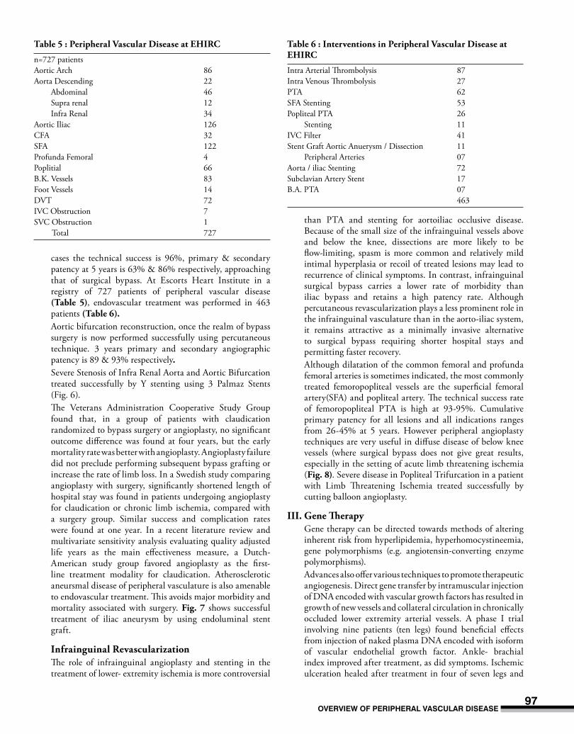
cases the technical success is 96%, primary & secondary patency at 5 years is 63% & 86% respectively, approaching that of surgical bypass. At Escorts Heart Institute in a registry of 727 patients of peripheral vascular disease (table 5), endovascular treatment was performed in 463 patients (table 6).
Aortic bifurcation reconstruction, once the realm of bypass surgery is now performed successfully using percutaneous technique. 3 years primary and secondary angiographic patency is 89 & 93% respectively.
Severe Stenosis of Infra Renal Aorta and Aortic Bifurcation treated successfully by Y stenting using 3 Palmaz Stents (Fig. 6).
The Veterans Administration Cooperative Study Group found that, in a group of patients with claudication randomized to bypass surgery or angioplasty, no significant outcome difference was found at four years, but the early mortality rate was better with angioplasty. Angioplasty failure did not preclude performing subsequent bypass grafting or increase the rate of limb loss. In a Swedish study comparing angioplasty with surgery, significantly shortened length of hospital stay was found in patients undergoing angioplasty for claudication or chronic limb ischemia, compared with a surgery group. Similar success and complication rates were found at one year. In a recent literature review and multivariate sensitivity analysis evaluating quality adjusted life years as the main effectiveness measure, a Dutch-American study group favored angioplasty as the first- line treatment modality for claudication. Atherosclerotic aneursmal disease of peripheral vasculature is also amenable to endovascular treatment. This avoids major morbidity and mortality associated with surgery. fig. 7 shows successful treatment of iliac aneurysm by using endoluminal stent graft.
infrainguinal revascularization The role of infrainguinal angioplasty and stenting in the
treatment of lower- extremity ischemia is more controversial
than PTA and stenting for aortoiliac occlusive disease. Because of the small size of the infrainguinal vessels above and below the knee, dissections are more likely to be flow-limiting, spasm is more common and relatively mild intimal hyperplasia or recoil of treated lesions may lead to recurrence of clinical symptoms. In contrast, infrainguinal surgical bypass carries a lower rate of morbidity than iliac bypass and retains a high patency rate. Although percutaneous revascularization plays a less prominent role in the infrainguinal vasculature than in the aorto-iliac system, it remains attractive as a minimally invasive alternative to surgical bypass requiring shorter hospital stays and permitting faster recovery.
Although dilatation of the common femoral and profunda femoral arteries is sometimes indicated, the most commonly treated femoropopliteal vessels are the superficial femoral artery(SFA) and popliteal artery. The technical success rate of femoropopliteal PTA is high at 93-95%. Cumulative primary patency for all lesions and all indications ranges from 26-45% at 5 years. However peripheral angioplasty techniques are very useful in diffuse disease of below knee vessels (where surgical bypass does not give great results, especially in the setting of acute limb threatening ischemia (fig. 8). Severe disease in Popliteal Trifurcation in a patient with Limb Threatening Ischemia treated successfully by cutting balloon angioplasty.
iii. Gene Therapy Gene therapy can be directed towards methods of altering
inherent risk from hyperlipidemia, hyperhomocystineemia, gene polymorphisms (e.g. angiotensin-converting enzyme polymorphisms).
Advances also offer various techniques to promote therapeutic angiogenesis. Direct gene transfer by intramuscular injection of DNA encoded with vascular growth factors has resulted in growth of new vessels and collateral circulation in chronically occluded lower extremity arterial vessels. A phase I trial involving nine patients (ten legs) found beneficial effects from injection of naked plasma DNA encoded with isoform of vascular endothelial growth factor. Ankle- brachial index improved after treatment, as did symptoms. Ischemic ulceration healed after treatment in four of seven legs and
table 5 : Peripheral Vascular disease at eHirc
n=727 patients Aortic Arch 86Aorta Descending 22 Abdominal 46 Supra renal 12 Infra Renal 34Aortic Iliac 126CFA 32SFA 122Profunda Femoral 4Poplitial 66B.K. Vessels 83Foot Vessels 14DVT 72IVC Obstruction 7SVC Obstruction 1 Total 727
table 6 : interventions in Peripheral Vascular disease at eHirc
Intra Arterial Thrombolysis 87Intra Venous Thrombolysis 27PTA 62SFA Stenting 53Popliteal PTA 26 Stenting 11IVC Filter 41Stent Graft Aortic Anuerysm / Dissection 11 Peripheral Arteries 07Aorta / iliac Stenting 72Subclavian Artery Stent 17B.A. PTA 07 463

MeDicine upDate 200598
Fig. 6 : Severe Stenosis of Infra Renal Aorta and Aortic Bifurcation treated successfully by Y stenting using 3 Palmaz Stents
Fig. 7 : Aneurysm of Right Common Iliac Artery treated by recreating the aortic bifurcation using PTFE covered stents (JOMED)
Aneurysm of RT Common Iliac Artery
three of these patients were able to avoid the amputation that had been recommended before DNA treatment. 21
Catheter- based delivery systems for gene therapy are being developed, in conjunction with adaptation of various vectors containing therapeutic genetic material (usually DNA, but RNA vectors are also being investigated). Other delivery systems include use of infective viruses (e.g. adenovirus),
which can incorporate genetic material into the host genome, and use of liposomes, which deliver encapsulated genetic messages to the host target cells. 22
other forms of Therapeutic Angiogenesis An alternative strategy to genetic alteration that is under
investigation is use of protein growth factors (i.e. vascular endothelial growth factor, acidic and basic fibroblast growth

overvieW of peripHeraL vascuLar Disease99
factors, transforming growth factor, platelet-derived growth factor, angiogenesis factor, interleukins, tumor necrosis factor). The ideal method of therapeutic angiogenesis awaits further study. In a thought provoking commentary, Henry recently reviewed the current state of therapeutic angiogenesis and noted that issues requiring further study are the possibility of pathologic angiogenesis, defining of the optimum therapeutic targets, development of better delivery systems and definitions of goals and endpoints of therapy.23
diSeASeS of tHe VenouS SySteM
VenouS tHroMBoSiSMay affect superficial veins (superficial thombophlebitis) or deep veins (deep vein thrombosis). Prolonged venous thrombosis may lead to chronic venous insufficiency, in which there is edema, pain, stasis pigmentation, stasis dermatitis and stasis ulceration. Thrombosis is virtually always accompanied by phlebitis, thus the terms thrombosis and thrombophlebitis are used interchangeably. DVT is usually benign but can cause lethal pulmonary emboli or chronic venous insufficiency. For DVT, the objectives of treatment are prevention of pulmonary embolism and chronic venous insufficiency. LMWH therapy is followed soon by oral warfarin therapy keeping INR between 2 and 3.
Varicose VeinsElongated, dilated and tortuous superficial veins(usually in the legs) with incompetent valves, permitting reversed flow are the hall mark of this condition. This condition is a fairly common in India. Valve failure at the saphenofemoral junction permits reflux into the saphenous vein, resulting in sequential descending valvular incompetence from the thigh to the calf.
Fig. 8 : Diffuse arteriopathy in below knee arteries treated successfully by cutting balloon angioplasty
referenceS1. Dutta. R; Vascular disease management plaqued by lack of awareness &
Research, Express Health Care Management, 1:2, Jan 1.15, 2003. 2. Shead GV, Oomen RM, et al. The pattern of non-diabetic peripheral
vascular disease in South India, Br J Surgery 1978 65: 49-53.3. Kinare SG, Kher YR, et al. Pattern of occlusive peripheral vascular disease
in India (clinicopathological study of cases), Angiology 1976, 27:165-80.4. Kurata A, Franke FE, et al. Thromboangiitis obliterans: classic and new
morphological features. Virchows Arch 2000; 436:59-67.5. Orchard Tj. Strandness DE: Assessment of peripheral vascular in diabetes.
Report and recommendations of an international workshop. Circulation, 1993; 88:819.
6. Rose GA, Blackburn H : Cardiovascular Survey Methods. Geneva, Switzerland, World Health Organization Monograph Series p. 172-75, 1968.
7. Siperstein ND, Unger RH, Madison LL. Studies of muscle capillary basement membranes in normal subjects, diabetic and prediabetic patients. J Clin Invest 1968; 47: 1973-1999.
8. Brownlee M. Cerami IA, Valassara H. Advanced glycosylation and products in tissue and the biochemical bases of diabetic complications. N Engl J Med 1988;318:1315-1321.
9. Conrad MC. Large and small artery occlusion in diabetics and non diabetics with severe vascular disease. Circulation 1967;36:83-91
10. Strandness DE Jr, Priest RE, Gibbons GE. Combined clinical pathological study of diabetic and non diabetic peripheral arterial disease. Diabetes 1964;13:366-372.
11. Menzoian JO, Lamorate WW, Paniszyn CC, et al. Symptomatology and anatomic patterns of peripheral vascular disease : differing impact of smoking and diabetes. Ann Vasc Surg 1989;3:224-228.
12. Goldenberg SG, Alex M, Joshi RA, Bluementhal HD. Non atheromatous peripheral vascular disease of the lower extremity in diabetes mellitus. Diabetes 1959;8:261-273.
13. Irwin ST, Gilmore J, McGrann S, et. al. Blood flow in diabetics with foot lesions due to small vessel disease. Br J Surg 1988;75:1201-1206.
14. Panja M. Kar AK. Dutta AL, Chhetri M. Kumar S. Panja S : Cardiac involment in non specific aorto arteritis. Int J Cardiol 1992;34:289.
15. Sen PK, Kinare SG, Kulkarni TP, Parulkar GB : Stenosing aortitis of unknown etiology. A study of four cases. Surgery 1962; 51: 317.
16. Chhetri M.K., Neelkantan Chandrika Bose, J. : A profile on non- specific arteritis as observed in Eastern India. Jr Assoc Phys India 1974;22:839-847.
17. Tyagi S, Kaul UA, Nair M et al : Balloon angioplasty of the aorta in Takayasu’s arteritis – initial and long term results. Am Heart J 1992;124:876.
18. Borssen B, Bergen Hein T, Lithner F : the epidemiology of foot lesions in diabetic patients. Diabetic Medicine 1990; 7:438-44.
19. Blakeman BM, Littooy FM, Baker WH, Intra-arterial digital subtraction angiography as a method to study peripheral vascular disease. J Vasc Surg 1986;4:168-173.
20. Dawson DL, Cutler BS, Meissner MH, et al. Cilostazol has beneficial effects in treatment of intermittent claudication: result from a multicenter, randomized, prospective, double-blind trial. Circulation 1998;98:678-86.
21. Svensson EC, Schwartz LB. Gene therapy for vascular disease. Curr Opin Cardiol 1998;13:369-74.
22. Baumgartner I, Pieczek A Manor O, et al. Constitutive expression of phVEGF165 after Intramuscular gene transfer promotes collateral vessel development in patients with critical limb ischemia. Circulation 1998;97:1114-23.
23. Henry TD. Can we really grow new blood vessels? (Commentary). Lancet 1998;351:1826-7.