overview of a mass analyzer - home - department of chemistry€¦ · · 2015-12-07electron...
TRANSCRIPT

Overview of a Mass Spectrometer

GC-MS Instrument
GC
MS
Instrument footprint

Electron Multiplier Detector
An electron multiplier consists of many dynodes. A progressively decreasing negative voltage (more positive) is applied to each dynode in series by dividing the high voltage applied to the first dynode with resisters. A few secondary electrons are emitted from the first dynode by ion impingement. An electron multiplier is put in an off-axis position to reduce noise signals by direct hit of neutral particles.

Different Mass Analyzers
A mass analyzer is the component of the mass spectrometer that takes ionized masses and separates them based on charge to mass ratios and outputs them to the detector where they are detected and later converted to a digital output.
There are five general types of mass analyzers that can be used for the separation of ions in a mass spectrometry.
1. Quadrupole Mass Analyzer 2. Time of Flight Mass Analyzer 3. Magnetic /Electrostatic Double Sector Mass Analyzer 4. Quadrupole Ion Trap Mass Analyzers 5. Ion Cyclotron Resonance
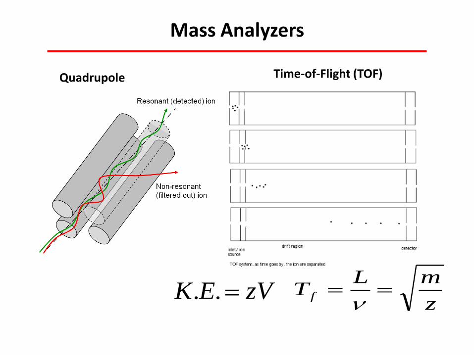
Mass Analyzers
Quadrupole Time-of-Flight (TOF)
zVEK ..z
mLT f

Quadrupole Ion Trap Mass Analyzers
The ions enter the area between the electrodes through one of the end caps. After entry, the electric field in the cavity due to the electrodes causes the ions of certain m/z values to orbit in the space. As the radio frequency voltage increases, heavier mass ion orbits become more stabilized and the light mass ions become less stabilized, causing them to collide with the wall, and eliminating the possibility of traveling to and being detected by the detector.
The quadrupole ion trap usually runs a mass selective ejection, where selectively it ejects the trapped ions in order of in creasing mass by gradually increasing the applied radio frequency voltage.
ChemWiki

Matrix-Assisted Laser Desorption Ionization
Laser radiation of this matrix-analyte preparation results in desorption of the matrix as a plume, which carries the analyte along with it into gas phase. Thus, the matrix plays a key role by strongly absorbing the laser light energy and causing, indirectly, the analyte to vaporize. The matrix also serves as a proton donor and acceptor, acting to ionize analyte in both positive and negative ionization modes, respectively.
Dept. of Chemistry, Univ. of Pittsburgh

LC-MS Instrument

Dual Ion Source for LC-MS (ESI and APCI)
Shimadzu Scientific Instruments
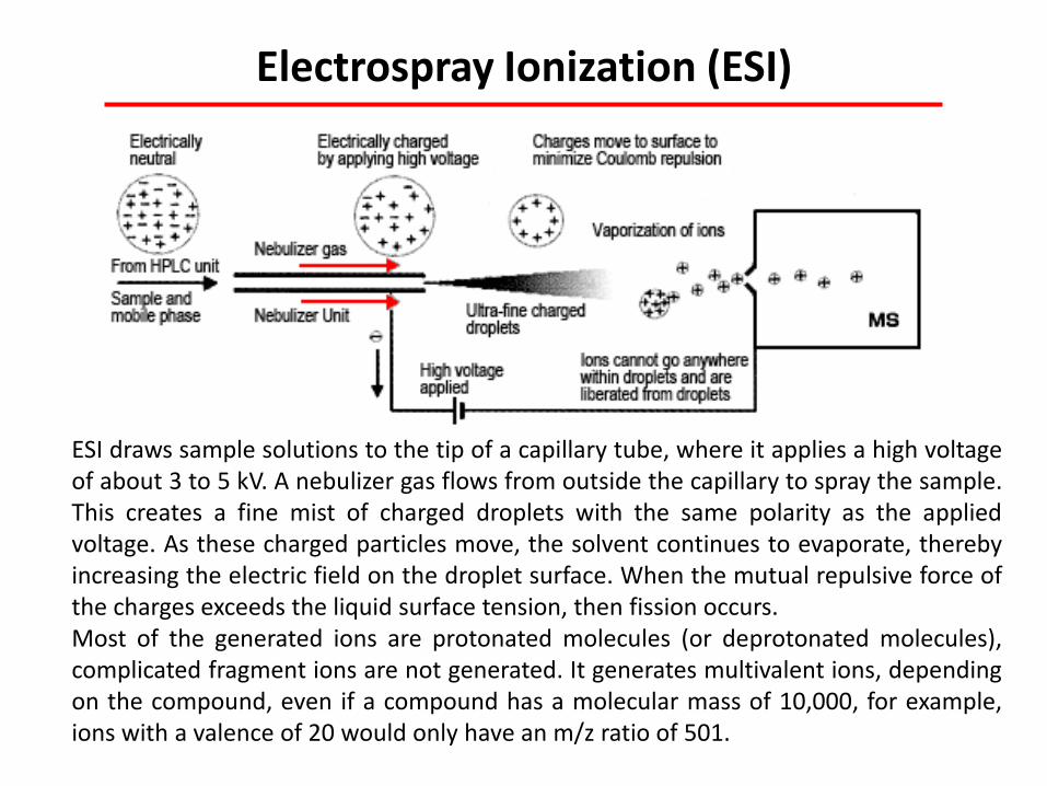
Electrospray Ionization (ESI)
ESI draws sample solutions to the tip of a capillary tube, where it applies a high voltage of about 3 to 5 kV. A nebulizer gas flows from outside the capillary to spray the sample. This creates a fine mist of charged droplets with the same polarity as the applied voltage. As these charged particles move, the solvent continues to evaporate, thereby increasing the electric field on the droplet surface. When the mutual repulsive force of the charges exceeds the liquid surface tension, then fission occurs. Most of the generated ions are protonated molecules (or deprotonated molecules), complicated fragment ions are not generated. It generates multivalent ions, depending on the compound, even if a compound has a molecular mass of 10,000, for example, ions with a valence of 20 would only have an m/z ratio of 501.

Electrospray Ionization (ESI)
ESI can produce singly or multiply charged ions. The number of charges retained by a particular analyte depends on several factors such as the size, chemical composition, and higher order structure of the analyte molecule, the solvent composition, the presence of co-solutes, and the instrument parameters. For small molecules (< 2000 Da) ESI typically generates singly, doubly, or triply charged ions, while for large molecules (> 2000 Da) ESI can produce a series of multiply charged ions.
ESI is very suitable for a wide range of biochemical compounds including peptides and proteins, lipids, oligosaccharides, oligonucleotides, bio-organic compounds, synthetic polymers.
Dept. of Chemistry, Univ. of Pittsburgh
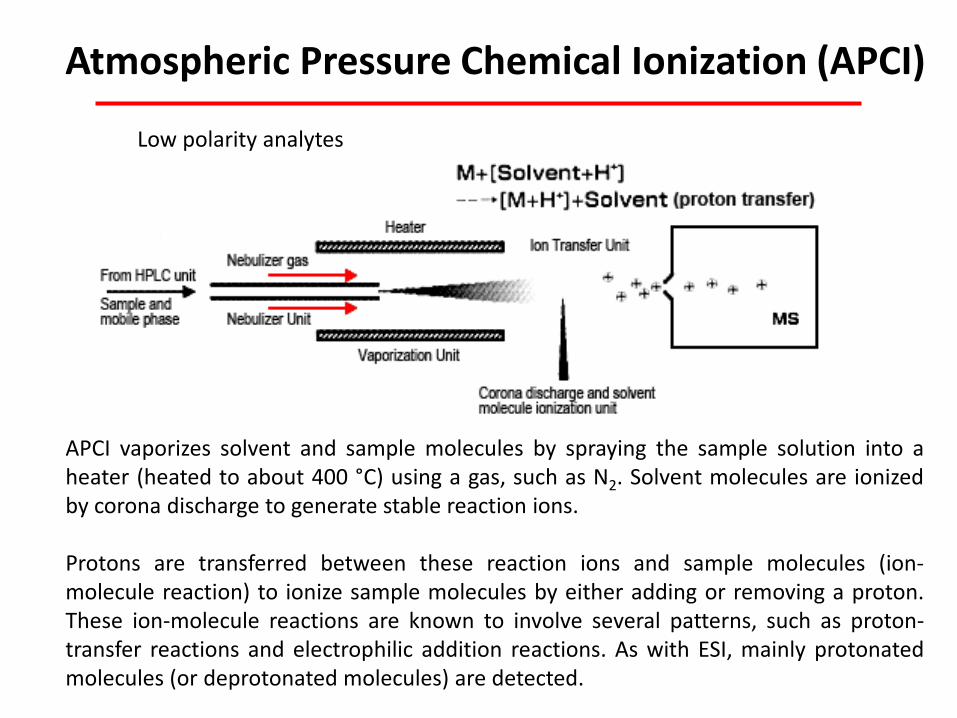
Atmospheric Pressure Chemical Ionization (APCI)
APCI vaporizes solvent and sample molecules by spraying the sample solution into a heater (heated to about 400 °C) using a gas, such as N2. Solvent molecules are ionized by corona discharge to generate stable reaction ions. Protons are transferred between these reaction ions and sample molecules (ion-molecule reaction) to ionize sample molecules by either adding or removing a proton. These ion-molecule reactions are known to involve several patterns, such as proton-transfer reactions and electrophilic addition reactions. As with ESI, mainly protonated molecules (or deprotonated molecules) are detected.
Low polarity analytes

Atmospheric Pressure Chemical Ionization (APCI)
Typically single charged analytes produced
Dept. of Chemistry, Univ. of Pittsburgh
Sample solution undergoes nebulization to form an aerosol spray of fine droplets and it is rapidly heated in a stream of nitrogen desolvation gas before emerging from the probe as a desolvated/vaporized sample plume. Solvent/Reagent ions are formed in the region of the corona discharge needle. These ions react with analyte molecules to form singly charged protonated or deprotonated analyte ions.

Dual ESI and APCI
Shimadzu

Mass Spectrometry Imaging
Shimadzu Scientific Instruments

Desorption Electrospray Ionization (DESI) -MS
+ electrolyte
Desorption electrospray ionization minimizes the requirements for sample preparation by enabling investigation of samples in their native environment.
Prosolia

Desorption Electrospray Ionization (DESI) -MS
Selected ion images of phosphatidylcholine (PC) species in a 10 μm coronal section of rat brain. A)PC(32:0) [M+K] m/z 772.3 B)PC(36:1) [M+K] m/z 826.4.
A B
MALDI is primarily suited for detection of large molecules such as peptides and proteins, and DESI is well suited for detection of small molecules such as lipids, metabolites and drug molecules.

Fast Scanning Speeds
0.1 m/z ~15,000 amu/s
Ultrafast HPLC requires fast detector response.

Fast Scanning Speeds

Scan and SIM Mode
You can use either scan mode or SIM mode for GC/MS analysis. The choice depends on the aim of the analysis. If you identify the sample components using a mass spectrum, scan mode is indispensable. SIM mode is suitable for quantitative analysis of trace components.When the mass spectra pf the trace components are known.