overview - dermpathmd.comdermpathmd.com/clinical dermatology/cosmetic dermatology.pdf · chemical...
TRANSCRIPT


Overview Chemical Peels Sclerotherapy Botox Soft Tissue Augmentation Future Trends

Chemical Peels improve skin texture reduce hyperpigmentation and mild wrinkling do not improve deep wrinkles or sagging skin useful as adjunctive treatment for acne, rosacea, and
melasma

Chemical Peels Categorized based on depth of the procedure:
superficial: induce necrosis of all or parts of the epidermis
medium: necrosis of the epidermis and part or all of the papillary dermis
deep: necrosis extends into reticular dermis

Superficial Peels alpha hydroxy acids (AHA) beta hydroxy acids (BHA) Jessner’s solution modified Jessner’s resorcinol trichloroacetic acid (TCA)

AHA’s and BHA Naturally occurring organic acids
high concentrations cause detachment of keratinocytes and epidermolysis
lower concentrations reduce keratinocyte cohesion above granular layer
2 major effects: quickens the cell cycle smoothes stratum corneum

AHA’s
Glycolic acid Lactic acid Citric acid Phytic acid
sugar cane sour milk citrus fruits rice

Glycolic Acid AHA most commonly used in peels “lunchtime peel” increases skin thickness and MPS’s in dermis improved quality of elastic fibers increased density of collagen

Lactic Acid
Found in many OTC and Rx moisturizers Lac-Hydrin is AHA used as rx for dry skin Not frequently used for in-office peels

BHA Aka salicylic acid (SA) derived from willow bark, wintergreen leaves, and
sweet birch in-office peels use 20-30% salicylic acid, OTC preps
contain ~2% SA helps decrease hyperpigmentation, decrease surface
roughness, and reduce fine lines

BHA increase exfoliation accelerate cell cycle exhibits anti-inflammatory capabilities, thus induce
less irritation than AHA’s. useful peel in rosacea and acne patients lipophilic and comedolytic does not increase collagen synthesis

Disadvantages of Hydroxy Acids unrealistic patient expectations decreased efficacy with continued use ? decrease natural skin barrier to UV light and harmful
environmental toxins
Important Considerations When Comparing Preparations pH and pKa buffered solutions
sodium bicarb sodium hydroxide vehicle

Performing a Superficial Peelwith AHA or BHA Cleanse the skin
4 x4 gauze with 0.25% triclosan rinse with water, then dry apply acetone gauze
Apply 40-70% glycolic acid with 2x2 gauze, and rinse with water or neutralize with 5%NaHCO3 after 2-4 min.
Apply SA with 2x2 gauze. It will precipitate (frost) in approx. 2 min. and does not need to be neutralized


Jessner’s Solution Combination of:
resorcinol 14g salicylic acid 14g lactic acid 14g ethanol 95% to make 100cc of solution
formulated to reduce concentration and toxicity of individual ingredients while increasing efficacy

Jessner’s Solution strength of the peel determined by how many layers
are applied does not need neutralization can be combined with other peels to increase efficacy
(ie. TCA) use cautiously in dark skin b/c of risks of post
inflammatory hyperpigmentation or contact dermatitis with resorcinol

Performing a Jessner’s Peel Cleanse skin Apply thin layer of petrolatum to naso-alar grooves
and lips Apply thin coat of Jessner’s to desired treatment
area First coat complete when frosting occurs (approx.
3-5 minutes) can apply more coats to deepen penetration patient will experience flaking for ~7days

Modified Jessner’s
Combinations including hydroquinone and kojic acid combination without resorcinol

Resorcinol Used as peeling agent since 1882 is m-dihydroxybenzene, a phenol derivative antipruritic, keratolytic, antimycotic, and antiseptic
properties used as treatment for pigmentary disorders, acne, and
in combo with other peel agents

Resorcinol must limit surface area treated due to risk of phenol-
like systemic toxicity prolonged use can be assoc. with myxedema and
methemoglobinemia can cause allergic contact dermatitis and post-
inflammatory hyperpigmentation

Medium Depth Peels Trichloroacetic Acid
10-20% used for superficial peels 35-40% used for medium peels
produces epidermal and papillary dermal necrosis can cause hyperpigmentation and scarring
usually used in combination with Jessner’s or 70% glycolic acid as priming agents

Medium Depth Peels Indications:
photoaging actinic keratoses pigmentary dyschromias mild acne scarring
improves fine lines and stimulates collagen remodeling for 3-4 months after the procedure

Performing a Jessner’s/TCA Peel Cleanse face, de-grease with acetone or Etoh Apply Jessner’s and wait 1-2 minutes for frosting to
occur Apply 35% TCA with 1-4 cotton tipped applicators.
Allow 30sec-2 min. for white-coated frosting with background erythema.
May re-apply to areas without adequate frosting May apply saline compresses for comfort after
frosting


Medium Depth Peels Healing time:
5-7 days with TCA alone 7-14 days with Jessner’s/TCA peel
Contraindications: dark skin types recent treatment with Accutane
Cost: $28-32/ 2 oz. Bottle ($1/ patient)

Deep-Depth Peels Create injury through papillary and into reticular
dermis TCA >50% or 88% phenol preparations largely supplanted by dermabrasions and laser
resurfacing due to high incidence of side effects

Post-Op Care Superficial Peels
minimal down time mild erythema and desquamation for 1-4 days post op wash face with mild cleanser use routine moisturizers and sunscreens
Post-Op Care Medium Depth Peel
apply soaks QID with warm compresses apply petrolatum or Aquaphor following each soak NSAIDs for pain control
What to Expect immediate edema with worsening for 48 hours erythema resolves within 2-4 weeks post op

Post Op Care Deep Depth Peels
biosynthetic dressing applied QD for the first 2-3 days post op
debridement with saline soaks and cotton tips D3-14 acetic acid soaks 4-6x/d, followed by ointment
What to expect edema for weeks, erythema for 2-4 months

Complications of Peels Excessive depth of tissue injury Infection Delayed wound healing and erythema Scarring Post-inflammatory hyperpigmentation
Sclerotherapy The art of using sclerosants to destroy endothelial cells
and cause vessel fibrosis Venous pathology occurs when venous return is
impaired for any reason primary muscle pump failure due to venous obstruction valvular incompetence


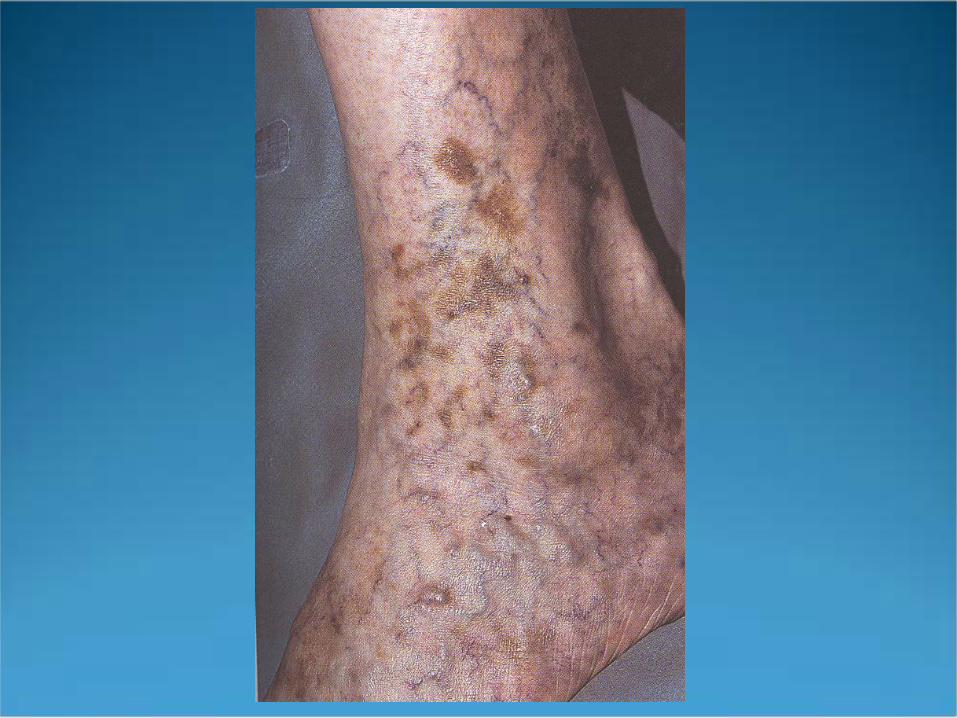
Sclerotherapy Telangectasias, reticular veins, and varicose veins are
influenced by heredity hormones static gravitational pressures incompotent valves
Physical Exam of Patient Goal is to determine where the primary or highest
points of reflux are located Grade insufficiency with Widmer classification
Stage I - presence of corona phlebectasia (telangectasias)
Stage II - hypo- or hyperpigmentation Stage III - presence of recent or healed ulcer


Vascular Testing Indicated for symptomatic patients when reflux source
is unclear Use of Doppler probe to detect frequency shifts of
blood coming towards or going away from probe


Sclerosing Solutions Optimal agent produces pan-endothelial destruction
without systemic toxicity if too weak, thrombosis without fibrosis and eventual
recanalization if too strong, hyperpigmentation, telangectatic matting,
and ulceration can occur


Sclerosing Agents Hyperosmotic agents
hypertonic saline and saline-dextrose (Sclerodex) endothelial damage through dehydration
hypertonic saline is FDA approved associated with burning and cramping on injection increased incidence of ulcerative necrosis

Sclerosing Agents Detergent sclerosants
sodium tetradecyl sulfate (Sotradecol), polidocanol, sodium morrhuate (Scleromate) vascular injury by altering surface tension around endothelial
cells
Sotradecol assoc. with allergic hypersensitivity and hyperpigmentation
Polidocanol foam
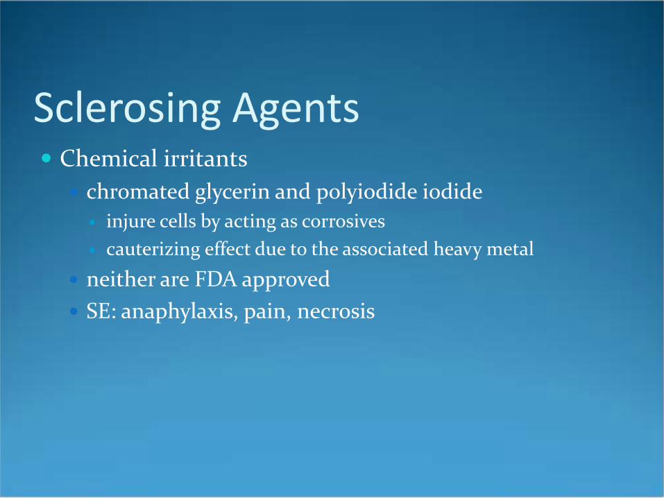
Sclerosing Agents Chemical irritants
chromated glycerin and polyiodide iodide injure cells by acting as corrosives cauterizing effect due to the associated heavy metal
neither are FDA approved SE: anaphylaxis, pain, necrosis


Technique for Telangectasias and Reticular Veins Telangectasias: flat red vessels 0.1-1mm Venulectasias: bluish vessels 1-2 mm Reticular veins: have a cyanotic hue, 2-4 mm treat proximal and larger vessels first with the minimal
sclerosant concentration (MSC)
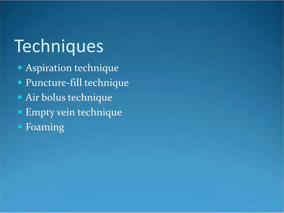
Techniques Aspiration technique Puncture-fill technique Air bolus technique Empty vein technique Foaming

Injection technique Choose one of four previous techniques Insert 30g needle at 30 degree angle, maintaining
hand traction Inject larger vessels first Inject 0.1-0.4 cc into each injection site at 3cm
intervals Wear 20 mmHg compression hose for 3 weeks post
op Wait 6-8 weeks between treatments

Treatment of Varicose Veins Must understand precise anatomy of varicosity to be
treated May need Duplex ultrasound to determine primary
source of reflux Sotradecol and hypertonic saline commonly used

Treatment of Varicose Veins Supine Direct Cannulation technique
map out injection sites while patient is standing inject 0.5-1.5 mL of sclerosant at sites separated by 3-4
cm along the vein Multiple Precannulation Sites technique
23g butterfly needles inserted into one proximal and distal site on vein
2-3 mL of sclerosant infused into cannulas


Treatment of Varicose Veins
Pt. Wears 30-40 mmHg compression hose for 3 weeks post op, and continuously for first 72 hours

Sclerotherapy Complications Hyperpigmentation (10-30%)
usually lasts for 6-12 months avoid NSAID’s and minocycline elevate leg during treatment use sclerosant concentration appropriate for vessel size apply compression immediately post-op


Sclerotherapy Complications Telangectatic Matting (5-14%)
usually resolves within 3-12 months risk factors: obesity, use of estrogen containing
medications, pregnancy, Fhx, excess post-op inflammation
use minimal sclerosant concentration may discontinue OCP’s for 1 month prior and 2 months
following treatment

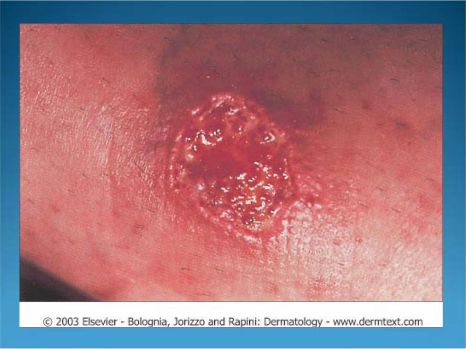
Sclerotherapy Complications Ulceration
due to extravasation of sclerosing agent, injection into dermal arteriole, or reactive vasospasm
hemorrhagic bulla may form within 12-24 hours may apply 2% nitroglycerin ointment to try and prevent
ulceration

Serious Complications Systemic allergic reaction
Sotradecol has low allergic potential (0.3%) Arterial Injection
produces sludge embolus most commonly occurs in posterior or medial malleolar
region immed. pain, decreased pulses, cyanosis, pallor tx with immediate periarterial 1% procaine, heparin for
7-10 days, and IV dextran for 3 days
Miscellaneous Complications Localized urticaria Compression ulcers, dermatitis, folliculitis Nerve damage Superficial thrombophlebitis

Botox 1895- Emile Pierre van Ermengem identified
Clostridium botulinum as an agent of food poisoning
1920- Herman Sommer attempted to purify the neurotoxin
1946- botulinum toxin A purified by Edward Schantz
1979- Alan Scott used botox to tx strabismus

History of Botox 1987- Alastair and Carruthers incidentally discovered
potential use in cosmetics when a patient treated for blepharospasm noticed a decrease in glabellar wrinkles
1989- botox accepted by FDA for treatment of strabismus, blepharospasm, and hemifacial spasm

Basic Science of Botox 8 distinct subtypes of botulinum neurotoxin
A, B, C, alpha, C beta, D, E, F, and G botox induces chemical denervation of straited muscle
by cleaving proteins required for release of acetylcholine
results in temporary flaccid paralysis of the injected muscles for 3-5 months

Basic Science of Botox Botox type A (BOTOX) is most common type used it cleaves the SNAP-25 protein (a component of the
SNARE complex) an intact SNARE complex is necessary for release of
Ach Botox B (Myobloc) cleaves synaptobrevin, another
component of SNARE


Clinical Indications
Prevention and amelioration of dynamic wrinkles (“wrinkles in motion”) and cessation of hyperhidrosis
Not useful for static wrinkles (“wrinkles at rest”)

Storage and Handling One vial of BOTOX contains 100 units of vacuum
dried type A toxin, human albumin, and sodium chloride
reconstitution procedures vary, but recommended is: 2.5 mL of 0.9% saline per vial results in 4.0 units per 0.1 mL
use by 48 hrs to up to 6 weeks, keep refrigerated


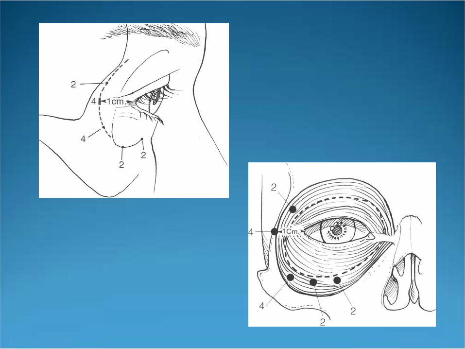
Glabellar Frown Lines Muscles involved include frontalis, procerus,
corrugator supercilli, and medial fibers of orbicularis oculi
contraction results in elevation of the brow and wrinkles of the forehead
corrugator contraction results in adduction of the eyebrow inferiorly and medially


Glabellar region Inject 4 units (0.1cc) into each corrugator and the
procerus muscle (IM injections) avoid hitting the periosteum after injection of the procerus, massage laterally to
ensure diffusion into the depressor supercilii portion of the corrugator


Side Effects of Treating the Glabellar Region
Blepharoptosis- occurs when toxin diffuses into upper eyelid levator muscle

Avoiding Side Effects Patient should remain vertical for 2-3 hours postop encourage patient to frown frequently, but not
manipulate the area avoid injection of the levator palpebrae superioris
muscle corrugator injection should be at least 1 cm above
supraorbital ridge do not inject closer than 1 cm above the central
brow

Treatment of the Forehead
Horizontal lines produced by action of the frontalis muscle
inject 4 units (0.1 cc) along the forehead at 2 cm intervals
injection of the forehead may dramatically affect eyebrow shape


Side Effects to Treatment of the Forehead
Unwanted eyebrow shape Brow ptosis Drooping of the eyelids


Avoiding Side Effects Do not over-inject the forehead avoid the area 1 cm above the eyebrows to reduce
chances of ptosis avoid forehead injections in patients with low-set
brows or excess eyelid skin ideal patient for forehead injections is 20-40 y.o.

Treating Crow’s Feet Result from the action of the orbicularis oculi inject 0.1 cc 1cm lateral to the lateral canthus, 0.05cc 1
cm above the first injection, and 0.1 cc 1 cm below

Side Effects of Treating Crow’s Feet
Bruising diplopia ectropion drooping lateral lower eyelid

Other Cosmetic Treatment Areas Brow-Lift Bunny Lines (Upper Nasalis) Lower Nasalis Orbicularis Oris (Vertical Lip Rhytides) Melolabial Folds Platysmal Bands Etc, Etc….

Post Procedure Care Patients should remain vertical for 2-4 hours avoidance of touching or rubbing of the treated sites
for 24 hours results take 12-96 hours to appear optimal effect develops within 7 days effectiveness declines after 3-4 months

Contraindications to Botox Presence of neuromuscular disorders such as
myasthenia gravis or ALS Pregnant or lactating women Patients taking aminoglycosides, penicillamine,
quinine, or CCB’s Evidence of active infection at injection site

General Complications Bruising (esp. in patients taking ASA or Vitamin E) acute Type I allergic reactions nausea, headache, fatigue, malaise, flulike symptoms,
and rashes at sites distant from injection have all been reported

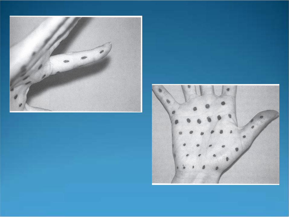
Hyperhidrosis Can treat axillary, palmar, and plantar areas reconstitute one 100 unit vial with 5 cc NS
2 units per 0.1 cc injections are intradermally (vs. IM for facial lines) nerve blocks are needed for anesthesia inject 0.05 cc at 1.5 cm intervals

Hyperhidrosis Perform Minor’s starch-iodine test
iodine solution (9 parts iodine with 1 part castor oil) applied to affected area
cover with starch powder areas producing sweat will turn blue-black provides map for injection sites


Hyperhidrosis 100 Botox units (5 cc) needed per palm or sole 50 Botox units needed per axilla effects last approx. 4 months side effects:
hematomas transient weakness of hand muscles
Botulinum Toxin Type B(Myobloc) Approved by FDA only for cervical dystonia may be useful in patients who develop antibodies
to Botox toxin type A onset of clinical effect more rapid than Type A diffuses more readily, therefore increased risk of
side effects is 100 times less potent than Botox Type A

Soft Tissue Augmentation 1893- Neuber used fat from arms and transplanted it
into facial defects 1899- Gersuny used paraffin as an augmentation
material 1940’s- silicone introduced 1970’s- researchers at Stanford introduced bovine and
human derived collagen


Classes of Fillers Heterograft/Xenograft
Bovine Collagen Zyderm and Zyplast
Hyaluronic Acid Derivatives Restylane Hylaform

Classes of Fillers Allografts (from human cadaveric tissue)
Human-Derived Collagen Dermalogen Cymetra Cosmoderm/Cosmoplast
Autografts autologous fat transplantation

Classes of Fillers Synthetic Materials
silicone Polytetrafluoroethylene (Gore-Tex)

Indications “wrinkles at rest” scars
acne scars traumatic scars
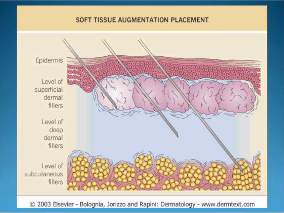
lip augmentation vermilion

General Technique Clean face to remove make-up surface anesthesia may be applied with ice or topical
preparation injection techniques
serial multiple punctures single-entry
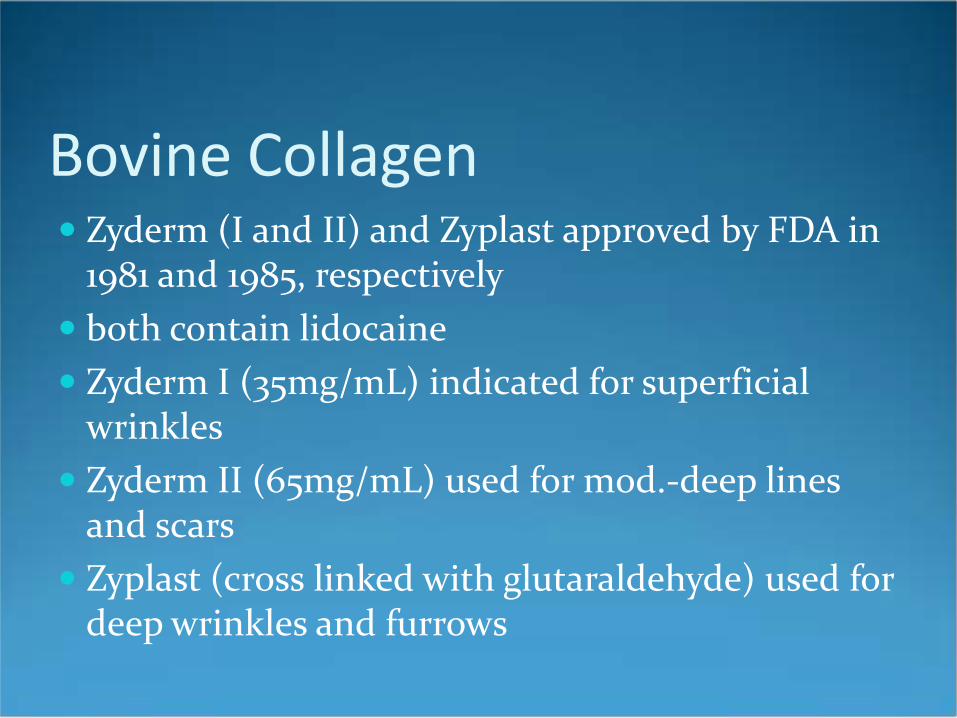
Bovine Collagen Zyderm (I and II) and Zyplast approved by FDA in
1981 and 1985, respectively both contain lidocaine Zyderm I (35mg/mL) indicated for superficial
wrinkles Zyderm II (65mg/mL) used for mod.-deep lines
and scars Zyplast (cross linked with glutaraldehyde) used for
deep wrinkles and furrows

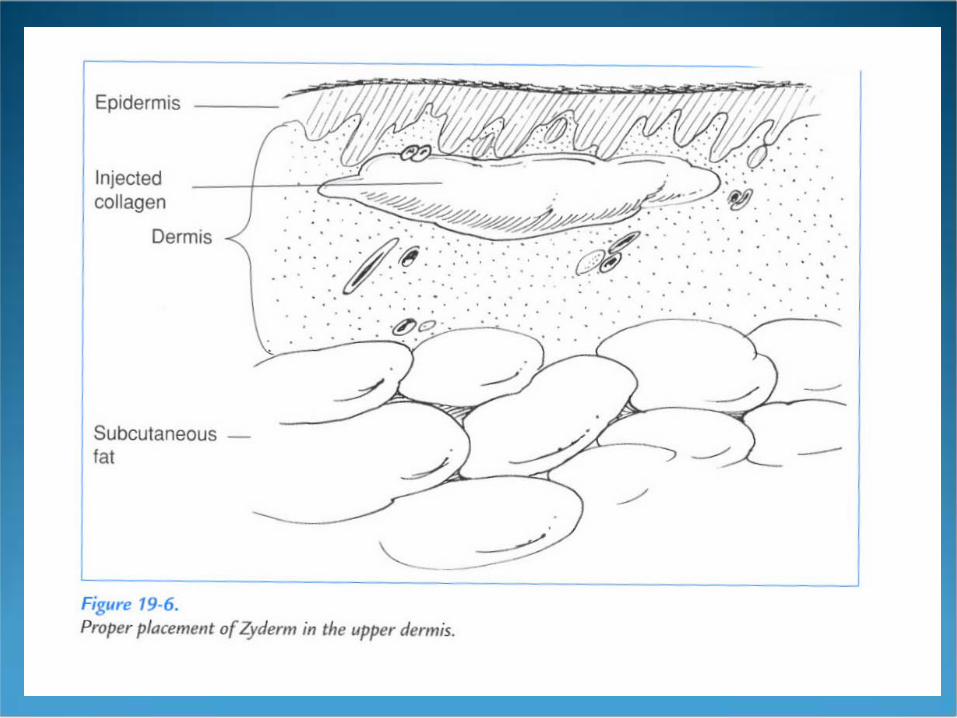
Bovine Collagen Must perform pre-tx skin testing x2 Zyderm I and II should be injected into the superficial
dermis at a 20-30 angle to produce blanching Zyplast is injected into the deep dermis at an angle of
45-90 results last 3-6 months


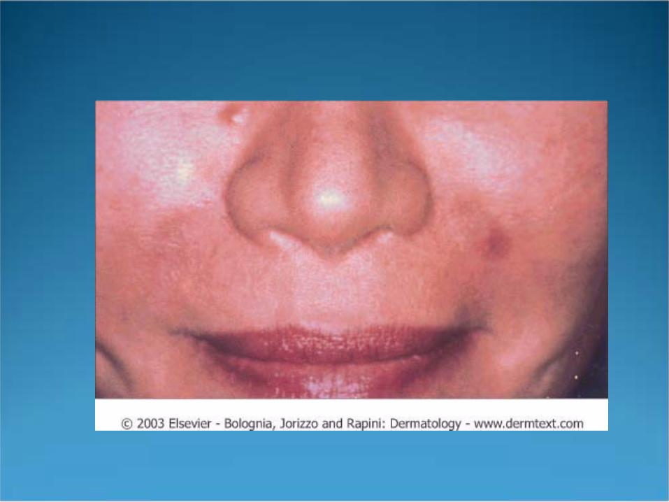
Complications of Bovine Collagen Ecchymosis Type IV hypersensitivity reactions granuloma
formation sterile abscesses (especially with Zyplast) Tissue necrosis with intravascular injection Re-activation of HSV


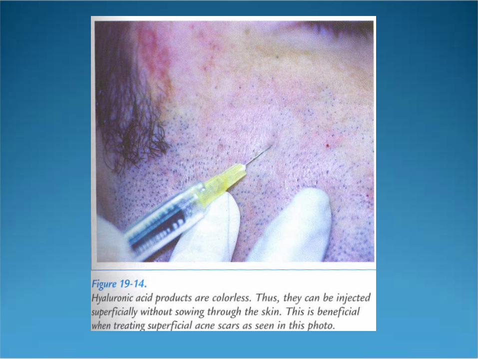
Hyaluronic Acid Derivatives
Restylane Hylaform

Hyaluronic Acid Derivatives Hyaluronic acid is composed of repeating dimers
of glucuronic acid and N-acetyl glucosamine These fillers are chemically altered forms of
hyaluronic acid, a GAG normally present in the dermis and identical in all species
Has capacity to bind water up to 1000x its volume Is insoluble, colorless, resists degradation, and
does not cause allergic reactions

Restylane FDA approved in 12/03 Produced by fermentation of streptococcal bacteria Less expensive than bovine collagen and able to be
stored at room temperature Less volume needed as compared to bovine collagen Results last up to 6 months

Restylane Three types that all contain 20mg/mL of hyaluronic
acid in a clear gel and vary based on the size of the particles Restylane Fine Lines: 0.4 mL syringe, inject into upper
dermis Restylane: 0.4 and 0.7 mL syringes, inject into mid
dermis Perlane: 0.7 mL syringe, inject into deep dermis
Restylane Treatment Cleanse face prior to injection Apply topical, local or block anesthesia Inject into the superficial dermis at an angle of 30
degrees Cost $210/vial
Restylane Side Effects Increased pain with injection Injection site reactions Edema following lip augmentation

Hylaform Not yet FDA approved, used in Europe for past 7 years hyaluronic acid derived from rooster combs concentration 6 mg/mL no allergic reactions reported injected into the deep dermis does not contain lidocaine and therefore anesthetic is
necessary

Hylaform Side effects
bruising, erythema, swelling ? Risk of use in patients with avian allergies Adequate studies lacking for use in African Americans

Human Derived Collagen
Dermalogen- human cadavers Alloderm/Cymetra- human cadavers Cosmoderm/Cosmoplast- neonatal foreskin
Human Derived Collagen Use began in the 1980’s eliminates need for pre-treatment skin testing no hypersensitivity reactions

Dermalogen Composed of intact collagen, elastin fibers, and GAG’s
harvested from the dermis of human cadaveric skin pre-screened for infectious diseases supplied in a 0.5 or 1.0cc syringe at a concentration of
3.5% pre-tx anesthesia necessary inject into mid-deep dermis
Cymetra/Alloderm Made from human cadaver dermis similar to Dermalogen, except is in powder form and
therefore requires reconstitution with lidocaine inject into mid-deep dermis effects last 4-6 months

Cosmoderm/Cosmoplast Derived from fibroblasts taken from neonatal foreskin no required skin testing injected into mid-deep dermis effects last 2-6 months
Autologous Fat Transplantation Performed since the 1980’s indicated for melolabial folds, lips, acne scarring, and
lipoatrophy involves removing 15-20 cc of fat with a 13-gauge needle
from various parts of the body, and re-injecting the fat into the SC using a 16-18-gauge needle

Autologous Fat Transplantation
More time consuming because harvesting and injection required
local anesthesia is necessary up to 50% of fat remains after 2 years of procedure

Synthetic Fillers Silicone
composed of dimethylsiloxane polymers is permanent not approved as a filler by the FDA physicians use silicone “off label” that is only approved
for ophthalmic use SE: hypersensitivity, granuloma formation, migration of
the material


New Fillers New-Fill (Sculptra)- polylactic acid, a component of
vicryl suture material Radiance- calcium hydroxyapatite (approved for vocal
cord paralysis and as a radiological soft tissue marker) Artecoll- polymethylmethacrylate microspheres
suspended in bovine collagen

Future Trends Non-Ablative Radiofrequency
Thermage Plasma Skin Rejuvenation Mesotherapy