over expression of iptg inducible gst protein in e.coli...
TRANSCRIPT
Over Expression of IPTG inducible GST protein in E.coli BL21 1Sugantha priya S, 1Gowri Shankar J, 1Thirumalaisamy R, 1Kavitha P, 1Prakash B
2Arunachalam G, 3S.Selvamuthukumar 1Department of Biotechnology, PGP College of Arts and Science
Namakkal - 637207.Tamilnadu, India. 2PGP College of Pharmaceutical Sciences and Research, Namakkal – 637207, Tamilnadu, India. 3Department of Pharmacy, Annamalai University, Annamalai Nagar – 608002, Tamilnadu, India
E-Mail: [email protected] Abstract:
ISSN:0975-542X
The objective of our work was to over express GST protein, from PGEX 3X in the BL21 strain of Escherichia coli. The GST protein was induced with 0.1mM of IPTG. Under induction condition PGEX 3X provides the ability to produce high level expression of fusion proteins. The expressed GST protein was purified through glutathione sepharose affinity resin as ten fractions. IPTG mediated GST induction was assayed with CDNB. Immunoblot analysis with anti-GST HRP conjugates shows functional expression of GST in the bacterial system. Keywords: Escherichia coli, PGEX and CDNB.
Introduction : Glutathione S-Transferase (GSTs) consist a family of multi functional enzymes that comprises a long list of cytosolic, mitochondrial, and microsomal proteins which are capable of multiple reactions with a multitude of substrates, both endogenous and xenobiotic [1].GSTs are involved in the detoxification of xenobiotic compounds and in the protection against degenerative disease such as cancer, the mechanism of these enzyme involves in nucleophilic attack by glutathione on an electrophilic substrate [2]. The resulting glutathione S conjugate are more soluble than the original substrate and thus more easily transported from the cell, mediated by ATP dependent MAPEG family membrane glycoprotein belonging to multiple drug resistant protein family [3]. Expression of genes in E.coli BL 21 offers a convenient system to produce large amount of recombinant protein that may otherwise be difficult to isolate from natural cell and tissues. Very often antibodies to these newly identified proteins are not available for purification or to study the biochemical and expression properties [4]. In order to circumvent this problem small proteins like bacterial GST are often cloned along with a target gene and are expressed as fusion protein [5]. GST fusion protein can bind to glutathione sepharose therefore the high degree of
purification of fusion protein can be achieved in just one affinity purification step. Antibodies to these fusion tags are already available to monitor fusion protein expression and purification. Fusion tags serves as universal tags much like secondary antibodies. Therefore the present work was undertaken to study the purification properties of GST protein induced by IPTG in a E.coli expression system like BL21 [6]. Materials and Methods: All the chemicals used in this work were scientific grade obtained from Himedia (Mumbai), E.coli PGEX 3X and BL21 from Promega scientific Ltd (USA) and Glutathione Sepharose kit from Qiagen (Germany). PGEX 3X vector isolation: The E.coli PGEX 3X vector was isolated by alkali lysis method. The following solution were used for isolation, like Resuspension buffer, Lysis buffer, Neutralization buffer, TE buffer, 70% ethanol, LB media. The isolated plasmid was checked in 0.8% agarose gel. The DNA was diluted by the dilution factor (Dilution factor = Total volume of sample / volume of preparation used) and quantified by using UV spectrophotometer (systronics) at 260 and 280nm [7]. Transformation: The quantified PGEX 3X vector DNA was transformed into the E.coli host BL21 for this the E.coli BL21 is made
Sugantha Priya et al / J Biomed Sci and Res., Vol 2 (1), 2010, 54-59.
54

competent enough by repeated treatment with 0.1M calcium chloride and made into 100μl aliquots and stored at 180°C for further transformation. By mixing 40μl of isolated PGEX 3X vector with one aliquots of BL21 competent cells [8], it was mixed gently and incubated in ice before exposing to heat shock at 42° C for transformation. After 90 seconds it was transferred to antibiotic free LB broth and allow for two and half hours in 37 °C. Then it was checked for the presence of transformed PGEX 3X vector in (100μg / ml) ampicillin containing LB agar plates by incubating at 37°C for overnight [9]. Induction with IPTG : Inoculated a colony from transformed LB ampicillin plates to 5 ml of LB broth with ampicillin and incubated at 37°C with 130rpm for overnight And 2ml of overnight culture was incubated in 100 ml of ampicillin(100μg / ml) containing LB broth. It was allowed until the OD reaches 0.5 at 600 nm from this 5ml of culture was labeled as Before Induction BI and the remaining 95ml of culture was mixed with 0.1mM IPTG and marked as After Induction (AI) sample. Both BI and AI cultures were incubated at 30°C at 220 rpm for overnight [6]. Cell Lyses: The incubated culture samples were lysed by two methods. Method I: 5ml of both BI and AI culture were centrifuged at 8k for 10 minutes and the supernatant was discarded. The pellet was resuspend in 100-150μl of cell lysis extraction buffer and boiled at 70-100°C for 20 minutes in serological water bath and checked in SDS PAGE with 25μl of sample loading buffer. Method II: Remaining 90ml of AI culture was pelleted at 5k under 4°C for 10 min added 2-5 parts of alumina for one part of pellet and grounded into paste. Added 2ml of precooked extraction cell lysis buffer per 100-500mg of cells and spinned at 5k under 4°C for 45 minutes carefully transferred the
supernatant to the clean pre chilled tubes. Any insoluble materials will be pelleted with cell debris and again to the supernatant 0.5ml of wash solution was added and centrifuged at 14k for 30 seconds under 4°C [6]. Purification in glutathione sepharose column: Immobilized glutathione column was equilibrated with 10ml of binding buffer till the O.D reaches 0.05 at 280nm, then 10ml of cell lysate was allowed to flow completely to the gel bed and collected in 10 fraction numbers with the 25ml of freshly prepared 1X elution buffer containing reduced glutathione. (Heike Berthold, Brigitte Frorath) Elution was assayed by the absorbance at 280nm and eluted fusion proteins were checked with anti-GST antibodies in western blotting [4]. Assay of GST activity by CDNB method: In a microfuge tube added 10μl of 100mM CDNB, 10μl of reduced glutathione 100μl of 10 x reaction buffer containing 1M potassium phosphate with pH 6, and made upto 100μl with millipore water. Capped and mixed by inverting several times transferred 50μl of above solution to the cuvettes one acts as a control with 50μl of 10X reaction buffer and to the other 50 μl of eluted sample to be assayed was added [10]. The absorbance of the blank and sample was read at 340nm with one minute regular interval for continuous 5 minutes and calculated by the following equation and the readings were plotted in graph 1 ∆A340/Min/ml = A340 (t2)-A340(t1)/(t2-t1)(ml of sample added) Immunodetection: Immuno blotted membrane was blocked with 1x blocking buffer at room temperature for two hours. Anti GST-HRP conjugate were incubated for 20 minutes and washed three times with 1x washing buffer to remove unbound anti GST-HRP conjugate then it was incubated in room temperature with substrate TMB H2O2 and allowed to react. Blue colour
Sugantha Priya et al / J Biomed Sci and Res., Vol 2 (1), 2010, 54-59.
55
development indicates the presence of GST protein specific for anti GST antibody [11]. Results: Isolated E.coli PGEX-3X host by alkali lysis method and was resolved on 0.8% agarose along with λ Hind III molecular weight marker, The PGEX 3X plasmid DNA was measured as 4900bp approximately. Quantification of DNA was done with ratio of absorbance at 260nm / 280 nm is 1.825 i.e. between 0.555/0.304 DNA concentration = 50*A260*dilution factor μg/ml Dilution factor = total volume of the sample /volume of DNA preparation used = 2000 μl/10 μl = 200 μl DNA concentration = 50*0.555*200 μg/ml = 5550 μg/ml Quantification of pure DNA was done in the ratio of absorbance at 1.825 and recovered. The absorbance ratio less or above was discarded as protein or RNA contamination. The transformed PGEX 3X vector showed growth in ampicillin (100 μg/ml) containing LB agar plates. This inferred the presence of transformed PGEX-3X in E.coli BL21.IPTG induced GST was collected by cell lysis method from both BI and AI and cell lysate was analyzed through SDS PAGE. By
performing SDS PAGE with both BI and AI reveals the presence of induced protein only in AI but not in BI. Before subjecting to the SDS PAGE the cell lysates were mixed with extraction buffer and centrifuged at 10k for 20minutes under 4°c to pellet out the insoluble proteins. The crude AI cell lysate was purified in glutathione cross linked agarose column .The trapped GST protein was eluted by elution buffer by checking with the absorbance 0.05 at 280nm indicates the glutathione CL agarose column contained only the expressed GST protein. The CDNB assay values are calculated by the following equation ∆ A340/min/ml = A340 (t0) - A340 (t1)/(t1-t0) (ml of sample added) A340 (t0) = Absorbance at 340nm at time t0 in minutes A340 (t1) = Absorbance at 340nm at time t1 in minutes (t1-t0) = time interval between the reading in minutes ∆ A340/min/ml = A340(t0)- A340 (t1)/(t1-t0) (ml of sample added) = 0.009-0.012/1*0.05 =0.06
Figure 1
Figure 2
Sugantha Priya et al / J Biomed Sci and Res., Vol 2 (1), 2010, 54-59.
56

Immunodetection: The nitrocellulose membrane was carefully taken and subjected for immunodetection. The non specific sites in membrane are blocked with blocking buffer for two hours, which blocks all the proteins except GST. Later incubation with anti-GST HRP conjugate antibody bind only to the GST in the membrane. On washing with the wash buffer it washes out all the
unbound proteins in the membrane. The substrate TMB H2O2 specific for this HRP was added in dark condition, the GST protein formed complex with anti GST HRP conjugate antibody when reacted with substrate solution blue colour bands were observed on nitrocellulose membrane and hence inferred that isolated proteins were GST proteins.
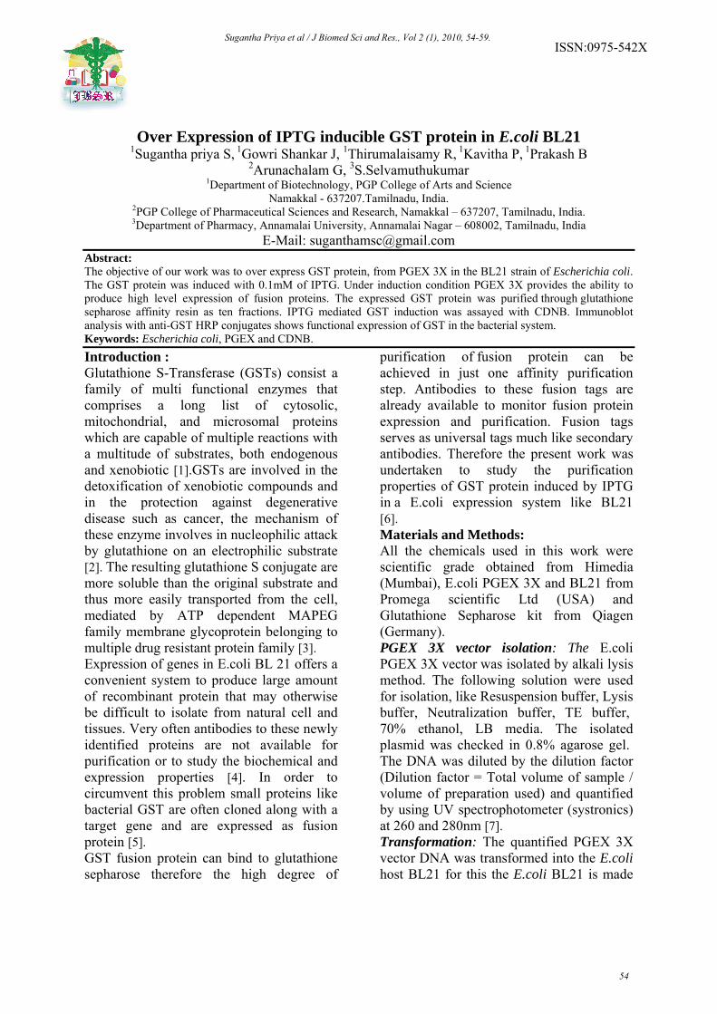
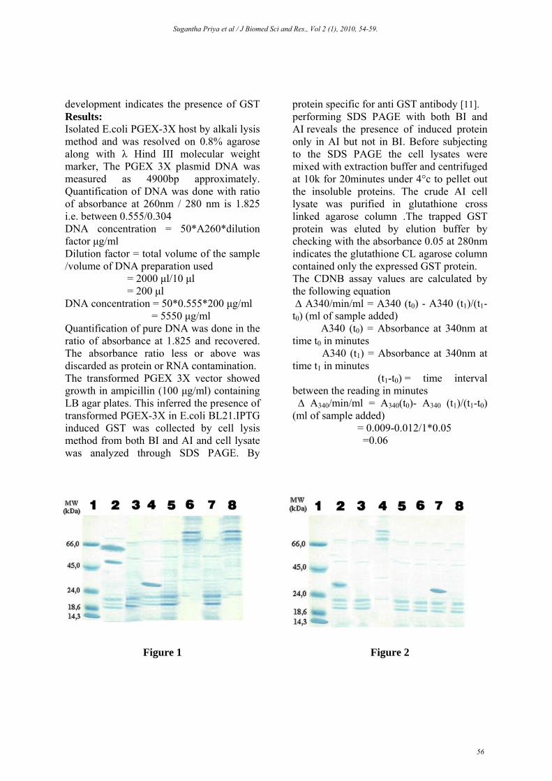
The GST activity is found to be increasing when the binding between glutathione and CDNB increases, and it was found to be decreasing gradually when binding decreases. (Table -1) SDS PAGE GEL : In figure 1 lane1 shows protein molecular weight marker , lane 2 shows AI (After induction ) with thick band indicating that IPTG has induced GST, lanes 3 contains BI(Before induction) sample which has no band, lane 4,5,6 were loaded with equilibrated sample EQ1,EQ2 and EQ3 and no bands were seen, which indicates the absence of GST and lane 7 were loaded with Eluate (E2) which shows sharp thin band measuring about 26KDa approximately. SDS PAGE GEL In figure 2 the lane1 shows protein molecular weight marker, lane 2 were loaded with crude lysate (L) which contains all bacterial proteins except GST and lanes 3,4,5,6 ,7 and 8 were loaded with eluate samples E2,E3,E4,E5 and E6 (E7was not shown). E2 shows sharp thin band and other samples E1, E3, E4, E5 and E6 show faint bands (not 26kDa GST).E2 was confirmed with immunodetection. Western blotting :In figure 3 the lane 1 with protein molecular weight marker, lane2 shows 26kDa GST Eluate (E2), lane3 were loaded with after induction sample(AI) containing induced protein and lane 4 contains crude cell lysate(BI) containing all other bacterial proteins except GST. Immunodetection: In figure 4 the lane MW shows protein molecular weight marker detected by specific antibodies (Promega,
USA), lane E2 shows sharp thin band approximately 26kDa GST protein detected by anti GST HRP conjugate antibody. Lane L was loaded with BI sample which has no band indicating the absence of GST. Discussion: Because of vast fund of knowledge about its genetics, biochemistry, and molecular biology, E.coli is the system of first choice for expression of many heterologous proteins. Genetic manipulation is straight forward and many foreign proteins were well tolerated and may be expressed at high levels. Expression of fused reading frames generates hybrid proteins in which the protein of interest is attached to carrier protein [12&13].Fusion proteins expressed from PGEX 3X contains the GST moiety and can therefore be purified to near homogeneity by affinity chromatography by glutathione agarose matrix. Bound GST proteins are readily displaced from the column by elution with buffers containing free glutathione. In order to further elucidate the mechanism of IPTG induced GST protein in PGEX3X vector within the expression host BL21 and to monitor for its presence in the transformed cells. BI cell lysate reveals the absence of GST protein whereas SDS PAGE reveals the presence of expressed GST as a 26KDa thin sharp band was visualized again in SDS PAGE and its enzymatic activity was confirmed by CDNB method. Immuno blotting analysis with anti GST HRP conjugate confirms the presence of GST protein in the blotted membrane.
Sugantha Priya et al / J Biomed Sci and Res., Vol 2 (1), 2010, 54-59.
57

50
Figure 3
Figure 4 Any fusion protein with the GST can be cleaved and can obtain protein in the native and a biologically active form the fusion proteins cellular localization can be detected either by immuno electron microscopy and its targeting to the nucleus will be studied through transgenic approaches either by purifying a fusion proteins tagged to fluorescent marker like GFP are creating transitional fusion of the protein to β-glucoronidase (GUS) reporter protein followed by histochemical and biochemical analysis of GUS activity can be done. Invitro phosphorylation assay of the purified protein with different kinases will also
assess whether phosphorylation mediates protein accumulation and function in different cellular compartments. Table 1: The GST activity of isolated Protein.
TIME OD AT 340nm GST activity t0 0.009 - t1 0.012 0.06
t2 0.016 0.08
t3 0.020 0.08
t4 0.022 0.04
t5 0.023 0.02
t6 0.024 0.02
Humans are exposed regularly to atleast 100 different chemicals which are naturally occurring which includes scientific pollutants that are highly mutagenic and carcinogens catalysed via conjugation of glutathione (GSH) to the electrophilic center of various carcinogens and mutagens. These belong to super family of multigene and multi functional dimeric proteins ubiquitously distributed in most of life forms i.e, animals, plants, insects, parasites, yeast, fungi, and bacteria. They occur in cytoplasm mitochondria microsomes and nuclei of each organ which possess a unique profile of GST. Acknowledgement : Dr.S.Padmavathi M.A.,M.Phil.,Ph.D, MBA, Principal and Management of PGP College of Arts and Science, Namakkal, Tamilnadu, India are gratefully acknowledged. References:
[1] Hayes, J.D. and Pulford, D.J. (1995). The glutathione S-transferase supergene family: regulation of GST and contribution of the isoenzymes to cancer chemoprevention and drug resistance. Crit. Rev. Biochem. Mol. Biol., 30, 445-600.
[2] Townsend DM, Tew KD, Tapiero H (2003). The importance of glutathione in human disease. Biomed Pharmacother. 57:145-55.
[3] Tew KD (2005). TLK-286: A novel glutathione-s-transferase-activated prodrug. Expert Opin Investig Drugs. 14:1047-54.
[4] Kapust R.B. and Waugh D.S. (1999) Escherichia coli maltose-binding protein is uncommonly effective at
Sugantha Priya et al / J Biomed Sci and Res., Vol 2 (1), 2010, 54-59.
58

promoting the solubility of polypeptides to which it is fused Protein Sci. 8:1668-1674.
[5] Heike Berthold, Brigitte Frorath, Mirko Scanarini, Charles C. Abney, Bruno Ernst and Wolfgang Northemann (1992). Plasmid pGEX-5T: An alternative system for expression and purification of recombinant proteins. Journal of Biotechnology Letters. 14, 245-250
[6] Swathi Arur and Sudhir Nayak (2005). Structural biology Protein preparation protocols, South Africa Structural Biology Initiative IPTG Induction and Extraction of Proteins from Bacteria Article from: Bioscience Technology.
[7] Brown TA (1999). Molecular biology lab fax II: gene analysis. Academic Press, San Diego, CA: 1998. Darbre P. Basic molecular biology: essential techniques. John Wiley & Sons, New York, NY.
[8] Chung, C. T. et al. (1989). One-step preparation of competent E. coli: Transformation and storage of
bacterial cells in the same solution. Proc. Natl. Acad. Sci. USA. 86:2172-2175.
[9] Rakesh C. Sharma and Robert T. Schimke (1996). "Preparation of Electro-competent E. coli Using Salt-free Growth Medium", Biotechniques. 20; 42-44.
[10] Habig WH, Pabst MJ, Jakoby WB (1974). Glutathione-S-transferases: The first enzymatic step in mercapturic acid formation. J Biol Chem. 249:7130-9
[11] Sambrook J, Russell DW (2001). Molecular Cloning: A Laboratory Manual, Vol. 1, 2 and 3. 3rd edition., Cold Spring Harbor Laboratory Press, Cold Spring Harbor, NY.
[12] Uhlen M, Moks T (1990). Gene fusions for purposes of expression: An introduction. Methods Enzymol. 185: 129-143.
[13] La Vallie ER, McCoy JM (1995). Gene fusion expression systems in Escherichia coli. Curr. Opin. Biotechnol. 6: 501-506.
Sugantha Priya et al / J Biomed Sci and Res., Vol 2 (1), 2010, 54-59.
59