outbreak surveillance and response - who...3 b. setting up dews the following steps should be...
TRANSCRIPT

Outbreak Surveillance and Response
Disease Early Warning System
Flooding Response in Pakistan
Operational Guidance August 2010

1
Table of Contents
I. Introduction
II. Disease Early Warning System
a) Risk Assessment
b) Setting up DEWS
c) Alert/Outbreak preparedness, response and control
Annex 1. Case definitions for 2010 flooding disaster Annex 2: Standard DEWS reporting forms: Provincial, District and Health Facility level Annex 3: Alert and Outbreak thresholds for DEWS Annex 4: Alert investigation form Annex 5. Guidelines for collection of specimens in emergency conditions

2
I. Introduction
The recent floods in Pakistan have affected millions of people and greatly increased the risk of disease outbreaks. This operational guidance was developed in order to strengthen surveillance using the Disease Early Warning System (DEWS), and to allow a coordinated approach to disease outbreak preparedness and response in the flood‐affected populations. The DEWS system has been functioning in Pakistan since 2005 and has relied on weekly reporting. The changes detailed in this document, including a daily reporting requirement, are a temporary modification in response to the increased risk of outbreaks in the flood‐affected populations. The daily reporting requirement will be relaxed as the risk of communicable diseases diminishes.
This document was developed through the National Infectious Disease Task Force, with support from the World Health Organization. The purpose of this document is to provide a standardized approach to surveillance and response to epidemic prone diseases in the flood‐affected areas of Pakistan. We hope that the plan may also provide a framework for coordinated response to other epidemics and public health emergencies.
II. Disease Early Warning System
The effectiveness of communicable disease control during emergencies relies on a comprehensive and robust disease surveillance system. A sensitive communicable disease surveillance/early warning and response system should be established at the beginning of public health activities set up in response to an emergency. The surveillance system should be simple, flexible, situation specific and widely accepted.
The Disease Early Warning System (DEWS) is a program by which health care workers can detect signs of an epidemic at an early stage in order to rapidly respond and to limit the impact. DEWS was first set up in 2005 in Pakistan in response to the earthquake crisis, and has since been expanded to other emergency affected areas in Pakistan. Currently, DEWS is expanding into the areas affected by the 2010 flooding disaster.
The overall goal of DEWS is to minimize morbidity and mortality due to communicable diseases. The objective is to detect potential outbreaks at its earliest possible stage and to facilitate timely interventions.
Disease early warning for rapid detection and prompt response to outbreaks is one of the priorities during a humanitarian crisis, as communicable diseases can be a major cause of morbidity and mortality in emergencies. The following document presents guidance for the implementation of DEWS in the flood affected areas of Pakistan.
a. Risk Assessment
The aim of the risk assessment is to identify the main communicable disease threats, outline the public health needs and plan priority interventions. Other important objectives of the risk assessment are to assess the extent of communicable disease risk and threats of outbreaks, and to define the type and size of interventions and priority activities needed.
The flood affected areas of Pakistan are at high risk of waterborne diseases (acute watery diarrhea, Hepatitis A and E, shigellosis, and typhoid), diseases associated with overcrowding (measles and meningitis), and vector borne diseases such as malaria and dengue.

3
b. Setting up DEWS The following steps should be followed in setting the surveillance system in the flood affected areas of Pakistan.
(i) Setting surveillance priorities
It is not possible to monitor everything in an emergency. In coordination with the health partners and local health authorities a limited number of priority communicable diseases that pose a threat to the health of the population have been identified. Experience from many emergency situations has shown that certain diseases/syndromes must be considered as priorities and monitored systematically. In the flood affected areas of Pakistan, the following diseases/syndromes are epidemic prone and deemed as priority illnesses:
• Acute watery diarrhea • Bloody diarrhea • Acute Respiratory Infection • Acute Flaccid Paralysis • Suspected Measles • Acute Jaundice Syndrome • Suspected Malaria • Suspected Meningitis • Suspected Haemorrhagic Fever • Unexplained Fever >38.5˚C • Unexplained cluster of health events • Other diarrheal diseases
(ii) Standard Case Definitions Standard operational case definitions must be used by all health providers to ensure consistency (See Annex 1). These are surveillance case definitions used to classify cases for reporting.
(iii) Planning and Implementation: DEWS should aim to include all health facilities, hospitals, and health care providers in the flood affected areas. The surveillance activities should not be limited to sentinel sites only.
• Identify the health facilities, hospitals, and mobile medical teams to include as reporting sites • Identify a responsible focal person in each health facility/ reporting site
(iv) Data Collection and Reporting The DEWS focal point at each reporting site must be oriented on the use of the form. DEWS reporting must capture the following categories of health‐related parameters: Morbidity (illness) mortality (deaths), both disaggregated by age (< 5 years, and > 5 years).
• DEWS surveillance is exhaustive, and aims to include every patient interaction occurring in the flood affected areas.
• At present, prompt daily reporting (including zero reporting) is required. The standard DEWS reporting forms should be used at each respective level (see Annex 2).
• The reporting day ends at 3:00 pm. Patients seen after 3:00 pm should be reported the following day.

4
• Each day at 4:00 pm, the information from each reporting site (health facility, mobile clinic, private practices, NGO clinics, etc.) should be compiled and the Health Facility Form completed (See Annex 2).
• By 4:00 pm the form should be sent from the reporting site to the district level (Executive District Officer Health, or District Surveillance Coordinator) where reports will be reviewed, compiled, analysed, and appropriate action taken as necessary. The District Reporting Form must be completed daily at each district.
• From district level the compiled daily data should be sent by 6:00 pm to the provincial level (Provincial Surveillance Officer) and to the central level for compilation and further analysis. The Provincial Reporting Form must then be completed at the provincial level.
• From the provincial level the data will be compiled and sent to the central level by 10:00 am the following day for analysis and inclusion in the daily bulletin to be issued at 4:00 pm (See Figure 1).
• Technical supervision and support for flow of information from the field should be provided at the district, provincial and central levels.
DEWS reporting standards at each level
Health facility focal point:
• Reporting period is 3:00 pm ‐ 3:00 pm. Review the register and medical records of patients seen by the facility during this period each day.
• Complete the Health Facility Reporting Form and send to the District Surveillance Coordinator by 4:00 pm daily.
District Surveillance Coordinator:
• Receive reports from the health facilities beginning at 4:00 pm. • Complete the District Reporting Form and send to the Provincial Surveillance Officer and Central level DEWS
coordinator by 6:00 pm daily. • Review and analyse the received data, and check to see if alert thresholds have been crossed. • Verify alerts, initiate outbreak investigations as necessary.
Provincial Surveillance Officer:
• Receive reports from the districts beginning at 6:00 pm. • Complete the Provincial Reporting Form and send to the Central level DEWS coordinator by 10:00 am the
next day. • Review and analyse the received data for inclusion in the Provincial epidemiological bulletin. • Provide technical and operational support for outbreak response as needed.
Central level DEWS coordinator:
• Receive reports from the provinces and districts beginning at 10:00 am. • Review and analyse the received data for inclusion in the EpiBulletin, to be published by 4:00 pm daily. • Provide technical and operational support for outbreak response as needed.

5
Figure 1. Flow of information and timing of reports for DEWS in flood affected areas of Pakistan
(v) Data analysis, interpretation and feedback The basic data analysis must be done at the field level by the District Surveillance Coordinators. The data is then forwarded to the provincial level and then to the central level for further interpretation, analysis and feedback. In the initial stages of an emergency, the most important data elements to be analyzed are the number of illnesses and deaths, the location of these events, and the comparison to usual disease trends.
District Surveillance Coordinators/Surveillance Officers
• Review, analyze and respond to the alerts on daily basis, with assistance from Rapid Response Teams. • Provide technical guidance on public health interventions needed to control the communicable diseases. • Provide feedback to reporting sites to keep them motivated for regular reporting and inform about main
health problems in the area. • Share the information with the decision makers for policy decisions and resource mobilization.
c. Alert / outbreak preparedness, response and control
Alerts are unusual health events that sometimes signal the early stages of an outbreak. Alerts must be quickly detected and investigated. The aim of the DEWS is to detect an outbreak as early as possible and to control the spread of disease among the population at risk. With DEWS, alert thresholds can allow early detection of threats
Central
District
Province
Health Facility
Health Facility
Health Facility
Health Facility
Health Facility
Health Facilities reporting by 4:00 pm same day
Districts reporting by 4:00 pm same day
Provinces reporting by 10:00 am next day
Patients seen at Health Facilities
EpiBulletin published by 4:00 pm next day Feedback from each level
6
of potential outbreaks.
Outbreaks can spread very rapidly in emergency situations and may lead to high rates of morbidity and mortality. Outbreaks can severely strain the health system, and frequently expand beyond the health sector to become multisectoral. Coordination among sectors is crucial to ensure a comprehensive and effective response.
Steps in the management of a communicable disease outbreak
a. Preparedness Outbreak preparedness is essential to respond most effectively to outbreaks. Key components include a multisectoral outbreak control team; an outbreak response plan; standard treatment protocols for key diseases, with training of clinical workers; stockpiles of essential treatment supplies (medication and material, e.g., oral rehydration salts, intravenous fluids, vaccination material, personal protective equipment, transport media and water purification supplies); laboratory sampling kits for the priority diseases and a competent laboratory identified for confirmation of cases; and pre‐identification of sites for isolation or surge patient capacity.
• Focused surveillance: daily DEWS reporting from all the health/reporting units and health partners to the MoH/WHO
• Outbreak response plans for each prioritized disease, including resources, skills and activities needed. • Sample collection and transportation kits, appropriate antimicrobial, intravenous fluids, vaccines,
disinfectants etc., should be stockpiled.
b. Detection DEWS provides an early warning mechanism for rapid detection of key epidemic prone diseases. By using alert thresholds for each disease, outbreaks and disease events can be quickly detected to allow rapid investigation and response.
Alert thresholds and actions needed for each of the priority communicable disease have been defined (see Annex 3). In situations where the alert threshold is passed, the District Surveillance officer and Executive District Officer Health should be informed as soon as possible; the health coordinator should inform the Ministry of Health and WHO. Alerts should be investigated using the Alert Investigation Form (See Annex 4).
c. Response i. Confirmation The District Surveillance officer investigates reported alerts to verify their validity and assess the likelihood of an outbreak. Clinical specimens can be collected and sent for laboratory testing (See Annex 5) as required.
ii. Investigation Investigation of outbreaks should be initiated at district level, with support as necessary from district resources (Executive District Officer Health office, Rapid Response Team, District Emergency Preparedness and Response Unit), provincial resources (Provincial Director General Health office, Provincial Emergency Preparedness and Response Unit), or central level resources (Epidemic Investigation Cell, National Infectious Diseases Task Force, FELTP).
The steps involved in outbreak investigation are summarized below. The steps often do not happen in sequence, and outbreak control measures should be implemented as soon as possible. In the initial stage of an outbreak, the causative agent may not be known and general control measures may be taken based on the best available data. Once the cause is confirmed, specific measures (e.g., vaccination) can be undertaken.

7
a) Establish the existence of an outbreak
• Are the initial reports verified? Has the threshold been crossed? b) Confirm the diagnosis
• Laboratory investigation to confirm clinical impressions; personally examine cases if possible c) Define a case
• The symptoms included in the outbreak case definition must be simple and easily applied, and must balance sensitivity and specificity
d) Count cases • Case finding and line‐listing of cases, to establish the extent of the outbreak (See Annex 4)
e) Perform descriptive epidemiology (time, person, place) and develop an epidemic curve • Determine who is at risk and where
f) Develop hypotheses explaining exposure & disease • Source of the outbreak? Mode of transmission? ‐ often obvious from descriptive epidemiology
g) Implement control measures as soon as possible • May change from general measures to specific measures as the investigation progresses and the
epidemiology is refined h) Evaluate hypotheses
• Formal epidemiological studies may be needed to further define risk and refine control methods i) Communicate findings
• Communicate public health messages • Prepare written report
An Outbreak Control Team (OCT) which represents a range of skills and stakeholders is a critical component of an effective outbreak response.
iii. Control: commonly used interventions to support outbreak control
• Interrupt environmental sources (safe water, sanitation, adequate shelter, standard infection control precautions in health‐care facilities)
• Remove persons from exposure (influenza and social distancing measures)
• Modify host response (vaccination, treatment of cases, prophylactic chemotherapy)
• Inactivate / neutralize the pathogen (water treatment measures)
• Isolate infected persons (VHF, cholera)
• Control vector transmission (IRS, larvicide, environmental hygiene ‐ depending on local vector species)
• Improve personal hygiene (health education, soap)
• Remove source of contamination (intoxication).
iv. Evaluation • Assess timeliness of outbreak detection and response. • Assess appropriateness and effectiveness of control measures. • Inform public health policy. • Write and disseminate outbreak report.
8
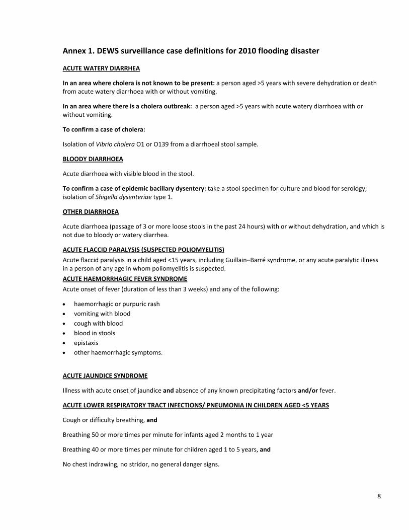
Annex 1. DEWS surveillance case definitions for 2010 flooding disaster
ACUTE WATERY DIARRHEA
In an area where cholera is not known to be present: a person aged >5 years with severe dehydration or death from acute watery diarrhoea with or without vomiting.
In an area where there is a cholera outbreak: a person aged >5 years with acute watery diarrhoea with or without vomiting.
To confirm a case of cholera:
Isolation of Vibrio cholera O1 or O139 from a diarrhoeal stool sample.
BLOODY DIARRHOEA
Acute diarrhoea with visible blood in the stool.
To confirm a case of epidemic bacillary dysentery: take a stool specimen for culture and blood for serology; isolation of Shigella dysenteriae type 1.
OTHER DIARRHOEA
Acute diarrhoea (passage of 3 or more loose stools in the past 24 hours) with or without dehydration, and which is not due to bloody or watery diarrhea.
ACUTE FLACCID PARALYSIS (SUSPECTED POLIOMYELITIS)
Acute flaccid paralysis in a child aged <15 years, including Guillain–Barré syndrome, or any acute paralytic illness in a person of any age in whom poliomyelitis is suspected.
ACUTE HAEMORRHAGIC FEVER SYNDROME
Acute onset of fever (duration of less than 3 weeks) and any of the following:
• haemorrhagic or purpuric rash • vomiting with blood • cough with blood • blood in stools • epistaxis • other haemorrhagic symptoms.
ACUTE JAUNDICE SYNDROME
Illness with acute onset of jaundice and absence of any known precipitating factors and/or fever.
ACUTE LOWER RESPIRATORY TRACT INFECTIONS/ PNEUMONIA IN CHILDREN AGED <5 YEARS
Cough or difficulty breathing, and
Breathing 50 or more times per minute for infants aged 2 months to 1 year
Breathing 40 or more times per minute for children aged 1 to 5 years, and
No chest indrawing, no stridor, no general danger signs.

9
Note: Severe pneumonia = cough or difficulty breathing + one or more of the following (inability to drink or breastfeed, severe vomiting, convulsions, lethargy or unconsciousness) or chest indrawing or stridor in a otherwise calm child
MALARIA
Person with current fever or history of fever within the past 48 hours (with or without other symptoms such as nausea, vomiting and diarrhoea, headache, back pain, chills, muscle pain) with positive laboratory test for malaria parasites (blood film (thick or thin smear) or rapid diagnostic test).
In children
Uncomplicated malaria
Fever AND no general danger signs such as lethargy or unconsciousness, convulsions, or inability to eat or drink. Where possible, confirm malaria with laboratory test.
Severe malaria
Fever AND general danger signs (lethargy or unconsciousness, convulsions, or inability to eat or drink).
MEASLES
Fever and maculopapular rash (i.e. non‐vesicular) and cough, coryza (i.e. runny nose) or conjunctivitis (i.e. red eyes) or Any person in whom a clinical health worker suspects measles infection.
To confirm a case of measles:
Presence of measles‐specific IgM antibodies.
MENINGITIS
Suspected case: Sudden onset of fever (>38.5 °C) with stiff neck.
In patients aged <12 months, a suspected case of meningitis occurs when fever is accompanied by a bulging fontanelle.
Probable case of bacterial meningitis: Suspected case of acute meningitis, as defined above, with turbid cerebrospinal fluid.
Probable case of meningococcal meningitis: Suspected case of meningitis, as defined above and Gram stain showing Gram‐negative diplococcus or ongoing epidemic or petechial or purpural rash.
Confirmed case of meningococcal meningitis: Suspected or probable case, as defined above, with either positive‐CSF antigen detection for Neisseria meningitidis or positive CSF culture or blood with identification of N. meningitidis.
UNEXPLAINED FEVER
Fever (body temperature >38.5 °C) for >48 hours and without other known etiology.
UNEXPLAINED CLUSTER OF HEALTH EVENTS
An aggregation of cases with similar symptoms and signs of unknown cause that are closely grouped in time and/or place.

10
Annex 2: Standard DEWS reporting forms: Provincial, District and Health Facility level ________________________________________________________________________
Pakistan Flood 2010
DEWS Daily Reporting Form
Provincial Reporting Form
Province:______________
Date: ___/___/2010 Total population under surveillance on the day of reporting: _________________
Total number of functioning reporting sites in the province
Number of sites reported
Total number of mobile medical teams/camps in the province
Number of mobile medical teams/camps reported
Disease Cases Deaths< 5 yrs > 5 yrs < 5 yrs >5 yrs
Acute Watery Diarrhea Bloody Diarrhea Other Diarrhea Acute Respiratory Infection Acute Flaccid Paralysis Suspected Measles Acute Jaundice Syndrome Suspected Malaria Suspected Meningitis Suspected Haemorrhagic fever
Unexplained Fever >38.5˚C Unexplained cluster of health events
Others (including skin diseases) Total

11
Pakistan Flood 2010
DEWS Daily Reporting Form
District Reporting Form
Province:______________ District: ___________________
Date: ___/___/2010 Total population under surveillance on the day of reporting: _________________
Total number of functioning reporting sites in the district
Number of sites reported
Total number of mobile medical teams/camps in the district
Number of mobile medical teams/camps reported
Disease Cases Deaths< 5 yrs > 5 yrs < 5 yrs >5 yrs
Acute Watery Diarrhea Bloody Diarrhea Other Diarrhea Acute Respiratory Infection Acute Flaccid Paralysis Suspected Measles Acute Jaundice Syndrome Suspected Malaria Suspected Meningitis Suspected Haemorrhagic fever
Unexplained Fever >38.5˚C Unexplained cluster of health events
Others (including skin diseases) Total

12
Pakistan Flood 2010
DEWS Daily Reporting Form
Health Facility Form
Province: _________________ District: ____________________ Date: ____/____/2010_________
Health facility name and Type (fix / Mobile) ______________________Location:_________________
Catchment Population: ____________________
Reported by: _____________________________ Contact Number: __________________________
Disease Cases Deaths< 5 yrs > 5 yrs < 5 yrs >5 yrs
Acute Watery Diarrhea Bloody Diarrhea Other Diarrhea Acute Respiratory Infection Acute Flaccid Paralysis Suspected Measles Acute Jaundice Syndrome Suspected Malaria Suspected Meningitis Suspected Haemorrhagic fever
Unexplained Fever >38.5˚C Unexplained cluster of health events
Others (including skin diseases) Total

13
Annex 3: Alert thresholds to trigger further investigation
Disease/Condition Alert Threshold Action suggested
Acute Watery Diarrhea One suspected case Reinforce appropriate case management; initiate investigation
Bloody Diarrhea Three or more cases in one location
Reinforce appropriate case management, including antibiotic usage; collect stool for culture and antimicrobial sensitivity; initiate investigation
Acute Respiratory Infection Twice the average number of cases seen in the previous three weeks for a given location
Reinforce appropriate case management; initiate investigation
Acute Flaccid Paralysis One suspected case Case investigation and specimen collection for laboratory diagnosis
Suspected Measles One case Immediate investigation and active case finding in coordination with the national immunization programme
Acute Jaundice Syndrome Three or more cases in one location
Initiate verification and investigation as required. Specimen collection for laboratory confirmation
Suspected Malaria Twice the mean number of cases seen in the previous three weeks for a given location
Active fever finding and specimen collection for laboratory confirmation
Suspected Meningitis One case Reinforce appropriate case management; initiate investigation
Acute Haemorrhagic Fever syndrome
One probable case Initiate verification and investigation as required
Unexplained Fever One death or two times the mean number of cases of the previous three weeks for a given location
Initiate investigation
Unknown diseases occurring in cluster
An aggregation of cases with related symptoms and signs of unknown cause that are closely grouped in time and/or place
Initiate verification and investigation as required.

14
Annex 4. Alert Investigation Form
District/Area: …………………………… Town/Village/Settlement/Camp: …………………………….
Health Facility: ……………………. Agency: ………………………………….
Date: ……/……/……….
Name of reporting officer: ………………………………………..
Suspected disease/syndrome:
(tick one box only)
Symptoms and signs:
(you can tick several boxes)
o Suspect cholera o Acute diarrhoea o Bloody diarrhoea o Acute Jaundice Syndrome o Suspected meningitis o Acute Respiratory Infection o Suspected measles o Unexplained fever o Suspected malaria o Acute Haemorrhagic Fever
Syndrome o Cluster of cases or deaths of
unknown origin o Acute flaccid paralysis /suspected
poliomyelitis (AFP) o Other
o Acute watery diarrhoea o Acute diarrhoea o Bloody diarrhoea o Fever o Rash o Other skin lesion o Cough o Vomiting o Jaundice o Neck stiffness o Convulsions/Seizures o Muscle weakness o Increased secretions (e.g. sweating, drooling) o Altered level of consciousness o Other (specify): _____________________________________
TOTAL NUMBER OF CASES REPORTED:
Line listing
Case No.
Age Address Sex (M/F)
Date of onset(dd/mm/YY)
Lab specimen taken¹
Treatment given(Yes/No)
Outcome² Final diagnosis
Epi‐linked?³(Yes/No)
¹Laboratory specimens: B=Blood, S=Stool, C=CSF, U=Urine, O = other ²Outcome: I = Currently ill, R= Recovering or recovered, D = died³ Known contact with previously identified case (list case no.)

15
Annex 5. Guidelines for collection of specimens in emergency conditions
A. BLOOD SPECIMEN COLLECTION
Blood and separated serum are the most common specimens taken in outbreaks of communicable disease. Venous blood can be used for isolation and identification of the pathogen in culture and by inoculation, or separated into serum for the detection of genetic material (e.g. by polymerase chain reaction), specific antibodies (by serology), antigens or toxins (e.g. by immunofluorescence). For the processing of most specimens for diagnosis of viral pathogens, serum is preferable to unseparated blood except where otherwise directed. When specific antibodies are being assayed, it is often helpful to collect paired sera, i.e. an acute sample at the onset of illness and a convalescent sample 1–4 weeks later. Whenever possible, blood specimens for culture should be taken before antibiotics are administered to the patient.
Venous blood samples
Materials for collection
• Skin disinfection: 70% alcohol (isopropanol, ethanol) or 10% povidone iodine, swabs, gauze pads,
adhesive dressings.
• Disposable latex or vinyl gloves.
• Tourniquet, Vacutainer or similar vacuum blood collection devices, or disposable syringes and needles.
• Vacutainer or sterile screw‐cap tubes (or cryotubes if indicated), blood culture bottles (50 ml for adults, 25 ml for children) with appropriate media.
• Labels and indelible marker pen.
Method of collection
Full infection control measures must be taken, with gowns, gloves, masks and boots for suspected viral haemorrhagic fever such as Lassa fever or Ebola.
• Place a tourniquet above the venepuncture site. Disinfect the tops of blood culture bottles.
• Palpate and locate the vein. The venepuncture site must be meticulously disinfected with 10% povidone iodine or 70% alcohol by swabbing the skin concentrically from the centre of the venepuncture site outwards. Let the disinfectant evaporate. Do not palpate the vein again. Perform venepuncture.
• If using conventional disposable syringes, withdraw 5–10 ml of whole blood from adults, 2–5 ml from children and 0.5–2 ml from infants. Using aseptic technique, transfer the specimen to the appropriate cap transport tubes and culture bottles. Secure caps tightly.

16
• If using a vacuum system, withdraw the desired amount of blood directly into each transport tube and culture bottle.
• Remove the tourniquet. Apply pressure to site until bleeding stops, and apply dressing.
• Label the tube, including the unique patient identification number, using indelible marker pen.
• Do not recap used sharps. Discard directly into the sharps disposal container.
• Complete the case investigation and the laboratory request forms using the same identification number.
Handling and transport
• Blood specimen bottles and tubes should be transported upright and secured in a screw‐cap container or in a rack in a transport box. They should have enough absorbent paper around them to soak up all the liquid in case of spill.
• For serum samples (e.g. measles, yellow fever, HIV), the blood cells must be separated from serum. Let the clot retract for 30 minutes then centrifuge at 2000 rpm for 10–20minutes and pour off serum. If no centrifuge is available, place sample in refrigerator overnight (4–6 hours) and pour off the serum for transport in a clean glass tube.
• Do not attempt this in case of suspected viral haemorrhagic fever unless you are a clinician/laboratory technician experienced in management of the disease. Full protection and infection control measures must be taken.
• If the specimen will reach the laboratory within 24 hours, most pathogens can be recovered from blood cultures transported at ambient temperature. Keep at 4–8 °C for longer transit periods, unless the bacterial pathogen is cold‐sensitive.
B. FAECAL SPECIMEN COLLECTION
Stool specimens are most useful for microbiological diagnosis if collected soon after onset of diarrhoea (for viruses <48 hours and for bacteria <4 days), and preferably before the initiation of antibiotic therapy. If required, two or three specimens may be collected on separate days. Stool is the preferred specimen for culture of bacterial, viral and parasitic diarrhoeal pathogens. Rectal swabs showing faeces may also be used from infants but are not useful for the diagnosis of viruses.
Materials for collection
• Tubes with Cary‐Blair transport medium
• Clean, dry, leak‐proof, screw‐cap container and tape if Cary‐Blair transport medium is not available.
• Appropriate bacterial transport media for transport of rectal swabs from infants (ideally Cary‐Blair).

17
• Parasitology transport pack: 10% formalin, polyvinyl isopropyl alcohol (PVA).
Method of collecting a stool specimen
If Cary‐Blair transport medium is available:
• Place sterile swab in freshly passed stool to allow it soak up stool.
• Place swab in the Cary‐Blair transport medium inside the tube.
• Break off the top part of the stick without touching the tube and tighten the screw cap firmly.
• Label the specimen tube.
If CARY‐BLAIR transport medium is not available, collect freshly passed stool, 5 ml liquid or 5 g solid (peasize), in a container. Label the container.
Method of collecting a rectal swab from infants
• Moisten a swab in sterile saline.
• Insert the swab tip just past the anal sphincter and rotate gently.
• Withdraw the swab and examine to ensure that the cotton tip is stained with faeces.
• Place the swab in sterile tube/container containing the appropriate transport medium.
• Break off the top part of the stick without touching the tube and tighten the screw cap firmly.
• Label the specimen tube.
Handling and transport
• Stool specimens should be transported in a cold‐box at 4–8 °C. Bacterial yields may fall significantly if specimens are not processed within 1–2 days of collection. Shigella is particularly sensitive to elevated temperatures. If transport medium is not available, do not allow specimen to dry – add few drops of 0.85% sodium chloride solution.
• Specimens to be examined for parasites should be mixed with 10% formalin or PVA, 3 parts stool to 1 part preservative. Transported at ambient temperature in containers sealed in plastic bags.
C. RESPIRATORY TRACT SPECIMEN COLLECTION
Specimens are collected from the upper or lower respiratory tract, depending on the site of infection. Upper respiratory tract pathogens (viral and bacterial) are found in throat and nasopharyngeal specimens. Lower respiratory tract pathogens are found in sputum specimens. For organisms such as Legionella, culture is difficult, and diagnosis is best based on the detection of antigen excreted in the urine. When acute epiglottitis is suspected, no attempt should be made to take throat or pharyngeal specimens since these procedures may

18
precipitate respiratory obstruction. Epiglottitis is generally confirmed by lateral neck Xray, but the etiological agent may be isolated on blood culture.
Materials for collection
• Transport media – bacterial (Trans Amies) and viral (Cellmatics)
• Dacron and cotton swabs
• Tongue depressor
• Flexible wire calcium alginate‐tipped swab (for suspected pertussis)
• Nasal speculum (for suspected pertussis – not essential)
• Suction apparatus or 20–50‐ml syringe
• Sterile screw‐cap tubes, and wide‐mouthed clean sterile jars (minimum volume 25 ml).
Upper respiratory tract specimens
Method of collecting a throat swab
• Hold the tongue down with the depressor. Use a strong light source to locate areas of inflammation and exudate in the posterior pharynx and the tonsillar region of the throat behind the uvula.
• Rub the area back and forth with a Dacron or calcium alginate swab. Withdraw the swab without touching cheeks, teeth or gums and insert into a screw‐cap tube containing transport medium.
• Break off the top part of the stick without touching the tube and tighten the screw cap firmly.
• Label the specimen containers.
• Complete the laboratory request form.
Method of collecting nasopharyngeal swabs (for suspected pertussis)
• Seat the patient comfortably, tilt the head back and insert the nasal speculum.
• Insert a flexible calcium alginate/Dacron swab through the speculum parallel to the floor of nose without pointing upwards. Alternatively, bend the wire and insert it into the throat and move the swab upwards into the nasopharyngeal space.
• Rotate the swab on the nasopharyngeal membrane a few times, remove it carefully and insert it into a screw‐cap tube containing transport medium.
• Break off the top part of the stick without touching the tube and tighten the screw cap firmly.
• Label the specimen tube, indicating left or right side.
• Complete the laboratory request form.
• Repeat on the other side.
Lower respiratory tract specimens

19
Method of collecting sputum
• Instruct patient to take a deep breath and cough up sputum directly into a wide‐mouthed sterile container. Avoid saliva or postnasal discharge. Minimum volume should be about 1 ml.
• Label the specimen container.
• Complete the laboratory request form.
Handling and transport
• All respiratory specimens except sputum are transported in appropriate bacterial/viral media.
• Transport as quickly as possible to the laboratory to reduce overgrowth by commensal oral flora.
• For transit periods up to 24 hours, transport bacterial specimens at ambient temperature and viruses at 4–8 °C in appropriate media.
D. URINE SPECIMEN COLLECTION
Material for collection
• Sterile plastic cup with lid (50 ml or more).
• Clean, screw‐top specimen transport containers ("universal" containers are often used).
• Gauze pads.
• Soap and clean water (or normal saline) if possible.
• Labels and indelible marker pen.
Method of collection
• Give the patient clear instructions to pass urine for a few seconds, and then to hold the cup in the urine stream for a few seconds to catch a mid‐stream urine sample. This should decrease the risk of
contamination from organisms living in the urethra.
• To decrease the risk of contamination from skin organisms, the patient should be directed to avoid touching the inside or rim of the plastic cup with the skin of the hands, legs or external genitalia. Tighten the cap firmly when finished.
• For hospitalized or debilitated patients, it may be necessary to wash the external genitalia with soapy water to reduce the risk of contamination. If soap and clean water are not available, the area may be rinsed with normal saline. Dry the area thoroughly with gauze pads before collecting the urine.

20
• Urine collection bags may be necessary for infants. If used, transfer urine from the urine bag to specimen containers as soon as possible to prevent contamination with skin bacteria. Use a disposable transfer pipette to transfer the urine.
• Label the specimen containers.
Handling and transport • Transport to the laboratory within 2–3 hours of collection. If this is not possible, do not freeze but keep the specimen refrigerated at 4–8 °C. Keeping the specimen refrigerated will reduce the risk of overgrowth of contaminating organisms.
• Ensure that transport containers are leak‐proof and tightly sealed.