osteomyelitis & septic arthritis - ksumsc.com. musculoskeletal block/team...objectives...
TRANSCRIPT
Osteomyelitis & Septic Arthritis
Black: original contentRed: important Green: ALRIKABI’s notesGrey: Explanation Blue: Only in the boys slidesPink: only in the girls slide
Objectives
● Understand the etiology, pathogenesis and clinical features
of osteomyelitis.
● Be familiar with some of the terminology used in bone
infections like: sequestrum, involucrum, Brodie abscess and
Pott’s disease.
● Understand the clinicopathological features of tuberculous
osteomyelitis.
● Identify the bacteria commonly involved in septic arthritis,
the clinicopathological features and the characteristics of the
joint fluid.
Osteomyelitis
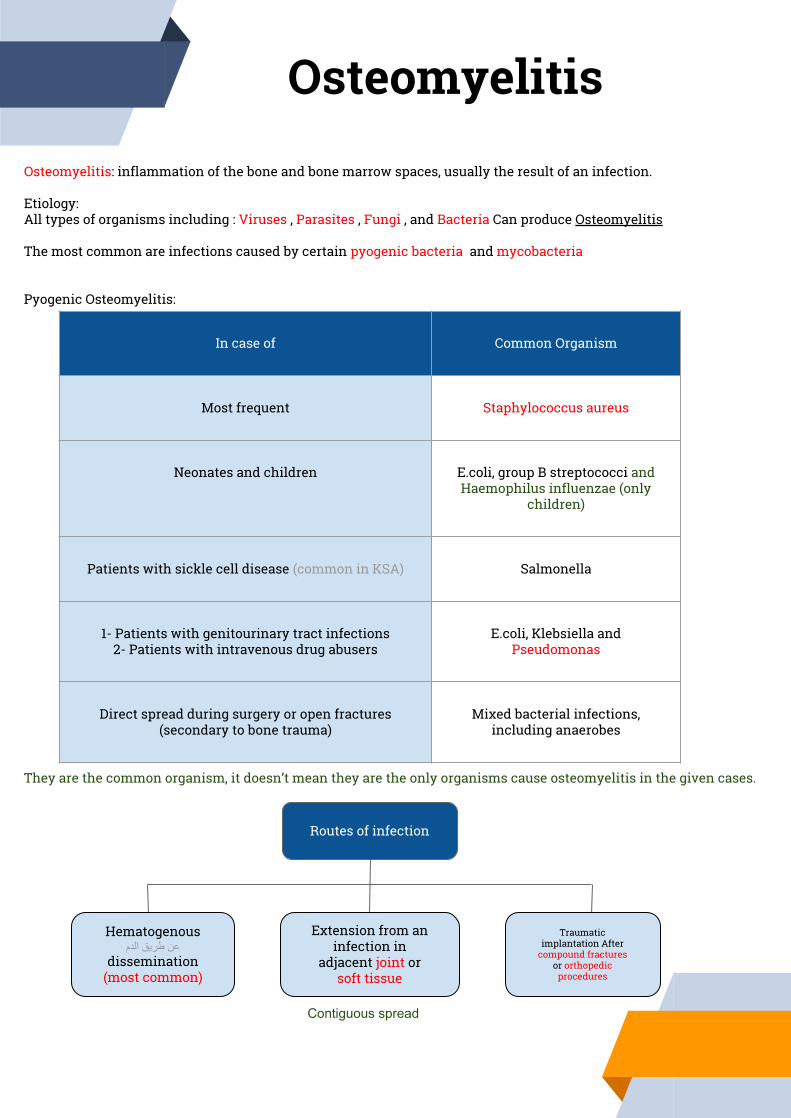
Osteomyelitis: inflammation of the bone and bone marrow spaces, usually the result of an infection.
Etiology:All types of organisms including : Viruses , Parasites , Fungi , and Bacteria Can produce Osteomyelitis
The most common are infections caused by certain pyogenic bacteria and mycobacteria
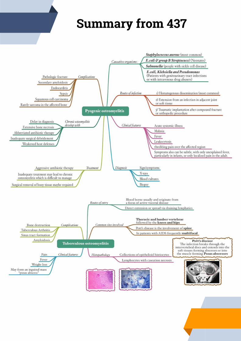
Pyogenic Osteomyelitis:
They are the common organism, it doesn’t mean they are the only organisms cause osteomyelitis in the given cases.
In case of Common Organism
Most frequent Staphylococcus aureus
Neonates and children E.coli, group B streptococci and Haemophilus influenzae (only
children)
Patients with sickle cell disease (common in KSA) Salmonella
1- Patients with genitourinary tract infections2- Patients with intravenous drug abusers
E.coli, Klebsiella and Pseudomonas
Direct spread during surgery or open fractures (secondary to bone trauma)
Mixed bacterial infections, including anaerobes
Routes of infection
Hematogenous عن طریق الدم
dissemination (most common)
Extension from an infection in
adjacent joint or soft tissue
Traumatic implantation After
compound fractures or orthopedic
procedures
Contiguous spread
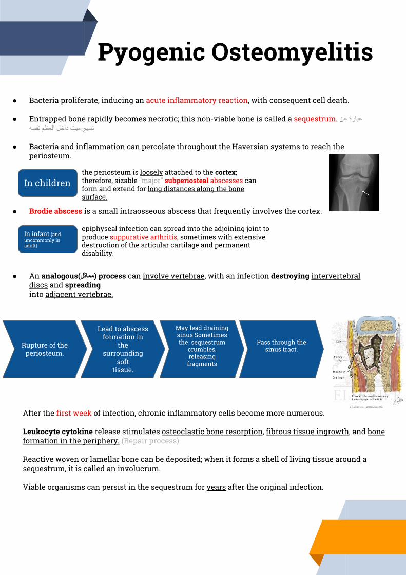
the periosteum is loosely attached to the cortex; therefore, sizable “major” subperiosteal abscesses can form and extend for long distances along the bone surface.
Pyogenic Osteomyelitis
● Bacteria proliferate, inducing an acute inflammatory reaction, with consequent cell death.
● Entrapped bone rapidly becomes necrotic; this non-viable bone is called a sequestrum. عبارة عن نسیج میت داخل العظم نفسھ
● Bacteria and inflammation can percolate throughout the Haversian systems to reach the periosteum.
● Brodie abscess is a small intraosseous abscess that frequently involves the cortex.
● An analogous(مماثل) process can involve vertebrae, with an infection destroying intervertebral discs and spreadinginto adjacent vertebrae.
In children
In infant (and uncommonly in adult)
epiphyseal infection can spread into the adjoining joint to produce suppurative arthritis, sometimes with extensive destruction of the articular cartilage and permanent disability.
Rupture of the periosteum.
Lead to abscess formation in
the surrounding
softtissue.
May lead draining sinus Sometimes the sequestrum
crumbles, releasing fragments
Pass through the sinus tract.
After the first week of infection, chronic inflammatory cells become more numerous.
Leukocyte cytokine release stimulates osteoclastic bone resorption, fibrous tissue ingrowth, and bone formation in the periphery. (Repair process)
Reactive woven or lamellar bone can be deposited; when it forms a shell of living tissue around a sequestrum, it is called an involucrum.
Viable organisms can persist in the sequestrum for years after the original infection.
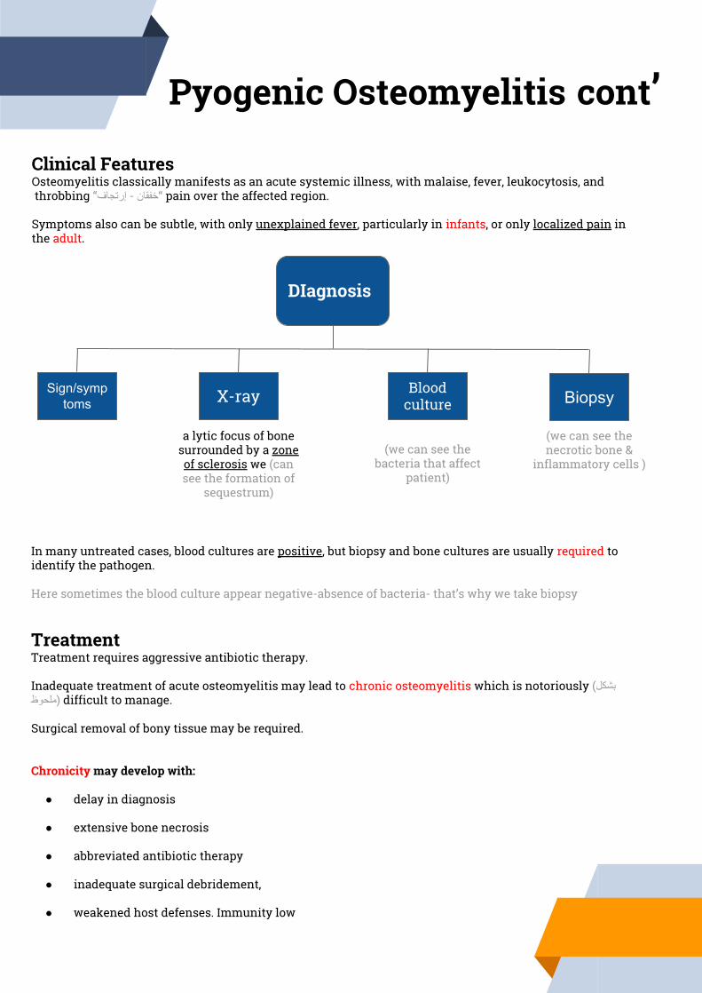
Pyogenic Osteomyelitis cont’Clinical FeaturesOsteomyelitis classically manifests as an acute systemic illness, with malaise, fever, leukocytosis, and throbbing “خفقان - إرتجاف“ pain over the affected region.
Symptoms also can be subtle, with only unexplained fever, particularly in infants, or only localized pain in the adult.
DIagnosis
Sign/symptoms X-ray Blood
culture Biopsy
a lytic focus of bone surrounded by a zone of sclerosis we (can see the formation of
sequestrum)
(we can see the bacteria that affect
patient)
In many untreated cases, blood cultures are positive, but biopsy and bone cultures are usually required to identify the pathogen.
Here sometimes the blood culture appear negative-absence of bacteria- that’s why we take biopsy
TreatmentTreatment requires aggressive antibiotic therapy.
Inadequate treatment of acute osteomyelitis may lead to chronic osteomyelitis which is notoriously (بشكل .difficult to manage (ملحوظ
Surgical removal of bony tissue may be required.
Chronicity may develop with:
● delay in diagnosis
● extensive bone necrosis
● abbreviated antibiotic therapy
● inadequate surgical debridement,
● weakened host defenses. Immunity low
(we can see the necrotic bone &
inflammatory cells )

Complications
*Any chronic inflammation in human body make amyloidosis after 15-20 years, it is protein secreted by the liver and accumulate in certain organs *kidney most important organ* the patient became with nephrosis secondary to Amyloidosis secondary to chronic osteomyelitis (can be in any other chronic diseases).
Tuberculous osteomyelitisCaused by hematogenous spread of mycobacterium tuberculosis from somewhere else (usually the lungs).It occurs when concentration of TB is high somewhere else. TB is microaerophilic: reacquired oxygen, that's why it go to the lung, it spread via the blood to single or multiple organs. It can affect the: lymph node, liver, spleen, bone, and joint.TB is acid fast bacilli.
The most common sites of skeletal involvement are: vertebral column (thoracic and lumbar vertebrae) followed by the knees and hips. And long bones.It go to body of the vertebrae and inters intervertebral disk, it can affect other vertebrae, sometimes it cause cold abscess (psoas abscess) and go down to inguinal (Groin), with bone lytic. inguinal (Groin) : is fold between the femur and pelvic.
In patients with AIDS frequently multifocal. When there is an associated osteomyelitis, vertebral collapse may result (pott’s disease of the spine).TB of spine is pott's disease, distraction and lytic lesion of vertebrae which extend to intervertebral disk and form psoas abscess: cold abscess not always because it don’t have acute symptoms *and present as growing mass *.
Clinical features: not acute
Complications:1- Bone destruction. 2- Sinus tract formation3- Amyloidosis 4- Tuberculous arthritis (if it extends to joint).
TB is tough bacteria because it has lipoprotein that resist to heat and dryness.Hypersensitivity type4 is the immune response that responsible for TB.At least we need 6 months (sometime go for 2 years) to heal from TB Sometimes we mix between TB and tumor because they have the same symptoms. To diagnose TB We take biopsy searching for granuloma.
EndocarditisReach to heart.
SepsisInfection in blood.
Formation of sinus and fistula.
Spread of infection.
Pathologic fractureBecause the bone is
weak.
Secondary amyloidosis*In any chronic.
Bone sarcomas it is rare.
Sinus when became chronic will cause
squamous cell carcinoma of the skin it
is rare.
Pain Mild fever may occur or may not.
Weight loss (always with TB)
Usually mild sweat.
Chills may occur or may not.
Mild Malaise.
May form an inguinal mass “psoas abscess”
Cold abscess do not produce pain or redness.
Pyogenic Osteomyelitis cont’

The infection breaks through the intervertebral discs and extends into the soft tissues forming abscesses.In Pott’s disease, the infection may breaks through the intervertebral discs and extends into the muscle forming Psoas abscesses. In psoas muscle
Cold abscesses
HistopathologyFormation of granuloma (collections of epithelioid histiocytes A, multinucleated giant cells (Langhans cells) B and lymphocytes C with caseation necrosis D )
CB
BD
A
Ziehl Neelsen stainSpecial stain used to identify the acid fast bacilli. Ziehl Neelsen may show a -ve result when there is small number of the pathogen in this case culture is needed even if it is -ve that does not exclude TB.
When TB enters the body it get phagocytosis by neutrophils and macrophages. it can live and multiply inside macrophages after that it rupture macrophages and go out and get stimulate by macrophages lymphokines specifically IL12, then stimulate Tcells(CD4), then activate TH1, then secrete IFNγ, that lead to transformation of more monocytes and macrophages to epithelioid cells, after that it surround the bacteria and form granuloma.
Tuberculous Osteomyelitis cont’

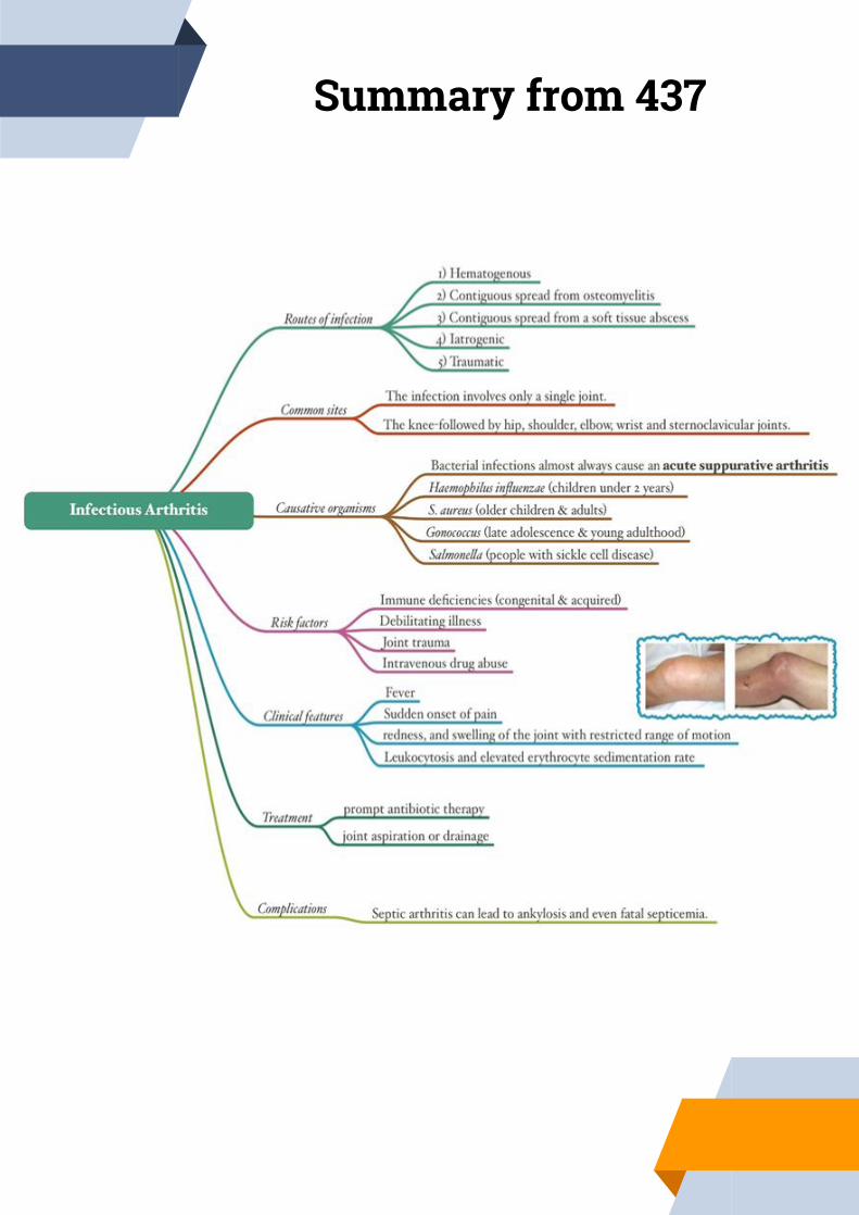
In children osteomyelitis can reach epiphyseal line and go to joint causing septic arthritis (it is common in children). In adult is little because the small vascularity in epiphyses.Septic arthritis is serious because it can cause rapid joint destruction and permanent deformities.Articular structures can also become infected by direct inoculation or from:contiguous spread from a soft-tissue abscess or focus of osteomyelitis.The infection involves only a single joint, usually the knee-followed in order by hip, shoulder, elbow, wrist, and sternoclavicular joints. Joint aspiration is typically purulent joint fluid has pus. Culture allows identification of the causal agent.Bacterial infections almost always cause an acute suppurative arthritis.
Any bacteria can be causal:
Haemophilus influenzae
Staphylococcus aureus
Neisseria gonorrhoeae (gonococcus)
Salmonella
predominates in children under age 2
years.
is the main causative agent in older children and
adults.
prevalent during late adolescence
and young adulthood.
Individuals with sickle cell disease
are prone to infection at any age.
Septic (infectious) Arthritis
Routes of infection
Direct inoculation: by trauma to the joint
where organisms go directly to the joint.
Contagious spread: it moves from the
bone (osteomyelitis) to
the joint.
Hematogenous spread: by the
blood.
Contiguous spread: from a soft tissue
abscess
Iatrogenic غلط من الدكتور أو الجراح
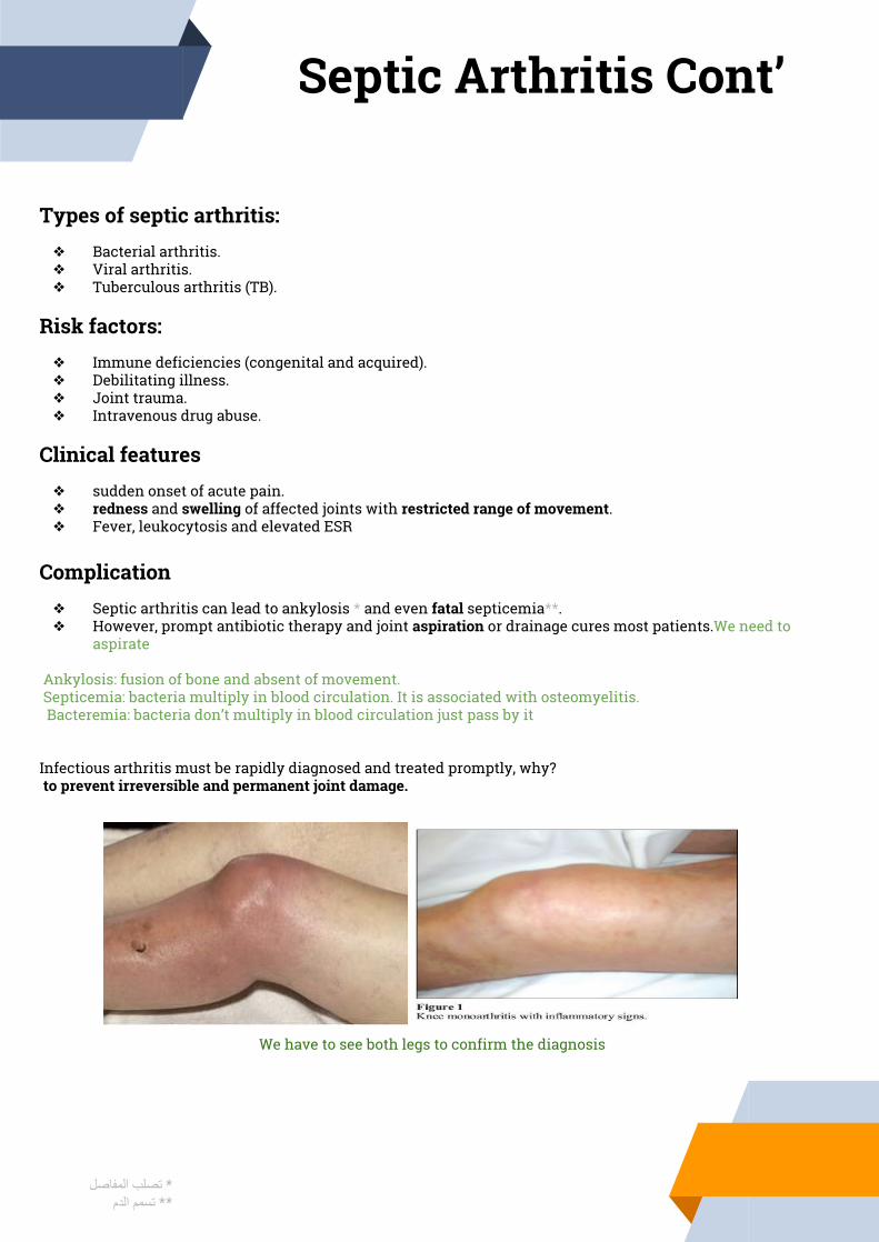
Types of septic arthritis: ❖ Bacterial arthritis.❖ Viral arthritis. ❖ Tuberculous arthritis (TB).
Risk factors: ❖ Immune deficiencies (congenital and acquired).❖ Debilitating illness.❖ Joint trauma. ❖ Intravenous drug abuse.
Clinical features❖ sudden onset of acute pain.❖ redness and swelling of affected joints with restricted range of movement. ❖ Fever, leukocytosis and elevated ESR
Complication❖ Septic arthritis can lead to ankylosis * and even fatal septicemia**. ❖ However, prompt antibiotic therapy and joint aspiration or drainage cures most patients.We need to
aspirate
Ankylosis: fusion of bone and absent of movement. Septicemia: bacteria multiply in blood circulation. It is associated with osteomyelitis. Bacteremia: bacteria don’t multiply in blood circulation just pass by it
Infectious arthritis must be rapidly diagnosed and treated promptly, why? to prevent irreversible and permanent joint damage.
* تصلب المفاصل** تسمم الدم
We have to see both legs to confirm the diagnosis
Septic Arthritis Cont’
Dr. Alrikabi’s notes
Osteomyelitis is infection of the bone and bone marrow spaces in a certain bone. this disease is quite common, seen in children, neonate and adult. There is no predilection(میل) for sex (it affects both sexes almost equally).
We defined it as an infection because the most of the time osteomyelitis caused by an organism, and the most organism causes osteomyelitis is bacteria, there are no absolute roles (each bacteria can cause osteomyelitis), but there is a distribution of the bacteria according to the ages and the medical condition.
Patient with diabetes can have osteomyelitis of the metatarsal bone and develop an inflammatory sinus discharging pus, they can have any organism and they can also have anaerobic bacteria and fungal specially in there foot, they develop osteomyelitis in there foot more than others. they have very weak phagocytosis and they are prone to infection especially in the lower limbs and they also prone to develop ischemia and secondary fungal and bacterial infection.
Other patients have osteomyelitis affect the metaphyseal part of the long bone (the distal end of the femur, proximal end of the tibia and the distal end of humerus).
The predisposing factors of osteomyelitis (usually):
● inflammatory focus somewhere in the body and infection (infective endocarditi)..● patient with immunocompromised (AIDs, undergoing treatment for cancer or diabetes).● compound fracture.● sickle cell anemia.● congenital immunodeficiency.● in war who injured by shrapnel (شظایا).● orthopedic surgical procedure, especially if there are prostheses(أطراف صناعیة)
The most common type of osteomyelitis the one which occur following compound fracture and orthopedic operation
Osteomyelitis usually occur in long bones, rarely occur in scapula and other bones.
People with chronic osteomyelitis may not present with fever but they present with sinus discharge pus.
In radiology it’s difficult to differentiate between osteomyelitis and tumors because both lysis the bone and we have take biopsy.
Children who have otitis (التھاب األذن) due to infection, the organism may spread to the temporal bone as an example of
contiguous root. Other example by compound fracture.
The pathogenesis of osteomyelitis:
● When the bacteria reaches the bone by one of the mentioned roots it usually goes from the periosteum to the Haversian canal (contain blood vessels)
● spread through vessel and accumulate in the metaphyseal part of the long bone, it can spread to the metaphysis and even may spread to the epiphysis, But in classical case it stay in the metaphyseal.
● Then it start proliferating and necrotizing the bone which cause acute and chronic inflammation. ● Lead to activate coagulation cascade which form septic thrombi (contain pyogenic bacteria).● lead to ischemia, as a result there will be a dead bone trabeculae (spicules), lacunae without osteocytes full of
sequestrum

Sequestrum (dead bone) is surrounded by reactive new bone formation, the newly formed bone name as involucrum.
Inflammatory start eroding the bone and reach the periosteum and cause subperiosteal abscess (pus under
periosteum) and do pressure to the bone and capillaries causing ischemia and cause formation of new sequestrum,
after that it became chronic.
Ischemia can cause by: pressure to capillaries, coagulation cascade and septic thrombi.
Subperiosteal Micro abscess after time they rapture and go the surrounding tissue and muscles and open through
the skin and form sinus ( inflammatory tract laid by inflammatory vascular granulation tissue and usually it has one
opening to another tissue or organ) sometime it do fistula.
Complication of osteomyelitis:
● Amyloidosis: any chronic inflammation in human body make amyloidosis after 15-20 years, it is protein secreted by the liver and accumulate in certain organs *kidney most important organ* the patient became with nephrosis secondary to Amyloidosis secondary to chronic osteomyelitis (can be in any other chronic diseases).
● Pathological fracture.● Sinus when became chronic will cause squamous cell carcinoma of the skin it is rare.● Bone sarcomas it is rare.
Ankylosis: fusion of bone and absent of movement.In children osteomyelitis can reach epiphyseal line and go to joint causing septic arthritis (it is common in children). In adult is little because the small vascularity in epiphyses.
Tuberculous osteomyelitis:
It is rare in Europe and USA but we still see it in Saudi arabia. Symptoms is not acute :
● Mild fever may occur or may not.● Cold abscess do not produce pain or redness.● Mild Malaise.● Chills may occur or may not.● Usually mild sweet.
Dr. Alrikabi’s notes (Cont’)
It occurs when concentration of TB is high somewhere else.
When TB enters the body it get phagocytosis by neutrophils and macrophages. it can live and multiply inside
macrophages after that it rupture macrophages and go out and get stimulate by macrophages lymphokinse
specifically IL12, then stimulate Tcells(CD4), then activate TH1, then secrete IFNγ, that lead to transformation of
more monocytes and macrophages to epithelioid cells, after that it surround the bacteria and form granuloma.
TB is tough bacteria because it has lipoprotein that resist to heat and dryness.
TB is acid fast bacilli.
It's microaerophilic: reacquired oxygen, that's why it go to the lung, it spread via the blood to single or multiple
organs. It can affect the: lymph node, liver, spleen, bone, and joint.
Sometimes we mix between TB and tumor because they have the same symptoms, to diagnose TB We take biopsy
searching for granuloma.
It go to body of the vertebrae and inters intervertebral disk, it can affect other vertebrae, sometimes it cause cold
abscess (psoas abscess) and go down to inguinal (Groin), with bone lytic.
inguinal (Groin) : is fold between the femur and pelvic.
Hypersensitivity type4 is the immune response that responsible for TB.
At least we need 6 months (sometime go for 2 years) to heal from TB
TB of spine is pott's disease, distraction and lytic lesion of vertebrae which extend to intervertebral disk and form
psoas abscess: cold abscess not always because it don’t have acute symptoms *and present as growing mass *.
Bacteremia: bacteria don’t multiply in blood circulation just pass by it.
septicemia: bacteria multiply in blood circulation. It is associated with osteomyelitis.
Dr. Alrikabi’s notes (Cont’)
Summary from 437
Summary from 437

Quiz
1- The main cause of OSTEOMYELITIS in adult is:A. S.aureus. B. Anthrax. C. Clostridium. D. Salmonella.
2- OSTEOMYELITIS is an inflammatory disease which occur in: A. BoneB. Bone marrow spaceC. MusclesD. Both A&B
3- Dead pieces of bone known as: A. Involucrum.B. Sequestrum. C. Fistula. D. All answers are correct
4- When TB affected the Spine that called: A. Osteomyelitis. B. Sickle cell anaemia. C. Diabetes mellitus. D. Pott’s disease.
5- People with Sickle cell anemia may develop Arthritis by unusual bacteria like :A. Streptococci aureusB. Salmonella C. E.coli D. T.B
6- osteomyelitis Etiology is:A.traumaB. genetic disorderC.All types of microorganisms, including bacteriaD. unknown etiology
1- A | 2- D | 3- B | 4- D | 5- B | 6- C

Thank you
Team Leaders ● Mohannad Ahmad● Raghad AlKhashan
Team members ● Leena Alnassar● Reema Alserhani● Taibah Alzaid● Lama Alzamil● Alhanouf Alhaluli● Sarah AlArif● Amirah Alzahrani ● Njoud AlAli ● Ghaida Alshehri ● Deana Awartani
● Naif Alsulais● Ibrahim Alshaqrawi● Alwaleed Alsaleh● Suhail Basuhail● Muhannad Makkawi● Tariq Aloqail
This lecture was done by
Special thanks to:Omar Alomar434 Pathology team437 Pathology team