osaka university knowledge archive : ouka ·...
TRANSCRIPT

TitleActivin A Binds to Perlecan through Its Pro-region That Has Heparin/Heparan Sulfate BindingActivity
Author(s)
Li, Shaoliang; Shimono, Chisei; Norioka, Naoko;Nakano, Itsuko; Okubo, Tetsuo; Yagi, Yoshiko;Hayashi, Maria; Sato, Yuya; Fujisaki, Hitomi;Hattori, Shunji; Sugiura, Nobuo; Kimata, Koji;Sekiguchi, Kiyotoshi
Citation Journal of Biological Chemistry. 285(47)P.36645-P.36655
Issue Date 2010-11
Text Version publisher
URL http://hdl.handle.net/11094/71421
DOI 10.1074/jbc.M110.177865
rights
Note
Osaka University Knowledge Archive : OUKAOsaka University Knowledge Archive : OUKA
https://ir.library.osaka-u.ac.jp/repo/ouka/all/
Osaka University

Activin A Binds to Perlecan through Its Pro-region That HasHeparin/Heparan Sulfate Binding Activity*
Received for publication, August 23, 2010 Published, JBC Papers in Press, September 15, 2010, DOI 10.1074/jbc.M110.177865
Shaoliang Li‡, Chisei Shimono‡, Naoko Norioka‡, Itsuko Nakano‡, Tetsuo Okubo‡, Yoshiko Yagi‡, Maria Hayashi‡,Yuya Sato‡, Hitomi Fujisaki§, Shunji Hattori§, Nobuo Sugiura¶, Koji Kimata¶, and Kiyotoshi Sekiguchi‡1
From the ‡Laboratory of Extracellular Matrix Biochemistry, Institute for Protein Research, Osaka University, 3-2 Yamadaoka, Suita,Osaka 565-0871, Japan, the §Nippi Research Institute of Biomatrix, 520-11 Kuwabara, Toride, Ibaraki 302-0017, Japan, and the¶Institute for Molecular Science of Medicine, Aichi Medical University, Nagakute, Aichi 480-1195, Japan
Activin A, a member of the transforming growth factor-�family, plays important roles in hormonal homeostasis andembryogenesis. In this study, we produced recombinant humanactivin A and examined its abilities to bind to extracellularmatrix proteins. Recombinant activinA expressed in 293-F cellswas purified as complexes of mature dimeric activin A with itspro-region. Among a panel of extracellular matrix proteinstested, recombinant activin A bound to perlecan and agrin, butnot to laminins, nidogens, collagens I and IV, fibronectin, andnephronectin. The binding of recombinant activinA to perlecanwas inhibited by heparin and high concentrations of NaCl andabolished by heparitinase treatment of perlecan, suggesting thatactivin A binds to the heparan sulfate chains of perlecan. Insupport of this possibility, recombinant activin Awas capable ofdirectly binding to heparin and heparan sulfate chains. Site-di-rectedmutagenesis of recombinant activin A revealed that clus-ters of basic amino acid residues, Lys259-Lys263 and Lys270-Lys272, in the pro-region were required for binding to perlecan.Interestingly, deletion of the peptide segment Lys259-Gly277
containing both basic amino acid clusters from the pro-regiondidnot impair the activity of activinA to stimulate Smad-depen-dent gene expressions, although it completely ablated the perle-can-binding activity. The binding of activin A to basementmembrane heparan sulfate proteoglycans through the basic res-idues in the pro-region was further confirmed by in situ activinA overlay assays using frozen tissue sections. Taken together,the present results indicate that activin A binds to heparan sul-fate proteoglycans through its pro-region and thereby regulatesits localization within tissues.
Extracellular matrix (ECM)2 is a supramolecular assemblycomposed of proteins and glycosaminoglycans (GAGs) that isdeposited outside cells. It serves as a scaffold for cell adhesion,and regulates cell survival, proliferation, migration, and geneexpression through interactions with cell surface receptorsincluding integrins and membrane-anchored proteoglycans
(1). The ECMalso binds to a variety of growth factors includingfibroblast growth factor (FGF), hepatocyte growth factor(HGF), and vascular endothelial growth factor (VEGF) andthereby modulates the availability and biological activities ofthese growth factors (2). The binding of growth factors toECM components, particularly heparan sulfate proteoglycans(HSPGs), is believed to protect them against proteolytic degra-dation, concentrate their activity in the vicinity of cells, andallow their rapid release during wound healing and tissueremodeling (2). The activities of such growth factors bound toHSPGs are considered to be regulated by heparanase, whichcleaves heparan sulfate chains and consequently releases thegrowth factors from the ECM (3).The interactions between growth factors and ECM compo-
nents can be categorized into protein-GAG interactions andprotein-protein interactions. For example, growth factorsincluding FGF, HGF, and VEGF bind to HSPGs in the ECM viaheparan sulfate chains (4–6) while transforming growth factor(TGF)-�1 interacts with fibronectin (7) and thrombospondin(8). Similarly, thrombospondin, fibronectin and collagen arebinding partners of HGF in the ECM (9, 10). Type IV collagenbinds to bone morphogenetic protein (BMP) and regulates itssignaling events in Drosophila (11). The TGF-� family mem-bers play central roles in the regulation of multiple physiologi-cal processes such as cell differentiation, mitogenesis, embryo-genesis, apoptosis, and inflammation (12). Many, if not all,TGF-� familymembers are produced as latent complexes of themature dimeric growth factor and its pro-region (13). The pro-region of TGF-�1, which is known to be a latency-associatedpeptide, forms two disulfide bonds with latent TGF-�-bindingproteins (LTBPs), thus targeting the latent TGF-�1 complexesto the ECM. The resulting latent TGF-�-LTBP complexes bindto fibrillins and fibronectins, and consequently facilitate theECM targeting of TGF-�1. Fibrillin-1 has also been shown tobind to the pro-regions of BMP-2, -4, -7, and -10 at its N-ter-minal region (14), thereby sequestering a panel of TGF-� familyproteins in the ECM. These observations indicate the impor-tance of the pro-regions of TGF-� family proteins for theirECM deposition.Activin A, a member of the TGF-� family, is a disulfide-
linked homodimer of inhibin �A. The inhibin �A-subunit issynthesized as a precursor and processed by a furin protease togive an N-terminal pro-region and a C-terminal mature �Aregion (15). Activin A participates not only in the release offollicle-stimulating hormone from anterior pituitary cells (16)
* This study was supported by Research Contract No. 06001294-0 with theNew Energy and Industrial Technology Development Organization ofJapan.
1 To whom correspondence should be addressed. Tel.: 81-6-6879-8617; Fax:81-6-6879-8619; E-mail: [email protected].
2 The abbreviations used are: ECM, extracellular matrix; GAG, glycosamino-glycan; HGF, hepatocyte growth factor; LTBP, latent TGF-�-binding pro-tein; BMP, bone morphogenetic protein; HSPG, heparan sulfate proteogly-can; mAb, monoclonal antibody; NTA, nitrilotriacetic acid.
THE JOURNAL OF BIOLOGICAL CHEMISTRY VOL. 285, NO. 47, pp. 36645–36655, November 19, 2010© 2010 by The American Society for Biochemistry and Molecular Biology, Inc. Printed in the U.S.A.
NOVEMBER 19, 2010 • VOLUME 285 • NUMBER 47 JOURNAL OF BIOLOGICAL CHEMISTRY 36645
at OSA
KA
UN
IVE
RSIT
Y on M
arch 3, 2019http://w
ww
.jbc.org/D
ownloaded from
but also in the regulation of development, mitogenesis, apopto-sis, and wound repair (17–20). Activin A has also been used tomaintain the pluripotency of embryonic stem cells (21) andinduce their differentiation into defined cell lineages (22). Byanalogy to TGF-�1 and some BMPs, the biological activity ofactivin A may also be modulated by the ECM, although theinteractions of activin A with ECM components remain unex-plored. In Xenopus, activin has been shown to act as a long-range dorsalizing signal to establish a concentration gradient(23), suggesting the potential associations of Xenopus activinwith ECM components. In this study, we examined the inter-actions of activin A with a panel of ECM proteins as a first steptoward addressing the roles of the ECM in the regulation ofactivin A function and tissue distribution. We found thatrecombinant activin A produced in human 293-F cells specifi-cally bound to perlecan and agrin through its pro-region thatwas capable of binding to heparin/heparan sulfate chains. Wemapped the regions responsible for the binding to perlecan toclusters of basic amino acid residues, Lys259-Lys263 and Lys270-Lys272, in the pro-region, although these residues were dispen-sable for the Smad-dependent signaling activity of activin A.
EXPERIMENTAL PROCEDURES
Chemical Reagents and Antibodies—KOD plus DNA poly-merase was purchased from Toyobo (Osaka, Japan). Human fol-listatin was purchased from BioVision (Mountain View, CA).Bovine activin A was obtained from Wako (Osaka, Japan).Monoclonal antibodies (mAbs) against activin A (MAB3381)and human follistatin (MAB669) were obtained fromR&D Sys-tems (Minneapolis, MN). A mouse anti-perlecan mAb (7B5)was obtained from Zymed Laboratories Inc. (South San Fran-cisco, CA). A horseradish peroxidase (HRP)-conjugated anti-pentaHis mAb and nickel-nitrilotriacetic (Ni-NTA) agarosewere purchased from Qiagen (Piscataway, NJ). Heparin andhyaluronidase were obtained fromSigma. Chondroitinase ABC(Proteus vulgaris), heparitinase (Flavobacterium heparinum),and mAbs against heparan sulfate (3G10 and 10E4) wereobtained from Seikagaku Kogyo (Tokyo, Japan). Heparin-Sepharose CL-6B dry resin was purchased fromGEHealthcare(Piscataway, NJ).ECM Proteins—Recombinant human laminins, agrin, nido-
gen 1, nidogen 2, and nephronectin were produced using aFreeStyleTM 293 Expression System (Invitrogen, Carlsbad, CA)and purified from conditioned medium by immunoaffinitychromatography or Ni-NTA agarose affinity chromatography.Perlecan was purified from conditionedmedium of JAR humanchoriocarcinoma cells by immunoaffinity chromatographyusing an anti-perlecan mAb (kindly provided by Dr. MasahikoKatayama, Eisai Co. Ltd., Tsukuba, Japan). Recombinant perle-can was also expressed and purified as described below.Fibronectin was purified from outdated human plasma bygelatin affinity chromatography (24). Type I collagen wasextracted from calf skin using 50mM acetic acid and purified bysalt fractionation under acidic and neutral conditions (25).Type IV collagen was extracted from bovine lens capsule using0.5 M acetic acid (26).Construction of Expression Vectors—pSecTag 2C (Invitro-
gen) was used to construct expression vectors for human
activin A and its mutants. A full-length cDNA encoding thehuman inhibin �A subunit (GeneBankTM accession number:BC007858) was purchased from Open Biosystems (Huntsville,AL) and inserted into pSecTag 2C to produce the expressionvector pSecTag-Act. Expression vectors for activin A with aHis6 tag at either theN- orC-terminal end and a panel of activinAmutants with amino acid substitutions or deletions were pre-pared as follows: forward and reverse primers flanking the sitesto bemutatedwere used to amplify pSecTag-Act by polymerasechain reaction (PCR), and the resulting linearized pSecTag-ActDNA fragments with added, deleted, or mutated nucleotides attheir 3�- or 5�-ends were ligated again to obtain expression vec-tors for the individualmutants. AHis6 tag was also added to theN terminus of the pro-region of the �A subunit (between Ser21and Pro22) and the C terminus of the �A subunit. The nucleo-tide sequences of the resulting expression vectors were verifiedby DNA sequencing. A full-length cDNA encoding human per-lecanwas constructed from a human fetus cDNA library (Clon-tech, Palo Alto, CA) by PCR and inserted into pSecTag 2B(Invitrogen) between the HindIII and NotI sites with a His6 tagat its C terminus.Expression and Purification of Recombinant Proteins—Re-
combinant activin A and its mutants were expressed and puri-fied using the FreeStyleTM 293 Expression System according tothe manufacturer’s instructions. Briefly, 3 � 107 293-F cellswere transfected with expression vectors and cultured for 3days at 37 °C. The medium was initially centrifuged at 1,000rpm for 5 min to remove the cells and then centrifuged at10,000 rpm for 10min. The supernatant wasmixedwith 1ml ofNi-NTA agarose and incubated overnight at 4 °C with gentleagitation. Next, the Ni-NTA agarose was packed into a columnand extensively washed with TBS (50mMTris-HCl, pH 7.4, 150mMNaCl) containing 40mM imidazole and 0.5 MNaCl. ActivinA was eluted from the column with 0.2 M imidazole and dia-lyzed against phosphate-buffered saline (PBS). The purity of thepurified activin A was verified by sodium dodecyl sulfate-poly-acrylamide gel electrophoresis (SDS-PAGE) using a 12% gel,followed by silver staining. Purified activin A and its mutantswere quantified by ELISA using bovine activin A as a standard.Expression and purification of recombinant perlecan were per-formed as described for activin A. Immunoblotting was carriedout using specific mAbs and signals were detected with achemiluminescence reagent (ECL;GEHealthcare) according tothe manufacturer’s protocol.ECM Protein Binding Assays—96-well plates (MaxiSorpTM;
Nunc, Roskilde, Denmark) were coated with individual ECMproteins at 10 nM overnight at 4 °C, followed by washing withTBS and blocking with 3% bovine serum albumin (BSA). Theplates were then incubatedwith 50 nM recombinant activinA atroom temperature for 1 h. After three washes with TBS con-taining 0.05% Tween-20, the bound recombinant activin A wasdetected with the HRP-conjugated anti-pentaHis mAb.GAG Binding Assays—Phosphatidylethanolamine-conju-
gated chondroitin sulfate-A, chondroitin sulfate-C, chon-droitin sulfate-D, chondroitin sulfate-E, dermatan sulfate, hy-aluronic acid, heparan sulfate, and heparin were synthesized asdescribed (27) and coated on 96-well MaxiSorpTM plates at 20�g/ml overnight at 4 °C, followed by blocking with 3% BSA for
Activin A Binds to Heparan Sulfate Proteoglycans
36646 JOURNAL OF BIOLOGICAL CHEMISTRY VOLUME 285 • NUMBER 47 • NOVEMBER 19, 2010
at OSA
KA
UN
IVE
RSIT
Y on M
arch 3, 2019http://w
ww
.jbc.org/D
ownloaded from

1 h at room temperature. The plates were then incubated with50 nM recombinant activinA at room temperature for 1 h. Afterthree washes with TBS containing 0.05% Tween-20, the boundrecombinant activin A was detected with the HRP-conjugatedanti-PentaHis mAb.Chondroitinase and Heparitinase Treatments—Perlecan
purified from JAR cells was coated on 96-well MaxiSorpTMplates at 10 nM overnight at 4 °C, followed by blocking with 3%BSA for 1 h. After washing with TBS, chondroitinase ABC (20mU) or heparitinase (5mU)was added, and the digestions wereconducted in 50 mM Tris-HCl (pH 8.0) containing 25 mM
sodium acetate and 5 mM CaCl2 at 37 °C for 3 h. The enzymeswere removed by washing with TBS, and the binding activitiesof the enzyme-treated perlecan toward recombinant activin Awere quantified by solid-phase binding assays as describedabove.Pull-down Assay of Activin A Binding to Heparin—Fifteen
microliters of recombinant activin A (300 ng) or bovinematureactivin A (300 ng) was mixed with 10 �l of heparin-Sepharosebeads and incubated on ice for 30 min. Unbound activin A wasseparated from the heparin-Sepharose beads by centrifugationat 10,000 rpm for 1 min. The heparin-Sepharose beads werewashed three times with TBS, and the activin A bound to thebeads was recovered in 15 �l of 2� SDS-PAGE sample loadingbuffer. The amounts of activin A in the supernatant and thoserecovered from the heparin-Sepharose beads were detected bySDS-PAGE using a 12% gel followed by silver staining.Luciferase Assay of Activin Activity—The activin-responsive
luciferase reporter plasmid p3TP-lux (Addgene, Cambridge,MA) and plasmid pSV�Gal (Promega,Madison,WI) were usedfor activin A activity assays after transfection into Chinesehamster ovary (CHO) DG44 cells (28). The CHO cells wereplated on 24-well plates (Falcon, San Jose, CA) at 150,000 cells/well and cultured in �-MEM containing 10% fetal bovineserum.After 24 h, the cells were transfectedwith p3TP-lux (600ng) and pSV�Gal (200 ng) using Lipofectamine LTX (Invitro-gen). At 24 h after the transfection, the cells were incubatedwith increasing concentrations of recombinant activin A orbovine activin A for 20 h in �-MEM containing 0.1% fetalbovine serum. The activity of the induced luciferase was mea-sured and normalized by the activity of �-galactosidase using aDual-Light� System (Applied Biosystems, Foster City, CA)according to the manufacturer’s instructions.Immunohistochemistry—Serial sections ofmouse embryos at
embryonic day 16.5 (E16.5) were air-dried, fixed with acetonefor 30min and digested with chondroitinase ABC (5 U/ml) andhyaluronidase (1 U/ml) for 30 min at 37 °C. The sections werethen incubated with recombinant activin A (5 �g/ml) in PBScontaining 0.5% goat serum and HRP-conjugated anti-penta-His mAb (1:100) at 4 °C overnight, followed by visualization ofthe bound HRP-conjugated mAbs with a DAKO Envision Kit(K3466;DAKO,Milan, Italy). For immunohistochemical detec-tion of heparan sulfate chains, the sections were incubated withanti-heparan sulfate mAb 10E4 (1:500) in PBS containing 0.5%goat serum overnight at 4 °C. The sections were then incubatedwith HRP-conjugated goat anti-mouse IgG, followed by visual-ization of the bound antibodies with a DAKO Envision Kit. Allsections were counterstained with hematoxylin.
RESULTS
Recombinant Activin A Binds to Perlecan—Activin A wasexpressed in 293-F cells with a C-terminal His6 tag and purifiedusing Ni-NTA-agarose. After SDS-PAGE under non-reducingconditions, recombinant activin A produced a 25-kDa bandthat corresponded to the disulfide-linked mature activin A(“�A/�A”) and two closely spaced bands in the 44–46 kDaregion that corresponded to the pro-region (Fig. 1A). The25-kDa bandwas recognized byMAB3381, a function-blockinganti-activin AmAb that recognizes a conformation-dependentepitope on mature activin A (Fig. 1B). Under reducing condi-tions, the mature activin A produced a 15 kDa band, thusendorsing its disulfide-linked dimeric nature. Amino acidsequencing of the two major bands migrating in the 44–46-kDa region revealed that both had the same N-terminalsequence, SPTPG,whichwas identical to the deducedN-termi-nal sequence of the pro-region. The mass difference betweenthese two bands could be caused by differentialN-glycosylationand/or partial protease degradation. These results indicatedthat recombinant activin A expressed in 293-F cells was prop-
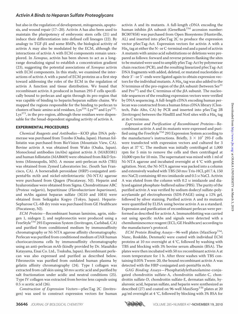
FIGURE 1. Purification of recombinant activin A and its interactions withECM proteins. A, SDS-PAGE of purified human recombinant activin A undernon-reducing (NR) and reducing (R) conditions. Mature dimeric activin A (�A/�A) is purified as complexes with its pro-region (Pro). The precursor proteinsthat failed to be processed by a furin protease are indicated by the asterisk.B, immunoblot analysis of purified recombinant activin A with the conforma-tional epitope-specific mAb MAB3381. C, solid-phase binding assays ofrecombinant activin A toward a panel of ECM proteins. ECM proteins werecoated at 10 nM and incubated with 50 nM recombinant activin A. Boundactivin A was quantified as described under “Experimental Procedures.” Eachcolumn and bar represent the mean and S.D. of triplicate assays, respectively.
Activin A Binds to Heparan Sulfate Proteoglycans
NOVEMBER 19, 2010 • VOLUME 285 • NUMBER 47 JOURNAL OF BIOLOGICAL CHEMISTRY 36647
at OSA
KA
UN
IVE
RSIT
Y on M
arch 3, 2019http://w
ww
.jbc.org/D
ownloaded from
erly processed to yield a mature dimer that remained in a com-plex with its pro-region.The ability of recombinant activin A to bind to the ECMwas
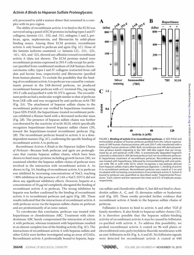
surveyed using a panel of ECMproteins including types I and IVcollagens, laminin-111, -332, and -511, nidogens 1 and 2, per-lecan, agrin, nephronectin, and fibronectin by solid-phasebinding assays. Among these ECM proteins, recombinantactivin A only bound to perlecan and agrin (Fig. 1C). None ofthe laminin isoforms examined, i.e. laminin-121, -211, -221,-411, -421, and -521, showed any affinities toward recombinantactivin A (data not shown). The ECM proteins tested wererecombinant proteins expressed in 293-F cells except for perle-can (purified from conditioned medium of JAR human chorio-carcinoma cells), types I and IV collagens (extracted from calfskin and bovine lens, respectively) and fibronectin (purifiedfrom human plasma). To exclude the possibility that the bind-ing of recombinant activinA to perlecanwas caused by contam-inants present in the JAR-derived perlecan, we producedrecombinant human perlecan with a C-terminal His6 tag using293-F cells and purified it with Ni-NTA agarose. The recombi-nant perlecan had amolecularweight similar to that of perlecanfrom JAR cells and was recognized by anti-perlecan mAb 7B5(Fig. 2A). The attachment of heparan sulfate chains to therecombinant perlecan was verified by heparitinase treatment.Upon SDS-PAGE, the heparitinase-treated recombinant perle-can exhibited a thinner band with a decreased molecular mass(Fig. 2B). The presence of heparan sulfate chains was furthercorroborated by the reactivity of mAb 3G10, which specificallyrecognizes heparitinase-cleaved heparan sulfate chains (29),toward the heparitinase-treated recombinant perlecan (Fig.2B). The recombinant perlecan bound to activin A in a dose-dependentmanner (Fig. 2C), confirming the specific binding ofrecombinant activin A to perlecan.Recombinant Activin A Binds to the Heparan Sulfate Chains
of Perlecan—Because both perlecan and agrin are proteogly-cans that contain heparan sulfate chains, which have beenshown to bind many proteins including growth factors (30), weexamined whether the heparan sulfate chains of perlecan wereinvolved in the interaction with recombinant activin A. Asshown in Fig. 3A, binding of recombinant activin A to perlecanwas inhibited by increasing concentrations of NaCl, reaching�80% inhibition in the presence of 1.64 M NaCl. EDTA did notshow any significant inhibitory effects. However, heparin at aconcentration of 10�g/ml completely abrogated the binding ofrecombinant activin A to perlecan. The strong inhibition byheparin was further confirmed by the binding of recombinantactivin A to recombinant perlecan and agrin (Fig. 3B). Theseresults indicated that the interactions of recombinant activin Awith perlecan occur via the heparan sulfate chains on perlecanand are predominantly of an ionic nature.To corroborate this conclusion, perlecan was treated with
heparitinase or chondroitinase ABC. Treatment with chon-droitinase ABC barely compromised the interaction of activinA with perlecan, whereas treatment with heparitinase resultedin an almost complete loss of the binding activity (Fig. 3C). Theinteractions of recombinant activin A with heparan sulfate andother GAGs were further investigated using an array of GAGs.Recombinant activin A preferentially bound to heparin, hepa-
ran sulfate and chondroitin sulfate-E, but did not bind to chon-droitin sulfate-A, -C, and -D, dermatan sulfate or hyaluronicacid (Fig. 3D). These results provided further evidence thatrecombinant activin A binds to the heparan sulfate chains ofperlecan.Follistatin is known to bind to activin A and other TGF-�
family members. It also binds to heparan sulfate chains (31).It is therefore possible that the heparan sulfate-bindingactivity of recombinant activin Amay be caused by follistatinco-purified with activin A. To address this possibility, weprobed recombinant activin A coated on 96-well plates orelectroblotted onto polyvinylidene fluoride membranes withan anti-follistatin mAb (Fig. 4,A and B). No follistatin signalswere detected for recombinant activin A coated at 640
FIGURE 2. Binding of activin A to recombinant perlecan. A, SDS-PAGE andimmunoblot analyses of human perlecan purified from the culture superna-tants of JAR human choriocarcinoma cells and 293-F cells transfected with afull-length human perlecan cDNA. Both recombinant and JAR-derived perle-cans produce bands migrating in the 500 – 600 kDa region that are recog-nized by an anti-perlecan mAb (7B5). A proteolytically degraded fragment(asterisks) is detected in JAR-derived perlecan but not recombinant perlecan.B, heparitinase treatment of recombinant perlecan. Recombinant perlecanwas treated with heparitinase, followed by immunoblotting with anti-perle-can mAb 7B5 or with mAb 3G10, which recognizes a neo-epitope derivedfrom heparan sulfate chains after heparitinase digestion. C, binding of activinA to recombinant perlecan. Recombinant perlecan was coated at 5 nM andincubated with increasing concentrations of recombinant activin A. Activin Abound to perlecan was quantified as described under “Experimental Proce-dures.” Each column and bar represent the mean and S.D. of triplicate assays,respectively.
Activin A Binds to Heparan Sulfate Proteoglycans
36648 JOURNAL OF BIOLOGICAL CHEMISTRY VOLUME 285 • NUMBER 47 • NOVEMBER 19, 2010
at OSA
KA
UN
IVE
RSIT
Y on M
arch 3, 2019http://w
ww
.jbc.org/D
ownloaded from

ng/well or loaded at 640 ng/lane, while control follistatin wasdetectable at concentrations as low as 1 ng/well, thus makingit unlikely that the heparan sulfate-binding activity ofrecombinant activin A was caused by follistatin copurifiedwith activin A.Clusters of Basic Amino Acid Residues in the Pro-region Are
Responsible for Perlecan Binding—It has been reported thatthe binding sites for heparin/heparan sulfate chains are com-
prised of clusters of basic amino acid residues (32). There arefour clusters of basic amino acid residues in the pro-region ofthe �A-subunit, comprising Lys69Lys70Arg71 (Cluster 1),Lys259Lys260Lys261Lys262Lys263 (Cluster 2), Lys270Lys271Lys272(Cluster 3), and Arg306Arg307Arg308Arg309Arg310 (Cluster 4)(Fig. 5A), suggesting that recombinant activinAmaybind to theheparan sulfate chains of perlecan through its pro-region. Toexplore the involvement of these basic amino acid clusters inthe binding of recombinant activin A to perlecan, we first com-pared the heparin binding activity of recombinant activin Awith that of bovine mature activin A without the pro-region bypull-down assays using heparin-Sepharose beads. SDS-PAGEanalyses of the bound and unbound fractions from the pull-down assays revealed that the recombinant activin A was fullyrecovered in the bound fraction,whilemature dimeric activinAwas largely recovered in the unbound fraction (Fig. 5B). These
FIGURE 3. Involvement of heparan sulfate chains in activin A binding toperlecan. A, inhibitory effects of high salt concentrations, the chelating agentEDTA and heparin on the perlecan-binding activity of activin A. Recombinantactivin A (50 nM) was incubated in 96-well microtiter plates coated with JAR-derived perlecan (10 nM) in the presence of increasing concentrations of NaCl(0.24 –1.64 M), EDTA (5 mM), or heparin (10 �g/ml) for 1 h, followed by quan-tification of bound activin A as described under “Experimental Procedures.”B, heparin (10 �g/ml) inhibits the binding of activin A to recombinant perle-can and agrin, both of which were expressed in 293-F cells. C, abrogation ofthe perlecan binding activity of activin A after heparitinase treatment of per-lecan. JAR-derived perlecan coated on 96-well microtiter plates at 10 nM wastreated with heparitinase or chondroitinase ABC at 37 °C for 3 h, followed byincubation with recombinant activin A (50 nM) for 1 h. D, solid-phase bindingassays of activin A with a panel of GAGs derivatized with phosphatidyletha-nolamine (PE-GAGs). PE-GAGs were coated at 20 �g/ml overnight and incu-bated with recombinant activin A (50 nM) for 1 h. Recombinant activin Abound to perlecan was quantified as described under “Experimental Proce-dures.” CS-A, chondroitin sulfate-A; CS-C, chondroitin sulfate-C; CS-D, chon-droitin sulfate-D; CS-E, chondroitin sulfate-E; DS, dermatan sulfate; HA, hyalu-ronic acid; HS, heparan sulfate; Hep, heparin. Each column and bar representthe mean and S.D. of triplicate assays, respectively.
FIGURE 4. Detection of follistatin in recombinant activin A. A, detection offollistatin in recombinant activin A by ELISA. Recombinant activin A wascoated on microtiter plates at 320 and 640 ng/well and probed with anti-follistatin mAb MAB669. Recombinant follistatin (1–16 ng/well) was analyzedby ELISA as a control. Each column and bar represent the mean and S.D. oftriplicate assays, respectively. B, detection of follistatin in recombinant activinA by immunoblotting. Recombinant activin A and recombinant follistatinwere loaded at 640 and 20 ng/lane, respectively, and detected with anti-follistatin mAb MAB669.
Activin A Binds to Heparan Sulfate Proteoglycans
NOVEMBER 19, 2010 • VOLUME 285 • NUMBER 47 JOURNAL OF BIOLOGICAL CHEMISTRY 36649
at OSA
KA
UN
IVE
RSIT
Y on M
arch 3, 2019http://w
ww
.jbc.org/D
ownloaded from

results indicated that the pro-region harbors the major hepa-rin-binding sites.The involvement of the clusters of Lys/Arg residues in per-
lecan binding was investigated by alanine-scanning mutagene-sis of these positively charged residues. Mutant proteins withindividual or entire alanine substitutions at these basic aminoacid clusters were expressed in 293-F cells and purified usingNi-NTA-agarose. Although individual alanine substitutions atCluster 2 did not compromise the expressions and secretions ofthe mutant proteins, alanine substitutions at Cluster 1 resultedin severe reductions in the amounts of recombinant proteinsrecovered fromNi-NTA-agarose (Fig. 5C).Notably, we failed toobtain any detectable amount of the mutant with alanine sub-stitution for Lys69. These results suggested that the basic aminoacid residues in Cluster 1, particularly Lys69, play critical rolesin the proper folding and/or subsequent secretion of activin A.On the other hand, amino acid substitutions at Clusters 3 and 4exhibited only partial, if any, reductions in the amounts of themutant proteins recovered from Ni-NTA-agarose, except thatentire substitutions of all five arginine residues in Cluster 4resulted in a failure of proteolytic processing by a furin proteaseinto the pro-region and mature activin A.The perlecan-binding activities of the mutant proteins were
examined by solid phase binding assays (Fig. 5D). Entire substi-tutions of the basic amino acid residues in Clusters 2 and 3caused�95 and�90% reductions in the perlecan binding activ-ity, respectively. In contrast, entire substitutions of all five argi-nine residues in Cluster 4 did not compromise the activity.These results suggested that the basic residues inClusters 2 and3, but not those in Cluster 4, are critically involved in the per-lecan-binding activity of recombinant activin A. Although themutantwith substitutions of all three basic residues inCluster 1failed to be expressed and its perlecan-binding activity couldnot be determined, individual substitutions at Lys70 and Lys71showed no significant decreases in the perlecan binding activ-ity, suggesting the possibility that Cluster 1 is not required forbinding to perlecan.Among the five mutant proteins with individual alanine sub-
stitutions at Cluster 2, the K259A mutant exhibited the mostpronounced reduction in the perlecan-binding activity, whilethe K260A, K261A, and K262A mutants showed partial reduc-tions and the K263A mutant showed only a moderate decreasein the activity. Among the three mutants with individual ala-nine substitutions in Cluster 3, the K272Amutant exhibited an�60% reduction in the perlecan-binding activity, while theK270A andK271Amutants showed�40 and�20% reductions,respectively. These results indicated that Lys259 inCluster 2 andLys272 inCluster 3 are likely candidates that comprise themajorheparan sulfate binding sites in the pro-region of activin A,although other residues in Clusters 2 and 3 also contribute tothe heparan sulfate binding ability to variable extents.Because activin A is expressed and secreted as a dimer, we
next examined whether the dimer formation is required forperlecan binding by activin A. To this end, we produced amutant protein in which Cys390 was substituted with serine todisrupt the disulfide bond required for dimer formation. TheC390S mutant produced a 15-kDa band of the mature �Aregion upon SDS-PAGE under non-reducing conditions (Fig.
FIGURE 5. Mapping of the perlecan-binding sites to basic amino acid clus-ters in the pro-region. A, schematic representation of the distributions of theclusters of basic amino acid residues in activin A. There are four clusters ofbasic amino acid residues, designated Clusters 1, 2, 3, and 4, in the pro-region(Pro). B, pull-down assays for the heparin-binding activity of activin A. Recom-binant activin A and control bovine activin A that was free from the boundpro-region were incubated with heparin-Sepharose beads, followed by sep-aration of the bound and unbound activin A by centrifugation and subse-quent SDS-PAGE under non-reducing conditions. Proteins were visualized bysilver staining. C, effects of amino acid substitutions within the basic aminoacid clusters on the secretion of recombinant activin A. Equal volumes of theculture supernatants of 293-F cells that had been transfected with activin Amutants with amino acid substitutions were subjected to purification of therecombinant activin A mutants using Ni-NTA-agarose, followed by SDS-PAGEof equal volumes of the eluates. Proteins in the eluates were visualized bysilver staining. D, solid-phase binding assays of the perlecan-binding activi-ties of the activin A mutants. Activin A mutants with amino acid substitutionswere incubated on microtiter plates coated with 10 nM JAR-derived perlecanfor 1 h, followed by quantification of bound activin A as described under“Experimental Procedures.” Each column and bar represent the mean and S.D.of triplicate assays, respectively.
Activin A Binds to Heparan Sulfate Proteoglycans
36650 JOURNAL OF BIOLOGICAL CHEMISTRY VOLUME 285 • NUMBER 47 • NOVEMBER 19, 2010
at OSA
KA
UN
IVE
RSIT
Y on M
arch 3, 2019http://w
ww
.jbc.org/D
ownloaded from
5C), confirming the requirement of Cys390 for the dimer forma-tion. Solid-phase binding assays revealed that the C390Smutant was barely capable of binding to perlecan (Fig. 5D),thereby demonstrating the importance of the dimeric structureof activin A for binding to perlecan.Deletion of the Heparan Sulfate Binding Sites Does Not
Impair the Biological Activity of Activin A—The human inhibin�A- and �B-subunits share 39% amino acid sequence similarity.A comparison of the amino acid sequences of the �A- and�B-subunits revealed that the peptide segment Lys259-Glu284containing Clusters 2 and 3 is absent from the �B-subunit (Fig.6A). However, the amino acid sequence of this segment ishighly conserved among different species (Fig. 6B). The signif-icant conservation of the amino acid sequence of the Lys259-Glu284 segment together with its absence from the inhibin�B-subunit prompted us to hypothesize that this segmentadopts an independent conformation and that the involvementof Clusters 2 and 3 in the heparan sulfate binding activity couldbe corroborated by deleting this segment from activin A. Weproduced a deletion mutant from which most of this segmentincluding Clusters 2 and 3 (Lys259-Gly277) was deleted andexamined its expression and perlecan-binding activity. Asexpected, this mutant was expressed and processed similar tothe case for wild-type activin A, but was barely active in bindingto perlecan (Fig. 6C).Next, we addressed whether the perlecan-binding activity of
recombinant activin A is involved in the biological activities ofactivin A. To this end, we employed an activin-responsive lucif-erase reporter gene assay using CHO cells (28). As the recom-binant activin A with a C-terminal His6 tag exhibited a lowluciferase-inducing activity (data not shown), we producedanother recombinant activin A with a His6 tag at the N termi-nus of the pro-region (designated activin ANHis). Activin ANHis
was expressed well compared with the activin A with a C-ter-minal His6 tag, and retained the ability to bind to perlecan (Fig.6C). Themoderate decrease in its perlecan binding activitymaybe caused by differential effects of the position of theHis6 tag oneither the accessibility of the anti-His tag antibody or the bind-ing affinity toward the heparan sulfate chains onperlecan.Dele-tion of the Lys259-Gly277 segment from activin ANHis abrogatedits perlecan-binding activity (Fig. 6C), thus confirming the crit-ical roles of Clusters 2 and 3 in the binding of activin A toperlecan.Incubation of CHOcells transfectedwith the activin-respon-
sive luciferase reporter gene with increasing concentrations ofactivin ANHis resulted in dose-dependent increases in the lucif-erase activity, reaching a maximum activity that was compara-ble to that of a commercially available mature activin A at �1.5nM (Fig. 7). Activin ANHis exhibited lower luciferase activitiesthan mature activin A at �1.5 nM, which mostly reflected thefact that only a fraction of activin ANHis gave rise to functionallyactive mature activin A upon dissociation, meaning that theeffective concentration of mature activin A was significantlylower for recombinant activin A than for the mature activin A.Interestingly, the mutant activin ANHis lacking the Lys259-Gly277 segment was equally as potent as control activin ANHis,exhibiting the same dose-dependent luciferase activity (Fig. 7).These results indicated that the heparan sulfate-binding sites
comprised ofClusters 2 and 3 in the pro-region are not requiredfor receptor binding or subsequent signal transduction. Fur-thermore, neither the association with the pro-region nor thepresence of theN-terminal His6 tag imposed deleterious effectson the biological activity of activin A.Activin A Binds to Heparan Sulfate in Tissues—Finally, we
addressed the biological relevance of the heparan sulfate bind-ing activity of the pro-region of activin A by in situ activin Aoverlay assays using frozen sections of mouse embryos, inwhich detection of bound activin A signals was facilitated bypretreatment of the tissue sections with chondroitinase ABC
FIGURE 6. Abrogation of the perlecan binding activity of activin A by dele-tion of the inhibin �A subunit-specific region harboring Clusters 2 and 3.A, amino acid sequence alignment between the inhibin �A- and �B-subunits.The peptide segment Lys259-Glu284 containing Clusters 2 and 3 in the inhibin�A-subunit is absent from the inhibin �B-subunit (highlighted in italics). Theconserved amino acid residues are shaded in gray. B, alignment of basic aminoacid clusters in the amino acid sequences of the inhibin �A-subunits fromdifferent species. The amino acid sequence of the peptide segment Lys259-Glu284 harboring Clusters 2 and 3 is well conserved among the different spe-cies. The clusters of basic amino acid residues are shown in white letters onblack whereas acidic amino acid residues are shaded in gray. C, the perlecanbinding activities of recombinant activin A with a His6 tag at either the Nterminus (N-His tag) or C terminus (C-His tag) and those with deletion of theLys259-Gly277 segment (designated �K259-G277) were determined by solid-phase binding assays. The SDS-PAGE profiles of purified activin A with andwithout deletion of Clusters 2 and 3 are shown in the inset. Note that thepro-regions of intact and mutant activin A differ in their sizes because of thepresence or absence of the Lys259-Gly277 segment.
Activin A Binds to Heparan Sulfate Proteoglycans
NOVEMBER 19, 2010 • VOLUME 285 • NUMBER 47 JOURNAL OF BIOLOGICAL CHEMISTRY 36651
at OSA
KA
UN
IVE
RSIT
Y on M
arch 3, 2019http://w
ww
.jbc.org/D
ownloaded from

and hyaluronidase. A significant proportion of the signals forbound activin A were confined to the areas corresponding tothe basementmembranes of the lungs, hair follicles and epider-mis (Fig. 8, A and B). The basement membrane regions of theretinas and kidneys were also positive for bound activin A sig-nals (data not shown). Binding of activinA to the tissue sectionswas abrogated by pretreatment of the sections with hepariti-nase (Fig. 8, C and D), confirming that the binding of activin Ato the tissues was heparan sulfate-dependent. The mutantactivin A lacking the Lys259-Gly277 segment did not produceany significant signals for bound activin A (Fig. 8, E and F),consistent with the requirement of the basic amino acid clus-ters in the pro-region for the binding of activinA to the heparansulfate chains of perlecan. Furthermore, the overall patterns ofthe activin A signals were reminiscent of those for mAb 10E4,which recognizes heparan sulfate chains (Fig. 8, G and H).Taken together, these results support the conclusion thatactivin A binds to heparan sulfate proteoglycans in basementmembranes through its pro-region in a heparan sulfate-depen-dent manner.
DISCUSSION
An important feature of activin A revealed in this study isthat activin A specifically binds to perlecan and agrin among avariety of ECM proteins. Our data clearly show that activin Abinds to perlecan through heparan sulfate chains. Conse-quently, the binding of activin A to perlecan was inhibited byhigh concentrations of NaCl and by heparin. Treatment withheparitinase, but not chondroitinase ABC, abolished the bind-ing. Furthermore, activin A directly bound to heparin andheparan sulfate chains in solid-phase binding assays. To thebest of our knowledge, this is the first report that a growthfactor in the TGF-� family binds to HSPGs via its pro-region,although BMP-2 and BMP-4 have been reported to bind to
HSPGs via theN-terminal regions of themature growth factors(30, 33). Given that the pro-region of TGF-�1 binds to LTBPs(13) and those of BMP-2, -4, -7, and -10 bind to fibrillins (14),the pro-regions of TGF-� family proteins have distinctive spec-ificities to localize these growth factors at target sites in theECM and regulate their activities in a spatiotemporal manner.It should be noted that perlecan and agrin are major basementmembrane components that are widely expressed in bothembryonic and adult tissues (34–36). In embryogenesis, base-ment membranes serve as structural as well as functional inter-faces of epithelial-mesenchymal interactions, through whichmany organs develop. Given the role of activin A as a morpho-gen, the associations of activin A with embryonic basementmembranes through perlecan and agrin may be instrumentalin regulating the morphogenetic activity of activin A duringembryonic development. In support of this possibility, we dem-onstrated that activin A predominantly bound to the basementmembrane regions of various tissues in a heparan sulfate-de-pendent manner.The interactions of proteins with heparin/heparan sulfate
chains aremainly contributed by ionic interactions between thenegatively charged sulfate groups and positively charged aminoand guanidinium groups of lysine and arginine residues, and insome cases aremediated byCa2� coordination (32). The stronginhibition of activin A binding to perlecan by high concentra-tions of NaCl and scarce inhibition by chelation of divalentcations with EDTA indicated that ionic interactions are morecritical for the binding of recombinant activin A to perlecan.Because the binding of recombinant activin A to perlecan wasnot completely blocked at the highest concentration of NaClexamined (1.64 M), other types of chemical bonds such ashydrogen bonds and hydrophobic interactions may partiallycontribute to the interactions between recombinant activin Aand perlecan (32). It should be noted that not only heparin andheparan sulfate but also chondroitin sulfate-E bound to activinA among the panel of GAGs tested in this study. The failures ofchondroitin sulfate-A, -C, and -D, dermatan sulfate, hyaluronicacid, and highly sialylated nephronectin (37) to bind to activinA suggest that the interactions are not simply electrostatic andpossibly require two closely positioned sulfate groups onrepeating disaccharide units. Among the different forms ofchondroitin sulfates, only chondroitin sulfate-E containsN-acetylgalactosaminewith two sulfate groups at carbons 4 and6. Consistent with these results, various heparin/heparan sul-fate-binding growth factors including midkine, pleiotrophin,FGF-2, FGF-10, FGF-16, FGF-18, and heparin-binding epider-mal growth factor are capable of binding to chondroitin sul-fate-E (38). Although chondroitin sulfate-E is a minor compo-nent of mammalian chondroitin sulfate chains (39), it maycontribute to the ECM deposition of activin A and the regula-tion of its biological functions.Our data show that the basic amino acid clusters in the pro-
region, which are well conserved among different species, func-tion differently. Cluster 1 (Lys69-Lys70-Arg71) is criticallyrequired for secretion, possibly because of its involvement inthe proper formation of the three-dimensional structure, while
FIGURE 7. Smad-dependent signaling activities of recombinant activinA lacking Clusters 2 and 3. CHO cells cotransfected with an activin-respon-sive luciferase reporter gene and a �-galactosidase gene were stimulatedwith increasing concentrations of N-terminally His-tagged activin A with andwithout deletion of the Lys259-Gly277 segment (designated activin ANHis andactivin ANHis(�K259-G277), respectively) or bovine activin A without the pro-region for 20 h in �-MEM containing 0.1% fetal bovine serum. The luciferaseactivities are expressed as the fold increases after normalization by the�-galactosidase activity. The results represent the means of duplicatedeterminations.
Activin A Binds to Heparan Sulfate Proteoglycans
36652 JOURNAL OF BIOLOGICAL CHEMISTRY VOLUME 285 • NUMBER 47 • NOVEMBER 19, 2010
at OSA
KA
UN
IVE
RSIT
Y on M
arch 3, 2019http://w
ww
.jbc.org/D
ownloaded from

Clusters 2 (Lys259-Lys263) and 3 (Lys270-Lys272) contribute toperlecan binding. Cluster 4 serves as a recognition site for pro-teolytic processing by a furin protease. All five basic amino acid
residues in Cluster 2 are fully con-served among different species,while Lys271 in Cluster 3 is replacedbyGlu in the chicken and Lys271 andLys272 are replaced by Ala in thenewt (see Fig. 6B). The stringentconservation of the stretch of fiveLys residues in Cluster 2 implies acritical role of Cluster 2 in heparansulfate binding. The requirementfor Clusters 2 and 3 in the heparansulfate binding was further demon-strated by the deletion experimentin which the segment containingboth Clusters 2 and 3 was deletedfrom the pro-region. There are fiveisoforms of inhibin � subunits,namely �A, �B, �C, �D, and �E (40).Among these, the �A- and �B-sub-units share 39% amino acidsequence similarity. The Lys259-Glu284 segment that contains Clus-ters 2 and 3 is �A-subunit-specificand does not show any significanthomology to other proteins includ-ing those with heparin bindingactivities (32). Because the deletionof a large part of this segment didnot impair the growth factor activityof activin A, it is tempting to specu-late that the Lys259-Glu284 segmentadopts an independent conforma-tion within the pro-region and thatthe �A-subunit could have acquiredthis extra segment during evolutionto endow the binding activitytoward heparan sulfate chains.Alternatively, this peptide segmentcould have been removed from theinhibin �B-subunit to attenuate thebinding to heparan sulfate chains. Itis interesting to note that there aretwo conserved clusters of negativelycharged amino acid residues in theextra segment, which may regulatethe heparan sulfate binding activityby antagonizing the positive elec-trostatic potential within the samesegment.The growth factors of the TGF-�
family are synthesized as precursorsand most of them are secreted ascomplexes of the N-terminal pro-region and the C-terminal maturegrowth factor (41). The pro-regions
not only facilitate proper folding of the mature growth factorsand their subsequent secretion from cells (15) but also conferlatency on TGF-�1 (41) and growth differentiation factor-8
FIGURE 8. In situ activin A overlay assays on tissue sections. A–F, frozen sections of mouse E16.5 embryoswere incubated with activin ANHis (A–D) or its mutant lacking the Lys259-Gly277 segment (E and F), followed bydetection of the bound proteins by immunohistochemistry. Sections treated with heparitinase (C and D) wereincluded in the overlay assays to confirm that the signals were heparan sulfate-dependent. G and H, additionalsections were subjected to immunohistochemistry with anti-heparan sulfate mAb 10E4 to clarify the localiza-tions of heparan sulfate chains. The signals for bound activin A are localized to the basement membranes ofvarious organs including the lungs (A) and skin (B), where the epidermis and hair follicles are labeled witharrowheads and asterisks, respectively. Scale bar, 100 �m.
Activin A Binds to Heparan Sulfate Proteoglycans
NOVEMBER 19, 2010 • VOLUME 285 • NUMBER 47 JOURNAL OF BIOLOGICAL CHEMISTRY 36653
at OSA
KA
UN
IVE
RSIT
Y on M
arch 3, 2019http://w
ww
.jbc.org/D
ownloaded from

(42) and target TGF-�1, BMP-2, -4, -7, and -10 to the ECM (14).The latent TGF-�1 complexes are activated by binding tothrombospondin-1 (43) and integrin�v�6 (44) and also by pro-teolytic cleavage of the pro-region (45, 46). The bioactivities ofgrowth differentiation factor-8 and BMP-4 are also regulatedby proteolytic cleavage of their pro-regions (45, 47–49). In con-trast, the pro-regions of BMP-7 and BMP-9 form stable com-plexes with themature growth factors but do not confer latencyon the complexes (50, 51). Our results showed that activin Aexpressed in 293-F cells was purified as complexes with its pro-region but was functionally active in its complex form, as hasbeen observed for BMP-7 andBMP-9 (50, 51). The failure of thepro-region to render activin A inactive may be caused by itsrelatively low binding affinity toward the mature growth factor(52), thus allowing activin A receptors on the cell surface tocompete with the pro-region for the mature ligand. Consistentwith this possibility, recombinant activin A exhibited lowerbiological activities at concentrations of �1.5 nM thanbovine mature activin A that was free from a pro-region.Dimerization of activin A is essential for its biological activ-
ity, since monomerization of activin A significantly decreasesits receptor binding affinity as well as its activity to enhancethe release of follicle-stimulating hormone (53). Our resultsshowed that the activin A mutant C390S, which was unable toform a dimer, was devoid of the perlecan binding activity,thereby indicating that dimerization was also required for itsheparan-sulfate binding activity. This finding is in striking con-trast to many other heparin/heparan sulfate-binding growthfactors including midkine, pleiotrophin, FGF-2, FGF-10, FGF-16, FGF-18, and heparin-binding epidermal growth factor,which can bind to heparin/heparan sulfates as monomers. Therequirement for the dimeric structure of activin A may becaused by the relatively low heparan sulfate-binding activity ofmonomeric activin A, which necessitates bivalency for stablebinding to heparan sulfate chains. Alternatively, the bindingsite for the heparan sulfate chains may be formed at the inter-face of the two pro-regions, thus requiring the juxtaposition oftwo monomers. The prerequisite of the dimeric structure forperlecan binding may also serve as a fail-safe quality controlthat rejects improperly folded monomeric activin A and pre-vents its deposition in the ECM near the target cells.The negatively charged heparan sulfate chains that are
covalently attached to perlecan, syndecan, agrin, and �-glycanhave been shown to participate in the regulation of cell signal-ing and morphogenesis via binding to a variety of growth fac-tors and/or their receptors (2, 54). Binding of heparan sulfate toFGF-2 stabilizes the complex formation with its receptor andprolongs its activity (55, 56). Release of the VEGF isoformsV165 and V189 fromHSPGs by plasmin enables them to act onendothelial cells, leading to enhancement of vascular perme-ability (5). Regarding the TGF-� family, cell surface HSPGsmediate BMP-2 internalization and modulate BMP-2 osteo-genic activity (57), while endogenous heparan sulfate chainsmodulate BMP-4 signaling and activity (58). The consequencesof the sequestration of growth factors in the ECM and theirsubsequent regulated release are to prolong their action, facili-tate their localization to the environment immediately adjacentto their target cells and tune their biological activity (56).
Because deletion of the heparan sulfate-binding sites fromrecombinant activin A did not impair its biological activity, it isconceivable that the HSPG-binding activity of activin A facili-tates the deposition of activin A in the ECM but does not reg-ulate the growth factor activity per se. The physiological rele-vance of the heparan sulfate binding by activin A remains to beaddressed in the context of histogenesis and organogenesisusing animalmodels inwhich the heparan sulfate binding activ-ity of activin A is ablated in particular cell lineages.
Acknowledgment—We thank Dr. Masahiko Katayama, Eisai Co.Ltd., for the generous gift of an anti-perlecan mAb.
REFERENCES1. Nelson, C. M., and Bissell, M. J. (2006) Annu. Rev. Cell Dev. Biol. 22,
287–3092. Macri, L., Silverstein, D., and Clark, R. A. (2007)Adv. Drug. Deliv. Rev. 59,
1366–13813. Vlodavsky, I., and Friedmann, Y. (2001) J. Clin. Invest. 108, 341–3474. Asada, M., Shinomiya, M., Suzuki, M., Honda, E., Sugimoto, R., Ikekita,
M., and Imamura, T. (2009) Biochim. Biophys. Acta 1790, 40–485. Houck, K. A., Leung, D. W., Rowland, A. M., Winer, J., and Ferrara, N.
(1992) J. Biol. Chem. 267, 26031–260376. Lyon, M., Deakin, J. A., Mizuno, K., Nakamura, T., and Gallagher, J. T.
(1994) J. Biol. Chem. 269, 11216–112237. Mooradian, D. L., Lucas, R. C., Weatherbee, J. A., and Furcht, L. T. (1989)
J. Cell. Biochem. 41, 189–2008. Schultz-Cherry, S., and Murphy-Ullrich, J. E. (1993) J. Cell Biol. 122,
923–9329. Lamszus, K., Joseph, A., Jin, L., Yao, Y., Chowdhury, S., Fuchs, A., Pol-
verini, P. J., Goldberg, I. D., and Rosen, E. M. (1996) Am. J. Pathol. 149,805–819
10. Schuppan, D., Schmid, M., Somasundaram, R., Ackermann, R., Ruehl, M.,Nakamura, T., and Riecken, E. O. (1998) Gastroenterology 114, 139–152
11. Wang, X., Harris, R. E., Bayston, L. J., and Ashe, H. L. (2008) Nature 455,72–77
12. Hogan, B. L. (1996) Curr. Opin. Genet. Dev. 6, 432–43813. Saharinen, J., Hyytiaïnen, M., Taipale, J., and Keski-Oja, J. (1999) Cytokine
Growth Factor Rev. 10, 99–11714. Sengle, G., Charbonneau, N. L., Ono, R. N., Sasaki, T., Alvarez, J., Keene,
D. R., Bachinger, H. P., and Sakai, L. Y. (2008) J. Biol. Chem. 283,13874–13888
15. Gray, A. M., and Mason, A. J. (1990) Science 247, 1328–133016. Vale, W., Rivier, J., Vaughan, J., McClintock, R., Corrigan, A., Woo, W.,
Karr, D., and Spiess, J. (1986) Nature 321, 776–77917. Matzuk, M. M., Kumar, T. R., Shou, W., Coerver, K. A., Lau, A. L., Be-
hringer, R. R., and Finegold, M. J. (1996) Recent Prog. Horm. Res. 51,123–154, discussion 155–157
18. Spencer, S. J., Rabinovici, J., Mesiano, S., Goldsmith, P. C., and Jaffe, R. B.(1992) J. Clin. Invest. 90, 142–149
19. Nishihara, T., Okahashi, N., and Ueda, N. (1993) Biochem. Biophys. Res.Commun. 197, 985–991
20. Munz, B., Tretter, Y. P., Hertel, M., Engelhardt, F., Alzheimer, C., andWerner, S. (2001)Mol. Cell. Endocrinol. 180, 169–177
21. Beattie, G.M., Lopez, A.D., Bucay,N., Hinton, A., Firpo,M.T., King, C. C.,and Hayek, A. (2005) Stem. Cells 23, 489–495
22. Asashima,M.,Michiue, T., and Kurisaki, A. (2008)Dev. Growth Differ. 50,Suppl. 1, S35–S45
23. McDowell, N., Zorn, A. M., Crease, D. J., and Gurdon, J. B. (1997) Curr.Biol. 7, 671–681
24. Sekiguchi, K., Hakomori, S., Funahashi, M., Matsumoto, I., and Seno, N.(1983) J. Biol. Chem. 258, 14359–14365
25. Sato, K., Ebihara, T., Adachi, E., Kawashima, S., Hattori, S., and Irie, S.(2000) J. Biol. Chem. 275, 25870–25875
26. Iwata, M., Imamura, Y., Sasaki, T., and Hayashi, T. (1995) J. Biochem. 117,
Activin A Binds to Heparan Sulfate Proteoglycans
36654 JOURNAL OF BIOLOGICAL CHEMISTRY VOLUME 285 • NUMBER 47 • NOVEMBER 19, 2010
at OSA
KA
UN
IVE
RSIT
Y on M
arch 3, 2019http://w
ww
.jbc.org/D
ownloaded from

1298–130427. Sugiura, N., Sakurai, K., Hori, Y., Karasawa, K., Suzuki, S., and Kimata, K.
(1993) J. Biol. Chem. 268, 15779–1578728. Arai, K. Y., Tsuchida, K., Li, C., Watanabe, G., Sugino, H., Taya, K., and
Nishiyama, T. (2006) Protein Expr. Purif. 49, 78–8229. David, G., Bai, X. M., Van der Schueren, B., Cassiman, J. J., and Van den
Berghe, H. (1992) J. Cell Biol. 119, 961–97530. Whitelock, J. M., Melrose, J., and Iozzo, R. V. (2008) Biochemistry 47,
11174–1118331. Innis, C. A., and Hyvonen, M. (2003) J. Biol. Chem. 278, 39969–3997732. Gandhi, N. S., and Mancera, R. L. (2008) Chem. Biol. Drug. Des. 72,
455–48233. Ohkawara, B., Iemura, S., ten Dijke, P., and Ueno, N. (2002)Curr. Biol. 12,
205–20934. Manabe, R., Tsutsui, K., Yamada, T., Kimura,M., Nakano, I., Shimono, C.,
Sanzen, N., Furutani, Y., Fukuda, T., Oguri, Y., Shimamoto, K., Kiyozumi,D., Sato, Y., Sado, Y., Senoo, H., Yamashina, S., Fukuda, S., Kawai, J.,Sugiura, N., Kimata, K., Hayashizaki, Y., and Sekiguchi, K. (2008) Proc.Natl. Acad. Sci. U.S.A. 105, 12849–12854
35. Murdoch, A. D., Liu, B., Schwarting, R., Tuan, R. S., and Iozzo, R. V. (1994)J. Histochem. Cytochem 42, 239–249
36. Groffen, A. J., Buskens, C. A., van Kuppevelt, T. H., Veerkamp, J. H.,Monnens, L. A., and van den Heuvel, L. P. (1998) Eur. J. Biochem. 254,123–128
37. Brandenberger, R., Schmidt, A., Linton, J.,Wang, D., Backus, C., Denda, S.,Muller, U., and Reichardt, L. F. (2001) J. Cell Biol. 154, 447–458
38. Deepa, S. S., Umehara, Y., Higashiyama, S., Itoh, N., and Sugahara, K.(2002) J. Biol. Chem. 277, 43707–43716
39. Ueoka, C., Kaneda, N., Okazaki, I., Nadanaka, S., Muramatsu, T., andSugahara, K. (2000) J. Biol. Chem. 275, 37407–37413
40. Mellor, S. L., Cranfield, M., Ries, R., Pedersen, J., Cancilla, B., de Kretser,D., Groome, N. P., Mason, A. J., and Risbridger, G. P. (2000) J. Clin. Endo-crinol. Metab. 85, 4851–4858
41. Rifkin, D. B. (2005) J. Biol. Chem. 280, 7409–741242. Lee, S. J., and McPherron, A. C. (2001) Proc. Natl. Acad. Sci. U.S.A. 98,
9306–931143. Crawford, S. E., Stellmach, V.,Murphy-Ullrich, J. E., Ribeiro, S.M., Lawler,
J., Hynes, R. O., Boivin, G. P., and Bouck, N. (1998) Cell 93, 1159–117044. Munger, J. S., Huang, X., Kawakatsu, H., Griffiths, M. J., Dalton, S. L., Wu,
J., Pittet, J. F., Kaminski, N., Garat, C., Matthay, M. A., Rifkin, D. B., andSheppard, D. (1999) Cell 96, 319–328
45. Lyons, R. M., Keski-Oja, J., and Moses, H. L. (1988) J. Cell Biol. 106,1659–1665
46. Ge, G., and Greenspan, D. S. (2006) J. Cell Biol. 175, 111–12047. Wolfman, N. M., McPherron, A. C., Pappano, W. N., Davies, M. V., Song,
K., Tomkinson, K. N., Wright, J. F., Zhao, L., Sebald, S. M., Greenspan,D. S., and Lee, S. J. (2003) Proc. Natl. Acad. Sci. U.S.A. 100, 15842–15846
48. Degnin, C., Jean, F., Thomas, G., and Christian, J. L. (2004)Mol. Biol. Cell15, 5012–5020
49. Sopory, S., Nelsen, S. M., Degnin, C., Wong, C., and Christian, J. L. (2006)J. Biol. Chem. 281, 34021–34031
50. Sengle, G., Ono, R. N., Lyons, K. M., Bachinger, H. P., and Sakai, L. Y.(2008) J. Mol. Biol. 381, 1025–1039
51. Brown, M. A., Zhao, Q., Baker, K. A., Naik, C., Chen, C., Pukac, L., Singh,M., Tsareva, T., Parice, Y., Mahoney, A., Roschke, V., Sanyal, I., and Choe,S. (2005) J. Biol. Chem. 280, 25111–25118
52. Walton, K. L.,Makanji, Y.,Wilce,M.C., Chan, K. L., Robertson, D.M., andHarrison, C. A. (2009) J. Biol. Chem. 284, 9311–9320
53. Husken-Hindi, P., Tsuchida, K., Park, M., Corrigan, A. Z., Vaughan, J. M.,Vale, W. W., and Fischer, W. H. (1994) J. Biol. Chem. 269, 19380–19384
54. Bishop, J. R., Schuksz, M., and Esko, J. D. (2007) Nature 446, 1030–103755. Schlessinger, J., Plotnikov, A. N., Ibrahimi, O. A., Eliseenkova, A. V., Yeh,
B. K., Yayon, A., Linhardt, R. J., and Mohammadi, M. (2000) Mol. Cell 6,743–750
56. Flaumenhaft, R., and Rifkin, D. B. (1991)Curr. Opin. Cell Biol. 3, 817–82357. Jiao, X., Billings, P. C., O’Connell, M. P., Kaplan, F. S., Shore, E. M., and
Glaser, D. L. (2007) J. Biol. Chem. 282, 1080–108658. Khan, S. A., Nelson, M. S., Pan, C., Gaffney, P. M., and Gupta, P. (2008)
Am. J. Physiol. Cell Physiol. 294, C1387–1397
Activin A Binds to Heparan Sulfate Proteoglycans
NOVEMBER 19, 2010 • VOLUME 285 • NUMBER 47 JOURNAL OF BIOLOGICAL CHEMISTRY 36655
at OSA
KA
UN
IVE
RSIT
Y on M
arch 3, 2019http://w
ww
.jbc.org/D
ownloaded from

Kimata and Kiyotoshi SekiguchiYagi, Maria Hayashi, Yuya Sato, Hitomi Fujisaki, Shunji Hattori, Nobuo Sugiura, Koji Shaoliang Li, Chisei Shimono, Naoko Norioka, Itsuko Nakano, Tetsuo Okubo, Yoshiko
Sulfate Binding ActivityActivin A Binds to Perlecan through Its Pro-region That Has Heparin/Heparan
doi: 10.1074/jbc.M110.177865 originally published online September 15, 20102010, 285:36645-36655.J. Biol. Chem.
10.1074/jbc.M110.177865Access the most updated version of this article at doi:
Alerts:
When a correction for this article is posted•
When this article is cited•
to choose from all of JBC's e-mail alertsClick here
http://www.jbc.org/content/285/47/36645.full.html#ref-list-1
This article cites 58 references, 25 of which can be accessed free at
at OSA
KA
UN
IVE
RSIT
Y on M
arch 3, 2019http://w
ww
.jbc.org/D
ownloaded from