orthopaedic manual physical therapy including thrust ... · pdf fileself-report outcome...
TRANSCRIPT

10 / The Journal of Manual & Manipulative Therapy, 2007
Jacqueline van Duijn, PT, DPT, OCSArie J. van Duijn, PT, EdD, OCSWanda Nitsch, PT, PhD
Abstract: It has been reported that in Western society as many as 16% of individuals experience cervi-cogenic headache, which can lead to signifi cant amounts of pain and perceived disability. Cervicogenic headache is characterized by unilateral occipital-temporal pain that is increased by neck movement; it is accompanied by cervical hypomobility, postural changes, and/or increased cervical muscle tone. This case report describes the physical therapy differential diagnosis, management, and outcomes of a patient with cervicogenic headache. The patient was a 40-year-old woman referred by her physiatrist with com-plaints of cervical pain and ipsilateral temporal headache. The patient presented with increased muscle tone, multiple-level joint hypomobility in the cervical and thoracic spine, muscle weakness, and postural changes. Self-report outcome measures included the Visual Analog Scale for headache pain intensity and the Neck Disability Index. Management consisted of various thrust and non-thrust manipulations, soft tissue mobilizations, postural re-education, and exercise to address postural defi cits and cervical and thoracic hypomobility and diminished strength. At discharge, the patient demonstrated clinically meaningful improvements with regard to pain, disability, and headache. This case report indicates that a multimodal physical therapy treatment program may be effective in the management of a patient diagnosed with cervicogenic headache.
Key Words: Cervicogenic Headache, Manipulation, Thrust, Exercise, Physical Therapy
Orthopaedic Manual Physical Therapy Including Thrust Manipulation and Exercise in the Management of a Patient with Cervicogenic Headache: A Case Report
Cervicogenic headaches are commonly encountered in physical therapy (PT) practice. Cervicogenic head-ache is a headache type validated recently by the In-
ternational Headache Society (IHS) that is hypothesized to originate due to nociception in the cervical area. In a Scan-dinavian population study, its prevalence was established at approximately 16%1. Table 1 outlines the diagnostic criteria for cervical headaches as described by the IHS2.
Horn and Smith3 reviewed the literature on approaches to management of cervicogenic headache using orthopedic
manual physical therapy (OMPT) principles. They recom-mended careful examination to identify all impairments, to provide a differential diagnosis, and to plan appropriate in-tervention. Recommended interventions included joint mo-bilization, soft tissue mobilization, retraining of specifi c pos-tural muscle groups, and patient education. The authors recommended that emphasis should be placed on home and self-management skills and on increasing patient under-standing of the predisposing factors with regard to cervico-genic headache. Jull4 also described the importance of an ac-curate differential diagnosis ascertaining the cervical musculoskeletal origin of the headache to assure success with management of a cervical headache patient. She de-scribed the history and symptomatic features of cervical headache following the description of the IHS2, and she noted the role of physical impairments in the articular, mus-cular, and nervous system. She suggested that the diagnosis
The Journal of Manual & Manipulative TherapyVol. 15 No. 1 (2007), 10–24
Address all correspondence and requests for reprints to:Jacqueline van Duijn12601 Strathmore LoopFort Myers, FL 33912E-mail: [email protected]
Orthopaedic Manual Physical Therapy Including Thrust Manipulation and Exercise in the Management of a Patient with Cervicogenic Headache: A Case Report / 11
of cervical headache was dependent on the presence of both articular and muscle impairments, accompanied by acute or chronic poor neuromotor control.
The presence of painful upper cervical joint dysfunction accompanied by impairments in the deep cervical fl exors, scapular postural muscles, and cervical kinaesthesia can in-dicate that a headache is cervical in origin4. Other physical impairments that are often present in a cervical headache patient include postural abnormalities, muscle tightness, and neural tissue mechanosensitivity. However, Jull4 also noted that the absence of the above impairments does not necessarily preclude a cervical headache diagnosis. Jull4 em-phasized the importance of specifi c retraining of upper cer-vical fl exor muscles, the lower trapezius, and serratus ante-rior, combined with postural retraining and ergonomic and lifestyle advice.
Various authors have identifi ed current approaches in the management of cervical headaches using OMPT princi-ples3,5–11. Intervention may include joint mobilization/ma-nipulation, soft tissue mobilization and stretching tech-niques, retraining specifi c postural muscle groups, and patient education. This literature suggests that emphasis should be placed on addressing joint mobility, posture, mus-cle strength, and imbalances in the upper back and neck with attention also directed toward life style, habits, and stress management.
The literature discussed above provides preliminary evi-dence to demonstrate the utility of a thorough clinical ex-amination as well as the effectiveness of OMPT intervention as a component of a multimodal approach to managing pa-tients with cervicogenic headaches. The purpose of this case report is to describe the PT differential diagnosis and man-agement of a patient with cervical headaches.
Case Description
The patient was referred by her physiatrist to an orthopaedic outpatient PT clinic with a diagnosis of cervical spondylosis, myofascial pain, and headaches. An ABPTS (American Board of Physical Therapy Specialties)-certifi ed orthopedic PT clin-ical specialist, who was also certifi ed in manual therapy and had 15 years of clinical experience, performed the standard-ized examination described below. The examination followed the spinal evaluation format as described by Paris and Lou-bert12 (Table 2).
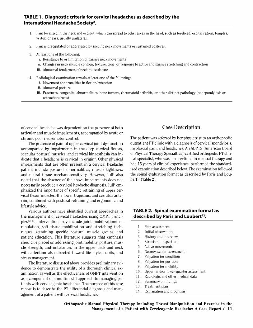
TABLE 1. Diagnostic criteria for cervical headaches as described by the International Headache Society2.
1. Pain localized in the neck and occiput, which can spread to other areas in the head, such as forehead, orbital region, temples, vertex, or ears, usually unilateral.
2. Pain is precipitated or aggravated by specifi c neck movements or sustained postures.
3. At least one of the following: i. Resistance to or limitation of passive neck movements ii. Changes in neck muscle contour, texture, tone, or response to active and passive stretching and contraction
iii. Abnormal tenderness of neck musculature
4. Radiological examination reveals at least one of the following: i. Movement abnormalities in fl exion/extension ii. Abnormal posture iii. Fractures, congenital abnormalities, bone tumors, rheumatoid arthritis, or other distinct pathology (not spondylosis or osteochondrosis)
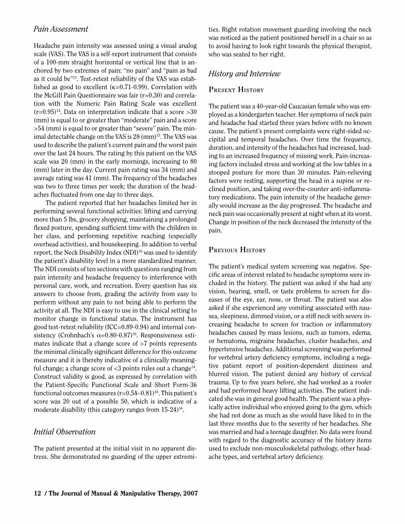
TABLE 2. Spinal examination format as described by Paris and Loubert12.
1. Pain assessment 2. Initial observation 3. History and interview 4. Structural inspection 5. Active movements 6. Neurovascular assessment 7. Palpation for condition 8. Palpation for position 9. Palpation for mobility 10. Upper- and/or lower-quarter assessment 11. Radiologic and other medical data 12. Summary of fi ndings 13. Treatment plan 14. Explanation and prognosis
12 / The Journal of Manual & Manipulative Therapy, 2007
Pain Assessment
Headache pain intensity was assessed using a visual analog scale (VAS). The VAS is a self-report instrument that consists of a 100-mm straight horizontal or vertical line that is an-chored by two extremes of pain: “no pain” and “pain as bad as it could be”13. Test-retest reliability of the VAS was estab-lished as good to excellent (κ=0.71-0.99). Correlation with the McGill Pain Questionnaire was fair (r=0.30) and correla-tion with the Numeric Pain Rating Scale was excellent (r=0.95)14. Data on interpretation indicate that a score >30 (mm) is equal to or greater than “moderate” pain and a score >54 (mm) is equal to or greater than “severe” pain. The min-imal detectable change on the VAS is 28 (mm)15. The VAS was used to describe the patient’s current pain and the worst pain over the last 24 hours. The rating by this patient on the VAS scale was 20 (mm) in the early mornings, increasing to 80 (mm) later in the day. Current pain rating was 34 (mm) and average rating was 41 (mm). The frequency of the headaches was two to three times per week; the duration of the head-aches fl uctuated from one day to three days.
The patient reported that her headaches limited her in performing several functional activities: lifting and carrying more than 5 lbs, grocery shopping, maintaining a prolonged fl exed posture, spending suffi cient time with the children in her class, and performing repetitive reaching (especially overhead activities), and housekeeping. In addition to verbal report, the Neck Disability Index (NDI)16 was used to identify the patient’s disability level in a more standardized manner. The NDI consists of ten sections with questions ranging from pain intensity and headache frequency to interference with personal care, work, and recreation. Every question has six answers to choose from, grading the activity from easy to perform without any pain to not being able to perform the activity at all. The NDI is easy to use in the clinical setting to monitor change in functional status. The instrument has good test-retest reliability (ICC=0.89-0.94) and internal con-sistency (Crohnbach’s α=0.80-0.87)16. Responsiveness esti-mates indicate that a change score of >7 points represents the minimal clinically signifi cant difference for this outcome measure and it is thereby indicative of a clinically meaning-ful change; a change score of <3 points rules out a change14. Construct validity is good, as expressed by correlation with the Patient-Specifi c Functional Scale and Short Form-36 functional outcomes measures (r=0.54–0.81)16. This patient’s score was 20 out of a possible 50, which is indicative of a moderate disability (this category ranges from 15-24)16.
Initial Observation
The patient presented at the initial visit in no apparent dis-tress. She demonstrated no guarding of the upper extremi-
ties. Right rotation movement guarding involving the neck was noticed as the patient positioned herself in a chair so as to avoid having to look right towards the physical therapist, who was seated to her right.
History and Interview
PRESENT HISTORY
The patient was a 40-year-old Caucasian female who was em-ployed as a kindergarten teacher. Her symptoms of neck pain and headache had started three years before with no known cause. The patient’s present complaints were right-sided oc-cipital and temporal headaches. Over time the frequency, duration, and intensity of the headaches had increased, lead-ing to an increased frequency of missing work. Pain-increas-ing factors included stress and working at the low tables in a stooped posture for more than 30 minutes. Pain-relieving factors were resting, supporting the head in a supine or re-clined position, and taking over-the-counter anti-infl amma-tory medications. The pain intensity of the headache gener-ally would increase as the day progressed. The headache and neck pain was occasionally present at night when at its worst. Change in position of the neck decreased the intensity of the pain.
PREVIOUS HISTORY
The patient’s medical system screening was negative. Spe-cifi c areas of interest related to headache symptoms were in-cluded in the history. The patient was asked if she had any vision, hearing, smell, or taste problems to screen for dis-eases of the eye, ear, nose, or throat. The patient was also asked if she experienced any vomiting associated with nau-sea, sleepiness, dimmed vision, or a stiff neck with severe in-creasing headache to screen for traction or infl ammatory headaches caused by mass lesions, such as tumors, edema, or hematoma, migraine headaches, cluster headaches, and hypertensive headaches. Additional screening was performed for vertebral artery defi ciency symptoms, including a nega-tive patient report of position-dependent dizziness and blurred vision. The patient denied any history of cervical trauma. Up to fi ve years before, she had worked as a roofer and had performed heavy lifting activities. The patient indi-cated she was in general good health. The patient was a phys-ically active individual who enjoyed going to the gym, which she had not done as much as she would have liked to in the last three months due to the severity of her headaches. She was married and had a teenage daughter. No data were found with regard to the diagnostic accuracy of the history items used to exclude non-musculoskeletal pathology, other head-ache types, and vertebral artery defi ciency.

Orthopaedic Manual Physical Therapy Including Thrust Manipulation and Exercise in the Management of a Patient with Cervicogenic Headache: A Case Report / 13
MEDICATION HISTORY
The patient was not taking any prescription medication. She used over-the-counter anti-infl ammatory medication when her symptoms were at their worst.
BEHAVIORAL ASPECTS
The patient’s primary concern was her loss of function re-sulting from the increasing symptoms she was experiencing. Her decreased ability to continue her active lifestyle and the impact on her family life were primary motivators for seek-ing PT treatment.
Structural Inspection
Visual postural observation revealed a slight degree of for-ward head posture with protracted shoulders (right more noticeable than left), decreased curvatures of the thoracic and cervical spine, and right scapular winging. The patient was unable to maintain a corrected posture for >30 (s) at the time of the initial evaluation due to reported muscle fatigue. Intrarater reliability of visual assessment of spinal curvature has been reported as moderate (κ= 0.50) and interrater reli-ability as poor (κ= 0.16)17.
Active Movements
Active range of motion assessment of the subcranial, cervi-cal, and thoracic spine was performed using visual estima-tion with a 4-point rating scale consisting of the rating points no, minimal, moderate, and considerable restriction. It in-cluded forward (FB) and backward bending (BB), rotation (ROT), and sidebending (SB) in the frontal plane. These tests revealed considerable restriction in subcranial FB and right ROT, and moderate restriction in left SB. For this case re-port, the subcranial region was defi ned as including the C0-C2 spinal segments. Mid-cervical frontal plane right SB and
ROT were moderately restricted; frontal plane left SB was minimally restricted. Moderate restriction was noted in right SB and ROT in the upper thoracic region. Youdas et al18 re-ported on interrater reliability of visual estimation of active range of motion in degrees. They reported ICC=0.42 for cer-vical fl exion and extension, 0.63 for SB, and 0.70-0.82 for ROT (one side at a time).
Palpation for Condition, Position, Mobility
Palpation for condition revealed increased muscle tone and tenderness of the suboccipital, bilateral upper and lower tra-pezius, levator scapulae, and rhomboid muscles, most pro-nounced on the right side. Palpation of the right-sided C1-C2 and C2-C3 facet joints produced a pain response. The interrater reliability of pain provocation with palpation has been reported as low (κ=0.14-0.31)19 to moderate-good (κ=0.42-0.79)14,20. Aprill et al21 found a 60% positive predic-tive value for occipital headaches originating in the C1-C2 joint with a combination of fi ndings including pain in the (sub)occipital region, tenderness on palpation of the lateral C1-C2 joint, and restricted C1-C2 rotation. The interrater reliability of palpation for muscle tone has been reported as low to moderate (κ=0.16-0.39)22.
Palpation for position revealed the spinous processes of T6 and T8 in left rotated positions. Interrater reliability of palpation for vertebral position has been reported as poor (κ=–0.04-0.03)23, indicating that these particular test fi nd-ings should be interpreted with caution.
Palpation for mobility by way of passive intervertebral motion (PIVM) testing was performed after the vertebral ar-tery test (discussed below) was found to be negative, using a 7-point rating scale (Table 3). Mid-cervical PIVM was per-formed using the segmental sidebending technique (downslide, Fig. 1) and the direct facet palpation (upslide, Fig. 2)24. Subcranial PIVM included the use of the FB and BB nodding technique, ROT of occiput around long axis of the neck, ROT of the occiput on axis with neck fi xed in SB posi-tion, and SB slide of the atlas assessment technique24. Upper
TABLE 3. Rating scale as described by Paris and Loubert12.
Grade Description Criteria
0 Ankylosed No detectable movement 1 Considerable hypomobility Signifi cant decrease in expected range and signifi cant resistance to movement 2 Slight hypomobility Slight decrease in mobility and resistance to movement 3 Normal Expected movement 4 Slight hypermobility Slight increase in expected mobility and less than normal resistance to movement 5 Considerable hypermobility Signifi cant increase in expected mobility, eventually restricted by periarticular structures 6 Unstable Signifi cant increase in expected mobility without restraint of periarticular structures

14 / The Journal of Manual & Manipulative Therapy, 2007
thoracic spine PIVM included the use of intervertebral palpa-tion for mobility while passively moving the cervical spine in FB, BB, ROT, and SB24. First rib depression mobility was also assessed by way of an inferior-medial glide of this rib. Mid-thoracic PIVM included the performance of spring tests via the transverse processes for postero-anterior (PA) glide25. Considerable joint hypomobility was found in C0-C1 FB, C1-2 right ROT, C7-T1 right ROT and SB, and right fi rst rib depression. Slight joint hypomobility was found in C0-C1 left SB and C2-3 right ROT and SB. Slight joint hypermobil-ity was found in C5-C6 right SB and ROT. Thoracic PA test-ing revealed slight hypomobility at T6-T8.
Smedmark et al26 reported poor to moderate interrater reliability for PIVM in rotation of the cervical spine and for assessment of fi rst rib mobility (κ=0.28-0.43). Pool et al27 re-ported similar fi ndings for interrater reliability of PIVM in cervical sidebending (κ=0.08-0.63). These authors also re-ported poor to substantial interrater reliability of pain provo-cation with PIVM testing (κ=0.22-0.80). These values are similar to the ranges reported by Huijbregts28 in a review of reliability studies of spinal motion palpation. This review also reported that the intrarater reliability of PIVM of the
cervical spine in the literature ranged from poor to excellent (κ=0.01-0.81) and that in the thoracic spine it was moderate (κ=0.43-0.55). Thoracic interrater reliability was reported as poor to fair (κ= –0.03-0.35). Jull et al29 studied the diagnostic accuracy of manual diagnosis by a trained manipulative therapist in determining symptomatic facet joints in a series of 20 patients with cervical pain. The therapist performed a subjective examination followed by PA glides and PIVM test-ing in fl exion, extension, SB, and ROT, noting end-feel, resis-tance to motion, and pain reproduction in order to identify the presence and level of symptomatic facet joints. The gold standard test was pain relief on image-guided diagnostic nerve blocks. With sensitivity and specifi city both at 100%, manual diagnosis was as accurate as diagnostic nerve blocks in the diagnosis of cervical facet joint. Zito et al30 reported that pain on manual examination of the upper cervical joints (C0-C3) could discriminate cervicogenic headache subjects from other subjects (controls and migraine-with-aura sub-jects) with an 80% sensitivity, compared to a gold standard of headache classifi cation based on the diagnostic criteria described by the IHS. Jull et al31 reported intrarater reliabil-ity of manual examination as excellent (κ=0.78-1.0).
Fig. 1. Segmental side-bending technique (downslide) as described by Paris24. With the patient supine and the head supported on a pillow in physiological neutral, the operator contacted the articular pillar with the MP II joint of the hand. Downslide of the right facet joint was assessed when the right side was pressed producing a sidebending to the right.
Fig. 2. Direct facet palpation (upslide) as described by Paris24. With the patient supine, head supported on a pillow, the operator supported the left side of the head with the left hand and placed the thenars of the right hand on the right side. The head was moved in a physiological side bending left and rotation left, and the facet upslide was palpated with the index fi nger of the right hand.

Orthopaedic Manual Physical Therapy Including Thrust Manipulation and Exercise in the Management of a Patient with Cervicogenic Headache: A Case Report / 15
Neurovascular Findings
Neurovascular assessment was unremarkable with normal upper-limb tension tests (ULTT) with median, ulnar, and ra-dial nerve bias, deep tendon refl exes, and sharp and dull sen-sation tests of the upper extremities. Wainner et al32 com-pared clinical tests for the diagnosis of cervical radiculopathy to the gold standard test of electrodiagnostic studies. They reported specifi city of 0.93-0.95 and sensitivity of 0.03-0.24 for upper extremity DTR testing; specifi city of 0.22 and sen-sitivity of 0.97 for the median nerve bias ULTT; specifi city of 0.33 and sensitivity of 0.72 for the radial nerve bias ULTT; and specifi city of 0.66-0.86 and sensitivity of 0.12-0.29 for dermatomal sensation testing of the upper extremity for the diagnosis of cervical radiculopathy.
Vertebral artery testing (VAT) was performed using the DeKleyn-Nieuwenhuyse (sustained extension-rotation) test and prolonged cervical extension24, both with negative re-sults. The validity of VAT for the diagnosis of clinically signifi -cant perfusion abnormalities involving the posterior circula-tion is debated in the literature. Rivett et al33 found that end-range positions of extension and rotation resulted in sig-nifi cant reductions in blood fl ow velocity of the vertebral ar-tery (as assessed by Doppler ultrasonography) in a sample of 20 symptomatic and asymptomatic patients. However, Licht et al34 did not fi nd changes in fl ow velocity when studying 11 subjects undergoing vertebral artery testing. Zaina et al35 also did not fi nd changes in volume fl ow rate of the vertebral ar-teries in 20 subjects in various degrees of cervical rotation.
Upper-Quarter Assessment
Upper-quarter strength assessment revealed muscle weak-ness of 3+/5 for the lower trapezius and serratus anterior muscles. Strength testing was performed using the 0-5 rat-ing scale as described by Kendall et al36. Intra- and interrater reliability of manual muscle testing have been reported by multiple authors37-40, with reported correlation coeffi cients for intrarater reliability ranging from 0.71-0.99 and interra-ter reliability ranging from 0.72-0.96, indicating good to ex-cellent reliability. The patient populations included in these studies were patients with muscular dystrophy, upper ex-tremity neuropathies, and a general orthopedic population. Muscles tested in these studies included both upper-extrem-ity and lower- extremity muscles.
Neck fl exor muscle endurance was tested as described by Harris et al41. For this test, the patient, positioned in supine hook lying, was asked to lift her head with the chin maximally retracted one inch above the plinth. The patient was timed until the chin position was no longer maintained or the head was no longer maintaining the one-inch distance to the plinth. The intrarater reliability for this test was good to ex-cellent (ICC=0.82-0.91) for subjects without neck pain, and
the interrater reliability was moderate to good (ICC=0.67-0.78) for subjects with and without neck pain. In this reliabil-ity study, Harris et al found that the results for a group with-out neck pain (mean=38.95 (sec), SD=26.4) were signifi cantly different from the results for a group with neck pain (mean=24.1 (sec), SD=12.8). The patient in this case study was able to maintain the position for 18 (sec). This score was well below the mean scores for both the symptomatic and as-ymptomatic group mean scores reported by Harris et al. Based on the assumption of a normal distribution, standard-ized scores were calculated to interpret the patient’s standing relative to symptomatic group (z= –0.46) and the asymptom-atic group (z= –0.80); this patient’s score was in the 32nd per-centile of the symptomatic group and in the 21st percentile of the asymptomatic group substantiating a score well below the established mean for both groups and indicating to the authors of this case report the need for specifi c intervention.
Muscle length testing as described by Kendall et al36 re-vealed muscle shortening in the right pectoralis minor and major, left pectoralis minor, right levator scapulae, and the bilateral upper trapezius muscles (right side more than left). Barr et al37 reported good interrater reliability (ICC=0.76) and moderate to excellent intrarater reliability (ICC=0.33-0.97) for manual muscle length testing using a rating scale of normal or tight. Zito et al30 reported a κ score range of 0.4-1.0 for muscle extensibility testing, classifying muscles either as normal or tight.
Imaging and Other Medical Data
Plain fi lm radiography using a lateral view of the cervical spine showed a loss of cervical lordosis and decreased disc space evident at C5-C6. Loss of cervical lordosis has been at-tributed to muscle spasm in response to underlying pathol-ogy42. McAviney et al43 found that subjects with a lordosis of 200 or less on cervical radiographs were signifi cantly more likely to have cervical symptoms. They also found a statisti-cally signifi cant relationship between the presence of cervi-cal pain and a radiographic lordosis of 00 or less. In this study, the odds that a patient with cervical pain had a lordosis of 00
or less was 18 times greater than for a patient without cervi-cal pain. However, Helliwell et al44 compared the incidence of loss of radiographic cervical lordosis in patients with and without cervical pain and did not fi nd a such a relationship. Decreased height of disc spaces on the lateral view may be a radiographic indication of degenerative disc disease42. Ket-tler et al45 found excellent agreement (κ=0.90) between the radiological observation of decreased disc height and macro-scopically observed degenerative changes of the cervical intervertebral disc. Osteophyte formation was noted both anteriorly and posteriorly around the osseous margin of the endplates of the vertebral bodies of C5 and C6 in our patient.

16 / The Journal of Manual & Manipulative Therapy, 2007
Summary of Findings
The patient presented with signs and symptoms characteris-tic of cervical headache as defi ned by the IHS2 (Table 2). This patient presented with the following pathophysiologic im-pairments relevant to subsequent treatment planning:
1. Unilateral headaches and suboccipital pain, aggravated by head movements and prolonged fl exed posture
2. C0-C1 FB and left SB restriction 3. C1-C2 right ROT restriction 4. C2-C3 right ROT and right SB restriction 5. C5-C6 right ROT and right SB hypermobility 6. C7-T1 right ROT and right SB restriction 7. Right fi rst rib depression restriction 8. Hypomobility on T6-T8 PA testing 9. Increased muscle tone in the suboccipital, bilateral up-
per and lower trapezius, levator scapulae, and rhomboid muscles, most pronounced on the right side
10. Shortening of bilateral pectoralis minor and right pec-toralis major, right levator scapulae, and the upper tra-pezius muscles
11. Decreased strength and endurance of trapezius, serratus anterior, and deep cervical fl exor musculature
12. Forward head posture, protracted shoulders, and slight right scapular winging
In our clinical opinion, the medical diagnosis of cervico-genic headache was supported by the fi ndings of the history and examination outlined above. Specifi cally, the headache pattern, the presence of cervical joint dysfunction, the pres-ence of muscle tenderness and decreased extensibility, the abnormal posture and pathology reported in the radiological examination supported this diagnosis. The patient met all three criteria established by Aprill et al21 and noted above for the diagnosis of cervicogenic headache, and the pain re-ported on manual examination of C0-C3 also satisfi ed the criteria established by Zito et al30 discussed above.
We recognize that limitations exist related to the psy-chometric data for the tests and measures used in the evalu-ation of this patient, especially with regard to low levels of reliability and/or validity reported in the literature and dis-cussed above. The absence of psychometric data, such as minimal detectable change, for a number of the tests and measures limits their use as diagnostic tests and specifi cally as outcome measures. Thus, caution should be exercised when making diagnostic decisions or inferences regarding treatment outcomes based on these tests and measures. More specifi cally, of the tests done in the neurovascular ex-amination, only the negative ULTT test would seem to have suffi cient sensitivity to confi dently exclude a diagnosis of cervical radiculopathy for this patient. Questionable con-struct validity and the complete absence of data on predictive validity with regard to adverse effects after thrust or non-
thrust manipulation for the VAT render interpretation of its results questionable at best.
Palpation fi ndings clearly played a major role in the di-agnosis of this particular patient. The reliability data for PIVM testing, palpation for condition, and palpation for ten-derness of cervical facet joints suggest that the reliability is poor to moderate when these tests are performed in isola-tion. However, most relevant to this case report are studies by Jull et al29, Zito et al30, and Aprill et al21. These studies sug-gest that when the manual examination incorporates infor-mation from the subjective evaluation and information re-garding joint mobility in multiple directions noting end-feel, resistance to motion, and pain reproduction, the psychomet-ric properties of this compound manual examination in the cervical spine (interrater reliability, sensitivity, and specifi c-ity) are such that confi dence can be placed in use of manual examination in the identifi cation of symptomatic cervical facet joints and in the discrimination of subjects with cervi-cogenic headaches from those with migraine headaches and control subjects. However, we again recognize that Zito et al30 and Aprill et all21 only established values for sensitivity (80%) and positive predictive value (60%), respectively, for their diagnostic criteria. Data on sensitivity are diagnosti-cally useful only when tests are negative and then only when sensitivity values are high to rule out a condition and not to rule in a condition as was done here. Positive predictive val-ues are only relevant if the prevalence in the study popula-tion is similar to the one encountered in the clinic.
Explanation and Prognosis
The results of a recent RCT46 that tested the effectiveness of therapeutic exercise and manipulation on subjects with cer-vicogenic headache indicated that 75% of subjects achieved at least a 50% reduction in headache frequency. This study did not reveal a consistent pattern of predictors of successful short- and long-term treatment outcomes (defi ned as an at least 50% reduction in headaches) from variables in the de-mographics and headache history of the subjects. Absence of lightheadedness was a relevant and consistent prognostic indicator of successful long-term treatment outcomes only. The patient’s age, the chronicity of the headache, and the presence of headaches of at least moderate intensity did not reduce the odds of successful treatment outcomes. Coeytaux and Spierings47 indicated that a diagnosis of migraine might predict a less favorable prognosis than other types of head-aches in patients receiving treatment in a specialized head-ache clinic. The authors did not further specify the exact type of treatment given to each patient. In this study, gender, age, age at headache onset, and the presence of a daily headache did not predict poor prognosis. Based on these studies, the patient in this case report would seem to have a good prognosis of achieving at least a 50% decrease in headache symptoms.
Orthopaedic Manual Physical Therapy Including Thrust Manipulation and Exercise in the Management of a Patient with Cervicogenic Headache: A Case Report / 17
The functional goals for the identifi ed impairments were as follows:
1. The patient reports a clinically signifi cant improvement, for her, in her ability to perform house work-activities.
2. The patient reports a clinically signifi cant improvement, for her, in her ability to perform lifting activities and grocery shopping.
3. The patient reports a clinically signifi cant improvement, for her, in her ability to perform work-related activities, such as working at the children’s low desks in a pro-longed fl exed posture.
4. The patient reports a clinically signifi cant improvement, for her, in her ability to perform recreational activities including reading and driving for several hours, and re-turning to a fi tness program.
Interventions
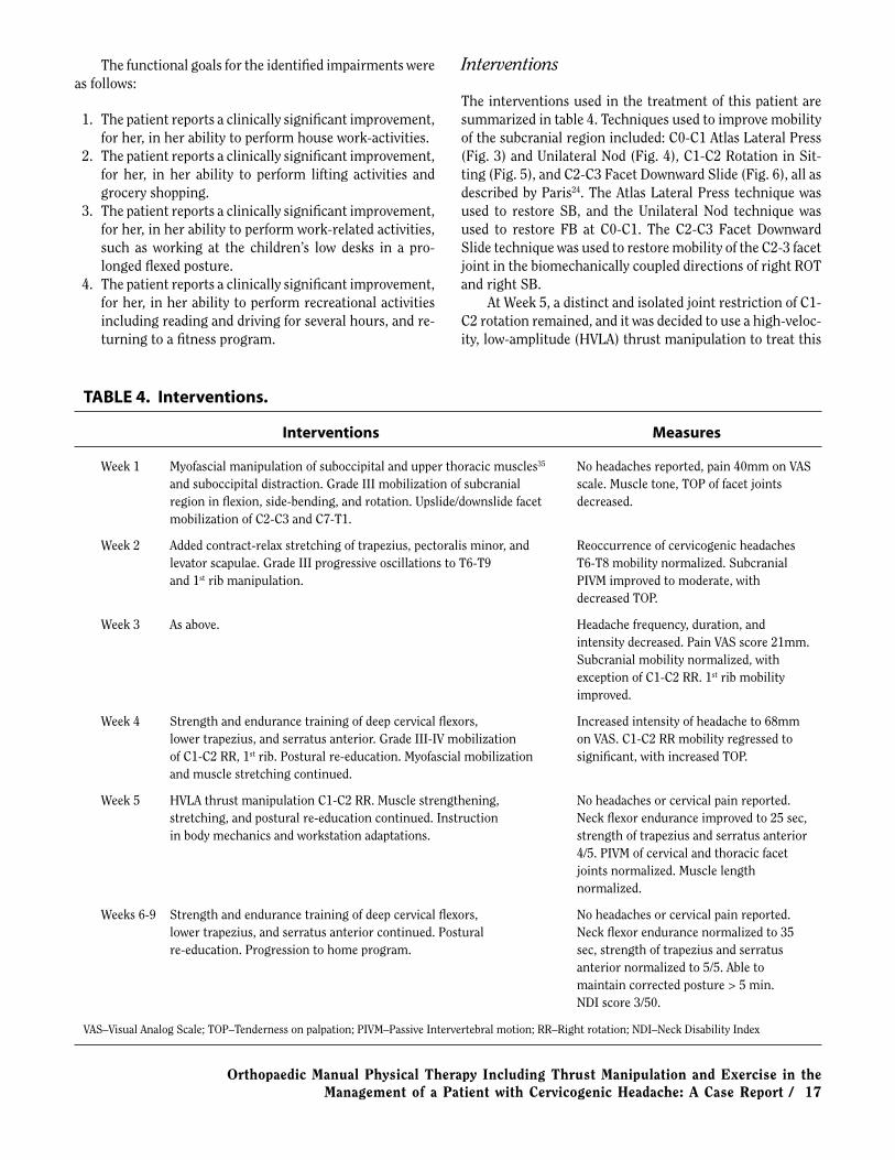
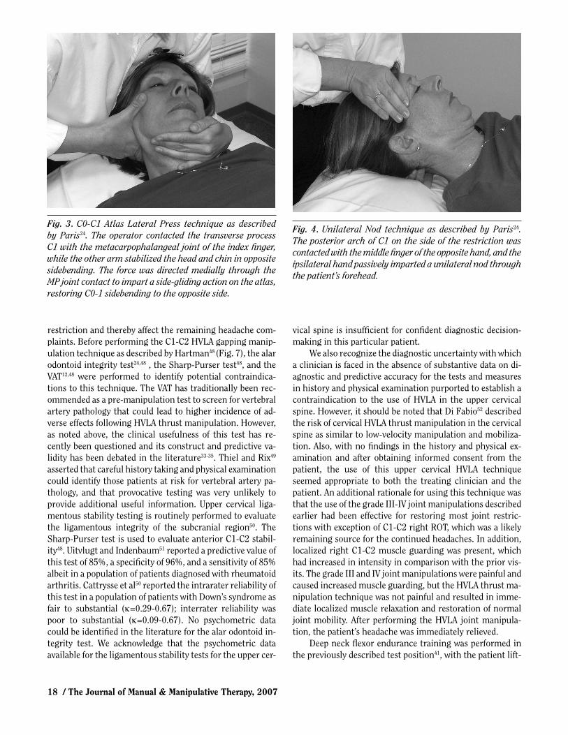
The interventions used in the treatment of this patient are summarized in table 4. Techniques used to improve mobility of the subcranial region included: C0-C1 Atlas Lateral Press (Fig. 3) and Unilateral Nod (Fig. 4), C1-C2 Rotation in Sit-ting (Fig. 5), and C2-C3 Facet Downward Slide (Fig. 6), all as described by Paris24. The Atlas Lateral Press technique was used to restore SB, and the Unilateral Nod technique was used to restore FB at C0-C1. The C2-C3 Facet Downward Slide technique was used to restore mobility of the C2-3 facet joint in the biomechanically coupled directions of right ROT and right SB.
At Week 5, a distinct and isolated joint restriction of C1-C2 rotation remained, and it was decided to use a high-veloc-ity, low-amplitude (HVLA) thrust manipulation to treat this
TABLE 4. Interventions.
Interventions Measures
Week 1 Myofascial manipulation of suboccipital and upper thoracic muscles35 No headaches reported, pain 40mm on VAS and suboccipital distraction. Grade III mobilization of subcranial scale. Muscle tone, TOP of facet joints region in fl exion, side-bending, and rotation. Upslide/downslide facet decreased. mobilization of C2-C3 and C7-T1.
Week 2 Added contract-relax stretching of trapezius, pectoralis minor, and Reoccurrence of cervicogenic headaches levator scapulae. Grade III progressive oscillations to T6-T9 T6-T8 mobility normalized. Subcranial and 1st rib manipulation. PIVM improved to moderate, with decreased TOP.
Week 3 As above. Headache frequency, duration, and intensity decreased. Pain VAS score 21mm. Subcranial mobility normalized, with exception of C1-C2 RR. 1st rib mobility improved.
Week 4 Strength and endurance training of deep cervical fl exors, Increased intensity of headache to 68mm lower trapezius, and serratus anterior. Grade III-IV mobilization on VAS. C1-C2 RR mobility regressed to of C1-C2 RR, 1st rib. Postural re-education. Myofascial mobilization signifi cant, with increased TOP. and muscle stretching continued.
Week 5 HVLA thrust manipulation C1-C2 RR. Muscle strengthening, No headaches or cervical pain reported. stretching, and postural re-education continued. Instruction Neck fl exor endurance improved to 25 sec, in body mechanics and workstation adaptations. strength of trapezius and serratus anterior 4/5. PIVM of cervical and thoracic facet joints normalized. Muscle length normalized.
Weeks 6-9 Strength and endurance training of deep cervical fl exors, No headaches or cervical pain reported. lower trapezius, and serratus anterior continued. Postural Neck fl exor endurance normalized to 35 re-education. Progression to home program. sec, strength of trapezius and serratus anterior normalized to 5/5. Able to maintain corrected posture > 5 min. NDI score 3/50.
VAS–Visual Analog Scale; TOP–Tenderness on palpation; PIVM–Passive Intervertebral motion; RR–Right rotation; NDI–Neck Disability Index
18 / The Journal of Manual & Manipulative Therapy, 2007
Fig. 3. C0-C1 Atlas Lateral Press technique as described by Paris24. The operator contacted the transverse process C1 with the metacarpophalangeal joint of the index fi nger, while the other arm stabilized the head and chin in opposite sidebending. The force was directed medially through the MP joint contact to impart a side-gliding action on the atlas, restoring C0-1 sidebending to the opposite side.
Fig. 4. Unilateral Nod technique as described by Paris24. The posterior arch of C1 on the side of the restriction was contacted with the middle fi nger of the opposite hand, and the ipsilateral hand passively imparted a unilateral nod through the patient’s forehead.
restriction and thereby affect the remaining headache com-plaints. Before performing the C1-C2 HVLA gapping manip-ulation technique as described by Hartman48 (Fig. 7), the alar odontoid integrity test24,48 , the Sharp-Purser test48, and the VAT12,48 were performed to identify potential contraindica-tions to this technique. The VAT has traditionally been rec-ommended as a pre-manipulation test to screen for vertebral artery pathology that could lead to higher incidence of ad-verse effects following HVLA thrust manipulation. However, as noted above, the clinical usefulness of this test has re-cently been questioned and its construct and predictive va-lidity has been debated in the literature33-35. Thiel and Rix49 asserted that careful history taking and physical examination could identify those patients at risk for vertebral artery pa-thology, and that provocative testing was very unlikely to provide additional useful information. Upper cervical liga-mentous stability testing is routinely performed to evaluate the ligamentous integrity of the subcranial region50. The Sharp-Purser test is used to evaluate anterior C1-C2 stabil-ity48. Uitvlugt and Indenbaum51 reported a predictive value of this test of 85%, a specifi city of 96%, and a sensitivity of 85% albeit in a population of patients diagnosed with rheumatoid arthritis. Cattrysse et al50 reported the intrarater reliability of this test in a population of patients with Down’s syndrome as fair to substantial (κ=0.29-0.67); interrater reliability was poor to substantial (κ=0.09-0.67). No psychometric data could be identifi ed in the literature for the alar odontoid in-tegrity test. We acknowledge that the psychometric data available for the ligamentous stability tests for the upper cer-
vical spine is insuffi cient for confi dent diagnostic decision-making in this particular patient.
We also recognize the diagnostic uncertainty with which a clinician is faced in the absence of substantive data on di-agnostic and predictive accuracy for the tests and measures in history and physical examination purported to establish a contraindication to the use of HVLA in the upper cervical spine. However, it should be noted that Di Fabio52 described the risk of cervical HVLA thrust manipulation in the cervical spine as similar to low-velocity manipulation and mobiliza-tion. Also, with no fi ndings in the history and physical ex-amination and after obtaining informed consent from the patient, the use of this upper cervical HVLA technique seemed appropriate to both the treating clinician and the patient. An additional rationale for using this technique was that the use of the grade III-IV joint manipulations described earlier had been effective for restoring most joint restric-tions with exception of C1-C2 right ROT, which was a likely remaining source for the continued headaches. In addition, localized right C1-C2 muscle guarding was present, which had increased in intensity in comparison with the prior vis-its. The grade III and IV joint manipulations were painful and caused increased muscle guarding, but the HVLA thrust ma-nipulation technique was not painful and resulted in imme-diate localized muscle relaxation and restoration of normal joint mobility. After performing the HVLA joint manipula-tion, the patient’s headache was immediately relieved.
Deep neck fl exor endurance training was performed in the previously described test position41, with the patient lift-

Orthopaedic Manual Physical Therapy Including Thrust Manipulation and Exercise in the Management of a Patient with Cervicogenic Headache: A Case Report / 19
ing the head one inch from the surface with the chin maxi-mally retracted. The contraction time was progressively in-creased based on patient tolerance.
Outcomes
The patient was seen for a total of 16 visits over a 9-week pe-riod of time. The initial frequency of treatment was 3 times per week and was gradually decreased to once weekly during Weeks 6-9 to monitor progression of the home program. At time of discharge, the patient had reported no headaches for three weeks. Her NDI score had decreased to 3/50 from an initial level of 20/50, which indicated that a clinically mean-ingful change had occurred based on the minimal clinically important difference of 7 points16. The VAS pain scale score ranged from 0-5 (mm) with some daily variation, compared to a maximal initial level of 80 (mm), which represented a true change based on a minimal detectable change of 28 (mm)16. The location of the remaining pain was in the upper thoracic region.
Active cervical range of motion assessed by visual esti-mation had normalized. Segmental PIVM tests of the subcra-nial, mid-cervical, and thoracic joints that were initially identifi ed as hypomobile now revealed normal range and
end-feel, and did not reproduce pain. Moderate hypermobil-ity of the C5-C6 vertebral segment on PIVM testing remained unchanged but as this passive test is not a test of functional stability, this test result was expected. Muscle length of up-per trapezius, pectoralis minor, and levator scapulae muscles
Fig. 5. C2-C3 Facet Downward Slide technique as described by Paris24. With the patient situated in supine, the operator contacted the lower aspect of the facet joint with the 2nd metacarpophalageal joint of the ipsilateral hand. The other hand was placed on the contralateral side of the head and imparted sidebending towards the involved side down to the restricted level. A progressive oscillatory force was imparted through the MP II joint in an inferior, medial, and posterior direction.
Fig. 6. C1-C2 Rotation in Sitting technique as described by Paris24. In order to improve right rotation, the operator stood on the right side, with the right forearm and hand contacting the head of the patient. The left thumb was placed against the lamina of C2. The patient’s head was rotated towards the right around a vertical axis using gentle progressive oscillations, while the left thumb blocked movement at C2.
Fig. 7. C1-C2 high-velocity, low-amplitude gapping manipulation technique as described by Hartman48.
20 / The Journal of Manual & Manipulative Therapy, 2007
was normal upon re-evaluation. Neck fl exor endurance41 had improved to 35 seconds compared to an initial score of 18 seconds, placing this patient’s score in the 44th percentile of the reported normative asymptomatic group (compared to 21st percentile initially), and in the 80th percentile of the symptomatic group (compared to 32nd percentile initially); both scores seem to indicate normalization of this impair-ment identifi ed on the initial evaluation. Strength of the lower trapezius and serratus anterior muscles had increased to 5/5. Postural awareness and control had improved, with the patient able to reproduce and maintain corrected pos-ture for extended periods of time, i.e., >5 (min) as compared to being unable to hold corrected posture for 30 (sec) on the initial evaluation. The patient was contacted 6 months and 1 year after discharge, and at both instances she reported that her status was essentially unchanged from the time of discharge indicating positive long-term effects in addition to the positive effects noted upon discharge from PT treatment as described above.
Discussion
This case report describes the PT differential diagnosis, man-agement, and outcomes of a patient with cervicogenic head-ache. The patient was a 40-year-old woman referred by her physiatrist with complaints of cervical pain and ipsilateral temporal headache. The patient presented with increased muscle tone, multiple-level cervical and thoracic joint hypo-mobility, muscle weakness, and postural changes. We used the VAS and NDI as self-report standardized and validated outcome measures. Management consisted of various thrust and non-thrust manipulations, soft tissue mobilizations, postural re-education, and exercise to address the identifi ed impairments noted above. At discharge, the patient demon-strated clinically meaningful improvements with regard to pain, disability, and headache intensity and frequency. The patient’s improvement on the NDI and VAS outcomes mea-sures exceeded the minimal clinically important difference
and minimal detectable change reported for these measures, respectively, and thus represented a clinically meaningful and true change, respectively, in patient status. This case re-port indicates that a multimodal PT treatment program in-cluding OMPT using thrust and non-thrust joint manipula-tion and soft tissue techniques, exercise, and education may be effective in the management of a patient diagnosed with cervicogenic headache.
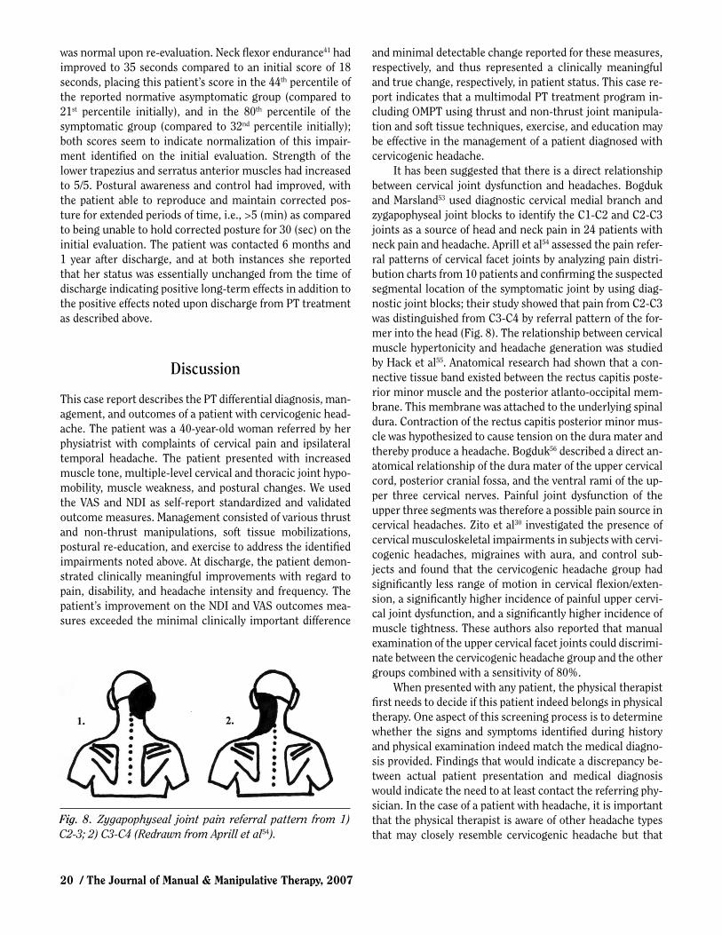
It has been suggested that there is a direct relationship between cervical joint dysfunction and headaches. Bogduk and Marsland53 used diagnostic cervical medial branch and zygapophyseal joint blocks to identify the C1-C2 and C2-C3 joints as a source of head and neck pain in 24 patients with neck pain and headache. Aprill et al54 assessed the pain refer-ral patterns of cervical facet joints by analyzing pain distri-bution charts from 10 patients and confi rming the suspected segmental location of the symptomatic joint by using diag-nostic joint blocks; their study showed that pain from C2-C3 was distinguished from C3-C4 by referral pattern of the for-mer into the head (Fig. 8). The relationship between cervical muscle hypertonicity and headache generation was studied by Hack et al55. Anatomical research had shown that a con-nective tissue band existed between the rectus capitis poste-rior minor muscle and the posterior atlanto-occipital mem-brane. This membrane was attached to the underlying spinal dura. Contraction of the rectus capitis posterior minor mus-cle was hypothesized to cause tension on the dura mater and thereby produce a headache. Bogduk56 described a direct an-atomical relationship of the dura mater of the upper cervical cord, posterior cranial fossa, and the ventral rami of the up-per three cervical nerves. Painful joint dysfunction of the upper three segments was therefore a possible pain source in cervical headaches. Zito et al30 investigated the presence of cervical musculoskeletal impairments in subjects with cervi-cogenic headaches, migraines with aura, and control sub-jects and found that the cervicogenic headache group had signifi cantly less range of motion in cervical fl exion/exten-sion, a signifi cantly higher incidence of painful upper cervi-cal joint dysfunction, and a signifi cantly higher incidence of muscle tightness. These authors also reported that manual examination of the upper cervical facet joints could discrimi-nate between the cervicogenic headache group and the other groups combined with a sensitivity of 80%.
When presented with any patient, the physical therapist fi rst needs to decide if this patient indeed belongs in physical therapy. One aspect of this screening process is to determine whether the signs and symptoms identifi ed during history and physical examination indeed match the medical diagno-sis provided. Findings that would indicate a discrepancy be-tween actual patient presentation and medical diagnosis would indicate the need to at least contact the referring phy-sician. In the case of a patient with headache, it is important that the physical therapist is aware of other headache types that may closely resemble cervicogenic headache but that
Fig. 8. Zygapophyseal joint pain referral pattern from 1) C2-3; 2) C3-C4 (Redrawn from Aprill et al54).

Orthopaedic Manual Physical Therapy Including Thrust Manipulation and Exercise in the Management of a Patient with Cervicogenic Headache: A Case Report / 21
either present an indication for medical management, pose a contraindication to PT intervention, or at the very least are not amenable to (sole) PT management. The pattern identi-fi ed in the review of the literature above of cervical joint dys-function, postural changes, muscular dysfunction, and cer-vical headaches was present in the case presented in this paper. Following the physical examination and analysis of data, we concluded that the patient had headache symptoms and cervical joint and muscular dysfunction that might be related to each other.
Headaches can be classifi ed in three major types: vascu-lar, traction (infl ammatory), and headache of musculoskele-tal origin24. Vascular headaches include migraines, cluster headaches, toxic vascular, and hypertensive headaches. The patient’s headache symptoms were not vascular in origin be-cause they did not follow the typical migraine or cluster headache description, and the patient did not show any signs of toxic vascular or hypertensive disease in her medical his-tory. Traction or infl ammatory headaches are caused by mass lesions, such as tumors, edema, haematoma, diseases of the eye, ear, nose, and throat, or by occlusive vascular disease. The patient’s medical history and physical examination did not reveal any warning signs associated with this type of headache, such as vomiting associated with nausea, sleepi-ness, dimmed vision, or a stiff neck with severe rapidly in-creasing headache.
Headaches of musculoskeletal origins are the tension-type headache and the cervicogenic or cervical headache as described by IHS2. The patient presented with signs and symptoms characteristic of cervical headache. The major fi ndings that led to this diagnosis were (a) unilateral head-aches and suboccipital pain, aggravated by certain head movements and prolonged fl exed posture; (b) cervical joint dysfunction; (c) muscle hypertonicity, tenderness, and de-creased extensibility; and (d) abnormal posture and pathol-ogy reported in the radiological examination. Other relevant clinical fi ndings in this case were hypermobility of one cervi-cal level (C5-C6), upper and mid-thoracic joint restrictions, and the presence of decreased strength and endurance in specifi c postural musculature. Tension-type headache crite-ria include headache lasting from 30 minutes to 7 days, two of the criteria of pressing/tightening quality, mild to moder-ate intensity (not inhibiting activity), bilateral location, not aggravated by walking or stair climbing, no vomiting, nau-sea, or presence of photophobia. Since this patient presented with unilateral, high-intensity headaches that were of long duration, were aggravated by activity, and affected the pa-tient’s function, it was felt that the criteria for tension-type headache were not met.
Physical therapy management within the evidence-based practice paradigm is decided by best available evidence. In many cases—including the case report discussed here—best evidence means a combination of pathophysiologic hy-potheses and research evidence. A limited number of studies
have investigated the outcomes of various interventions for the treatment of cervicogenic headaches. We performed a review of the literature using computerized literature searches of the following databases: CINAHL, FirstSearch (incl. MedLine), OVID, ProQuest, and ScienceDirect. We used MeSH terms and other keywords including cervico-genic headaches, physical therapy, manual therapy, manipu-lation, mobilization, and exercise. The results of this litera-ture review supported the diagnosis and management of this patient. The effectiveness of treatment of this case is compa-rable with the outcomes described in the literature. A recent clinical trial by Jull et al6 involving 200 patients with cervi-cogenic headaches, compared the effect of various combina-tions of specifi c strengthening and endurance exercises of the deep neck fl exors and other postural muscles, and man-ual therapy treatment of the cervical joints. That study used four treatment groups: manipulative therapy group, exercise group, combined manipulative therapy and exercise group, and a control group. Outcomes measures included the fre-quency of headaches, headache intensity and duration, the Northwick Park Neck Pain Index, medication intake, and pa-tient satisfaction. Patients who received active exercise, manual therapy, or a combination of active exercise and manual therapy displayed signifi cantly better outcomes (fre-quency of headaches, pain intensity, neck pain) than the con-trol group. This effect was maintained at the 12-month follow-up. There was no statistically signifi cant difference in outcomes among the different treatment groups; however, 10% more patients experienced relief with the combination of manipulative therapy and exercise than with any of these interventions alone. Nilsson57 conducted an RCT of 53 sub-jects with cervicogenic headache, comparing the outcomes of HVLA manipulation of the cervical spine to a control group who received low-level laser treatment and deep fric-tion massage. The intensity, duration, and frequency of headaches were signifi cantly reduced in the HVLA manipula-tion group immediately following the 3-week treatment pro-tocol; however, long-term follow-up was not performed. Gross et al58 performed a Cochrane systematic review of 33 randomized clinical trials evaluating the effectiveness of ma-nipulation and mobilization compared to other forms of conservative therapy for the treatment of mechanical neck disorders. These authors concluded that strong evidence ex-ists for the effectiveness of multimodal care (manipulation and/or mobilization in combination with exercise) in the treatment of mechanical neck disorders with or without headache; however, they did not fi nd evidence for the effec-tiveness of manipulation and/or mobilization done as the only intervention. Molina8 performed a review of clinical tri-als, descriptive studies, and case reports regarding the treat-ment effi cacy of manipulation related to upper cervical spine dysfunction and cervical headaches. Effi cacy studies were described measuring the effect of manipulation on symp-toms, cervical mobility, and proprioception-dependent per-

22 / The Journal of Manual & Manipulative Therapy, 2007
formance. The outcome of these studies showed a moderate to good effectiveness of treatment measured during or im-mediately post-treatment and moderate long-term (3 to 6 months post-treatment) effectiveness; however, the author noted that the descriptions of the study designs and specifi c-ity of the description of interventions used were insuffi cient in a number of the studies reviewed, and the number of qual-ity studies was limited. A recent study59 evaluating the meth-odological quality of RCTs of spinal manipulation and mobi-lization in the treatment of headaches reached a similar conclusion that the number of studies in this area is limited, and that the methodological quality of these papers is typi-cally low. The authors identifi ed the clear need for additional high-quality RCTs in this area.
A forward head posture and weakness of the upper cervi-cal fl exors has been commonly observed in headache pa-tients60,61. Watson and Trott5 investigated the relationship between forward head posture and weakness of the upper cervical fl exor musculature in cervical headache patients. In this study, 60 female subjects, aged 25-40, were divided in two groups (headache and non-headache group). The proto-col to examine the subjects consisted of lateral photographs of natural head posture, isometric strength and endurance testing of upper cervical fl exor muscle group, passive inter-vertebral movement testing, and the completion of a ques-tionnaire (by the headache group). The headache group was found to be signifi cantly different from the non-headache group with respect to increased forward head posture, less isometric strength, and less endurance of the upper cervical fl exor musculature. It was concluded that cervical headache patients exhibited forward head posture and demonstrated weakness and lack of endurance of the upper cervical fl exor musculature. The authors found a signifi cant relationship of endurance and forward head posture in cases of cervical headaches as analyzed by the Chi-square test (χ2 = 13.7, p <0.01), in that lower endurance values corresponded with a more pronounced forward head posture. They con-cluded that management of patients with cervical headaches should emphasize postural correction and re-education in addition to specifi c endurance training of upper cervical fl exor musculature.
Primary impairments (abnormalities of structure or function62) can result in the development of secondary im-pairments. For this patient, we considered it likely that sig-nifi cant restrictions of the cervico-thoracic junction and mid-thoracic region, in combination with postural defi cits (primary impairment), resulted in the subcranial dysfunc-tion (secondary impairment). Restrictions in both subcra-nial and thoracic spine could then have caused increased stress to the C5-C6 segment, resulting in its hypermobility. Prolonged positioning of the head in forward head position and of the shoulders in a rounded position could have re-sulted in the observed adaptive shortening of the pectoralis minor and major, the upper trapezius muscles, and the right
levator scapulae muscle, and decreased strength of the scap-ular stabilizing muscles. The muscle weakness and the re-sulting inability to maintain a corrected posture for any length of time might also have been one of the underlying causes of the patient’s postural defi cits.
This case report describes a multimodal treatment ap-proach to management of cervical headaches patients by physical therapists. This multimodal treatment approach was based on a combination of the evidence provided in the literature and pathophysiologic hypotheses. Manipulation techniques, including HVLA thrust, appear to be effective interventions for the management of cervical joint dysfunc-tion when integrated in a comprehensive treatment ap-proach. Several studies described the importance of postural correction and the specifi c endurance training of the deep cervical fl exors3-6, 8. It is essential that underlying impair-ments of decreased mobility, strength, endurance, and pos-tural control be addressed in addition to managing subjec-tive symptoms. A comprehensive treatment approach emphasizing the restoration of normal joint mobility, strengthening of specifi c postural muscle groups, postural retraining, and patient education in self-management skills may provide long-term treatment success. Of course, the format of a case report does not allow us to infer a cause-and-effect relationship between intervention and reported out-comes and indicates the need for further outcome studies in this area. This case report also clearly identifi ed the need of further research in the area of psychometric properties of a number of tests and measures used in the evaluation of pa-tients with cervicogenic headaches, including upper cervical ligament stability testing, evaluation of muscle tone, pos-tural analysis, and methods used for manual diagnosis.
Conclusion
This case report illustrates the PT differential diagnosis, management, and outcomes of a patient diagnosed with cer-vicogenic headaches. This patient presented with headache symptoms matching the classifi cation of cervicogenic head-ache as defi ned by the IHS2, dysfunctions in the subcranial, the mid-cervical, and thoracic joints, decreased strength and endurance in postural musculature, and postural defi cits. A multimodal PT treatment approach was used based on evi-dence in the literature and pathophysiologic hypotheses, and it included the use of non-thrust and thrust manipulation techniques, therapeutic exercise, and postural correction. Clinically meaningful short- and long-term improvements with regard to pain, disability, and headache were reported with at least a temporal relation to this treatment approach. Physical therapy management of the cervical headache pa-tient should address all identifi ed impairments with inter-ventions including non-thrust joint manipulation, HVLA thrust manipulation, soft tissue manipulation and stretch-

Orthopaedic Manual Physical Therapy Including Thrust Manipulation and Exercise in the Management of a Patient with Cervicogenic Headache: A Case Report / 23
ing techniques, retraining specifi c postural muscle groups, and patient education. A need for further research exists in the areas of psychometric properties of a number of the tests
and measures commonly used during the diagnostic process for these patients and in the area of outcomes of PT treat-ment of patients with cervicogenic headaches. ■
REFERENCES
1. Nilsson N. The prevalence of cervicogenic headache in a random population sample of 20–59 year olds. Spine 1995;20:1884–1888.
2. Headache Classifi cation Subcommittee of the International Head-ache Society. The international classifi cation of headache disorders. 2nd edition. Cephalalgia 2004;24:suppl 1.
3. Horn C, Smith KL. Cervicogenic headache Part II: Clinical examina-tion, fi ndings and approaches to management. J Manual Manipula-tive Ther 1997;5:171–175.
4. Jull G. Management of cervical headache. Man Ther 1997;2:182–190. 5. Watson DH, Trott PH. Cervical headache: An investigation of natu-
ral head posture and upper cervical fl exor performance. Cephalalgia 1993;13:272–284.
6. Jull G, Trott P, Potter H, et al. A randomized controlled trial of ex-ercise and manipulative therapy for cervicogenic headache. Spine 2002;27:1835–1843.
7. Vernon H. Spinal manipulation in the management of tension-type migraine and cervicogenic headaches: The state of evidence. Topics Clin Practice 2002; 9:14–20.
8. Molina P. Management of upper cervical spine disorders and cervico-genic headache. Orthop Phys Ther Clinics N Am 1999;8(1):45–67.
9. Vernon H, Steiman I, Hagino C. Cervicogenic dysfunction in muscle contraction headache and migraine: A descriptive study. J Manipula-tive Physiol Ther 1992;15:418–429.
10. Jensen O, Nielsen F, Vosmar L. An open study comparing manual therapy with the use of cold packs in the treatment of post-traumatic headache. Cephalalgia 1990;10:241–250.
11. Schoensee SK, Jensen G, Nicholson G, Gossman M, Katholi C. The effect of mobilization on cervical headaches. J Orthop Sports Phys Ther 1995; 21:184–196.
12. Paris SV, Loubert PV. Foundation of Clinical Orthopedics. 3rd ed. St. Augustine, FL: Institute Press, 1999:308–312.
13. Huskinsson EC. Visual analogue scales. In: Melzack R, ed. Pain Mea-surement and Assessment. New York, NY: Raven Press, 1983: 33–37.
14. Finch E, Brooks D, Stratford PW, Mayo NE. Physical Rehabili-tation Outcomes Measures. 2nd ed. Hamilton, ON: BC Decker, 2000: 173–174.
15. Bertilson B, Grunnosjo M, Strender L. Reliability of clinical tests in the assessment of patients with neck/shoulder problems: Impact of history. Spine 203;28:2222–2231.
16. Vernon H, Mior S. The neck disability index: A study of reliability and validity. J Manipulative Physiol Ther 1991;14:411.
17. Fedorak C, Ashworth N, Marshall J, Paull H. Reliability of the visual assessment of cervical and lumbar lordosis: How good are we? Spine 2000;28:1857–1859.
18. Youdas J, Cary J, Garrett T. Reliability of measurement of cervical spine ROM: Comparison of three models. Phys Ther 1991;71:98–106.
19. Van Suijlekom H, deVet H, van den Berg S, Weber W. Interobserver reliability in physical examination of the cervical spine in patients with headache. Headache 2000;40:581–586.
20. Viikari-Juntura E. Interexaminer reliability of observations in physi-cal examinations of the neck. Phys Ther 1987;67:1526–1532.
21. Aprill C, Axinn MJ, Bogduk N. Occipital headaches stemming from the lateral atlanto-axial (C1-2) joint. Cephalalgia 2002;22: 15–22.
22. Schoeps P, Pfi ngsten M, Siebert U. Reliabilität manualmedizinischer Untersuchungstechniken an der Halswirbelsäule. Studie zur Qual i-täts s ich erung in der manuellen Diagnostik. Z Orthop Ihre Grenzgeb 2000;138:2–7.
23. Keating J, Bergmann T, Jacobs G, Finer B, Larson K. Interexaminer reliability of eight evaluative dimensions of lumbar segmental ab-normality. J Manipulative Physiol Ther 1990;13:463–470.
24. Paris SV. Course notes S3. Atlanta, GA: Institute Press; 1988.25. Paris SV, Nyberg R, Irwin M. Course notes S2. St.Augustine, FL:
Institute of Physical Therapy: 1993.26. Smedmark V, Wallin M, Arvidsson I. Inter-examiner reliability in as-
sessing passive intervertebral motion of the cervical spine. Man Ther 2000;5:97–101.
27. Pool J, Hoving J, de Vet H, van Memeren H, Bouter L. The interex-aminer reproducibility of physical examination of the cervical spine. J Manipulative Physiol Ther 2003;27:84–90.
28. Huijbregts PA. Spinal motion palpation: A review of reliability stud-ies. J Manual Manipulative Ther 2002;10:24–39.
29. Jull G, Bogduk N, Marsland A. The accuracy of manual diagno-sis for cervical zygapophysial joint pain syndromes. Med J Aust 1988;148(5):233–236.
30. Zito G, Jull G, Story I. Clinical tests of musculoskeletal dys-function in the diagnosis of cervicogenic headache. Man Ther 2006;11:118–129.
31. Jull G, Zito G, Trott P, Potter H, Shirley D, Richardson C. Interex-aminer reliability to detect painful upper cervical joint dysfunction. Aust J Physiother 1997;43:125–129.
32. Wainner R, Fritz J, Irrgang J, Boninger M, Deltto A, Allison S. Reliability and diagnostic accuracy of the clinical examination and patient self-report measures for cervical radiculopathy. Spine 2003;28:52–62.
33. Rivett DA, Sharples KJ, Milburn PD. Effect of premanipulative tests on vertebral artery and internal carotid artery blood fl ow: A pilot study. J Manipulative Physiol Ther 1999;22:368–375.
34. Licht PB, Chistensen HW, Hoilund-Carlsen PF. Carotid artery blood fl ow during premanipulative testing. J Manipulative Physiol Ther 2002;25:568–572.
35. Zaina C, Grant R, Johnson C, Dansie B, Taylor J, Spyropolous P. The effect of cervical rotation on blood fl ow in the contralateral vertebral artery. Man Ther 2003;8:103–109.
36. Kendall FP, McCreary EK, Provance PG. Muscles: Testing and Func-tion. 4th ed. Baltimore, MD: Williams and Wilkins, 1995.
37. Barr AE, Diamond BE, Wade CK, Harashima T, Pecorella WA, Potts CC, Rosenthal H, Fleiss JL, McMahon DJ. Reliability of testing mea-sures in Duchenne or Becker muscular dystrophy. Arch Phys Med Rehabil 1991;72:315–319.
38. Brandsma JW, Schreuders TA, Birke JA, Piefer A, Oostendorp R. Manual muscle strength testing: Intraobserver and interobserver reliabilities for the intrinsic muscles of the hand. J Hand Ther 1995;8:185–190.
39. Wadsworth CT, Krishnan R, Sear M, Harrold J, Nielsen DH. Intra-

24 / The Journal of Manual & Manipulative Therapy, 2007
rater reliability of manual muscle testing and hand-held dynametric muscle testing. Phys Ther 1987;67:1342–1347.
40. Florence JM, Pandya S, King WM, Robison JD, Baty J, Miller JP, Schierbecker J, Signore LC. Intrarater reliability of manual muscle test (Medical Research Council scale) grades in Duchenne’s muscular atrophy. Phys Ther 1992;72:115–122.
41. Harris KD, Heer DM, Roy TC, Santos DM, Whitman JM, Wainner RS. Reliability of a measurement of neck fl exor muscle endurance. Phys Ther 2005;85:1349.
42. McKinnis LN. Fundamentals of Orthopedic Radiology. Philadelphia, PA: FA Davis Company; 1997.
43. McAviney J, Schulz D, Bock R, Harrison D, Holland B. Determining the relationship between cervical lordosis and neck complaints. J Manipulative Physiol Ther 2005;28:187–193
44. Helliwell PS, Evans PF, Wright V. The straight cervical spine: Does it indicate muscle spasm? J Bone Joint Surg Br 1994;76: 103–106.
45. Kettler A, Rolmann F, Neidlinger-Wilke C, Werner K, Claes, Wilke HJ. Validity and interobserver agreement of a new radiographic grading system for intervertebral disc degeneration. Part II: Cervical spine. Eur Spine J 2006;15:732–741.
46. Jull GA, Stanton WR. Predictors of responsiveness to physio-therapy management of cervicogenic headache. Cephalalgia 2004; 25:101–108.
47. Coeytaux RR, Spierings ELH. Prognostic factors, disability, and functional status among patients in a headache specialty practice. Cephalalgia 2005;26:7–13.
48. Hartman L. Handbook of Osteopathic Technique. 3rd ed. Cheltenham, UK: Stanley Thornes Ltd., 1998:185–186.
49. Thiel H, Rix E. Is it time to stop functional pre-manipulation testing of the cervical spine? Man Ther 2005;10:154–158.
50. Cattrysse E, Swinkels RAHM, Oostendorp RAB, Duquet W. Upper cer-vical instability: Are clinical tests reliable? Man Ther 1997;2:91–97.
51. Uitvlugt G, Indenbaum S. Clinical assessment of atlantoaxial insta-bil ity using the Sharp-Purser test. Arthritis Rheum 1988;31; 918–922.
52. Di Fabio RP. Manipulation of the cervical spine: Risks and benefi ts. Phys Ther 1999;79:50–65.
53. Bogduk N, Marsland A. The cervical zygapophysial joints as a source of neck pain. Spine 1988;13:610–617.
54. Aprill C, Dwyer A, Bogduk N. Cervical zygapophyseal joint pain pat-terns II: A clinical evaluation. Spine 1990;15(6):458–461.
55. Hack G, Koritzer T, Robinson W, Hallgren R, Greenman P. Anatomic relation between the rectus capitus posterior minor muscle and the dura mater. Spine 1995;20(23):2484–2486.
56. Bogduk N. Cervical causes of headache and dizziness. In: Boyling J, Palastanga N, eds. Grieve’s Modern Manual Therapy of the Vertebral Column. Edinburgh, UK: Churchill Livingstone, 1994:317–332.
57. Nilsson N, Christensen HW, Hartvigsen J. The effect of spinal ma-nipulation in the treatment of cervicogenic headache. J Manipulative Physiol Ther 1997;20:326–330.
58. Gross AR, Hoving JL, Haines TA, et al. A Cochrane review of ma-nipulation and mobilization for mechanical neck disorders. Spine 2004;29(14):1541–1548.
59. Fernandez-de-las-Penas C, Alonso-Blanco C, San-Roman J, Miango-larra-Page JC. Methodological quality of randomized controlled trials of spinal manipulation and mobilization in tension-type headache, migraine, and cervicogenic headaches. J Orthop Sports Phys Ther 2006;36(3):160–169.
60. Janda V. Muscles and cervicogenic pain syndromes. In: Grant R (ed). Physical therapy of cervical and thoracic spine. Edinburgh, UK: Churchill Livingston; 1988.
61. Paris V. Cervical symptoms of forward head posture. Topics Geriatric Rehabil 1990;5:11–19.
62. Guide to Physical Therapist Practice, 2nd Ed. Phys Ther 2001;81: 9–744.