original articles feeling sounds after a thalamic lesion
TRANSCRIPT

Feeling Sounds after a Thalamic LesionTony Ro, PhD,1,2 Alessandro Farne, PhD,1,3 Ruth M. Johnson, MA,1 Van Wedeen, MD,4 Zili Chu, PhD,5
Zhiyue J. Wang, PhD,5 Jill V. Hunter, MD,5 and Michael S. Beauchamp, PhD6
Objective: The ventrolateral nucleus of the thalamus (VL), based on its connectivity with the cerebellum and motor cortex, haslong been considered to be involved with motor functions. We show that the human VL also plays a prominent role in sensoryprocessing.Methods: Structural magnetic resonance imaging and diffusion tensor imaging were used to localize a small lesion restricted tothe right VL in a patient with contralesional sensory processing deficits. Systematic assessments of anatomic brain organizationand behavioral measurements of somatosensory and visual processing were conducted at several time points after stroke.Results: Initially, the patient was more likely to detect events on the contralesional side when a simultaneous ipsilesional eventwas presented within the same, but not different, sensory modality. This perceptual phenomenon, which we refer to as unisen-sory antiextinction, persisted for several months before transforming into a form of synesthesia in which auditory stimuli pro-duced tactile percepts. Tractography performed on the diffusion tensor imaging data showed altered connections from thelesioned thalamus to the cerebral cortex, suggesting a neural basis for these sensory changes.Interpretation: These results demonstrate a role for the VL in sensory processing and suggest that reorganization of thalamo-cortical axonal connectivity can lead to major changes in perception.
Ann Neurol 2007;62:433–441
In [other patients] the thought, sight, or noise of a scratch-ing, rubbing or scraping motion evoked severe tingling in theaffected hand. —M. B. Bender (1945)
The influences of one sensory stimulus on the detec-tion of another simultaneously occurring target stimu-lus can often be dramatic, with target detection eitherimpaired or facilitated depending on the nature of thetask.1–5 This is especially true in patients with lateral-ized sensory and attentional deficits consequent to uni-lateral brain damage. For example, patients with hemis-patial neglect after right temporal/parietal damage (forreviews, see Driver and Mattingley,6 Rafal,7,8 and Val-lar9) frequently show extinction, that is, the inability todetect and report a contralesional event when simulta-neously presented with an ipsilesional one.10–12 Extinc-tion can occur after either cortical or subcortical dam-age,12,13 including thalamic lesions,14 is considered tobe a high-level attentional deficit rather than a low-level sensory disorder, and can affect visual, somatosen-sory, and auditory processing. Recently, multisensoryextinction has also been demonstrated (eg, a visualevent on the ipsilesional side extinguishing a contrale-
sional tactile one15–17), supporting the idea that extinc-tion occurs at a high level after initial sensory encod-ing.
Unilateral brain damage can also produce mispercep-tions of a single sensory stimulus, as in the phenomenaof allesthesia and intermanual sensory referral. Allesthe-sia occurs when a unilateral tactile stimulus deliveredto the impaired side is felt in a corresponding area onthe intact side.12 In contrast, intermanual sensory re-ferral occurs when a tactile stimulus delivered to theintact side is felt in a corresponding area on the af-fected side.12,18 In both cases, a unilateral tactile stim-ulus is mislocalized to the corresponding portion of theopposite side of the body, suggesting cross talk betweensomatotopic representations in the two hemispheres ofthe brain. As with extinction, allesthesia and inter-manual sensory referral can each occur after either cor-tical or subcortical damage.12,18
Another related perceptual phenomenon has been re-ferred to as antiextinction, in which detection of con-tralesional events is better when a simultaneous ipsile-sional stimulus is presented.19,20 Whereas the
From the 1Department of Psychology, Rice University; 2Depart-ment of Physical Medicine and Rehabilitation, Baylor College ofMedicine, Houston, TX; 3Institut National de la Sante et de la Re-cherche Medicale, U864, Espace et Action, Bron, France; 4Athi-noula A. Martinos Center for Biomedical Imaging, MassachusettsGeneral Hospital, Boston, MA; 5Department of Radiology, BaylorCollege of Medicine and Texas Children’s Hospital; and 6Depart-ment of Neurobiology and Anatomy, University of Texas HealthScience Center-Houston, Houston, TX.
Received May 22, 2007, and in revised form Jul 6. Accepted forpublication Jul 27.
This article includes supplementary materials available via the Inter-net at http://www.interscience.wiley.com/jpages/0364-5134/supp-mat
Published online Sep 24, 2007 in Wiley InterScience(www.interscience.wiley.com). DOI: 10.1002/ana.21219
Address correspondence to Dr Ro, Department of Psychology,MS25, Rice University, 6100 Main Street, Houston, TX 77005-1892. E-mail: [email protected]
ORIGINAL ARTICLES
© 2007 American Neurological Association 433Published by Wiley-Liss, Inc., through Wiley Subscription Services

psychological and neural mechanisms underlying anti-extinction remain unclear, the consistent modulationof antiextinction by task requirements suggests thattop-down modulation of sensory processing plays arole.19,20 Although antiextinction has been reported inonly two patients, both patients appeared to have dam-age to the parietal cortex and thalamus.
In this article, we describe the results of several be-havioral and neuroimaging studies conducted on a pa-tient with unique sensory phenomena after a small anddiscrete lesion to the right ventrolateral nucleus of thethalamus (VL; Fig 1). In the initial poststroke experi-ments, we observed multisensory neglect with unisen-sory antiextinction in this patient. More recently, how-ever, we measured a new brain-damage–inducedphenomenon where a stimulus in one sensory modalityinduces sensations in another. Specifically, the patientdemonstrated a striking form of sensory reorganizationafter thalamic damage: approximately 18 months afterthe initial antiextinction phenomena, the patient beganto feel tactile sensations in response to sounds.
Patient and MethodsPatientThe patient was a 36-year-old, right-handed female professorwho approximately 9 months before the first examinationhad suffered a lacunar infarct to the right VL. Subsequent tothe infarct, she reported noticeable changes in her sensoryand attentional abilities, consistent with hemispatial neglect.For example, she reported occasionally walking into andbumping the left sides of doorways and tended to veer right-ward when driving. Neuropsychological and neurological as-sessments conducted approximately 1 year after stroke, how-ever, demonstrated no measurable signs of visual neglect asher line bisection, line and shape cancellation, and doublevisual simultaneous stimulation performance were all normal.Somatosensory functions were abnormal on the contrale-sional half of her body, with decreased sensations to fingertaps and pricks on her left foot, hand, arm, and face. Apartfrom the decreased sensations contralesionally, the patient re-ported no other sensory or motor disturbances. The patientreported no form of synesthesia before the infarct.
Apparatus and StimuliFor all testing sessions, an Intel-based personal computer wasused for stimulus timing and presentation. For the first test-ing session, the computer controlled the presentation of thevisual stimuli, which were light emitting diodes (LEDs) thatwere illuminated for 5 milliseconds, and the tactile stimuli,which were 2cm filaments that were attached on solenoids.The tactile stimuli were delivered by a computer-interfaced,custom-made relay switchboard, which controlled the sole-noids. The LEDs and solenoids were mounted onto a verti-cal Plexiglas sheet placed approximately 57cm from the pa-tient’s eyes and directly above her outstretched hands thatwere resting on a table. A small filled square (0.1°) served asthe fixation point in the center of the display, which was20cm above and 15cm to the left and right of the LEDs and
the right and left hands. On each trial, either a unilateraltactile (left or right), unilateral visual (left or right), bilateraltactile, bilateral visual, multisensory with tactile left/visualright, multisensory with visual left/tactile right, or no stim-ulus (catch trials) was delivered. Because we were interestedin the magnitude of extinction that this patient exhibited,there were twice as many bilateral trials as unilateral trials ineach session. The patient was tested in 6 separate sessions,with each session containing 70 trials (10 trials for each bi-lateral condition and the catch trials and 5 trials for eachunilateral condition). Her task was to report whether some-thing was seen or felt on either the left, the right, both, orneither sides.
The second behavioral experiment was identical to thefirst, except for the following changes. A 200-millisecondwarning tone (500Hz) was delivered by the computerthrough its speaker and was used to signal the start of a trial.At 500 milliseconds after the tone, an electrical cutaneousstimulus (0.3-millisecond square wave; intensity set at 50%detection for each hand as assessed before the experiment)was delivered to the left hand alone, the right hand alone, toboth hands simultaneously, or was not delivered on the catchtrials. Each of these four trial types occurred with equalprobability and was randomly presented throughout the ex-periment with the only constraint being that three consecu-tive trials could not be from the same condition. No visualstimuli were used in this experiment, but the patient wasasked to fixate a mark on the table that was placed in be-tween and equidistant from each hand. Eight sessions of 40trials each were administered, for a total of 320 trials.
The third behavioral experiment used a piezoelectric stim-ulator that was attached to the dorsal surface of the lefthand. The tactile stimulus was a 1,000-millisecond, 200Hzoscillation of the piezoelectric stimulator. The low-frequencytone was a 1,000-millisecond, 200Hz pure-frequency sine-wave auditory tone. The higher frequency tone was a 1,000-millisecond, 750Hz pure-frequency sine-wave auditory tone.A total of 45 trials for each of the 4 conditions (tactile, lowtone, high tone, and no stimluus catch trials) was collectedin one testing session.
Magnetic Resonance ImagingThe first diffusion tensor imaging (DTI) scans were acquiredat Massachusetts General Hospital on a 3.0-Tesla SiemensAllegra head-only scanner (Siemens Medical Systems,Malvern, PA). Twenty 4mm-thick whole-brain axial sliceswere acquired using 6 gradient directions with a maximum bvalue of 700 sec/mm2. The field of view was set at 256 �256mm, for an in-plane resolution of 2 � 2mm. The secondset of imaging data was acquired at Texas Children’s Hospi-tal using a six-channel parallel receiver array head coil on aPhilips 1.5-Tesla whole-body scanner (Philips Medical Sys-tems, Bothell, WA). The Philips 32-direction diffusion en-coding scheme (high angular resolution) was used withoutgradient overplus. A total of 55 transverse slices were ac-quired using a maximum b value of 860 sec/mm2. The fieldof view for this scan was 240 � 240mm, with 2.5mm slicethickness and no gap, yielding a nominal spatial resolution of2.5 � 2.5 � 2.5mm. The third set of imaging data wasacquired using a Philips 3.0-Tesla whole-body scanner at the
434 Annals of Neurology Vol 62 No 5 November 2007

Fig 1. (A) A proton density (PD)–weighted, 3-Tesla structural magnetic resonance imaging (MRI) scan of the patient 1 year afterher stroke. A ventral-to-dorsal series of axial slices through the brain are shown (right is right in all images). The lesion, located inright ventrolateral nucleus of the thalamus (VL), is visible on only three sequential 2mm-thick axial slices (yellow arrowheads).Squares outline the seed regions of interest (ROI) used for diffusion tensor imaging (DTI) fiber tracking from the affected right andunaffected left VL. (B) Single axial slices from structural MRI scans taken at three different poststroke time intervals show nochanges in the lesion size or volume (yellow arrowheads). T1 � T1-weighted imaging sequence; T2 � T2-weighted imaging se-quence; FLAIR � flip-angle inversion recovery imaging sequence. (C) Results of the DTI tractography. Fiber bundles leaving thenormal left VL ROI are shown in green, whereas fiber bundles leaving the damaged right VL ROI are shown in red. The underly-ing image is the b0 (null direction) DTI image. The 1-year poststroke DTI shows no asymmetry between the left and right hemi-spheres; fiber bundles in both hemispheres are artificially shortened because of the magnetic resonance acquisition parameters. Thesagittal view (top) of the 3-year DTI data illustrates normal focal projections from unaffected VL to the motor and premotor cortex.The axial view for the 3-year DTI scan shows reduced and disjointed projections from the right VL to cortex (cyan arrow), relativeto the left VL. The DTI data from the 6-year poststroke scan also show different projection patterns between the right and left VL.Projection patterns from the normal left VL to cortex are similar between the 3- and 6-year DTI datasets, whereas projections fromlesioned right VL to cortex show differences.
Ro et al: Sensation in the VL 435

University of Texas Health Sciences Center in Houston,equipped with a six-channel parallel receiver array head coil.The Philips 32-direction diffusion encoding scheme (highangular resolution) was used with the gradient overplus op-tion. Sixty transverse slices were acquired using a 224 �224mm field of view with 2mm slice thickness and a maxi-mum b value of 800 sec/mm2. Unfortunately, because ofscanner and patient availability, it was not possible to scanthe patient in the same scanner with the same pulse se-quence.
All DTI data were analyzed using DTI Studio Software(https://www.mristudio.org). DTI and ADC images werecreated by removing all background noise pixels with inten-sity values lower than 30 for the Siemens scan and 10 for thePhilips scans. A 7 � 7-voxel region of interest (ROI), cen-tered on the lesion in the right VL, was then outlined on theDTI images (with reference to the high-resolution anatomicimages) for all slices in which the lesion was visible, and forone slice dorsal and one slice ventral to the lesion. A controlROI of the same size was created in the left VL by mirroringthe right ROI. Fiber tracts were computed from these ROIsusing the fiber assignment by fractional anisotropy continu-ous tracking algorithm21 implemented in DTI Studio with afractional anisotropy threshold of 0.25 (tracts containingvoxels with fractional anisotropy � 0.25 were discarded) anda turning angle threshold of 70 degrees (tracts containingangles greater than 70 degrees were discarded from the anal-ysis). All fiber tracts meeting these criteria were superim-posed on the b0 image as colored streamlines.
High-resolution structural scans were acquired at the sametime points as the DTI scans (see Fig 1). Both a protondensity–weighted and a T2-weighted scan were acquired 1year after the patient’s stroke. At 3 years after stroke, a T1-weighted scan was performed. At 6 years after stroke, a T1-weighted scan and a flip-angle inversion recovery scan wereacquired. The T1 anatomy of the patient’s brain from the6-year time point was normalized to the International Con-sortium for Brain Mapping 452 T1 Atlas using AFNI soft-ware (http://afni.nimh.nih.gov) to measure the location ofthe lesion in standard space.22
ResultsOne Year after StrokeIn the first experiment, conducted approximately 1year after the patient’s stroke, we found multisensoryneglect6,7,9 for vision and touch in this patient. In con-trast with extinction, which usually accompanies ne-glect,8 we measured the much rarer behavioral phe-nomenon of antiextinction, in which an ipsilesionalstimulus aids detection of a contralesional stimulus,within both the visual and somatosensory modalities(Fig 2). When an identical ipsilesional stimulus waspresented together with a contralesional one, detectionwas systematically increased for the contralesional stim-ulus (92% for tactile stimuli and 88% for visual stim-uli; p � 0.05 for both). However, when a unilateralcontralesional stimulus was presented, the patient de-tected only 50% of the mechanical tactile stimuli and73% of the visual stimuli that were delivered (see Fig
2). These contralesional detection rates were signifi-cantly worse than detection of the identical sensorystimuli delivered unilaterally to the ipsilesional side(100% and 93% detection for the ipsilesional tactileand visual stimuli, respectively; p � 0.05 for both).These results illustrate the multisensory nature of herneglect for a unilateral stimulus presented on the con-tralesional side of space or her body. This demonstratesthat the impaired detection with unilateral stimuli can-not be attributed to a primary sensory loss (eg, hemi-anesthesia or hemianopsia) because she was able to de-tect these same stimuli when an identical stimulus waspresented on the ipsilesional side.
Antiextinction was not observed for multisensorystimuli. When a contralesional tactile stimulus was pre-sented together with an ipsilesional visual stimulus, de-tection rates for the contralesional tactile stimulus werethe same as for the unisensory contralesional tactilecondition (p � 0.20; see Fig 2). When a contralesional
Fig 2. (A) Data demonstrating unisensory antiextinction fortouch. The multisensory condition used a tactile stimulus onthe left hand and a visual stimulus on the right. (B) Datademonstrating unisensory antiextinction for vision. The multi-sensory condition had a visual stimulus on the left hand and atactile stimulus on the right. For both vision and touch, notethe better contralesional target detection performance (ie, bilat-eral reports) when it was simultaneously presented with anipsilesional target. *p � 0.05.
436 Annals of Neurology Vol 62 No 5 November 2007

visual stimulus was paired with an ipsilesional tactilestimulus, detection rates for the visual stimuli wereslightly greater in this multisensory condition (85%)than for the unisensory contralesional visual condition(73%) (p � 0.058). None of these results can be at-tributed to response biases because the patient was100% correct on the catch trials, reporting that she didnot feel or see anything when no stimulus was pre-sented. These findings were replicated in an experi-ment conducted 3.5 months later using electrical-cutaneous rather than mechanical stimulation (seeSupplementary information).
To examine the neural substrates of these behavioraleffects, we conducted structural magnetic resonanceimaging (MRI) and DTI23,24 1.3 years after stroke (ie,2.5 months after the first behavioral experiment). Theneuroimaging data showed an extremely small lesionconfined to the right thalamus. The lesion was clearlyvisible as reduced signal intensity on a T1 image andincreased signal intensity on a T2 image, because ofcell death and resulting replacement of neuronal tissuewith interstitial and cerebrospinal fluid. As shown inFigure 1, and based on a thorough review by a con-sulting neurologist of the original clinical MRI scans,the lesion was in the VL. Tractography was performedon the DTI data to measure changes in white matterpathways passing through the lesioned and contrale-sional thalamus. Although the lesion is clearly visible inthe structural images, there was no asymmetry in theDTI tracts between the lesioned and nonlesioned sideat this time point (see Fig 1C, left).
Three Years after StrokeApproximately 3 years after the patient’s stroke, and 2years after the previous experiments, we completed sev-eral additional behavioral and neuroimaging studies. Asimilar behavioral experiment as the previous one wasconducted to assess any changes in sensory perceptionthat may have resulted from any neural reorganizationand recovery. At this stage, the patient had a strongtendency to report feeling tactile sensations on thehands from a centrally located 500Hz, 200-millisecondwarning tone provided before each trial (Table 1). Onsome trials, the patient reported this synesthetic phe-nomenon of feeling tactile sensations on both of herhands after a sound. The proportion of these bilateralillusory sensations was highly significant during thesound only/no tactile stimulation trials (p � 0.001), aswell as in each of the sound with unilateral tactile stim-ulation conditions (p � 0.001 each for bilateral reportsafter unilateral right and unilateral left hand stimula-tion). On other trials when no tactile stimulation andonly auditory stimulation was provided, the patient re-ported feeling a sensation only on the contralesionalleft hand after the sound (p � 0.01), but not on theipsilesional right hand (p � 0.10). This difference be-
tween the hands was statistically significant (p � 0.05).We first found evidence for this auditory-tactile synes-thesia in a testing session performed 1.5 years afterstroke (see Supplementary information), which is whenthe patient also recalls first feeling sensations in re-sponse to certain sounds (eg, the voice of a particularradio announcer).
Two months after this behavioral experiment illus-trating a brain-damage induced synesthesia, another setof structural MRI and high-resolution DTI scans wereacquired (see Fig 1B). The lesion location, shape, andvolume were indistinguishable from the scans per-formed 1 year after stroke and were still confined tothe right VL, despite her major changes in sensory pro-cessing. To determine whether brain plasticity mightaccount for the patient’s behavioral changes, we per-formed tractography on this newly acquired set of DTIdata (see Fig 1C, middle). Unlike the previous DTIdata, there were clear asymmetries between the left andright hemispheres. In the unaffected left hemisphere,the DTI tracking from the thalamic ROI showed ro-bust, direct, and focal projections to the motor/premo-tor cortex.25–27 These fiber bundles provide further ev-idence that this patient’s lesion was restricted to theVL. In contrast, fiber bundles from the lesioned rightVL were disorganized and diffuse, and statisticallysmaller in number compared with the normal VL (p �0.001). These disorganized and statistically fewer fiberbundles may be the neural basis for the sound-inducedfeelings of touch in this patient.
Six Years after StrokeApproximately 6 years after stroke, an additional set ofbehavioral and imaging data was collected to furtherinvestigate the auditory-tactile synesthesia experiencedby this patient. Before the main behavioral experimentand to obtain a qualitative sense of which sounds pro-duced tactile percepts, we recorded the patient’s self-reported percepts to 110 different natural and artificialsounds across three separate testing sessions that wereseparated by 35 and 15 days. A total of 23 sounds wererepeated in at least 2 of the 3 testing sessions to allow
Table 1. Percentage of Responses from anExperiment Illustrating the Development of IllusorySensations of Touch from a Warning Tone
Response Stimulus
Left Both Right None
“Left” 35 41.25 5 11.25a
“Both” 28.75a 33.75 32.5a 21.25a
“Right” 2.5 2.5 16.25 2.5“None” 33.75 22.5 46.25 65
aIllusory responses at p � 0.05.
Ro et al: Sensation in the VL 437
for an informal measure of consistency, which is a hall-mark of synesthesia.28–31 Note that we cannot defini-tively rule out any consistencies in performance as be-ing due to memory retrieval rather than actualsynesthesia because of the lack of a memory-matchedcontrol group. However, the large list of stimulus ma-terials, the length of time between the tests, and theexceptionally high consistency performance makes amemory retrieval explanation unlikely. Of the 110sounds that were presented to the patient, 73% ofthem evoked a somatosensory percept in the patient(see Table 2 for representative examples). All of the feltsensations in response to the sounds were basic ratherthan complex (eg, shapes) in nature and included tin-gling sensations, “hair-raising” ones, and “pressure.”Importantly, the majority (91%) of the repeatedsounds produced consistent percepts, or lack thereof,across the different testing sessions. Specifically, the pa-tient consistently reported subjectively that somesounds were more intense than others, and that differ-ent sounds produced sensations on different parts ofthe left upper half of her body (eg, she felt one soundaround her left ear, another on her upper arm and face,and others on the back of her hand and forearm), al-though the consistency in somatotopy was not as ro-bust as the consistency in subjective intensity. Thesetouch-inducing sounds included pure-frequency tones,which she felt primarily on her left hand and forearm(as the previous experiment demonstrated).
To investigate this auditory-tactile synesthesia moredirectly, we performed a behavioral experiment inwhich on any given trial, no stimulus was presented, alow (200Hz) or high (750Hz) sine-wave tone was pre-sented, or a vibrotactile stimulus was presented to thepatient’s left (contralesional) hand. The patient re-ported the presence or absence of any sensation on herleft hand after each trial. On a large and statistically
significant proportion of the trials, the patient reportedfeeling tactile sensations in response to the pure fre-quency tones (p � 0.001 for both tones), with no dif-ference between the low and high tones used in thisexperiment (Fig 3).
Neuroimaging data were again collected. The struc-tural images showed no change in the location, extent,or shape of the lesion (see Fig 1B). To further confirmthat the lesion remained confined to the VL, we nor-malized the high-resolution MRI scan into standardcoordinates and the center of the lesion was located(14mm R, �12mm P, 13mm S). Comparisons of thelesion location to standard brain atlases and the on-lineTalairach demon32 showed that the entirety of the le-sion was confined to the right VL (Fig 4). As in the3-year poststroke data, tractography performed on theDTI data showed a significant asymmetry, with appar-ently normal connectivity from the unaffected left VLto cortex, but disorganized and diffuse bundles fromthe affected right VL to cortex. The right VL projec-tions 6 years after stroke appeared even less organizedand specific than in the previous tractography studyperformed 3 years after stroke (see Fig 1C).
DiscussionSystematic investigations of the human ventrolateralthalamic nucleus have heretofore been limited, partlybecause of a lack of tools available to examine the iso-lated function of this compact structure in vivo and thetypically larger lesions that affect more than one nu-cleus of the thalamus. We were able to examine a pa-tient with a rare focal lesion of a single nucleus of thethalamus, the VL, to assess the role of this nucleus inhuman brain function. By taking advantage of recentdevelopments that allow for in vivo delineation of thehuman thalamic nuclei using structural MRI andDTI,25–27 we explored the perceptual consequences
Table 2. Examples of Some of the Stimuli and Spontaneous Verbal Responses Regarding the SomatosensoryPercepts from the Test-Retest Sessions That Were Conducted 6 Years after Stroke
Auditory Stimulus Response
Session I Session II Session III
Choir singing “A lot! That’s enough!” DNT “Yes”Computer beep “Yeah” DNT “Around my ear”Growl DNT “Yes” “Yes”Gorilla “A little” DNT “Not much”Missile launch “Yes” DNT “Yes”Pouring liquid DNT “Yes, constant” “Yes”Radio announcer “Yes” DNT “Yes”Woman’s laughter DNT “Yes, forearm” “Yes”500Hz tone “Yes” “Yes” “Yes”1,500Hz tone “Hand” “Yes” DNT
DNT � did not test.
438 Annals of Neurology Vol 62 No 5 November 2007

and axonal connectivity changes of restricted braindamage to the VL. Our longitudinal results suggestthat a significant amount of functional and neural re-organization occurs after a lesion confined to the VL ofthe thalamus.
The initial damage and subsequent neural reorgani-zation systematically influenced sensory perception.First, the patient demonstrated antiextinction, in whichthe patient detected contralesional somatosensoryevents significantly more when paired with an ipsile-sional one. This antiextinction phenomenon, whichlasted approximately 1.5 years after stroke, may reflectthe initial stages of sensory coupling and reorganizationvia strengthened cortical callosal connections or alteredthalamic ones. Subsequently, the patient experienced il-lusory tactile sensations induced by sound, which mayreflect even further weakened thalamocortical path-ways, as our DTI results suggest, or altered corticocor-tical pathways as a consequence of reorganization fromdeafferentation. For example, the deprived cortical in-put from the VL may have consequently strengthenedsome direct connections between the auditory and so-matosensory cortices,33 thereby producing the sound-touch synesthesia. This account of strengthened oroveractive connections between different cortical struc-tures in producing synesthesia is consistent with otherneural accounts of this perceptual phenomenon.34 Re-gardless of the exact neural mechanisms, this phenome-non of brain-damage–induced feelings of sound suggeststhat other forms of synesthesia, in which reportedly neu-rologically normal individuals feel,30,31,35,36 taste,37–39
or see34,40–42 something qualitatively different than theactual sensory input, may be due to cross-wiring in thebrain, especially subcortically.30
However, the acquired synesthetic percepts experi-enced by this patient differ in many ways from thosereported in most developmental forms of synesthesia.
First, the sensations experienced by the patient tend tobe simpler than the complex sensations experienced bydevelopmental synesthetes. Developmental synesthetesmay feel complex shapes, such as pyramids in responseto tastes,30 or experience complex tastes, such as “ba-con” or “apple” in response to words.38,39 In contrast,our patient reports only basic somatosensations such astingling or pressure. Second, the sensory modalities af-fected in our patient differ from those usually involvedin developmental synesthesia. Namely, our patient ex-periences touch in response to sounds (auditory-somatosensory), whereas most forms of developmentalsynesthetes experience color from letters (visual-visual)or vision from sound (visual-auditory).31,40,43 In fact,feeling touch from another sensory modality is ex-tremely rare in developmental synesthesia, with onlythree cases to our knowledge in the literature (one de-scribed in Luria,36 another case in Cytowic,30 and thethird case in Simner and colleagues31 and Simner andHolenstein35). Interestingly, a recent report demon-strates that developmental digit-color synesthetes refertouch sensations to distant parts of their bodies morethan nonsynesthetes.44 In conjunction with other re-ports of acquired synesthesia (eg, simple visual perceptsevoked by sound45–48), our results suggest that ac-quired forms of synesthesia may influence the affectedsensory modality only in limited ways and only aftermonths to years following the initial brain damage(compare with a patient who perceived visual events inresponse to touch49).
In addition to demonstrating a previously unknownrole for the VL in sensory processing, the results fromthis patient suggest that connections between the thal-amus and other brain areas may be important for sen-sory plasticity after brain damage. Our results suggestthat local disruption of the thalamus causes large-scalechanges in remotely connected regions of the brain,perhaps including enhanced excitatory connections be-tween auditory and somatosensory cortex leading tothe patient’s synesthesia. The synesthesia may have alsobeen caused by altered interthalamic connections (ortranscallosal connections originating in the thalamus)that facilitate the processing of afferent sensory infor-mation when normal processing is impaired. Despitethe exact nature of these altered connections betweenbrain regions, such changes can clearly have a widespectrum of behavioral effects, from antiextinction tosynesthesia. Although further converging evidence willbe necessary to definitively demonstrate whether suchremote changes take place, as well as the more preciserole of the VL in sensory processing, this single-casestudy nonetheless provides tremendous insight into theconsequences of VL thalamic brain damage. As best de-scribed by this patient, she is “sensitive to sound, onmy skin as it were. This is true of the left side (theaffected side) and it’s especially noticeable in the hand
Fig 3. Data from an experiment illustrating illusory sensationsof touch from different frequency tones. Asterisks indicate illu-sory tactile sensations in response to sounds at p � 0.05.
Ro et al: Sensation in the VL 439

maybe because that part of the body is more involvedin everyday activities (of an academic).…Maybe theleft side is using this sensitivity to vibration to extractinformation it can’t get in other ways.” In future stud-ies, we plan to use other behavioral and neuroimagingmethods, such as functional MRI, to shed light on theneural substrates of this fascinating perceptual phe-nomenon.
This research was supported by the NIH (NIMH R03 MH64606)(T.R.), the National Science Foundation (0642801, T.R.; 0642532,M.S.B.), and a Human Frontiers Postdoctoral Fellowship (SF-0022/2000-B). TR designed and programmed all of the experiments, col-lected and analyzed the data, and wrote the manuscript; AF helpeddesign, collect, and analyze the anti-extinction data; RMJ helpedcollect the first set of synesthesia data; VW helped collect and ana-lyze the first set of DTI data; ZC, ZJW, and JVH helped collect
and analyze the second set of DTI data; MSB helped collect andanalyze the third set of DTI data. The authors have no conflict ofinterests.
We thank R. Rafal for reviewing the structural MRI scans and S.Mori, H. Jiang, and J. Farrell for assistance with data analysis usingDTI Studio.
References1. Rafal R, Smith J, Krantz J, et al. Extrageniculate vision in
hemianopic humans: saccade inhibition by signals in the blindfield. Science 1990;250:118–121.
2. Ro T, Shelton DJM, Lee OL, Chang E. Extrageniculate medi-ation of unconscious vision in transcranial magneticstimulation-induced blindsight. Proc Natl Acad Sci U S A2004;101:9933–9935.
3. Roser M, Corballis MC. Interhemispheric neural summation inthe split brain: effects of stimulus colour and task. Neuropsy-chologia 2003;41:830–846.
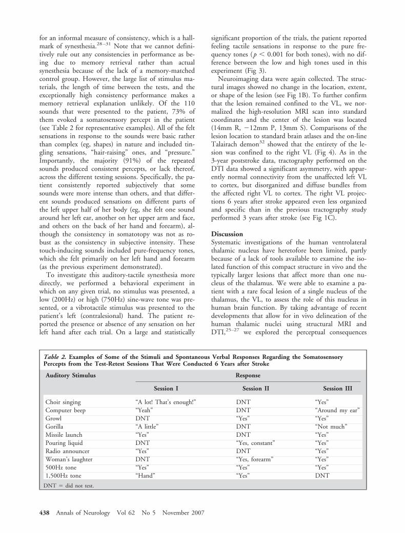
Fig 4. Relationship between the thalamic nuclei and the patient’s lesion. (A) Parcellation of the patient’s thalamus in Talairachspace. Her brain was normalized and the thalamic nuclei were color coded in the axial (top; x � 15mm), coronal (middle; y ��11mm), and sagittal planes (bottom; z � 14mm). (B) Enlarged view of the thalamic parcellation. (C) The thalamic parcellation(colored outlines) and lesion location (dashed black line). The lesion falls entirely within ventrolateral nucleus (red outline).ANT � anterior nucleus; LDN � laterodorsal nucleus; LPN � lateroposterior nucleus; MD � mediodorsal nucleus; MID �midline nucleus; PUL � pulvinar nucleus; VA � ventroanterior nucleus; VL � ventrolateral nucleus; VPL � ventral posterolat-eral nucleus; VPM � ventral posteromedial nucleus.
440 Annals of Neurology Vol 62 No 5 November 2007

4. Savazzi S, Marzi CA. The superior colliculus subserves inter-hemispheric neural summation in both normals and patientswith a total section or agenesis of the corpus callosum. Neuro-psychologia 2004;42:1608–1618.
5. Walker R, Deubel H, Schneider WX, Findlay JM. Effect ofremote distractors on saccade programming: evidence for an ex-tended fixation zone. J Neurophysiol 1997;78:1108–1119.
6. Driver J, Mattingley JB. Parietal neglect and visual awareness.Nat Neurosci 1998;1:17–22.
7. Rafal RD. Neglect. Curr Opin Neurobiol 1994;4:2312–2316.8. Rafal RD. Neglect II: cognitive neuropsychological issues. In:
Farah MJ, Feinberg TE, eds. Patient-based approaches to cog-nitive neuroscience. Cambridge, MA: The MIT Press, 2000:125–141.
9. Vallar G. Spatial hemineglect in humans. Trends Cogn Sci1998;2:87–97.
10. Oppenheim H. Diseases of the nervous system. Mayer EE,translator. Philadelphia: Lippincott, 1900.
11. Poppelreuter W. Die psychischen Schadigungen durch Kopfs-chuss im Kriege 1914/16. Band 1: Die Storungen der niederenund hoheren Sehleistungen durch Verletzungen des Okzipital-hirns. Leipzig, Germany: Voss Leopold, 1917.
12. Bender MB. Extinction and precipitation of cutaneous sensa-tions. Arch Neurol Psychiatry 1945;54:1–9.
13. Vallar G, Rusconi ML, Bignamini L, et al. Anatomical corre-lates of visual and tactile extinction in humans: a clinical CTscan study. J Neurol Neurosurg Psychiatry 1994;57:464–470.
14. Staines WR, Black SE, Graham SJ, McIlroy WE. Somatosen-sory gating and recovery from stroke involving the thalamus.Stroke 2002;33:2642–2651.
15. di Pellegrino G, Ladavas E, Farne A. Seeing where your handsare. Nature 1997;388:730.
16. Mattingley JB, Driver J, Beschin N, Robertson IH. Attentionalcompetition between modalities: extinction between touch andvision after right hemisphere damage. Neuropsychologia 1997;35:867–880.
17. Ladavas E, di Pellegrino G, Farne A, Zeloni G. Neuropsycho-logical evidence of an integrated visuotactile representation ofperipersonal space in humans. J Cogn Neurosci 1998;10:581–589.
18. Sathian K. Intermanual referral of sensation to anesthetichands. Neurology 2000;54:1866–1868.
19. Goodrich SJ, Ward R. Anti-extinction following unilateral pa-rietal damage. Cogn Neuropsychol 1997;14:595–612.
20. Humphreys GW, Riddoch MJ, Nys G, Heinke D. Transientbinding by time: neuropsychological evidence from anti-extinction. Cogn Neuropsychol 2002;19:361–380.
21. Mori S, Crain BJ, Chacko VP, van Zijl PC. Three-dimensionaltracking of axonal projections in the brain by magnetic reso-nance imaging. Ann Neurol 1999;45:265–269.
22. Cox RW. AFNI: software for analysis and visualization of func-tional magnetic resonance neuroimages. Comput Biomed Res1996;29:162–173.
23. Basser PJ, Mattiello J, LeBihan D. MR diffusion tensor spec-troscopy and imaging. Biophys J 1994;66:259–267.
24. Mori S, Zhang J. Principles of diffusion tensor imaging and itsapplications to basic neuroscience research. Neuron 2006;51:527–539.
25. Sherman SM, Guillery RW. Exploring the thalamus and its rolein cortical function. 2nd ed. Cambridge, MA: MIT Press,2006.
26. Behrens TE, Johansen-Berg H, Woolrich MW, et al. Non-invasive mapping of connections between human thalamus andcortex using diffusion imaging. Nat Neurosci 2003;6:750–757.
27. Wiegell MR, Tuch DS, Larsson HB, Wedeen VJ. Automaticsegmentation of thalamic nuclei from diffusion tensor magneticresonance imaging. Neuroimage 2003;19:391–401.
28. Baron-Cohen S, Wyke MA, Binnie C. Hearing words and see-ing colours: an experimental investigation of a case of synaes-thesia. Perception 1987;16:761–767.
29. Dixon MJ, Smilek D, Cudahy C, Merikle PM. Five plus twoequals yellow. Nature 2000;406:365.
30. Cytowic RE. The man who tasted shapes. Cambridge, MA:MIT Press, 2000.
31. Simner J, Mulvenna C, Sagiv N, et al. Synaesthesia: the prev-alence of atypical cross-modal experiences. Perception 2006;35:1024–1033.
32. Lancaster JL, Woldorff MG, Parsons LM, et al. Automated Ta-lairach atlas labels for functional brain mapping. Hum BrainMapp 2000;10:120–131.
33. Budinger E, Heil P, Hess A, Scheich H. Multisensory process-ing via early cortical stages: connections of the primary auditorycortical field with other sensory systems. Neuroscience 2006;143:1065–1083.
34. Hubbard EM, Ramachandran VS. Neurocognitive mechanismsof synesthesia. Neuron 2005;48:509–520.
35. Simner J, Holenstein E. Ordinal linguistic personification as avariant of synesthesia. J Cogn Neurosci 2007;19:694–703.
36. Luria AR. The mind of a mnemonist. New York: Basic Books,1968.
37. Simner J, Ward J. Synaesthesia: the taste of words on the tip ofthe tongue. Nature 2006;444:438.
38. Ward J, Simner J. Lexical-gustatory synaesthesia: linguistic andconceptual factors. Cognition 2003;89:237–261.
39. Ward J, Simner J, Auyeung V. A comparison of lexical-gustatory and grapheme-colour synaesthesia. Cogn Neuropsy-chol 2005;22:28–41.
40. Mattingley JB, Rich AN, Yelland G, Bradshaw JL. Unconsciouspriming eliminates automatic binding of colour and alphanu-meric form in synaesthesia. Nature 2001;410:580–582.
41. Rich AN, Mattingley JB. Anomalous perception in synaesthesia:a cognitive neuroscience perspective. Nat Rev Neurosci 2002;3:43–52.
42. Ramachandran VS, Hubbard EM. Psychophysical investigationsinto the neural basis of synaesthesia. Proc R Soc Lond B BiolSci 2001;268:979–983.
43. Hubbard EM, Arman AC, Ramachandran VS, Boynton GM.Individual differences among grapheme-color synesthetes: brain-behavior correlations. Neuron 2005;45:975–985.
44. Burrack A, Knoch D, Brugger P. Mitempfindung insynaesthetes: co-incidence or meaningful association? Cortex2006;42:151–154.
45. Vike J, Jabbari B, Maitland CG. Auditory-visual synesthesia.Report of a case with intact visual pathways. Arch Neurol 1984;41:680–681.
46. Rao A, Nobre AC, Alexander I, Cowey A. Auditory evokedvisual awareness following sudden ocular blindness: an EEGand TMS investigation. Exp Brain Res (in press).
47. Lessell S, Cohen MM. Phosphenes induced by sound. Neurol-ogy 1979;29:1524–1526.
48. Jacobs L, Karpik A, Bozian D, Gothgen S. Auditory-visualsynesthesia: sound-induced photisms. Arch Neurol 1981;38:211–216.
49. Armel KC, Ramachandran VS. Acquired synesthesia in retinitispigmentosa. Neurocase 1999;5:293–296.
Ro et al: Sensation in the VL 441