original article primary poorly differentiated … · primary poorly differentiated monophasic...
TRANSCRIPT

Int J Clin Exp Pathol 2017;10(11):11143-11146www.ijcep.com /ISSN:1936-2625/IJCEP0066533
Original ArticlePrimary poorly differentiated monophasic synovial sarcoma of ileum mesenteries with pulmonary metastasis: a case report
Li Ding, Anjia Han
Department of Pathology, The First Affiliated Hospital of Sun Yat-sen University, Guangzhou, China
Received September 30, 2017; Accepted October 21, 2017; Epub November 1, 2017; Published November 15, 2017
Abstract: Primary intra-abdominal synovial sarcomas are rare soft tissue malignancies. Herein, we present a case of poorly differentiated monophasic synovial sarcomain ileum mesenterieswith pulmonary metastasis. The patient was a 47-year-old female with a history of cough with variable expectoration and paroxysmal abdominal pain for two months. The tumor was located in ileum mesenteries and composed of monophasic spindle tumor cells with active mitosis and massive necrosis. Tumor cells were positive for vimentin and BCL-2 by immunohistochemistry staining and positive for SYT gene break apart by dual color break apart fluorescence in situ hybridization assay. The differ-ential diagnosis includes gastrointestinal stromal tumour and other mesenchymal tumour.
Keywords: Synovial sarcoma, poorly differentiated, ileum mesenteries, pulmonary metastasis
Introduction
Synovial sarcomais a rare neoplasm that accounts for 5-10% of all soft-tissue tumors and occurs mainly near tendons/tendon sheaths and next to joint capsules. It rarely arises in head and neck, orbit, brain, esopha-gus, kidney, and prostate [1-6]. Primary intra-abdominal synovial sarcoma is very rare and mainly occurs in omentum [7-18]. The tumor has the specific chromosomal translocation t(X;18) (p11; q11) that leads to formation of a SS18-SSX fusion gene detected in more than 95% of cases [19]. Herein, we present a case of poorly differentiated monophasic synovial sar-coma in ileum mesenteries which was an unusual site and analyzed its clinicopathologi-cal features and the differential diagnosis.
Case presentation
A 47-year-old female presented cough with vari-able expectoration and paroxysmal abdominal painfor two months. She had no diarrhea, nau-sea, vomiting, and serious medical or surgical history. All laboratory examination including blood tests, biochemical tests, and urinalysis were within normal limits.
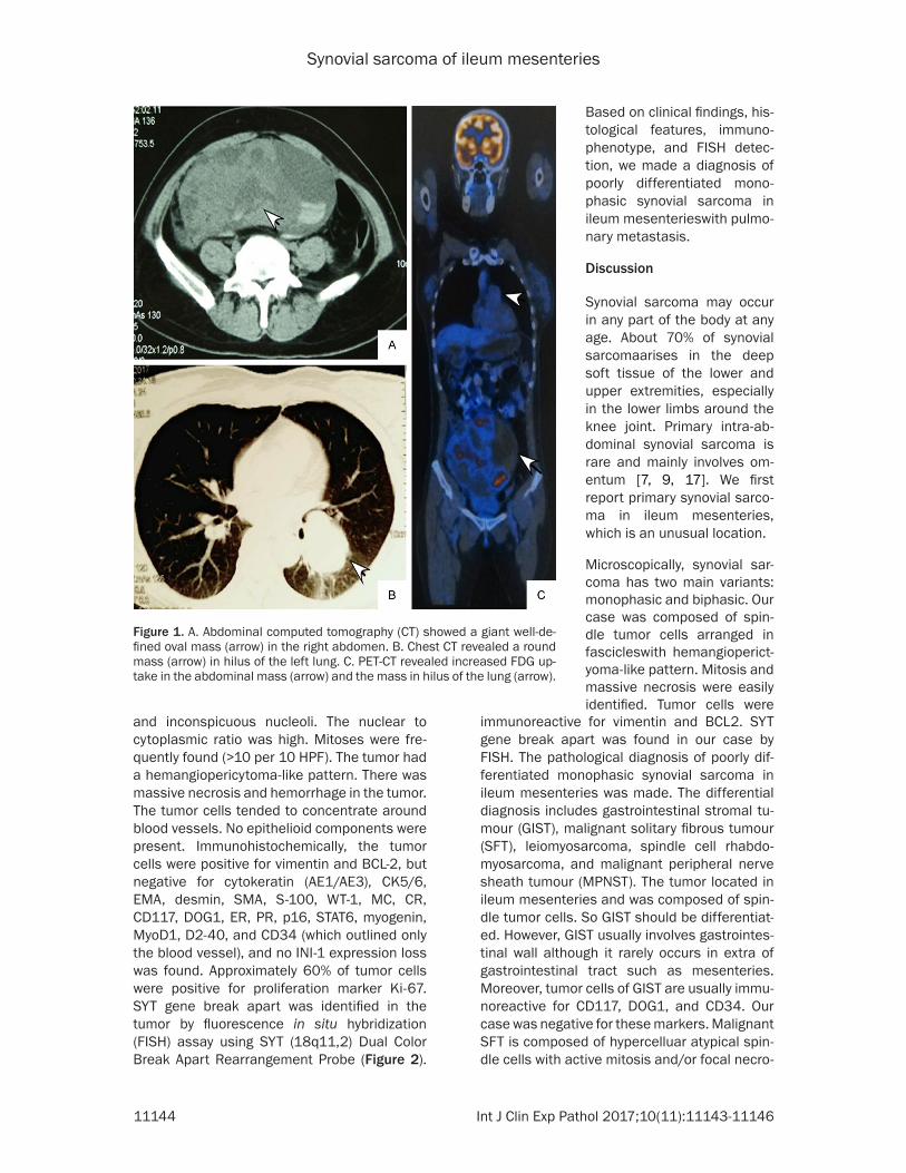
Abdominal computed tomography (CT) and magnetic resonance imaging (MRI) showed a giant well-defined ovalmass in the right abdo-men. The masswas a cystic-solid and 16.5 cm×8.6 cm in maximum plane size. A small amount of pelvic fluid was visible (Figure 1). Chest CT revealed several round masses in the left upper and lower lungs. The largest mass was 5.5 cm×3.2 cm and well-circumscribed. F-18 fluorodeoxyglucose positron emission tomography-CT (18F FDG PET/CT) revealed an increased FDG uptake in the abdominal mass and the lung mass, with a maximum standard-ized uptake value of 13.0 (Figure 1).
The patient received operation for abdomen mass resection. The mass located in the ileum mesenteries and had an apparently well cir-cumscribed with 19 cm×16 cm×9 cm in size. Grossly, the mass was well circumscribed and had a yellow-grayish color with necrotic and hemorrhagic areas on cut surface.
Microscopically, the tumor was composed of monomorphic spindle cells growing in tight fas-cicles. The spindle tumor cellswere uniform and had scant eosinophilic cytoplasm and round or ovoid nuclei with a finely dispersed chromatin
Synovial sarcoma of ileum mesenteries
11144 Int J Clin Exp Pathol 2017;10(11):11143-11146
and inconspicuous nucleoli. The nuclear to cytoplasmic ratio was high. Mitoses were fre-quently found (>10 per 10 HPF). The tumor had a hemangiopericytoma-like pattern. There was massive necrosis and hemorrhage in the tumor. The tumor cells tended to concentrate around blood vessels. No epithelioid components were present. Immunohistochemically, the tumor cells were positive for vimentin and BCL-2, but negative for cytokeratin (AE1/AE3), CK5/6, EMA, desmin, SMA, S-100, WT-1, MC, CR, CD117, DOG1, ER, PR, p16, STAT6, myogenin, MyoD1, D2-40, and CD34 (which outlined only the blood vessel), and no INI-1 expression loss was found. Approximately 60% of tumor cells were positive for proliferation marker Ki-67. SYT gene break apart was identified in the tumor by fluorescence in situ hybridization (FISH) assay using SYT (18q11,2) Dual Color Break Apart Rearrangement Probe (Figure 2).
immunoreactive for vimentin and BCL2. SYT gene break apart was found in our case by FISH. The pathological diagnosis of poorly dif-ferentiated monophasic synovial sarcoma in ileum mesenteries was made. The differential diagnosis includes gastrointestinal stromal tu- mour (GIST), malignant solitary fibrous tumour (SFT), leiomyosarcoma, spindle cell rhabdo-myosarcoma, and malignant peripheral nerve sheath tumour (MPNST). The tumor located in ileum mesenteries and was composed of spin-dle tumor cells. So GIST should be differentiat-ed. However, GIST usually involves gastrointes-tinal wall although it rarely occurs in extra of gastrointestinal tract such as mesenteries.Moreover, tumor cells of GIST are usually immu-noreactive for CD117, DOG1, and CD34. Our case was negative for these markers. Malignant SFT is composed of hypercelluar atypical spin-dle cells with active mitosis and/or focal necro-
Figure 1. A. Abdominal computed tomography (CT) showed a giant well-de-fined oval mass (arrow) in the right abdomen. B. Chest CT revealed a round mass (arrow) in hilus of the left lung. C. PET-CT revealed increased FDG up-take in the abdominal mass (arrow) and the mass in hilus of the lung (arrow).
Based on clinical findings, his-tological features, immuno-phenotype, and FISH detec-tion, we made a diagnosis of poorly differentiated mono-phasic synovial sarcoma in ileum mesenterieswith pulmo-nary metastasis.
Discussion
Synovial sarcoma may occur in any part of the body at any age. About 70% of synovial sarcomaarises in the deep soft tissue of the lower and upper extremities, especially in the lower limbs around the knee joint. Primary intra-ab- dominal synovial sarcoma is rare and mainly involves om- entum [7, 9, 17]. We first report primary synovial sarco-ma in ileum mesenteries, which is an unusual location.
Microscopically, synovial sar-coma has two main variants: monophasic and biphasic. Our case was composed of spin-dle tumor cells arranged in fascicleswith hemangioperict- yoma-like pattern. Mitosis and massive necrosis were easily identified. Tumor cells were

Synovial sarcoma of ileum mesenteries
11145 Int J Clin Exp Pathol 2017;10(11):11143-11146
sis and also has hemangioperictyoma-like pat-tern and is immunoreactive for BCL-2 and CD99. These features of malignant SFT are easily confused with poorly differentiated monophasic synovial sarcoma. However, malig-nant SFT is commonly immunoreactive for STAT6 and CD34 except for BCL-2 and CD99. Our case was negative for STAT6 and CD34 by immunohistochemistry staining. Leiomyosar- coma and spindle cell rhabdomyosarcoma are composed of spindle tumor cells. However, leio-myosarcoma is immunoreactive for SMA and desmin. Spindle cell rhabdomyosarcoma is immunoreactive for myogenin and MyoD1. The
coma in ileum mesenteries was confirmed. As for new therapeutic strategy, Yamda et al. have reported TAS-115 monotherapy may benefit synovial sarcoma patients whose tumour are dependent upon either c-MET or PDGFRα sig-naling by functioning as a multiple tyrosine kinase inhibitor to suppress c-MET as well as PDGFRα pathways [21].
Disclosure of conflict of interest
None.
Address correspondence to: Dr. Anjia Han, Depart- ment of Pathology, The First Affiliated Hospital, Sun
Figure 2. Microscopic features of monophasic synovial sarcoma. A. Tumor had hemangiopericytoma-like pattern. HE×100. B. The tumor cells tended to concentrate around blood vessels with massive necrosis. HE×200. C. Spindle tumor cells arranged in tight fascicles with active mitosis at high power magnification. HE×400. D, E. Tumor cells were immunoreactive for vi-mentin (D×200) and Ki-67 (E×400) in about 60% of tumor cells. Immunohis-tochemistry staining. F. SYT gene break apart was identified in tumor cells by dual color break-apart fluorescence in situ hybridization (FISH) assay. FISH×400.
tumor cells tended to concen-trate around blood vessels in our case, which is the feature of MPNST. However, MPNST usually arises from a periph-eral nerve or from a pre-exist-ing benign nerve tumour (usu-ally neurofibroma) and is im- munoreactive for S-100 pro-tein and GFAP. Tumor cellsin our case were negative for S-100 protein. Most impor-tantly, specific SYT gene break apart was found in our report-ed tumorby FISH. As our kn- owledge, GIST and the above mesenchymal tumour are lack of such specific chromosomal translocation.
Surgical resection and adju-vant chemotherapy are the preferred treatments for syno-vial sarcoma. But the progno-sis of synovial sarcoma is usu-ally poor. Histology and gr- ading are important prognos-tic factors in synovial sarco-ma. Sarcoma specific survival (OS) was 56.6% at 5 years and 46.9% at 10 years. Prognosis was worse in those patients monophasic synovial sarcoma as in those with grade 3 tu- mors [20]. Metastasis occurs most commonly in lungs and bone. The patient in our case had multiple pulmonary me- tastases by chest CT examina-tion as primary synovial sar-

Synovial sarcoma of ileum mesenteries
11146 Int J Clin Exp Pathol 2017;10(11):11143-11146
Yat-sen University, 58 Zhongshan Road II, Guang- zhou 510080, China. Tel: 8620-87332235; Fax: 8620-87332235; E-mail: [email protected]
References
[1] Gopalakrishnan V, Amini B, Wagner MJ, Nowell EN, Lazar AJ, Lin PP, Benjamin RS, Araujo DM. Synovial sarcoma of the head and neck: a sin-gle institution review. Sarcoma 2017; 2017: 2016752.
[2] Sharma S, Sharma A, Lobo G, Nayak M, Prad-han D, Samriti, Bindal S, Gogoi K, Katara R, Kini L, Mohanty SK. Primary dura-based syno-vial sarcoma of the parafalcine region of brain. Pathol Res Pract 2017; 213: 868-71.
[3] Stagner AM, Jakobiec FA, Fay A. Primary orbital synovial sarcoma: a clinicopathologic review with a differential diagnosis and discussion of molecular genetics. Surv Ophthalmol 2017; 62: 227-36.
[4] Chandrasekaran D, Narayanaswamy K, Sun-dersingh S, Senniappan K, Raja A. Primary sy-novial sarcoma of the kidney with inferior vena caval thrombus. Indian J Surg Oncol 2016; 7: 345-48.
[5] Doroudinia A, Bakhshayesh KM, Dorudinia A, Mehrian P, Agha-Hosseini F. Synovial sarcoma of the esophagus: a case report and review of literature. Middle East J Dig Dis 2017; 9: 111-13.
[6] Olofson AM, Linos K. Primary intraprostatic sy-novial sarcoma. Arch Pathol Lab Med 2017; 141: 301-04.
[7] Alcala SF, Hernandez HJ, Montenegro DT, Lo-pez-Tomassetti FE. Monophasic synovial sar-coma of the greater omentum: case report and review of literature. Ann R Coll Surg Engl 2017; 99: e172-73.
[8] Hu S, Wong K, Ramesh KH, Villanueva-Siles E, Panarelli N, In H. Diffuse, aggressive metastat-ic progression after minimally invasive local resection of primary gastric synovial sarcoma: a case report and systematic review of the lit-erature. J Gastrointest Cancer 2017.
[9] Iwahashi N, Deguchi Y, Horiuchi Y, Ino K, Furu-kawa K. Omental synovial sarcoma mimicking an ovarian malignancy: a case report. Mol Clin Oncol 2017; 6: 688-90.
[10] Indranil G, Divya M. Synovial sarcoma of the omentum: a rare entity. Indian J Cancer 2015; 52: 166-67.
[11] Travaglini G, Biagetti S, Alfonsi S, Bearzi I, Mar-morale C. Primary intra-abdominal synovial sarcoma: a case report. Ann Ital Chir 2013; 84.
[12] Rao L, Jaiprakash P, Palankar N, Gowda V. In-tra-abdominal primary monophasic synovial sarcoma with hemangiopericytoma-like areas. J Cancer Res Ther 2013; 9: 102-04.
[13] Wang YJ, Wen SC, Chien ST, Sheu JW, Hsuea CW, Feng NH. Primary intra-abdominal synovi-al sarcoma. J Chin Med Assoc 2006; 69: 492-95.
[14] Vera J, Garcia MD, Marigil M, Abascal M, Lopez JI, Ligorred L. Biphasic synovial sarcoma of the abdominal wall. Virchows Arch 2006; 449: 367-72.
[15] Buiga-Potcoava R, Crisan D, Olinici CD. Primary intraabdominal synovial sarcoma: a case re-port. Rom J Gastroenterol 2005; 14: 67-69.
[16] Fisher C, Folpe AL, Hashimoto H, Weiss SW. Intra-abdominal synovial sarcoma: a clinico-pathological study. Histopathology 2004; 45: 245-53.
[17] Hemmings C, Fisher C. Primary omental syno-vial sarcoma: a case with cytogenetic confir-mation. Pathology 2004; 36: 208-11.
[18] Ko SF, Chou FF, Huang CH, Ng SH, Wan YL, Lee TY, Lin JW, Chen WJ. Primary synovial sarcoma of the gastrocolic ligament. Br J Radiol 1998; 71: 438-40.
[19] Clark J, Rocques PJ, Crew AJ, Gill S, Shipley J, Chan AM, Gusterson BA, Cooper CS. Identifica-tion of novel genes, SYT and SSX, involved in the t(X;18)(p11.2;q11.2) translocation found in human synovial sarcoma. Nat Genet 1994; 7: 502-08.
[20] Bianchi G, Sambri A, Righi A, Dei Tos AP, Picci P, Donati D. Histology and grading are impor-tant prognostic factors in synovial sarcoma. Eur J Surg Oncol 2017; 43: 1733-1739.
[21] Yamada S, Imura Y, Nakai T, Nakai S, Yasuda N, Kaneko K, Outani H, Takenaka S, Hamada K, Myoui A, Araki N, Ueda T, Itoh K, Yoshikawa H, Naka N. Therapeutic potential of TAS-115 via c-MET and PDGFRalpha signal inhibition for synovial sarcoma. BMC Cancer 2017; 17: 334.