original article outer membrane protein 31 of brucella promotes … · 2017-03-28 · outer...
TRANSCRIPT

Int J Clin Exp Pathol 2017;10(3):3006-3014www.ijcep.com /ISSN:1936-2625/IJCEP0043326
Original Article Outer membrane protein 31 of Brucella promotes apoptosis of astrocytes via the p38MAPK/MK2 signaling pathway
Xiangyang Li1,2, Lihua Song3, Jiabao Zhang1
1College of Animal Science, Jilin University, Changchun, China; 2College of Animal Science and Technology, 3The First Clinical Hospital, Inner Mongolia University for Nationalities, Tongliao, China
Received October 31, 2016; Accepted January 13, 2017; Epub March 1, 2017; Published March 15, 2017
Abstract: Neurobrucellosis is an inflammatory disorder caused by infection of Brucella in central nervous sys-tem (CNS). The Brucella infection induced the secretion of cytokines in astrocytes, such as TNF-α, IL-6, and IL-1β, which played an important role in the immunophathogenesis of neurobrucellosis. The outer-membrane protein 31 (Opm31) in Brucella melitensis functions as an immunodominant and protective antigen during the course of Bru-cella infection. The purpose of this study was to investigate the potential pathogenic mechanism of neurobrucellosis induced by Brucella in astrocytes. ELISA assay showed that the concentrations of TNF-α, IL-6, IL-1β, and IL-10 in astrocytes in Opm31 groups were significantly increased compared with control group. Flow cytometry with annexin V-FITC/PI staining indicated that Opm31 protein significantly promoted the apoptosis of astrocytes via the mitochon-drial pathway. Moreover, Opm31 significantly increased the phosphorylations of p38MAPK and MK2. Si-p38MAPK and p38MAPK inhibitor SB203580 significantly decreased Opm31-induced apoptotic rates. Opm31 of Brucella promoted the apoptosis of astrocytes via the p38MAPK/MK2 signaling pathway, providing a possible pathogenesis of neurobrucellosis infected by Brucella in astrocytes.
Keywords: Apoptosis, astrocytes, Brucella, outer membrane protein 31, p38MAPK/MK2
Introduction
Brucellosis caused by Brucellae is a wide-spread infectious and zoonotic disease that affecting humans and animals, and it is charac-terized by undulant fever, arthritis, abortion and infertility in a variety of animals [1]. Brucellosis is an inflammatory disease and has caused dramatic economic losses for livestock indus-try, agriculture and public health, especially in developing countries. Though great efforts have been made to develop effective vaccines against brucellosis, there is still no satisfactory progress to defend this disease in human [2]. Neurobrucellosis is a crucial complication of systemic brucellosis infection in the central nervous system (CNS) and could cause inflam-matory disorders and tissue destruction, such as meningitis, meningoencephalitis and polyra-diculitis [3]. Thus, it is extremely helpful to understand the potential pathogenesis of neu-robrucellosis to improve its therapeutic effect.
Innate immune in the CNS is essential to de- tect and clear bacterial pathogens and is regu-larly activated by various pathways when recog-nizing invasive pathogens [4]. Astrocytes, the most numerous cell type in the CNS, play an important role in maintaining innate immunity, blood-brain barrier (BBB) integrity, and function of CNS [5]. The infection of astrocytes caused the secretion of cytokines TNF-α, IL-6, and IL-1β and chemokines such as CCL2 and CXCL1 [6].
Brucellae, with an extensive host range, are Gram-negative, facultative intracellular and coccobacillus bacteria which belong to the alpha subclass of Proteobacteria. Brucellae lacks classical virulence factors, such as inva-sive proteases, virulence plasmids and lysogen-ic phages, and instead expresses virulence fac-tors associated with the capacity to invade and propagate within host cells [7]. Brucellae trig-gered multiple system damages in human and

Omp31 inhibits apoptosis of astrocytes
3007 Int J Clin Exp Pathol 2017;10(3):3006-3014
[18]. Briefly, human astrocytes of cortical origin were used to dissociate and produce single cell suspension by enzymatic hydrolysis and then the cell suspension was cultured on negative immunopanning plates to isolate astrocytes. The isolated astrocytes were grown in a Dulbecco’s modified Eagle’s medium-high glu-cose (DMEM-HG) (Hyclone, Logan, Utah, USA) containing 10% fetal calf serum (Invitrogen, Grand Island, NY, USA), Nutrient Mixture F-12, 100 units/ml penicillin, 100 μg/ml streptomy-cin, 1 mM sodium pyruvate, and 2 mM L-glutamine at 37°C in humidified 5% CO2 and 95% air. When the cells were grown to 80% confluency, the culture media of astrocytes were replaced with serum-free media.
Cell transfection
Small interfering RNA against p38MAPK (si-p38MAPK) and corresponding negative con- trol (si-control) were purchased from Gene- Pharma Biotech (Shanghai, China). Cells were plated into six-well plates and cultured for 24 h, and then 100 pmol/L of these molecular products were transfected into astrocytes by LipofectamineTM 2000 (Invitrogen, Grand Island, NY, USA).
Purification of recombinant Omp31 expression
The primer of Opm31 gene was designed and synthesized depending on the nucleotide sequences of Brucella outer membrane protein gene in GenBank. The Omp31 gene was then amplified with Opm31 primers by polymerase chain reaction (PCR) and cloned into NotI and NdeI sites of PET-30a plasmid to construct the recombinant plasmid PET-30a-Opm31. Subsequently, the recombinant plasmid was transformed into competent cells Escherichia coli BL21 (DE3) to overexpress recombinant proteins Opm31 induced by 0.1 mM IPTG. The recombinant proteins were purified by affinity chromatography with Ni (II)-conjugated Sepha- rose at 4°C and quantified by BCA protein assay kits (Bio-Rad Laboratories, Inc. Hercules, CA, USA). SDS-polyacrylamide gel (SDS-PAGE) was performed to confirm the expression of Opm31.
Cytokine detection by ELISA assays
Astrocytes were seeded in 6-well plates at a density of 1 × 10⁶ cells/ml and incubated in DMEM-HG medium at an incubator of 5% CO2
animals, among which the CNS injury approxi-mately accounted for 1.7%-10% [8].
The major outer membrane proteins (Omps) of Brucella are mainly divided into two groups: group 2 (Omp2a and Omp2b) and group 3 (Omp25 and Omp31). Omp31 is found in almost all Brucella species, except for Brucella abor-tus [9]. Opm31 protein was able to form oligo-mers resistant to denaturation by sodium dodecyl sulfare (SDS) at low temperatures in B. melitensis [10]. Omp31 gene was initially cloned and sequenced from B. melitensis 16 M and shared 34% identity with Opm25 [10]. It is reported that group 3 proteins, especially Omp31, functions as an immunodominant and protective antigen of B. ovis infection in rams [10]. Gupta et al. reported that vaccines of recombinant invasive Escherichia coli express-ing B. melitensis Opm31 provided the protec-tion for mice against virulent B. melitensis and promoted cellular immune responses [11]. However, the relationship between Opm31 and the damage caused by Brucella remains unclear.
p38 mitogen-activated protein kinase (p38- MAPK) is a common intracellular signaling transduction pathway which is always activated by phosphorylation of p38MAPK [12]. MAPKAP kinase 2 (MK2), a protein kinase activating the downstream of p38MAPK, is a main regulator of stress- and cytokine-induced post-transcrip-tional gene expression [13, 14]. The p38MAPK signaling pathway is closely related to the regu-lation of neuronal growth, proliferation, apopto-sis, and pro-inflammatory cytokine secretion [15, 16]. Experimental evidences have showed that the activation of p38MAPK is strongly associated with astrocyte senescence-related secretory phenotype [17].
In this study, the effect of Opm31 on apoptosis of astrocytes was investigated and the poten-tial relationships between Opm31 and the p38MAPK/MK2 signaling pathway was further studied in astrocytes.
Materials and methods
Primary astrocytes culture
Human astrocytes of cortical origin (ScienCell, San Diego, CA USA) were used for the primary cultures of astrocytes as previously described

Omp31 inhibits apoptosis of astrocytes
3008 Int J Clin Exp Pathol 2017;10(3):3006-3014
Assessment of apoptosis
Astrocytes (1 × 10⁵ cells/ml) or astrocyte cells with si-p38MAPK or si-control were treated with different concentrations of Opm31 (0, 25 and 50 ng/ml) for 24 h and 48 h, or SB203580 (0, 0.05 and 0.1 μM) for 48 h. The treated cells were then stained with FITC conjugated Annexin V and propidium iodide (PI) using an Annexin V-FITC Apoptosis Detection kit (Beyotime Institute of Biotechnology, Nantong, China) as described previously [19, 20]. The stained sam-ples were analyzed by a flow cytometer (FACSCanto; BD Bioscience, San Jose, CA, USA) to evaluate the percentage of apoptotic cells.
Statistical analysis
All data were showed as mean ± standard devi-ation (SD) and analyzed using the GraphPad Prism Software version 5. Differences among multiple groups were analyzed by one-way anal-ysis of variance (ANOVA). A P value <0.05 was considered to be statistically significant.
Results
Opm31 promoted production of cytokines in astrocytes
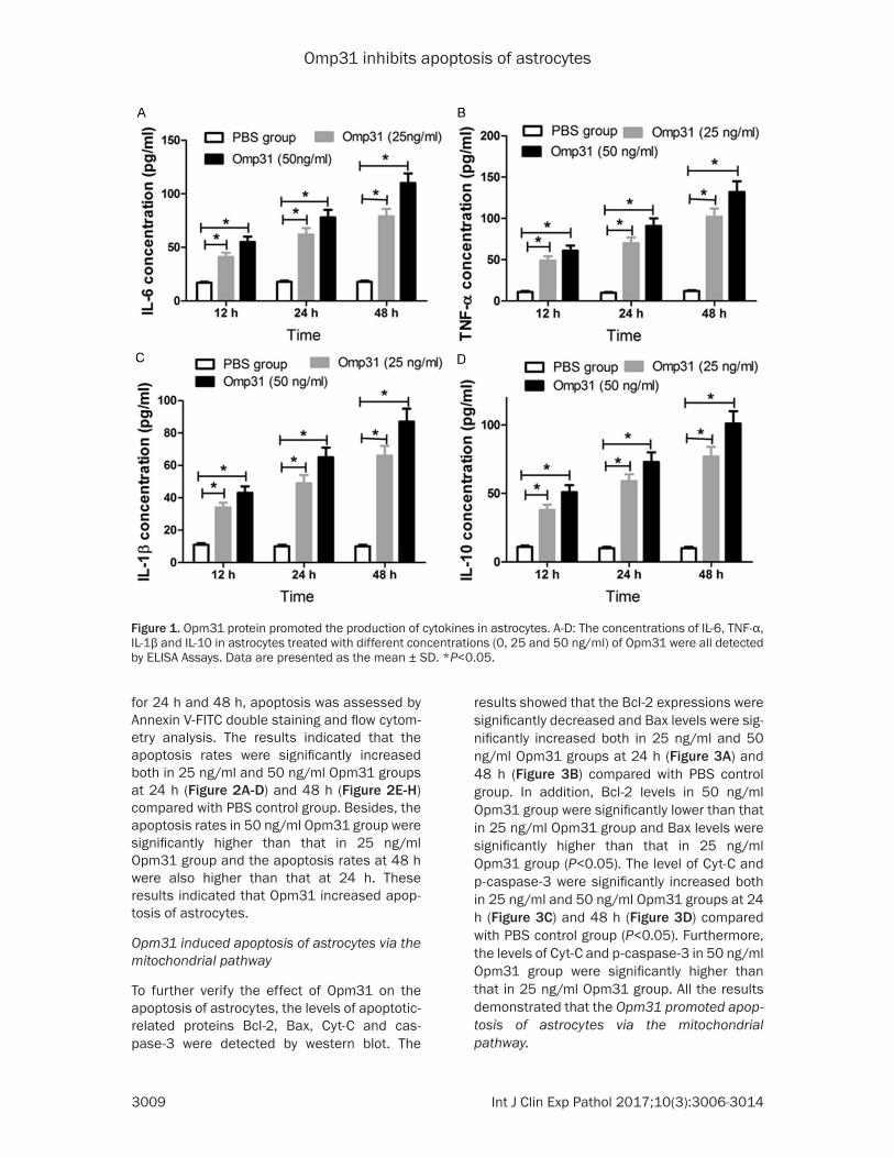
The recombinant protein Opm31 was purified (one signal band on SDS-PAGE) and the con-centration of purified Opm31 proteins by Ni-NTA affinity column was approximately 0.23 mg/ml. To investigate the effect of Opm31 on the expression of cytokines including IL-6, TNF-α, IL-1β, and IL-10, astrocytes were treated with different concentrations (0, 25 and 50 ng/ml) of Opm31 for 12 h, 24 h, and 48 h. The cells treated with PBS were used as control. The con-centrations of IL-6, TNF-α, L-1β, and IL-10 in the supernatant of astrocytes were detected by ELISA Assays. As shown in Figure 1A-D, the concentrations of IL-6, TNF-α, IL-1β, and IL-10 in Opm31 groups were significantly increased in a time-dependent manner compared with PBS control group. In addition, the concentrations of IL-6, TNF-α, IL-1β, and IL-10 in 50 ng/ml Opm31 group were all significantly higher than that in 25 ng/ml Opm31 group. These data suggested that Opm31 induced the section of cytokines IL-6, TNF-α, IL-1β, and IL-10 in astrocytes.
Opm31 promoted apoptosis of astrocytes
After astrocytes were treated with different concentrations (0, 25 and 50 ng/ml) of Opm31
at 37°C for 2 h. Then the cells were collect- ed, washed three times and resuspended in DMEM-HG medium treated with different con-centrations (0, 25 and 50 ng/ml) of Opm31. After incubation for 12 h, 24 h, and 48 h, the concentrations of cytokines (TNF-α, IL-6, IL-1β, and IL-10) produced in culture supernatant of each sample were detected based on the stan-dard curves of known concentrations of recom-binant cytokines by ELISA antibody kits (AlerChek, Portland, Maine, USA) following the manufacturer’s protocol.
Western blot analysis
Astrocytes were seeded into 6-well plates and treated with different concentrations (0, 25 and 50 ng/ml) of Opm31 for 24 h and 48 h in a 5% CO2 incubator at 37°C. After incubation for 48 h, cells were collected and lysed by ice-cold RIPA buffer (Zhong-Shan Jinqiao, Beijing, China) for 30 min. The extracted protein con-centrations were determined by BCA protein assay kits (Bio-Rad Laboratories, Inc. Hercules, CA, USA). Equal amounts of prepared protein samples (50 mg per sample) were mixed with 5 × loading buffer, denatured at 100°C for 5 min in a water bath and then analyzed by 10% SDS-polyacrylamide gels (SDS-PAGE) electrophore-sis. After electrophoresis, the protein samples were transferred to polyvinylidene fluoride (PVDF; Millipore, Bedford, MA, USA) and then blocked with 5% non-fat milk in TBST at room temperature. The membrane was washed three times and probed with rabbit anti-Opm31, rab-bit anti-Bcl-2, rabbit anti-Bax, rabbit anti-Cyt-C, rabbit anti-total cascase-3, rabbit anti-phos-phorylated caspase-3, mouse anti-phosphory-lated p38MAPK, mouse anti-phosphorylated MK2 (1:1000 dilution; Santa Cruz Biote- chnology, Santa Cruz, CA, USA), and rabbit anti-β-actin (1:2500 dilution; Santa Cruz Biote- chnology) on a rocking platform overnight at 4°C. Then the membrane was washed three times with TBST buffer and incubated with horseradish peroxidase (HRP)-conjugated sec-ondary antibody (1:5000 dilution; Santa Cruz Biotechnology) for 2 h at room temperature. The protein bands were visualized by using enhanced chemiluminescence detection rea- gent (Super-Signal West Pico Chemiluminesc- ent Substrate; Thermo Scientific, Waltham, MA, USA) and normalized to β-actin.
Omp31 inhibits apoptosis of astrocytes
3009 Int J Clin Exp Pathol 2017;10(3):3006-3014
results showed that the Bcl-2 expressions were significantly decreased and Bax levels were sig-nificantly increased both in 25 ng/ml and 50 ng/ml Opm31 groups at 24 h (Figure 3A) and 48 h (Figure 3B) compared with PBS control group. In addition, Bcl-2 levels in 50 ng/ml Opm31 group were significantly lower than that in 25 ng/ml Opm31 group and Bax levels were significantly higher than that in 25 ng/ml Opm31 group (P<0.05). The level of Cyt-C and p-caspase-3 were significantly increased both in 25 ng/ml and 50 ng/ml Opm31 groups at 24 h (Figure 3C) and 48 h (Figure 3D) compared with PBS control group (P<0.05). Furthermore, the levels of Cyt-C and p-caspase-3 in 50 ng/ml Opm31 group were significantly higher than that in 25 ng/ml Opm31 group. All the results demonstrated that the Opm31 promoted apop-tosis of astrocytes via the mitochondrial pathway.
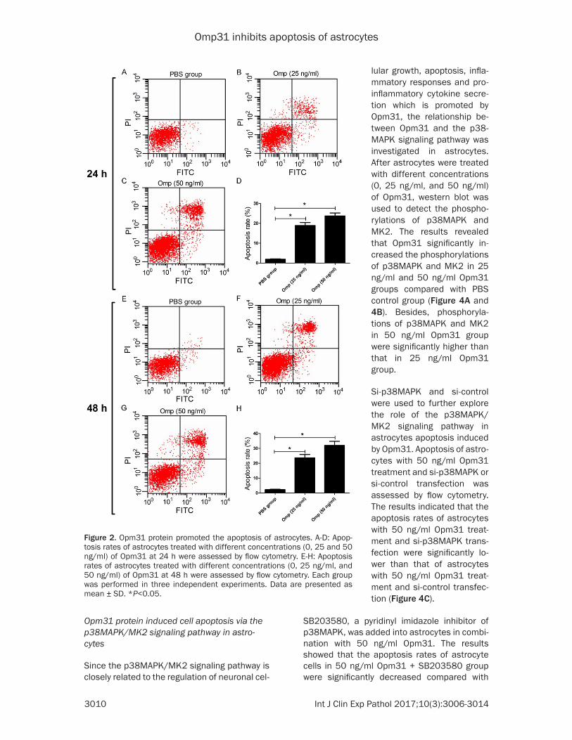
for 24 h and 48 h, apoptosis was assessed by Annexin V-FITC double staining and flow cytom-etry analysis. The results indicated that the apoptosis rates were significantly increased both in 25 ng/ml and 50 ng/ml Opm31 groups at 24 h (Figure 2A-D) and 48 h (Figure 2E-H) compared with PBS control group. Besides, the apoptosis rates in 50 ng/ml Opm31 group were significantly higher than that in 25 ng/ml Opm31 group and the apoptosis rates at 48 h were also higher than that at 24 h. These results indicated that Opm31 increased apop-tosis of astrocytes.
Opm31 induced apoptosis of astrocytes via the mitochondrial pathway
To further verify the effect of Opm31 on the apoptosis of astrocytes, the levels of apoptotic-related proteins Bcl-2, Bax, Cyt-C and cas-pase-3 were detected by western blot. The
Figure 1. Opm31 protein promoted the production of cytokines in astrocytes. A-D: The concentrations of IL-6, TNF-α, IL-1β and IL-10 in astrocytes treated with different concentrations (0, 25 and 50 ng/ml) of Opm31 were all detected by ELISA Assays. Data are presented as the mean ± SD. *P<0.05.
Omp31 inhibits apoptosis of astrocytes
3010 Int J Clin Exp Pathol 2017;10(3):3006-3014
SB203580, a pyridinyl imidazole inhibitor of p38MAPK, was added into astrocytes in combi-nation with 50 ng/ml Opm31. The results showed that the apoptosis rates of astrocyte cells in 50 ng/ml Opm31 + SB203580 group were significantly decreased compared with
Opm31 protein induced cell apoptosis via the p38MAPK/MK2 signaling pathway in astro-cytes
Since the p38MAPK/MK2 signaling pathway is closely related to the regulation of neuronal cel-
Figure 2. Opm31 protein promoted the apoptosis of astrocytes. A-D: Apop-tosis rates of astrocytes treated with different concentrations (0, 25 and 50 ng/ml) of Opm31 at 24 h were assessed by flow cytometry. E-H: Apoptosis rates of astrocytes treated with different concentrations (0, 25 ng/ml, and 50 ng/ml) of Opm31 at 48 h were assessed by flow cytometry. Each group was performed in three independent experiments. Data are presented as mean ± SD. *P<0.05.
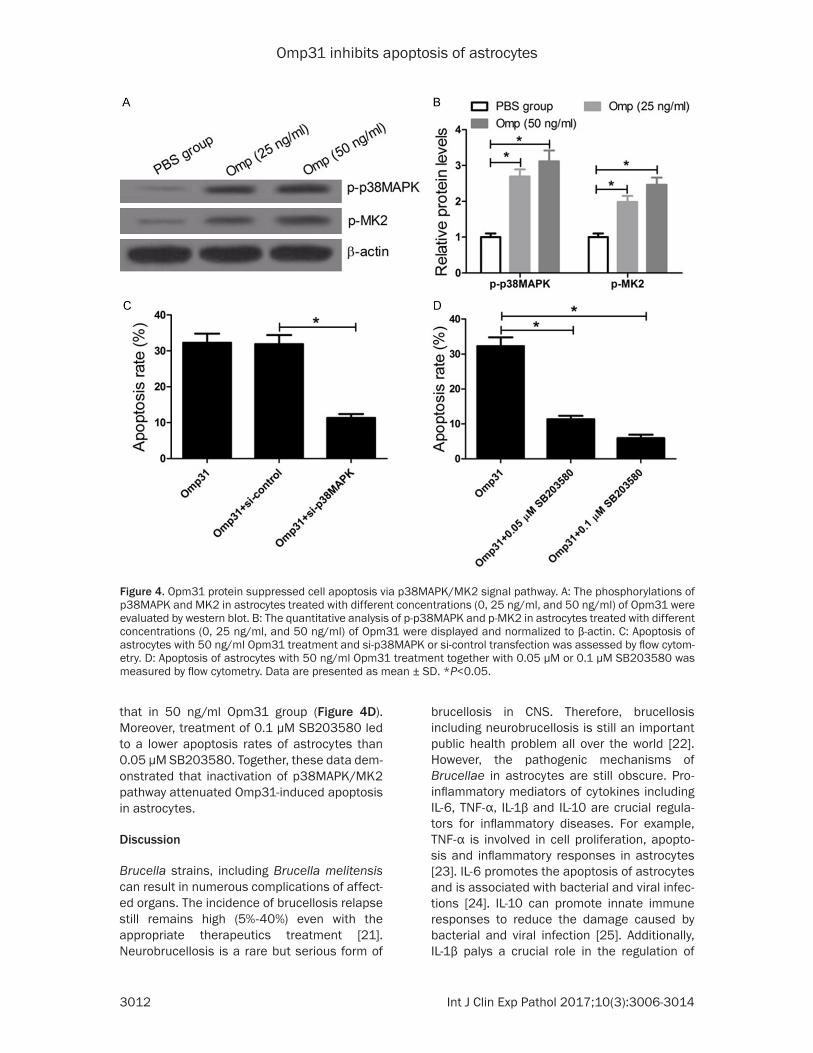
lular growth, apoptosis, infla- mmatory responses and pro-inflammatory cytokine secre- tion which is promoted by Opm31, the relationship be- tween Opm31 and the p38- MAPK signaling pathway was investigated in astrocytes. After astrocytes were treated with different concentrations (0, 25 ng/ml, and 50 ng/ml) of Opm31, western blot was used to detect the phospho- rylations of p38MAPK and MK2. The results revealed that Opm31 significantly in- creased the phosphorylations of p38MAPK and MK2 in 25 ng/ml and 50 ng/ml Opm31 groups compared with PBS control group (Figure 4A and 4B). Besides, phosphoryla-tions of p38MAPK and MK2 in 50 ng/ml Opm31 group were significantly higher than that in 25 ng/ml Opm31 group.
Si-p38MAPK and si-control were used to further explore the role of the p38MAPK/MK2 signaling pathway in astrocytes apoptosis induced by Opm31. Apoptosis of astro-cytes with 50 ng/ml Opm31 treatment and si-p38MAPK or si-control transfection was assessed by flow cytometry. The results indicated that the apoptosis rates of astrocytes with 50 ng/ml Opm31 treat-ment and si-p38MAPK trans-fection were significantly lo- wer than that of astrocytes with 50 ng/ml Opm31 treat-ment and si-control transfec-tion (Figure 4C).

Omp31 inhibits apoptosis of astrocytes
3011 Int J Clin Exp Pathol 2017;10(3):3006-3014
Figure 3. Opm31 promoted the apoptosis of astrocytes via the mitochondrial pathway. The levels of Bcl-2 and Bax in astrocytes treated with different concentrations (0, 25 ng/ml, and 50 ng/ml) of Opm31 at 24 h (A) and 48 h (B) were detected by western blot. Cyt-C levels and phosphorylation of caspase-3 were evaluated by western blot after astrocytes were treated with different concentrations (0, 25 ng/ml, and 50 ng/ml) of Opm31 at 24 h (C) and 48 h (D). Data are presented as mean ± SD. *P<0.05.
Omp31 inhibits apoptosis of astrocytes
3012 Int J Clin Exp Pathol 2017;10(3):3006-3014
brucellosis in CNS. Therefore, brucellosis including neurobrucellosis is still an important public health problem all over the world [22]. However, the pathogenic mechanisms of Brucellae in astrocytes are still obscure. Pro-inflammatory mediators of cytokines including IL-6, TNF-α, IL-1β and IL-10 are crucial regula-tors for inflammatory diseases. For example, TNF-α is involved in cell proliferation, apopto- sis and inflammatory responses in astrocytes [23]. IL-6 promotes the apoptosis of astrocytes and is associated with bacterial and viral infec-tions [24]. IL-10 can promote innate immune responses to reduce the damage caused by bacterial and viral infection [25]. Additionally, IL-1β palys a crucial role in the regulation of
that in 50 ng/ml Opm31 group (Figure 4D). Moreover, treatment of 0.1 μM SB203580 led to a lower apoptosis rates of astrocytes than 0.05 μM SB203580. Together, these data dem-onstrated that inactivation of p38MAPK/MK2 pathway attenuated Omp31-induced apoptosis in astrocytes.
Discussion
Brucella strains, including Brucella melitensis can result in numerous complications of affect-ed organs. The incidence of brucellosis relapse still remains high (5%-40%) even with the appropriate therapeutics treatment [21]. Neurobrucellosis is a rare but serious form of
Figure 4. Opm31 protein suppressed cell apoptosis via p38MAPK/MK2 signal pathway. A: The phosphorylations of p38MAPK and MK2 in astrocytes treated with different concentrations (0, 25 ng/ml, and 50 ng/ml) of Opm31 were evaluated by western blot. B: The quantitative analysis of p-p38MAPK and p-MK2 in astrocytes treated with different concentrations (0, 25 ng/ml, and 50 ng/ml) of Opm31 were displayed and normalized to β-actin. C: Apoptosis of astrocytes with 50 ng/ml Opm31 treatment and si-p38MAPK or si-control transfection was assessed by flow cytom-etry. D: Apoptosis of astrocytes with 50 ng/ml Opm31 treatment together with 0.05 μM or 0.1 μM SB203580 was measured by flow cytometry. Data are presented as mean ± SD. *P<0.05.

Omp31 inhibits apoptosis of astrocytes
3013 Int J Clin Exp Pathol 2017;10(3):3006-3014
Acknowledgements
The work was supported by National Natural Science Foundation of China (Grant No. 31260608).
Disclosure of conflict of interest
None.
Address correspondence to: Lihua Song, The First Clinical Hospital, Inner Mongolia University for Nationalities, East Street of Huolinhe, Keerqin District, Tongliao 028000, China. Tel: +86-475-8314248; E-mail: [email protected]
References
[1] Elfaki MG, Alaidan AA, Al-Hokail AA. Host re-sponse to Brucella infection: review and future perspective. J Infect Dev Ctries 2015; 9: 697-701.
[2] Fugier E, Pappas G, Gorvel JP. Virulence factors in brucellosis: implications for aetiopathogen-esis and treatment. Expert Rev Mol Med 2007; 9: 1-10.
[3] Kim EJ, Lee SJ. Relapsed brucellosis present-ing as neurobrucellosis with cerebral vasculitis in a patient previously diagnosed with Brucel-lar spondylitis: a case report. Infect Chemother 2015; 47: 268-271.
[4] Hanamsagar R, Hanke ML, Kielian T. Toll-like receptor (TLR) and inflammasome actions in the central nervous system. Trends Immunol 2012; 33: 333-342.
[5] Sreekanthreddy P, Gromnicova R, Davies H, Phillips J, Romero IA, Male D. A three-dimen-sional model of the human blood-brain barrier to analyse the transport of nanoparticles and astrocyte/endothelial interactions. Version 2. F1000Res 2015; 4: 1279.
[6] Juric DM, Krzan M, Lipnik-Stangelj M. Hista-mine and astrocyte function. Pharmacol Res 2016; 111: 774-783.
[7] Seleem MN, Boyle SM, Sriranganathan N. Bru-cella: a pathogen without classic virulence genes. Vet Microbiol 2008; 129: 1-14.
[8] Turel O, Sanli K, Hatipoglu N, Aydogmus C, Hatipoglu H, Siraneci R. Acute meningoen-cephalitis due to Brucella: case report and re-view of neurobrucellosis in children. Turk J Pe-diatr 2010; 52: 426-429.
[9] Vizcaino N, Cloeckaert A, Zygmunt MS, Fernan-dez-Lago L. Characterization of a Brucella spe-cies 25-kilobase DNA fragment deleted from Brucella abortus reveals a large gene cluster related to the synthesis of a polysaccharide. Infect Immun 2001; 69: 6738-6748.
BBB permeability, inflammatory activation of resident glia and human brain microvascular endothelial cells (HBMEC) in CNS [23]. In the present study, Opm31 significantly promoted secretion of cytokines (IL-6, TNF-α, IL-1β and IL-10) in the supernatant of astrocytes com-pared with PBS control group.
Bcl-2 and Bax have the opposite effects and play vital roles in the mitochondrial pathway of apoptosis [26]. Cyt-C, distributed in the mito-chondrial membrane, is an important aspect of the intrinsic pathway of apoptosis [27]. Zhao et al. reported that the levels of Cyt-C, cas-pase-9 and caspase-3 were closely related to pentavalent vanadium-induced neuronal apop-tosis [28]. In this study, flow cytometry results showed that Opm31 significantly suppressed astrocytes apoptosis by the decrease of Bcl-2/Bax ratio. In addition, the release of Cyt-C from the mitochondrial membrane into the cyto-plasm was also induced by Opm31. Besides, the apoptosis was further confirmed by the activation of caspase-3. All the results suggest-ed that Opm31 significantly inhibited apoptosis of astrocytes.
p38MAPK, a serine/threonine protein kinase, is a crucial signaling pathway activated by extracellular stimulation. p38MAPK can regu-late various cellular biological activities, such as inflammatory reactions, cell growth and apoptosis, and control cellular responses to cytokines and stress [29-31]. In the present study, Opm31 significantly improved the levels of p-38MAPK and p-MK2 and activated the p38MAPK signaling pathway. To further demon-strate the relationships between the p38MAPK signaling pathway and Opm31, p38MAPK gen- etic silencing by si-p38MAPK was used to inhib-it p38MAPK expression, which significantly decreased Opm31-induced apoptosis rates. SB203580, a selective p38MAPK inhibitor, also significantly suppressed apoptosis rates triggered by Opm31 in astrocytes. All the results suggested that Opm31 induced cell apoptosis via the p38MAPK/MK2 signaling pathway in astrocytes.
Taken together, the present study suggested that B. melitensis Opm31 promoted the secre-tion of inflammatory cytokines and induced the apoptosis of astrocytes via p38MAPK/MK2 signal pathway, providing a potential pathogen-esis of neurobrucellosis infected by Brucella in astrocytes.

Omp31 inhibits apoptosis of astrocytes
3014 Int J Clin Exp Pathol 2017;10(3):3006-3014
[21] Leong KN, Chow TS, Wong PS, Hamzah SH, Ah-mad N, Ch’ng CC. Outbreak of human brucel-losis from consumption of raw goats’ milk in Penang, Malaysia. Am J Trop Med Hyg 2015; 93: 539-541.
[22] Ojo KK, Ranade RM, Zhang Z, Dranow DM, My-ers JB, Choi R, Nakazawa Hewitt S, Edwards TE, Davies DR, Lorimer D, Boyle SM, Barrett LK, Buckner FS, Fan E, Van Voorhis WC. Bru-cella melitensis methionyl-tRNA-synthetase (MetRS), a potential drug target for brucello- sis. PLoS One 2016; 11: e0160350.
[23] Garcia Samartino C, Delpino MV, Pott Godoy C, Di Genaro MS, Pasquevich KA, Zwerdling A, Barrionuevo P, Mathieu P, Cassataro J, Pi-tossi F, Giambartolomei GH. Brucella abortus induces the secretion of proinflammatory me-diators from glial cells leading to astrocyte apoptosis. Am J Pathol 2010; 176: 1323-1338.
[24] Peri F, Piazza M, Calabrese V, Damore G, Cighetti R. Exploring the LPS/TLR4 signal path-way with small molecules. Biochem Soc Trans 2010; 38: 1390-1395.
[25] Ouyang W, Rutz S, Crellin NK, Valdez PA, Hy-mowitz SG. Regulation and functions of the IL-10 family of cytokines in inflammation and dis-ease. Annu Rev Immunol 2011; 29: 71-109.
[26] Gao CK, Liu H, Cui CJ, Liang ZG, Yao H, Tian Y. Roles of MicroRNA-195 in cardiomyocyte apop-tosis induced by myocardial ischemia-reperfu-sion injury. J Genet 2016; 95: 99-108.
[27] Logotheti S, Pavlopoulou A, Galtsidis S, Vojte-sek B, Zoumpourlis V. Functions, divergence and clinical value of TAp73 isoforms in cancer. Cancer Metastasis Rev 2013; 32: 511-534.
[28] Zhao J, Wang J, Wu J. [Roles of cytochrome c, caspase-9, and caspase-3 in pentavalent va-nadium-induced neuronal apoptosis]. Zhong-hua Lao Dong Wei Sheng Zhi Ye Bing Za Zhi 2014; 32: 664-667.
[29] Ashraf MI, Ebner M, Wallner C, Haller M, Khalid S, Schwelberger H, Koziel K, Enthammer M, Hermann M, Sickinger S, Soleiman A, Steger C, Vallant S, Sucher R, Brandacher G, Santer P, Dragun D, Troppmair J. A p38MAPK/MK2 sig-naling pathway leading to redox stress, cell death and ischemia/reperfusion injury. Cell Commun Signal 2014; 12: 6.
[30] Hopker K, Hagmann H, Khurshid S, Chen S, Schermer B, Benzing T, Reinhardt HC. Putting the brakes on p53-driven apoptosis. Cell Cycle 2012; 11: 4122-4128.
[31] Ahn EY, Choi YH, Kim EJ, Kim TH, Lee SJ, Ryu DG. Relapsed brucellosis presenting as neuro-brucellosis with cerebral vasculitis in a patient previously diagnosed with brucellar spondyli-tis: a case report. Infect Chemother 2015; 47: 268-271.
[10] Cloeckaert A, Vizcaino N, Paquet JY, Bowden RA, Elzer PH. Major outer membrane proteins of Brucella spp.: past, present and future. Vet Microbiol 2002; 90: 229-247.
[11] Gupta VK, Radhakrishnan G, Harms J, Splitter G. Invasive Escherichia coli vaccines express-ing Brucella melitensis outer membrane pro-teins 31 or 16 or periplasmic protein BP26 confer protection in mice challenged with B. melitensis. Vaccine 2012; 30: 4017-4022.
[12] Tsuchiya T, Tsuno NH, Asakage M, Yamada J, Yoneyama S, Okaji Y, Sasaki S, Kitayama J, Osada T, Takahashi K, Nagawa H. Apoptosis induction by p38 MAPK inhibitor in human colon cancer cells. Hepatogastroenterology 2008; 55: 930-935.
[13] Gaestel M. What goes up must come down: molecular basis of MAPKAP kinase 2/3-de- pendent regulation of the inflammatory re-sponse and its inhibition. Biol Chem 2013; 394: 1301-1315.
[14] Fiore M, Forli S, Manetti F. Targeting mitogen-activated protein kinase-activated protein ki-nase 2 (MAPKAPK2, MK2): medicinal chemis-try efforts to lead small molecule inhibitors to clinical trials. J Med Chem 2016; 59: 3609-3634.
[15] Liao H, Xu J, Huang J. FasL/Fas pathway is in-volved in dengue virus induced apoptosis of the vascular endothelial cells. J Med Virol 2010; 82: 1392-1399.
[16] Liu XW, Ji EF, He P, Xing RX, Tian BX, Li XD. Pro-tective effects of the p38 MAPK inhibitor SB203580 on NMDA-induced injury in primary cerebral cortical neurons. Mol Med Rep 2014; 10: 1942-1948.
[17] Mombach JC, Vendrusculo B, Bugs CA. A mod-el for p38MAPK-induced astrocyte senes-cence. PLoS One 2015; 10: e0125217.
[18] Foo LC, Allen NJ, Bushong EA, Ventura PB, Chung WS, Zhou L, Cahoy JD, Daneman R, Zong H, Ellisman MH, Barres BA. Development of a method for the purification and culture of rodent astrocytes. Neuron 2011; 71: 799-811.
[19] Leitch AE, Riley NA, Sheldrake TA, Festa M, Fox S, Duffin R, Haslett C, Rossi AG. The cyclin-de-pendent kinase inhibitor R-roscovitine down-regulates Mcl-1 to override pro-inflammatory signalling and drive neutrophil apoptosis. Eur J Immunol 2010; 40: 1127-1138.
[20] Stellos K, Sauter R, Fahrleitner M, Grimm J, Stakos D, Emschermann F, Panagiota V, Gn-erlich S, Perk A, Schonberger T, Bigalke B, Langer HF, Gawaz M. Binding of oxidized low-density lipoprotein on circulating platelets is increased in patients with acute coronary syn-dromes and induces platelet adhesion to vas-cular wall in vivo--brief report. Arterioscler Thromb Vasc Biol 2012; 32: 2017-2020.