original article efficacy of royal jelly extract on ... dfct3.pdf · was to study the efficacy of...
TRANSCRIPT
9Jareemit et al., 2017
Original Article
Efficacy of Royal Jelly Extract on Inhibition of Candida Albicans
Adherence on Various Types of Denture Base Material
Danai Jareemit1, Duangporn Srisuparbh2 and Pavinee Padipatvuthikul Didron1
1Department of General Dentistry, Faculty of Dentistry, Srinakharinwirot University, Bangkok2Department of Stomatology, Faculty of Dentistry, Srinakharinwirot University, Bangkok
Abstract
Denture stomatitis is a common disease found in 60 percent of denture wearers. The causes are denture
trauma and the high concentration of Candida albicans adherence on the inner surface of denture. Microbial
adherence is the initial stage and the most important process, which causes the disease. The aim of this research
was to study the efficacy of crude royal jelly extract on inhibition of Candida albicans adherence on various types
of denture base material. The specimens of heat-cured acrylic resin, self-cured acrylic resin and tissue conditioners
were placed in a various concentration of crude royal jelly extract solution, using Sabouraud Dextrose Broth as a
negative control group and Nystatin as a positive control group. The standard cell suspension was added in each
well and incubated at 37ºC for 24 hours. The adherence of Candida albicans was determined using MTT assay
(3-(4,5-dimethylthiazol-2-yl)-2,5-diphenyltetrazolium bromide) and SEM (scanning electron microscopy). Adherence
of Candida albicans was found on both heat-cured and self-cured denture base acrylic for the negative control
group, but was less found on both types of tissue conditioners. In addition, crude royal jelly extract solution at a
concentration of 50 mg/mL and 25 mg/mL could significantly inhibited the adherence of Candida albicans when
compare with the negative controls group (P < 0.05). The increase of royal jelly concentration further reduced the
adherence of Candida albicans on both types of denture acrylic, which was consistent with the SEM result. There
was no statistical significance (P > 0.05) between the type of acrylic resin and the adherence of Candida albicans.
The results obtained from this research can be used as a baseline information for further development of royal
jelly products as an antimicrobial agent especially for those who wear denture.
Keywords: Acrylic resin, Candida albicans, Royal jelly, Tissue conditioner
Received Date: APR 27, 2017 Accepted Date: JUL 17, 2017
doi:
Correspondence to:
Danai Jareemit. Post-graduate student, Master of Science in General Dentistry, Srinakharinwirot University 114 Sukhumvit 23, Wattana
District, Bangkok 10110 Thailand. Tel: 0982631026 E-mail: [email protected]
JDAT DFCT Supplement Issue VOL.67 201710
Introduction
Denture stomatitis is one of the most common
diseases in denture wearers, affecting 60 % of the
population.1-5 Denture stomatitis is caused by poor-fitting
denture, improper denture border extension, and improper
denture cleansing which could lead to microbial adherence
and colonization.6-9 C. albicans is often found as the
cause of denture stomatitis.10-12 The adherence of C.
albicans on the inner surface of denture is the initial
stage and the most important process, which causes
the disease.13 However, adherence processes are different
depending on the type of denture base material, for
example; heat-cured acrylic resin, self-cured acrylic
resin and tissue conditioner, they have varying degrees
of porosity, surface free energy, hydrophobicity and
roughness. Acrylic resin is currently the most widely
used denture base material. Introduction of PMMA
(Polymethyl methacrylate) for using as denture base
material dates back to the year 1937 when Dr. Walter
Wright clinically evaluated PMMA and found that it
fulfilled all the requirements of an ideal denture base
material.14 Since its introduction, PMMA has been
continuously used because of its favorable working
characteristics, processing ease, accurate fit, stability in
oral environment, good color stability, dimensional
stability, superior esthetics, repairing ease and it can be
used with inexpensive equipment, however, Candida
can adhere to the inner surface of PMMA dentures.15
Heat-cured acrylic resin utilizes heat from hot water or
ultraviolet light to activate the polymerization process,
while self-cured acrylic resin utilizes chemical activator
such as Dimethyl-para-toluidine.16,17 Therefore the difference
between heat-cured acrylic resin and self-cured acrylic
resin is the activation process that causes free radicals.
However, The polymerization of self-cured acrylic is not
completed when compared to heat-cured acrylic, hence
some unpolymerized monomers is left after the reaction.18
The consequence is the reduced strength and tissue
irritation although self-cured acrylic resin causes less
contraction which results in more dimensional accuracy.
Self-cured acrylic is suitable for repairing denture base
because of its convenience. It also takes much less time
for denture repair and can be done in one visit in dental
clinic. Tissue conditioner has been developed in order
to reduce and redistribute occlusal stress especially in
patients who have thin, sharp, or badly resorbed residual
alveolar ridges or chronic tissue irritation from denture
forces that might damage the underlying mucosal tissues.19
The problems of tissue conditioner is the colonization
of C. albicans on and within it. Fungal growth is known
to destroy the surface properties of tissue conditioner
and this may lead to irritation of the oral tissues. This
is due to a combination of increased surface roughness
and high concentrations of exotoxins and metabolic
products produced by the fungal colonies.20 Unfortunately,
conflicting adherence results are reported on tissue
conditioner. Some in vitro studies reported significant
inhibitory effects on C. albicans.21 However, some studies
showed only limited antifungal properties and no
significant reduction on Candida adherence.22 C. albicans
adhere to polymeric surfaces by Van der Waals and
electrostatic forces.5,23,24 The development of yeast biofilm
on acrylic resin occurs in 3 distinct stages after colonization.
The initial stage (up to 11 hours), forming of
micro-colonies, the intermediated stage (12 hours to 30
hours), extracellular matrix accumulates over colonies,
and the maturation stage (38 hours to 72 hours), forming
of biofilm. The forming of yeast biofilm on the inner
surface of denture is the initial stage which causes the
denture stomatitis.13 Thus the prevention of C. albicans
adherence to acrylic resin could be a possible method
for prevention of denture stomatitis.25
In general, denture stomatitis often be treated
by application of topical antifungal drug, Nystatin which
is commonly used for the treatment of local fungal
infection is therefore often used for treating this disease.
The mechanism of Nystatin starts when forming
11Jareemit et al., 2017
complexes with the ergosterol, a major component of
the fungal cell membrane. When present in sufficient
concentrations, it forms pores in the membrane that
lead to K+ leakage, acidification, and death of the fungus.26
Despite aforementioned benefit, the antifungal
medications are chemically synthesized and possibly
lead to drug-resistance when used continuously. Nowadays,
the interest in medicinal nature as a source of antimicrobial
agents has grown dramatically. Recently, there were
sequentially reports of the in vitro and in vivo antibacterial
action of Royal jelly, which is a natural product.27,28 It is
a milky secretion produced by young worker honeybees,
containing numerous compounds such as water, proteins,
amino acids, minerals and vitamins. It was also found
to contain 10-HDA (Trans-10-hydroxy-2-decenoic acid),
which is an efficient bacteriostatic against gram-positive
and gram-negative bacterias.29 The aim of this study was
to investigate the efficacy of CRJE (Crude Royal Jelly
Extract) on the inhibition of C. albicans adherence on
various types of denture base material.
The sample size calculation
The sample size was calculated using G* Power
3.0 for Windows XP program.30 The obtained number
of sample in each group was eight specimens. Four
experimental groups are heat-cured acrylic resin, self-cured
acrylic resin, Soft-liner and Dura conditioner. Nystatin
(23 mg/mL) (Tystatin Oral Suspension, T.O. Phama
Co.,Ltd., Thailand) was used as a positive control and
SDB (Sabouraud Dextrose Broth) (Himedia, USA) was
used as the negative control.
Preparation of acrylic resin specimens
Brass metal mold was used to fabricate samples
of 10 mm in diameter and 3 mm in thickness. A thin
layer of Vaseline (Unilever, Thailand) was applied inside
the mold as a lubricant, self-cured acrylic resin (ProBase
Cold, Ivoclar-Vivadent AG, Liechtenstein) and tissue
conditioners (Soft liner, GC corporation, Tokyo, Japan /
Dura conditioner, Dental Mfg., Worth, IL) were prepared
according to the manufacturer’s recommended ratio
shown in Table 1 and placed in the mold, the surface
was finished with a flat mirror to obtain a flat surface. The
heat-cured acrylic resin (Vertex-Dental, B.V., Netherlands)
was prepare by pouring pink wax into the mold, the
pink wax samples were flasked and heat-cured acrylic
was packed to obtain heat-cured acrylic samples of the
same dimension. The Vaseline on specimens’ surface
was cleaned off using dishwashing liquid (Sunlight®,
Unilever, Thailand) and the specimens were soaked in
distilled water for 24 hours to get rid of the residual
monomer. They were then sterilized by ethylene oxide gas.
Preparation of C. albicans
The Candida strains used in this study was C.
albicans (ATCC 90028), which cultured on SDA
(Sabouraud Dextrose Agar) (Himedia, USA) by incubation
at 37ºC for 24 hours. Then the colonies were grown in
SDB and incubated at 37ºC for 24 hours and the cell
suspension was adjusted to 0.5 McFarland (1x10⁶ CFU
(Colony forming unit))
Preparation of royal jelly extract
Royal jelly powder (Su Pha Bee Farm, Chiang
Mai, Thailand) was extracted with 20 percent ethanol
at a concentration of initial solution royal jelly equals
100 mg/mL. The supernatant was collected after
centrifugation (TOMY® MX-160, American Laboratory
Trading, USA) at a temperature of 4ºC, 10,000 rpm for
10 minutes and freeze dried (FreeZone 2.5, LABCONCO.,
USA). Then CRJE (Crude Royal Jelly Extract) was weighed
and dissolved in SDB for the initial concentration of 100
mg/mL. The clear solution was sterilized through a
membrane filter paper with a pore size of 0.20 micrometers.
(Minisart®, Sigma-Aldrich Pte Ltd., Singapore)
Materials and methods

JDAT DFCT Supplement Issue VOL.67 201712
Products Manufacturers Polymerization
Method
Composition
Heat-cured acrylic resin
Vertex Rapid
Simplified
Powder
Lot.XR135P03
Liquid
Lot.XR15L01
Vertex-Dental
B.V.
Netherlands
Heat-cured Powder Polymethyl methacrylate, Accelerator,
Color agents
Liquid Methyl methacrylate,
Cross linker, Accelerator
Self-cured acrylic resin
ProBase Cold
Pink NO.5
Powder
Lot. R82188
Liquid
Lot. S03282
Ivoclar-Vivadent AG
Liechtensten
Self-cured Powder Polymethyl methacrylate, Softening
agent, Benzoyl peroxide, Catalyst,
Pigments
Liquid Methyl methacrylate, Dimethacrylate,
Catalyst
Tissue conditioners
1.Soft-liner
(Soft denture reline
material) Powder
Lot.1406101
Liquid
Lot.1406052
GC corporation
Tokyo, Japan
Self-cured Powder Polymethyl methacrylate
Liquid Butylphthalyl butylglycolate, Ethanol
2.Dura conditioner
(Reliance)
Powder
Lot.022305
Liquid
Lot.062309
Dental Mfg.Co.
Worth, IL
Self-cured Powder Polymethyl methacrylate
Liquid 2-Ethylhexyl diphenyl
phosphate, Bis(2-Ethylhexyl) phenyl
phosphate, Triphenyl phosphate
Table 1 Samples of two acrylic resins and two tissue conditioners
MIC (Minimum Inhibitory Concentration) and MFC
(Minimum Fungicidal Concentration)
The sterile CRJE solution was diluted by Two-fold
dilution at a concentration of 100 mg/mL, 50 mg/mL,
25 mg/mL, 12.5 mg/mL, 6.25 mg/mL and 3.125 mg/mL
respectively; at the volume of 1 mL. C. albicans suspension
1 mL was then added. So, a final concentration of CRJE
in the treatment group was 50 mg/mL, 25 mg/mL. 12.5
mg/mL, 6.25 mg/mL, 3.125 mg/mL, 1.0625 mg/mL,
respectively. A negative control group was used SDB 2
mL. These tubes were incubated at 37ºC temperature
for 24 hours. After that, the yeast colonies were observed
to get the MIC and MFC value.
Adhesion assay and analysis
The candida adherence to the acrylic resin
specimens was assayed in Broth dilution method and
MTT assay. Each specimen was placed in a well containing
500 µl of CRJE at a concentration of 12.5 mg/mL, 25
mg/mL and 50 mg/mL, SDB used as negative control
group and Nystatin of concentration 23 mg/mL used as
13Jareemit et al., 2017
Results
The fungal inhibition and fungicidal effect of
CRJE against C. albicans can be expressed in MIC and
MFC values, which were 12.5 mg/mL and 50 mg/mL
respectively. Therefore, the concentration of CRJE which
used in this study were 12.5, 25 and 50 mg/mL. To
determine whether the correlation between the types
of cured acrylic and concentration of CRJE solution
affect the adherence of C. albicans, two-way ANOVA
and Levene’s Test were used. Tests of Between-Subjects
Effects showed no correlation between the type of
positive control group. The 500 µl of the standard cell
suspension was then added in each well and incubated
at 37ºC for 24 hours to allow the cells to attach to the
surface of the specimens.31 After the incubation, the
specimens were washed in a standard manner by dipping
in sterile PBS (Phosphate Buffered Saline) to remove
loosely attached cells, then placed in a new 24-well
plates with a 600 µl volume of SDB and a 150 µl volume
of MTT Stock. Shake and then incubated at 37ºC for 4
hours to find the Formazan purple crystals stuck on the
specimens. The specimens were placed in a new 24-well
plates with a 700 µl volume of DMSO (Dimethyl sulfoxide)
to dissolve the crystals and then into the shaker (Rocker-
Shaker®, Biosan, Latvia) for 15 minutes. It has a purple
solution, were determined in terms of optical density
at a wavelength of 570 nm using DMSO solution as a
blank to analyze.
Scanning electron microscopy for C. albicans-attached
specimens
C. albicans were adhered to the acrylic resin
as described above and were incubated at 37ºC for 24
hours, specimen were washed with PBS and then soaked
in 2.5 percent Glutaralaldehyde in PBS for two hours,
25ºC, then washed with PBS, dehydrated with Alcohol
and sputter coated with platinum for investigation with
scanning electron microscopy (JEOL, USA) at a
magnification of 400 times.
Statistical analysis
The adherence of C. albicans on acrylic resin
surfaces were analyzed using one-way and two-way
ANOVA (analysis of variance). The analysis was done
with a statistic package for social science (SPSS for
Windows® version 22). For all of the statistic analysis, a
P-value below 0.05 was considered statistically significant.
C. albicans adherence was investigated in four
types of denture base materials. The results were
statistically analyzed using one-way ANOVA and Welch
test. It was found that the groups of denture base material
were significantly different on the C. albicans adherence.
Subsequent Post-hoc test revealed that both types of
acrylic resin had more C. albicans adherence while both
of tissue conditioners had less C. albicans adherence
as shown in Table 2. We therefore decided to continue
the study on the efficacy of various concentration of
CRJE on inhibition of C. albicans adherence on both
acrylic resin materials.
Denture materials OD570
(Mean ± SD) (n=8)
Heat-cured acrylic resin 0.4155 ± 0.0996a
Self-cured acrylic resin 0.4289 ± 0.1191a
Dura conditioner 0.1255 ± 0.0103b
Soft-liner 0.1196 ± 0.0090b
Table 2 Adherence of C. albicans in SDB to different types of denture base material
JDAT DFCT Supplement Issue VOL.67 201714
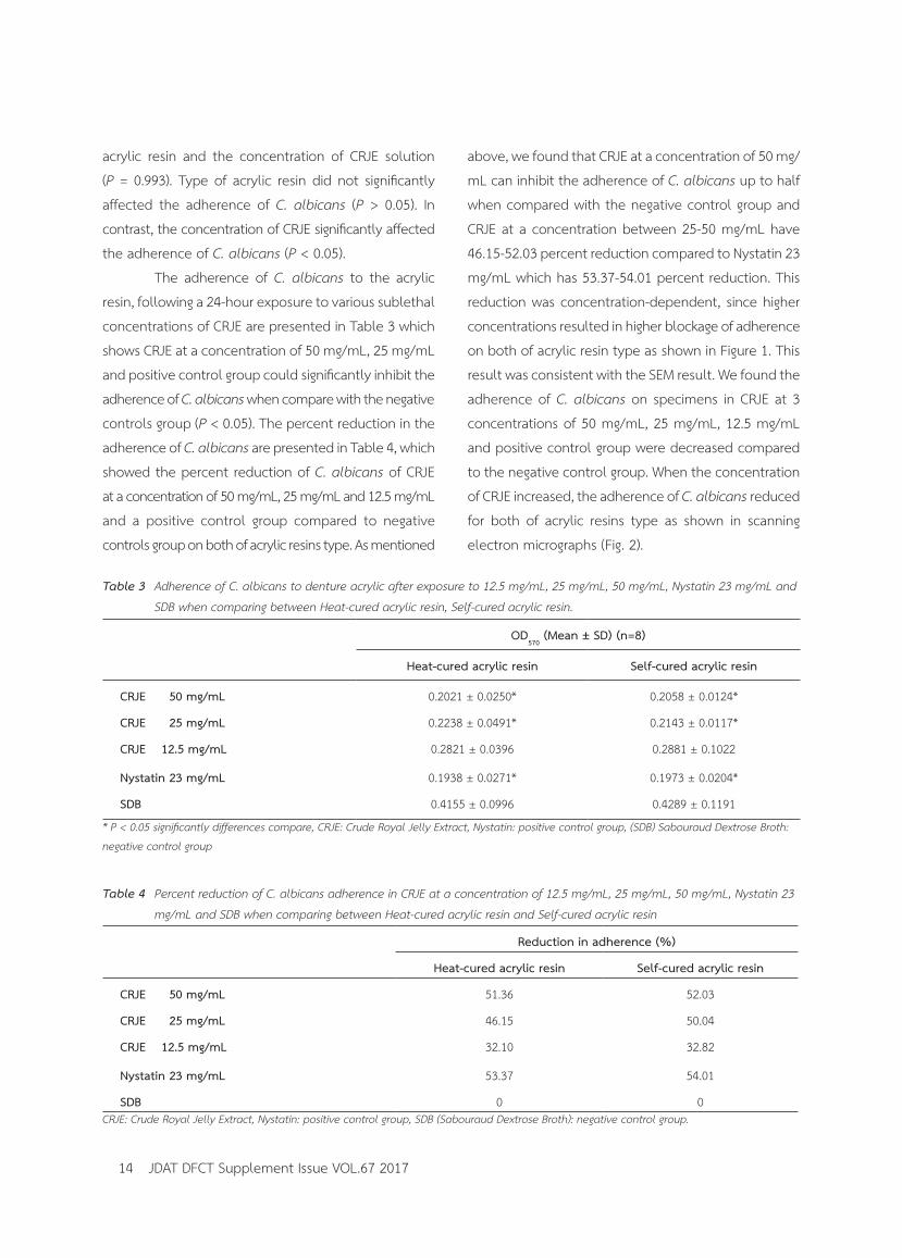
acrylic resin and the concentration of CRJE solution
(P = 0.993). Type of acrylic resin did not significantly
affected the adherence of C. albicans (P > 0.05). In
contrast, the concentration of CRJE significantly affected
the adherence of C. albicans (P < 0.05).
The adherence of C. albicans to the acrylic
resin, following a 24-hour exposure to various sublethal
concentrations of CRJE are presented in Table 3 which
shows CRJE at a concentration of 50 mg/mL, 25 mg/mL
and positive control group could significantly inhibit the
adherence of C. albicans when compare with the negative
controls group (P < 0.05). The percent reduction in the
adherence of C. albicans are presented in Table 4, which
showed the percent reduction of C. albicans of CRJE
at a concentration of 50 mg/mL, 25 mg/mL and 12.5 mg/mL
and a positive control group compared to negative
controls group on both of acrylic resins type. As mentioned
above, we found that CRJE at a concentration of 50 mg/
mL can inhibit the adherence of C. albicans up to half
when compared with the negative control group and
CRJE at a concentration between 25-50 mg/mL have
46.15-52.03 percent reduction compared to Nystatin 23
mg/mL which has 53.37-54.01 percent reduction. This
reduction was concentration-dependent, since higher
concentrations resulted in higher blockage of adherence
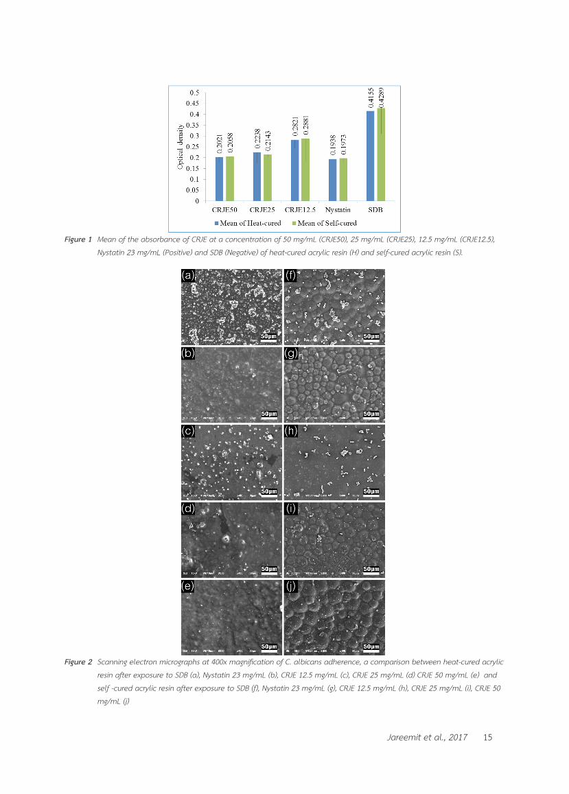
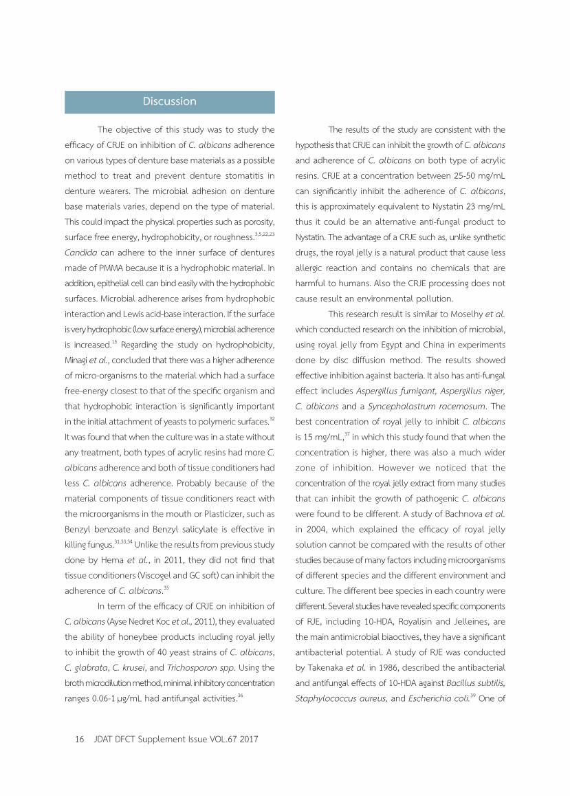
on both of acrylic resin type as shown in Figure 1. This
result was consistent with the SEM result. We found the
adherence of C. albicans on specimens in CRJE at 3
concentrations of 50 mg/mL, 25 mg/mL, 12.5 mg/mL
and positive control group were decreased compared
to the negative control group. When the concentration
of CRJE increased, the adherence of C. albicans reduced
for both of acrylic resins type as shown in scanning
electron micrographs (Fig. 2).
Table 3 Adherence of C. albicans to denture acrylic after exposure to 12.5 mg/mL, 25 mg/mL, 50 mg/mL, Nystatin 23 mg/mL and
SDB when comparing between Heat-cured acrylic resin, Self-cured acrylic resin.
OD570
(Mean ± SD) (n=8)
Heat-cured acrylic resin Self-cured acrylic resin
CRJE 50 mg/mL 0.2021 ± 0.0250* 0.2058 ± 0.0124*
CRJE 25 mg/mL 0.2238 ± 0.0491* 0.2143 ± 0.0117*
CRJE 12.5 mg/mL 0.2821 ± 0.0396 0.2881 ± 0.1022
Nystatin 23 mg/mL 0.1938 ± 0.0271* 0.1973 ± 0.0204*
SDB 0.4155 ± 0.0996 0.4289 ± 0.1191
* P < 0.05 significantly differences compare, CRJE: Crude Royal Jelly Extract, Nystatin: positive control group, (SDB) Sabouraud Dextrose Broth:
negative control group
Table 4 Percent reduction of C. albicans adherence in CRJE at a concentration of 12.5 mg/mL, 25 mg/mL, 50 mg/mL, Nystatin 23
mg/mL and SDB when comparing between Heat-cured acrylic resin and Self-cured acrylic resin
Reduction in adherence (%)
Heat-cured acrylic resin Self-cured acrylic resin
CRJE 50 mg/mL 51.36 52.03
CRJE 25 mg/mL 46.15 50.04
CRJE 12.5 mg/mL 32.10 32.82
Nystatin 23 mg/mL 53.37 54.01
SDB 0 0CRJE: Crude Royal Jelly Extract, Nystatin: positive control group, SDB (Sabouraud Dextrose Broth): negative control group.
15Jareemit et al., 2017
Figure 1 Mean of the absorbance of CRJE at a concentration of 50 mg/mL (CRJE50), 25 mg/mL (CRJE25), 12.5 mg/mL (CRJE12.5),
Nystatin 23 mg/mL (Positive) and SDB (Negative) of heat-cured acrylic resin (H) and self-cured acrylic resin (S).
Figure 2 Scanning electron micrographs at 400x magnification of C. albicans adherence, a comparison between heat-cured acrylic
resin after exposure to SDB (a), Nystatin 23 mg/mL (b), CRJE 12.5 mg/mL (c), CRJE 25 mg/mL (d) CRJE 50 mg/mL (e) and
self -cured acrylic resin after exposure to SDB (f), Nystatin 23 mg/mL (g), CRJE 12.5 mg/mL (h), CRJE 25 mg/mL (i), CRJE 50
mg/mL (j)
JDAT DFCT Supplement Issue VOL.67 201716
Discussion
The objective of this study was to study the
efficacy of CRJE on inhibition of C. albicans adherence
on various types of denture base materials as a possible
method to treat and prevent denture stomatitis in
denture wearers. The microbial adhesion on denture
base materials varies, depend on the type of material.
This could impact the physical properties such as porosity,
surface free energy, hydrophobicity, or roughness.3,5,22,23
Candida can adhere to the inner surface of dentures
made of PMMA because it is a hydrophobic material. In
addition, epithelial cell can bind easily with the hydrophobic
surfaces. Microbial adherence arises from hydrophobic
interaction and Lewis acid-base interaction. If the surface
is very hydrophobic (low surface energy), microbial adherence
is increased.15 Regarding the study on hydrophobicity,
Minagi et al., concluded that there was a higher adherence
of micro-organisms to the material which had a surface
free-energy closest to that of the specific organism and
that hydrophobic interaction is significantly important
in the initial attachment of yeasts to polymeric surfaces.32
It was found that when the culture was in a state without
any treatment, both types of acrylic resins had more C.
albicans adherence and both of tissue conditioners had
less C. albicans adherence. Probably because of the
material components of tissue conditioners react with
the microorganisms in the mouth or Plasticizer, such as
Benzyl benzoate and Benzyl salicylate is effective in
killing fungus.31,33,34 Unlike the results from previous study
done by Hema et al., in 2011, they did not find that
tissue conditioners (Viscogel and GC soft) can inhibit the
adherence of C. albicans.35
In term of the efficacy of CRJE on inhibition of
C. albicans (Ayse Nedret Koc et al., 2011), they evaluated
the ability of honeybee products including royal jelly
to inhibit the growth of 40 yeast strains of C. albicans,
C. glabrata, C. krusei, and Trichosporon spp. Using the
broth microdilution method, minimal inhibitory concentration
ranges 0.06-1 µg/mL had antifungal activities.36
The results of the study are consistent with the
hypothesis that CRJE can inhibit the growth of C. albicans
and adherence of C. albicans on both type of acrylic
resins. CRJE at a concentration between 25-50 mg/mL
can significantly inhibit the adherence of C. albicans,
this is approximately equivalent to Nystatin 23 mg/mL
thus it could be an alternative anti-fungal product to
Nystatin. The advantage of a CRJE such as, unlike synthetic
drugs, the royal jelly is a natural product that cause less
allergic reaction and contains no chemicals that are
harmful to humans. Also the CRJE processing does not
cause result an environmental pollution.
This research result is similar to Moselhy et al.
which conducted research on the inhibition of microbial,
using royal jelly from Egypt and China in experiments
done by disc diffusion method. The results showed
effective inhibition against bacteria. It also has anti-fungal
effect includes Aspergillus fumigant, Aspergillus niger,
C. albicans and a Syncephalastrum racemosum. The
best concentration of royal jelly to inhibit C. albicans
is 15 mg/mL,37 in which this study found that when the
concentration is higher, there was also a much wider
zone of inhibition. However we noticed that the
concentration of the royal jelly extract from many studies
that can inhibit the growth of pathogenic C. albicans
were found to be different. A study of Bachnova et al.
in 2004, which explained the efficacy of royal jelly
solution cannot be compared with the results of other
studies because of many factors including microorganisms
of different species and the different environment and
culture. The different bee species in each country were
different. Several studies have revealed specific components
of RJE, including 10-HDA, Royalisin and Jelleines, are
the main antimicrobial biaoctives, they have a significant
antibacterial potential. A study of RJE was conducted
by Takenaka et al. in 1986, described the antibacterial
and antifungal effects of 10-HDA against Bacillus subtilis,
Staphylococcus aureus, and Escherichia coli.39 One of

17Jareemit et al., 2017
the studies that evaluated the antibacterial activity of
Royalisin has been reported against B. subtilis. This inhibition
was equal to that of tetracycline at 50 µg/mL.40 Jelleines
are small peptides, which have antimicrobial properties
against several gram-positive cocci (S. aureus, S. saprophyticus,
and B. subtilis) and gram-negative rods (E. coli, E. cloacae,
K. pneumoniae, and P. aeruginosa), as well as yeast (C.
Albicans).41
A study on the ability of other natural extracts that
inhibit the adherence of C. albicans by Taweechaisupapong
et al. in 2006, they studied an Inhibitory effect of Stre-
blus asper leaf-extract on adherence of C. albicans to
denture acrylic, using various sublethal concentrations of
Streblus asper leaf ethanolic extract. The experiments
were performed on self-cured acrylic resin by Broth
dilution method, combined with A colorimetric
tetrazolium assay using XTT ((2, 3)-bis (2-methoxy-4-nitro-
5-sulfophenyl)-5-(12)-2H-tetrazolium hydroxide). The
results showed that the Streblus asper leaf-extract
concentration of 31.2 mg/mL, 62.5 mg/mL and 125 mg/
mL, led to a significant reduction (P < 0.05) of C. albicans
compared to the control group. The reduction was
concentration-dependent, since higher concentrations
resulted in higher blockage of adherence. The Streblus
asper leaf-extract could affect the cell wall of fungi,
such as creating extracellular components and chemical
surface adherence inhibiting effect.42
In a study using synthetic substances to inhibit
the adherence of C. albicans on various surfaces. Lin
Zhou et al. used Parylene® to inhibit the adherence of
C. albicans on the surface of denture made of heat-cured
acrylic resin. The result showed that Parylene® had the
ability to reduce the adherence of pathogenic C. albicans
on the acrylic resin. Cell counts and XTT assay also
showed significant reduction.43
When comparing properties and surface
characteristics of heat-cured acrylic resin and self-cured
acrylic resin, we found that heat-cured acrylic resin has
less surface roughness than self-cured acrylic resin which
is consistent with the less adherence of C. albicans on
heat-cured resin.44 However, the results from this study
found no difference in the adherence of C. albicans in
both of acrylic resins type. This is probably because of
the specimens preparation process
This research is only an in vitro study, therefore
the results from this study may differ from results from
an experiment conducted in oral condition. This is an
important factor encouraging the growth of fungus. Thus
more studies are still needed to provide more information
regarding the mechanism of the adherence of C. albicans.
In addition, a study on CRJE pre-coating application to
the denture base material could also be done instead
of soaking application.
The study concluded that CRJE has the ability
to inhibit the adherence of C. albicans on the surface
of both heat-cured acrylic resin and self-cured acrylic
resin. This is an alternative development the product
to patients who wear dentures which made from acrylic
resin. Especially the elderly or patients with restrictions
on the hand movement.
This work was supported by a research grant
from the Faculty of Dentistry, Srinakrinwirot University.
Authors would also like to thank Dr. Waiyawut Yoonisil
for his advices on the Statistical analysis.
1. He XY, Meurman JH, Kari K, Rautemaa R, Samaranayake
LP. In vitro adhesion of Candida species to denture base
materials. Mycoses 2006;49:80-4.
2. Jain D, Shakya P. An in vitro study on effect of Delmopinol
application on Candida albicans adherence on heat
cured denture base acrylic resin: a thorough study. Indian
J Dent Res 2013;24:645.
Conclusion
Acknowledgments
References
JDAT DFCT Supplement Issue VOL.67 201718
3. Uzunoglu E, Yildirim Bicer AZ, Dolapci I, Dogan A.
Biofilm-forming ability and adherence to poly-
(methyl-methacrylate) acrylic resin materials of oral
Candida albicans strains isolated from HIV positive
subjects. J Adv Prosthodont 2014;6:30-4.
4. Nawasrah A, AlNimr A, Ali AA. Antifungal Effect of
Henna against Candida albicans Adhered to Acrylic
Resin as a Possible Method for Prevention of Denture
Stomatitis. Int J Environ Res Public Health 2016;13.
5. Gebremedhin S, Dorocka-Bobkowska B, Prylinski M,
Konopka K, Duzgunes N. Miconazole activity against
Candida biofilms developed on acrylic discs. J Physiol
Pharmacol 2014;65:593-600.
6. Waters MG, Williams DW, Jagger RG, Lewis MA. Adherence
of Candida albicans to experimental denture soft lining
materials. J Prosthet Dent 1997;77:306-12.
7. Hoshi N, Mori H, Taguchi H, Taniguchi M, Aoki H, Sawada
T, et al. Management of oral candidiasis in denture wearers.
J Prosthodont Res 2011;55:48-52.
8. Iseri U, Uludamar A, Ozkan YK. Effectiveness of different
cleaning agents on the adherence of Candida albicans
to acrylic denture base resin. Gerodontology 2011;28:
271-6.
9. Skupien JA, Valentini F, Boscato N, Pereira-Cenci T.
Prevention and treatment of Candida colonization on
denture liners: a systematic review. J Prosthet Dent
2013;110:356-62.
10. Pereira-Cenci T, Del Bel Cury AA, Crielaard W, Ten
Cate JM. Development of Candida-associated denture
stomatitis: new insights. J Appl Oral Sci 2008;16:86-94.
11. Kanathila H, Bhat AM, Krishna PD. The effectiveness
of magnesium oxide combined with tissue conditioners
in inhibiting the growth of Candida albicans: An in vitro
study. Indian J Dent Res 2011;22:610.
12. Kang SH, Lee HJ, Hong SH, Kim KH, Kwon TY. Influence
of surface characteristics on the adhesion of Candida
albicans to various denture lining materials. Acta Odontol
Scand 2013;71:241-8.
13. Tari BF, Nalbant D, Dogruman Al F, Kustimur S. Surface
roughness and adherence of Candida albicans on soft
lining materials as influenced by accelerated aging. J
Contemp Dent Pract 2007;8:18-25.
14. Peyton FA. History of resins in dentistry. Dent Clin
North Am 1975;19:211-22.
15. Azuma A, Akiba N, Minakuchi S. Hydrophilic surface
modification of acrylic denture base material by silica
coating and its influence on Candida albicans adherence.
J Med Dent Sci 2012;59:1-7.
16. Winkler S, Wood R, Facchiano AM, Boberick KG, Patel
AR. Prosthodontic Self-treatment with Acrylic Resin Super
Glue: A Case Report. J Oral Implantol 2006;32:132-6.
17. Rashid H, Sheikh Z, Vohra F. Allergic effects of the
residual monomer used in denture base acrylic resins.
Eur J Dent 2015;9:614-9.
18. Chaves CA, Machado AL, Vergani CE, de Souza RF,
Giampaolo ET. Cytotoxicity of denture base and hard
chairside reline materials: a systematic review. J Prosthet
Dent 2012;107:114-27.
19. McCabe JF. A polyvinylsiloxane denture soft lining
material. J Dent 1998;26:521-6.
20. Masella RP, Dolan CT, Laney WR. The prevention of
the growth of Candida on silastic 390 soft liner for dentures.
J Prosthet Dent 1975;33:250-7.
21. Wright PS. The effect of soft lining materials on the
growth of Candida albicans. J Dent 1980;8:144-51.
22. Pereira-Cenci T, Cury AA, Cenci MS, Rodrigues-Garcia
RC. In vitro Candida colonization on acrylic resins and
denture liners: influence of surface free energy, roughness,
saliva, and adhering bacteria. Int J Prosthodont 2007;
20:308-10.
23. Oncul B, Karakis D. The effect of two artificial salivas
on the adhesion of Candida albicans to heat-polymerized
acrylic resin. J Adv Prosthodont 2015;7:93-7.
24. Akalin-Evren B, Kulak-Ozkan Y, Ozcan M, Kadir T.
Candida albicans adhesion on reinforced polymethyl-
methacrylate denture resin: effect of fibre architecture
and exposure to saliva. Gerodontology 2014;31:194-201.
25. Chandra J, Kuhn DM, Mukherjee PK, Hoyer LL, McCormick
T, Ghannoum MA. Biofilm formation by the fungal pathogen
Candida albicans: development, architecture, and drug
19Jareemit et al., 2017
resistance. J Bacteriol 2001;183:5385-94.
26. Ellepola AN, Samaranayake LP. The effect of limited
exposure to antifungal agents on the germ tube formation
of oral Candida albicans. J Oral Pathol Med 1998;27:213-9.
27. Bilikova K, Huang SC, Lin IP, Simuth J, Peng CC.
Structure and antimicrobial activity relationship of royalisin,
an antimicrobial peptide from royal jelly of Apis mellifera.
Peptides 2015;68:190-6.
28. Bilikova K, Kristof Krakova T, Yamaguchi K, Yamaguchi
Y. Major royal jelly proteins as markers of authenticity
and quality of honey. Arh Hig Rada Toksikol 2015;66:
259-67.
29. Fujii A, Kobayashi S, Kuboyama N, Furukawa Y, Kaneko
Y, Ishihama S, et al. Augmentation of wound healing by
royal jelly (RJ) in streptozotocin-diabetic rats. Jpn J
Pharmacol 1990;53:331-7.
30. Faul F, Erdfelder E, Lang AG, Buchner A. G*Power 3:
A flexible statistical power analysis program for the
social, behavioral, and biomedical sciences. Behav Res
Methods 2007;39:175-191.
31. Gautam R, Singh RD, Sharma VP, Siddhartha R, Chand
P, Kumar R. Biocompatibility of polymethylmethacrylate
resins used in dentistry. J Biomed Mater Res B Appl
Biomater 2012;100:1444-50.
32. Minagi S, Miyake Y, Inagaki K, Tsuru H, Suginaka H.
Hydrophobic interaction in Candida albicans and Candida
tropicalis adherence to various denture base resin materials.
Infect Immun 1985;47:11-14.
33. Bulad K, Taylor RL, Verran J, McCord JF. Colonization
and penetration of denture soft lining materials by
Candida albicans. Dent Mater 2004;20:167-75.
34. Serrano-Granger C, Cerero-Lapiedra R, Campo-Trapero
J, Del Rio-Highsmith J. In vitro study of the adherence of
Candida albicans to acrylic resins: relationship to surface
energy. Int J Prosthodont 2005;18:392-8.
35. Kanathila H, Bhat AM, Krishna PD. The effectiveness
of magnesium oxide combined with tissue conditioners
in inhibiting the growth of Candida albicans: an in vitro
study. Indian J Dent Res 2011;22:613.
36. Koc AN, Silici S, Kasap F, Hormet-Oz HT, Mavus-Buldu
H, Ercal BD. Antifungal activity of the honeybee products
against Candida spp. and Trichosporon spp. J Med Food
2011;14:128-34.
37. Moselhy A FA, Kamel A. An evaluation of the potent
antimicrobial matter analysis of the royal jelly. Life Sci
2013;10.
38. Bachanová K, Klaudiny J, Kopernicky J, Simúth J.
Identification of honeybee peptide active against
Paenibacillus larvae larvae through bacterial growth-
inhibition assay on polyacrylamide gel. Apidologie
2002;33:259-69.
39. Takenaka T, Yatsunami K, Echigo T. Changes in Quality
of Royal Jelly during Storage. Nippon Shokuhin Kogyo
Gakk 1986;33:1-7.
40. Bilikova K, Wu GS, Simuth J. Isolation of a peptide
fraction from honeybee royal jelly as a potential
antifoulbrood factor. Apidologie 2001;32: 275-83.
41. Fontana R, Mendes MA, de Souza BM, Konno K, César
LM, Malaspina O, et al. Jelleines. A family of antimicrobial
peptides from the Royal Jelly of honeybees (Apis mellifera).
Peptides 2004;25:919-28.
42. Taweechaisupapong S, Klanrit P, Singhara S, Pitiphat
W, Wongkham S. Inhibitory effect of Streblus asper
leaf-extract on adhesion of Candida albicans to denture
acrylic. J Ethnopharmacol 2006;106:414-7.
43. Zhou L, Tong Z, Wu G, Feng Z, Bai S, Dong Y, et al.
Parylene coating hinders Candida albicans adhesion to
silicone elastomers and denture bases resin. Arch Oral
Biol 2010;55:401-9.
44. He XY, Meurman JH, Kari K, Rautemaa R, Samaranayake
LP. In vitro adhesion of Candida species to denture base
materials. Mycoses 2006;49:80-4.