original article alleviative effects of yukmijihwang-tang ...ijcem.com/files/ijcem0022373.pdf ·...
TRANSCRIPT

Int J Clin Exp Med 2016;9(6):10093-10108www.ijcem.com /ISSN:1940-5901/IJCEM0022373
Original ArticleAlleviative effects of Yukmijihwang-tang on cholesterol related disease in a postmenopausal rat model and lipid accumulation in HepG2 cells
Hiroe Go1, Jin Ah Ryuk1, Hye Won Lee1, Ki-Jung Kil2, Byoung Seob Ko1
1Korean Medicine Convergence Research Division, Korea Institute of Oriental Medicine, Republic of Korea; 2Col-lege of Oriental Medicine, Joongbu University, Republic of Korea
Received December 21, 2015; Accepted April 3, 2016; Epub June 15, 2016; Published June 30, 2016
Abstract: Yukmijihwang-tang (YMJT), known as Liu-wei-di-huang-tang in Chinese and Lokumijio-to in Japanese, has been used as a traditional herbal formula for the treatment of various diseases. In Korean traditional medicine, YMJT is a well-known herbal prescription used to relieve Yin-Deficiency. In this study, we examined the effects of YMJT on Nonalcoholic steatohepatitis (NASH) and on diseases associated with cholesterol in a rat model of post-menopausal hyperlipidemia and the methyl-β-cyclodextrin (MβCD)-induced hepatic steatosis model in HepG2 cells. Twenty-five rats were ovariectomized (OVX), and five rats were sham-operated (Sham). Then, the OVX rats were randomly assigned to three groups (n = 5): OVX-Con, OVX with simvastatin, and OVX with YMJT (50, 150, 450 mg/kg) for 8 weeks. NASH and a number of targets associated with cholesterol were examined to confirm the effects of YMJT. Oil Red O staining and intracellular cholesterol analyses were used to quantify cellular cholesterol levels. The levels of phosphorylated AMP-activated protein kinases (AMPK) and the products of genes involved in cholesterol synthesis were measured via Western blotting. In OVX rats, YMJT reduced retroperitoneal and peri-renal fat accumu-lation, serum lipids, the atherogenic index, cardiac risk factors, intima-media thickness, and NASH. YMJT decreased lipid accumulation, total cholesterol, and low-density/very-low-density lipoprotein levels in HepG2 cells. Moreover, YMJT reversed the effects of MβCD on cholesterol synthesis regulators. Phosphorylation of AMPK was stimulated by YMJT. These results indicated that YMJT has cholesterol-lowering effects both in vivo and in vitro. Therefore, YMJT may have potential as a therapeutic agent for the treatment of hyperlipidemia in postmenopausal females.
Keywords: Yukmijihwang-tang (YMJT), cholesterol, hyperlipidemia, menopause, lipid, ovariectomized
Introduction
In Donguibogam, aging is said to result from the decline of blood [1]. Yukmijihwang-tang (YMJT) is used to improve health and has been used for aging care. Aging negatively affects the health of women. Hyperlipidemia is known as high lipid levels in the blood. Cholesterol is car-ried in the blood as lipoproteins, such as high-density lipoproteins (HDL) and low-density lipo-proteins (LDL). In our body, an excess of cholesterol formation leads to an increased risk of developing cardiovascular disease (CVD) [2]. Hyperlipidemia leads to the development of fatty liver diseases, including nonalcoholic fatty liver disease (NAFLD) and nonalcoholic steato-hepatitis (NASH) [3].
Postmenopausal females are at particular risk, with the accelerated progression of NASH and the accumulation of visceral fat being more common in these patients [4-6]. In a report about patients with NASH, premenopausal women are shown to be at a decreased risk of severe fibrosis compared with similarly aged men, but the risk is equivalent in postmeno-pausal women and similarly aged men [6]. NASH can be ameliorated by hormone replace-ment therapy (HRT) or estrogen administration [5]. HRT and estrogen replacement therapy (ERT) cause a slight increase in the risk of developing heavy diseases such as breast can-cer [7]. Therefore, the development of a safe, effective method of treating or preventing NASH is needed [8].

The effects of Yukmijihwang-tang in post-menopausal disorder
10094 Int J Clin Exp Med 2016;9(6):10093-10108
Recent studies indicate that increased hepatic free cholesterol is an important risk factor in NASH patients [9]. In addition, hypercholester-olemia is often observed in postmenopausal females [5]. Management of increased choles-terol may be associated with a reduced risk for NASH in postmenopausal females.
The synthesis and utilization of cholesterol is a tightly regulated system mediated by the sterol regulatory element binding proteins (SREBPs), which prevent its over-accumulation and abnor-mal deposition within the body [10, 11]. The SREBPs are a family of transcription factors consisting of three members, namely SREBP-1a, SREBP-1c, and SREBP2. SREBP2 regulates cholesterol metabolism primarily through its regulation of genes related to cholesterol stim-ulation and synthesis, such as low-density lipo-protein receptor (LDLR) and 3-hydroxy-3-methyl glutaryl coenzyme A reductase (HMGCR) [12-14]. When cells are deprived of cholesterol, the SREBP2-NH2-terminal fragment is translocated from the endoplasmic reticulum (ER) mem-brane to the nucleus and binds to sterol regula-tory elements in the promoter regions of the LDLR and HMGCR genes, resulting in an increase in cholesterol uptake and synthesis [11, 15]. When cholesterol levels are high, SREBP2 is retained in the ER, resulting in the downregulation of intracellular cholesterol uptake and synthesis [11].
LDLR is a transmembrane glycoprotein that plays a critical role in the homeostatic control of blood cholesterol by mediating the turnover of LDL particles in circulation [16]. In humans, the liver contains approximately 70% of the total LDLR. The abundance of LDLR has been shown to be closely associated with NASH [17, 18].
HMGCR catalyzes a rate-limiting step of the mevalonate pathway, the cholesterol biosyn-thesis pathway, and is responsible for the syn-thesis of mevalonate from hydroxyl-methylglu-taryl-coenzyme A (HMG-CoA) [19]. As the expr- ession of HMGCR is increased in patients with NASH, HMGCR inhibitors, such as lovastatin, have been proposed as a potential treatment for NASH, although their efficacy has yet to be established.
AMP-activated protein kinase (AMPK) is seen as a mediator of the cellular adaptation to the
environment, a metabolic master switch or a factor of nutritional stress [20]. The activated AMPK leads to simultaneous inhibition of ana-bolic pathways, such as cholesterol, fatty acids, and triglyceride synthesis, as well as to the stimulation of fatty acid oxidation and ketogen-esis [17, 21]. Because AMPK plays a central role in lipid metabolism, it was considered to be an important therapeutic target for the treat-ment of fatty liver disease [22].
There is increasing interest in the use of tradi-tional herbal medicines to prevent and treat various diseases [23]. YMJT, a traditional Ko- rean remedy, is composed of six oriental herbs (Rehmannia glutinosa Liboschitz ex Steudel, Dioscorea japonica Thunberg, Cornus officina-lis Siebold et Zuccarini, Alisma orientale Juze- pzuk, Poria cocos Wolf, Paeonia suffruticosa Andrews) and has been used to treat diseases associated with a decline in kidney. According to Traditional Chinese Medicine, kidney energy levels are low at the onset of menopause. It has been reported that YMJT is able to prevent and cure osteoporosis induced by ovariectomy in rat models of osteoporosis [24]. In addition, in rat models of diabetes, YMJT and Discoreae Radix decreased total serum cholesterol and triglyceride [25]. Thus, we thought that YMJT might have effects on the symptoms of meno-pause, especially on cholesterol related diseases.
We aimed to assess whether YMJT has choles-terol-lowering effects and is effective against NASH and diseases associated with cholester-ol in a postmenopausal model. Moreover, we also investigated whether YMJT has cholester-ol-lowering effects in the MβCD-induced hepat-ic steatosis model in vitro.
Materials and methods
YMJT preparation
The formula of YMJT consist of 6 herbs, includ-ing R. glutinosa (165 g), C. officinalis (82.5 g), D. japonica (82.5 g), P. suffruticosa (61.88 g), A. orientale (61.88 g), P. cocos (61.88 g). The water extract of YMJT were prepared using an S-20,000 extractor (Sak IK Medical Company). Briefly, mixture of 6 herb (515.64 g) were extracted by heating for 2 h in a 10-fold volume of water. Then extractions were freeze-dried and the resulting YMJT powder (148.3 g, yield:

The effects of Yukmijihwang-tang in post-menopausal disorder
10095 Int J Clin Exp Med 2016;9(6):10093-10108
28.76%) was collected. YMJT extract powder was stored at 4°C until use. The YMJT extract (KIOM PH 130003) was stored at Korea Institute of Oriental Medicine (KIOM, Daejeon, Korea) until used in this experiment.
Chromatographic conditions of high-perfor-mance liquid chromatography (HPLC)-DAD
Quantitative analysis of the reference com-pounds solutions; 5-HMF, morroniside, loganin, paeoniflorin, and paeonol (1,000 μg/mL) were prepared in 100% methanol and stored at 4°C. The standard solutions were prepared by six concentrations of diluted solutions (methanol). All calibration curves were attained by evaluat-ing the peak areas at six concentrations in the range of 10-500 μg/mL for all reference com-pounds. The linearity of the peak area (y) vs. concentration (x, μg/mL) curve for each compo-nent was used to calculate the contents of the main YMJT. 104.0 mg of YMJT extract powder was subsequently resuspended in 10 mL dis-tilled water for HPLC analysis. The contents of 5-HMF, morroniside, loganin, paeoniflorin, and paeonol in the YMJT water extract were ana-lyzed using an 1100 series HPLC instrument (Agilent Technologies, USA) with a Gemini C18 column (4.6 × 250 mm, 5 μm; Phenomenex, USA). The mobile phase consisted of the sol-vents, distilled water (A) and acetonitrile (B). The following gradient was used: 0 min, A:B 95:5 (v/v); 30 min, A:B 60:40; and 40 min, A:B 0:100. The mobile phase flow rate was 1.0 mL/min, the column temperature was 25°C, the injection volume was 10 uL, and UV detection was at 240 nm (morroniside, loganin, and pae-oniflorin) and 280 nm (5-HMF, paeonol).
Cell line and culture conditions
HepG2 human hepatocellular carcinoma cells were purchased from the Korean cell line bank (Seoul, Korea). HepG2 cells were maintained in Dulbecco’s modified Eagle’s medium (DMEM) supplemented with 10% fetal bovine serum, 100 U/mL penicillin, and 100 mg/mL strepto-mycin. Cells were incubated at 37°C in humidi-fied atmosphere of 5% CO2 in air (v/v). To induce cholesterol accumulation, HepG2 cells were exposed to MβCD mixed with palmitic acid. When cells reached 70% confluence, they were incubated in 0.2% BSA-DMEM containing 20 μg/mL MβCD mixed with palmitic acid (MβCD). The cells were exposed to 30 μM simvastatin or
various concentrations (250, 500, 750 μg/mL) of YMJT for 8 h. In this study, we used same data and pictures that were obtained from three groups (control, MβCD, and simvastatin) that were included in previous study [26].
Animals and experimental design
All experimental protocols and animal mainte-nance procedures used in this study were treat-ed in accordance with the Guide for Care and Use of Laboratory Animals by the Institutional Animal Care and Use Committee (IACUC) of KIOM. Six week-old female Sprague-Dawley rats (weight 225±25 g) were obtained from Samtako (Osan-si, Korea). Animals were main-tained at a regular 12 h light/dark cycle, at a controlled temperature (24±2°C) with a relative humidity of 50±5 % in the Laboratory Animals Center at KIOM (Approval No. 13-024). After 1 week of acclimation, the rats were performed ovariectomy (n = 25) or sham operation (n = 5) under general anesthesia by intramuscular injection. One week after the surgery, twenty-five OVX rats were randomly divided into three group: (1) high-fat (45%), high-cholesterol (1%) diet (OVX-Con) (n = 5), (2) high-fat (45%), high-cholesterol (1%) diet with simvastatin (OVX-SV; 20 mg/kg) (n = 5), (3) high-fat (45%), high-cho-lesterol (1%) diet with YMJT (OVX-YMJT; 50, 150, 450 mg/kg) (n = 5 per each dose). Five sham-operated rats were not assigned to a high-fat (45%), high-cholesterol (1%) diet (Sham). Simvastatin and YMJT were dissolved in phosphate-buffered saline (PBS) for daily oral administration. After eight weeks, rats were sacrificed under anesthesia and blood was collected for the estimation of total-choles-terol (TC), triglycerides (TG), HDL, LDL, athero-genic index, and cardiac risk factor. Liver tis-sue, retroperitoneal fat, peri-renal fat, and arterial tissue were also collected. In this study, we used same data and pictures that were obtained from three groups (Sham-operated, OVX-Con, and OVX-SV) that were included in previous study [26].
Lipid parameters
Measurements of serum TC, TG, and HDL levels were performed using a BS220 instrument (Mindary, Shenxhen, China). LDL levels were determined using the following equation: LDL = TC-HDL-(TG/5). TC and LDL/very low- density lipoproteins (VLDL) contents in HepG2 cells

The effects of Yukmijihwang-tang in post-menopausal disorder
10096 Int J Clin Exp Med 2016;9(6):10093-10108
were determined using an HDL and LDL/VLDL cholesterol assay kit according to the manufac-turer’s instructions. After treatment with 30 μM simvastatin or various concentrations of YMJT (250, 500, 750 μg/mL) with MβCD in 0.2% BSA-DMEM for 8 h, cells were washed with PBS and cellular lipids were extracted with chloro- form:isopropanol: NP-40 (7:11:0.1). Then extr- acts were centrifuged for 10 min at 15,000 g and supernatants were transfer to a new tube. To remove chloroform, supernatants were dried at 50°C and 200 μL cholesterol assay buffer were added to resuspend dried lipids. TC and LDL/VLDL levels were assayed by measuring absorbance at 570 nm using a spectrophoto- meter.
Atherogenic index and cardiac risk factor
Atherogenic index and cardiac risk factor were determined using the following equations: ath-erogenic index = (TC-HDL)/HDL, cardiac risk factor = TC/HDL.
Histology
Rat livers were fixed with 4% neutral buffered formalin (NBF) and embedded in paraffin. Four-micron sections were then cut and stained with hematoxylin and eosin (H&E) for histological analysis.
Cytotoxicity
The cell viability was examined by (4-[3-(4-io- dophenyl)-2-(4-nitrophenyl)-2H-5-tetrazolio]-1,3-benzene disulfonate) (WST) assay. Briefly, HepG2 cells were plated at a density of 2 × 10⁴ cells/well in 96-well plates and treated with 30 μM simvastatin or various concentrations of YMJT (250, 500, 750 μg/mL) with or without MβCD in 0.2% BSA-DMEM for 8 h. Then WST solution was added to each well. After 2 h of exposure at 37°C, the absorbance was mea-sured at 490 nm.
Oil Red O staining
To detect and quantify cellular lipid content, HepG2 cells were stained using the Oil Red O staining. Briefly, HepG2 cells were seeded at a density of 1 × 10⁵ cells/well in 24-well plates and treated with 30 μM simvastatin or various concentrations of YMJT (250, 500, 750 μg/mL) with MβCD in 0.2% BSA-DMEM for 8 h. Then
cells were gently washed with PBS and fixed using 10% formalin for 10 min at room temper-ature. Subsequently, cells were washed with PBS and 60% isopropanol, and stained for 1 h in a freshly diluted Oil Red O solution (stock solution, 3 mg/mL in isopropanol; working solu-tion, 60% Oil Red O stock solution diluted in water). After staining, cells were washed with 60% isopropanol and PBS, and photographed. To quantify cellular lipids, Oil Red O stain was extracted using isopropanol and its absorbance was measured at 500 nm using a spectropho- tometer.
Western blotting
Cells were cultured to 80% confluence at 37°C, incubated in 0.2% BSA-DMEM containing 20 μg/mL MβCD, and treated with 30 μM simvas-tatin or various concentrations of YMJT (250 and 500 μg/mL) for 8 h. Cell lysates were pre-pared using RIPA buffer containing protease inhibitors. Briefly, lysates were incubated at 4°C for 30 min and then centrifuged at 14,000 rpm for 15 min at 4°C to remove detergent-insoluble material.
Protein concentration was determined using a Bio-Rad protein assay. Protein samples (30-90 µg) were then separated on 4-15% Mini-Protean TGX Precast Gels and transferred to a polyvi-nylidene fluoride membrane. The membranes were blocked with 1% bovine serum albumin in PBS-T (137 mM NaCl, 2.7 mM KCl, 4.3 mM Na2HPO4, 1.4 mM KH2PO4, 0.1% Tween 20). After blocking, the membrane was incubated with the following first antibodies: anti-HMG- CR (C-1; 1:1,000), anti-SREBP2 (1C6; 1:1,000), anti-β-actin (C4; 1:1,000), anti-phospho-AM- PKα (Thr172; 1:1,000), anti-AMPKα (1:1,000), anti-LDL Receptor (1:1,000) antibodies for overnight at 4°C with gentle shaking. Secondary antibodies included HRP-conjugated goat anti-mouse, goat anti-rabbit, and donkey anti-goat (1:10,000) antibodies, as appropriate. Protein bands were visualized by using an Immun-Star WesternC kit.
Statistical analyses
All of the experiments were performed in tripli-cate. Data are expressed as a mean ± standard deviation. Statistical analysis was performed using the SPSS software version 12.0 (SPSS, Inc., Chicago, IL) and data were evaluated for
The effects of Yukmijihwang-tang in post-menopausal disorder
10097 Int J Clin Exp Med 2016;9(6):10093-10108
statistical significance using one-way ANOVA followed by a Duncan’ test (P < 0.05). The OVX and Sham groups; control and MβCD groups were compared using two-sample t-tests (P < 0.05).
Results
HPLC analysis of reference compounds in YMJT
The reference standard of calibration curves for the 5-HMF, morroniside, loganin, paeoniflo-rin, and paeonol were y = 97.8022x + 372.6578 (R2 0.998), y = 70.2673x + 65.4569 (R2 0.999),
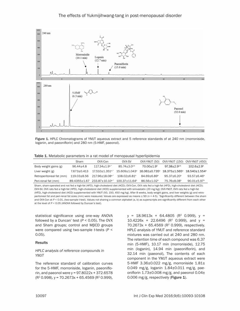
y = 18.9613x + 64.4805 (R2 0.999), y = 10.4226x + 22.6496 (R2 0.999), and y = 70.2673x + 65.4569 (R2 0.999), respectively. HPLC analysis of YMJT and reference standard mixtures was carried out at 240 and 280 nm. The retention time of each compound was 6.37 min (5-HMF), 10.17 min (morroniside), 12.75 min (loganin), 14.94 min (paeoniflorin), and 32.14 min (paeonol), The contents of each component in the YMJT aqueous extract were 5-HMF 3.36±0.022 mg/g, morroniside 1.81± 0.049 mg/g, loganin 1.84±0.011 mg/g, pae-oniflorin 1.73±0.008 mg/g, and paeonol 0.04± 0.006 mg/g, respectively (Figure 1).
Figure 1. HPLC Chromatograms of YMJT aqueous extract and 5 reference standards of at 240 nm (morroniside, loganin, and paeoniflorin) and 280 nm (5-HMF, paeonol).
Table 1. Metabolic parameters in a rat model of menopausal hyperlipidemiaSham OVX-Con OVX-SV OVX-YMJT (50) OVX-YMJT (150) OVX-YMJT (450)
Body weight gains (g) 96.44±4.8 117.34±1.9a,* 85.74±3.0a,b 70.00±1.9b 97.38±2.9a,b 102.6±2.9b
Liver weight (g) 7.673±0.413 17.510±1.351a,* 15.609±1.043a 16.061±0.735a 18.375±1.565a 18.540±1.554a
Retroperitoneal fat (mm) 119.03±8.56 217.96±18.08a,* 108.02±6.81b 84.69±6.89b 95.37±6.20b 93.57±6.46b
Peri-renal fat (mm) 89.4355±1.67 233.87±10.10a,* 100.37±11.64b 86.56±1.02b 75.76±8.08b 90.01±5.97b
Sham, sham-operated and not fed a high-fat (HFD), high-cholesterol diet (HCD); OVX-Con, OVX rats fed a high-fat (HFD), high-cholesterol diet (HCD); OVX-SV, OVX rats fed a high-fat (HFD), high-cholesterol diet (HCD) supplemented with simvastatin (20 mg/kg); OVX-YMJT, OVX rats fed a high-fat (HFD), high-cholesterol diet (HCD) supplemented with YMJT (50, 150, 450 mg/kg). After 8 weeks, body weight gains, and liver weights (g) and retro-peritoneal fat and peri-renal fat sizes (mm) were measured. Values are expressed as means ± SD (n = 4-5). *Significantly different between the sham and OVX-Con at P < 0.01, (two-sample t-test). Values not sharing a common alphabet (a, b) as superscripts are significantly different from each other at the level of P < 0.05 (ANOVA followed by Duncan’s test).

The effects of Yukmijihwang-tang in post-menopausal disorder
10098 Int J Clin Exp Med 2016;9(6):10093-10108
Effect of YMJT on the body weight and abdomi-nal fat accumulation
We established a rat model of menopausal hyperlipidemia using OVX rats that were fed a high-fat, high-cholesterol diet. Body weight gains of all rats after 8 weeks of treatments increased in the rats from the OVX group (OVX-Con) compared with those from the Sham group (Sham) (Table 1). In the YMJT treated groups (OVX-YMJT 50, 150, 450 mg/kg), YMJT inhibited the OVX-induced weight gain. Food intake remained the same in all groups (data not shown). All rats were sacrificed at week 8, and the liver weights of each group were com-pared (Table 1). In the OVX-Con group, liver weights were significantly increased compared with the Sham group; however, no significant differences were evident in the OVX-SV and OVX-YMJT groups relative to the OVX-Con group.
We also measured overall fat volumes in retro-peritoneal and peri-renal fat deposits (Table 1). The OVX-Con group significantly increased both retroperitoneal and peri-renal fat volumes com-pared with the Sham group. Furthermore, retro-peritoneal fat volumes and peri-renal fat vol-umes in all OVX-YMJT groups and the OVX-SV group decreased equally.
Effect of YMJT on serum lipids
TC, TG, and LDL levels were markedly higher in the OVX-Con group compared with the Sham group (Table 2). In contrast, the OVX-Con group showed decreased HDL levels compared with the Sham group (Table 2). In the OVX-SV group and OVX-YMJT groups (except for the group treated with 50 mg/kg), OVX-induced changes in TC and LDL levels decreased. TG levels were altered only in the OVX-SV and 450 mg/kg OVX-
YMJT groups. The OVX-SV group and OVX-YMJT groups exhibited increased HDL levels com-pared with the OVX-Con group (Table 2).
Effect of YMJT on the risk of arterial sclerosis
Arterial sclerosis is a medical condition in which fatty plaques build up along the interior wall of any artery in the body as a result of fat accumu-lation. Increased levels of total plasma choles-terol and obesity represent marked risk factors for atherosclerosis and increased rates of car-diovascular death [27, 28]. Because levels of fat accumulation in retroperitoneal and peri-renal spaces and serum cholesterol in meno-pausal hyperlipidemic rats were decreased by YMJT treatment (Tables 1 and 2), we next examined the effect of YMJT on the risk of arte-rial sclerosis by measuring the atherogenic index, cardiac risk factors, lumen diameter, and intima-media thickness (Table 3). The OVX-Con group showed a markedly increased atherogen-ic index and cardiac risk factor scores com-pared to the Sham group. The OVX-SV and OVX-YMJT groups (except for the group treated with 50 mg/kg) exhibited a lower atherogenic index. Moreover, cardiac risk factor scores were de- creased in the OVX-SV and all OVX-YMJT groups relative to the OVX-Con group (Table 3). Lumen diameter and intima-media thickness were al- so determined. The OVX-Con group showed a lower overall lumen diameter than the Sham group. In the OVX-YMJT and OVX-SV groups, lumen diameter increased but not significantly. Intima-media thickness was increased in the OVX-Con group relative to the Sham group (Table 3). All OVX-YMJT groups had a decreased intima-media thickness, similar to that of the OVX-SV group (Table 3).
Table 2. Serum lipid levels in a rat model of menopausal hyperlipidemia Sham OVX-Con OVX-SV OVX-YMJT (50) OVX-YMJT (150) OVX-YMJT (450)
T-CHO (mg/dL) 113.50±2.179 221.25±6.142a,** 178.50±35.624b 238.00±13.934a 181.00±11.712b 202.50±9.152a,b
TG (mg/dL) 46.6±7.80 64.06±6.06a,* 48.33± 3.21a,b 74.67±15.3a 64.33±7.75a 36.00±12.83b
HDL (mg/dL) 67.2±4.03 31.8±4.5a,** 41.6±3.14c 37.2±4.53a,b 52.8±6.3c 50.4±3.33c
LDL (mg/dL) 37.52±3.83 176.64± 0.43a,** 127.23± 31.842b 186.32±11.907a 115.56±10.856b 145.20±8.037b
Sham, sham-operated and not fed a high-fat (HFD), high-cholesterol diet (HCD); OVX-Con, OVX rats fed a high-fat (HFD), high-cholesterol diet (HCD); OVX-SV, OVX rats fed a high-fat (HFD), high-cholesterol diet (HCD) supplemented with simvastatin (20 mg/kg); OVX-YMJT, OVX rats fed a high-fat (HFD), high-cholesterol diet (HCD) supplemented with YMJT (50, 150, 450 mg/kg). After 8 weeks, the serum levels of TC, TG, HDL, and LDL were measured. Values are expressed as means ± SD (n = 3-5). *Significantly different between the sham and OVX-Con at P < 0.01, **P < 0.001 (two-sample t-test). Values not sharing a common alphabet (a, b, c) as superscripts are significantly different from each other at the level of P < 0.05 (ANOVA followed by Duncan’s test).

The effects of Yukmijihwang-tang in post-menopausal disorder
10099 Int J Clin Exp Med 2016;9(6):10093-10108
Table 3. Atherogenic index, cardiac risk factor, lumen diameter, and media thickness in a rat model of menopausal hyperlipidemia Sham OVX-Con OVX-SV OVX-YMJT (50) OVX-YMJT (150) OVX-YMJT (450)
Atherogenic index 0.69±0.09 6.05±0.89a,* 3.31±0.80b 5.46±0.76a 2.46±0.406b 3.03±0.281b
Cardiac risk factor 1.69±0.09 7.05±0.89a,* 4.31±0.80c 6.46±0.76b 3.46±0.406c 4.03±0.281b
Lumen diameter (μm) 1931.25±2.86 847.78±55.50a,** 885.39±74.43a 940.72±95.35a 976.41±98.49a 994.92±121.69a
Intima media thickness (μm) 97.65±12.65 217.66±24.01a,* 118.62±11.93b 107.05±4.13b 127.48±21.83b 107.08±9.60b
Lumen diameter/Intima media thickness 20.00±2.26 3.75±0.16a,* 7.51±0.86b 8.8±0.83b 7.81±1.36b 9.34±1.15b
Sham, sham-operated and not fed a high-fat (HFD), high-cholesterol diet (HCD); OVX-Con, OVX rats fed a high-fat (HFD), high-cholesterol diet (HCD); OVX-SV, OVX rats fed a high-fat (HFD), high-cholesterol diet (HCD) supplemented with simvastatin (20 mg/kg); OVX-YMJT, OVX rats fed a high-fat (HFD), high-cholesterol diet (HCD) supplemented with YMJT (50, 150, 450 mg/kg). After 8 weeks, their serum lipid levels were measured, followed by calculation of atherogenic index and cardiac risk factor scores (atherogenic index = (TC-HDL)/HDL, cardiac risk factor = TC/HDL). And lumen diameter and intima-media thickness were measured. Values are expressed as means ± SD (n = 4-5). *Significantly different between the sham and OVX-Con at P < 0.01, **P < 0.001 (two-sample t-test). Values not sharing a common alphabet (a, b, c) as superscripts are significantly different from each other at the level of P < 0.05 (ANOVA followed by Duncan’s test).

The effects of Yukmijihwang-tang in post-menopausal disorder
10100 Int J Clin Exp Med 2016;9(6):10093-10108
YMJT ameliorates hepatic steatosis of menopausal hyperlipidemic rats fed a high-fat, high-cholesterol diet
Obesity, diabetes, and hyp- erlipidemia are important risk factors for NASH; pa- tients with fatty liver dis-ease or an excessive accu-mulation of fat in liver are more likely to develop NA- SH [3, 8]. NASH is charac-terized by the presence of steatosis, lobular inflam-mation, and hepatocellular ballooning [29]. We exam-ined the effect of YMJT on NASH development by his-tological analysis of the liver. In the OVX-Con group, a greater number of fat vac-uoles were observed com-pared to the Sham group. However, both the OVX-SV and OVX-YMJT groups had fat accumulation levels si- milar to those of the Sham group (Figure 2).
YMJT has no toxicity against HepG2 cells
Because YMJT showed po- sitive effects on improving hepatic steatosis, the eff- ect and cellular mechanis- ms of YMJT on hepatic lipid accumulation in HepG2 he-
Figure 2. Histological analysis of livers in a rat model of menopausal hyperlipidemia. Sham, sham-operated and not fed a high-fat (HFD), high-cholesterol diet (HCD); Con, OVX rats fed a high-fat (HFD), high-cholesterol diet (HCD); SV, OVX rats fed a high-fat (HFD), high-cholesterol diet (HCD) supplemented with simvastatin (20 mg/kg); YM-50, OVX rats fed a high-fat (HFD), high-cholesterol diet (HCD) supplemented with YMJT (50 mg/kg). After 8 weeks, livers were extracted and stained with H&E (×100 magnification).
Figure 3. Cytotoxicity of MβCD and YMJT in HepG2 cells. HepG2 cells were treat-ed with several concentrations of YMJT (250, 500, 750 μg/mL) for 8 h in 0.2% BSA-DMEM, and the cell viability was determined using WST assay (A). HepG2 cells were treated with 20 μg/mL MβCD and 30 μM simvastatin or several con-centrations of YMJT (250, 500, 750 μg/mL) with 20 μg/mL MβCD for 8 h in 0.2% BSA-DMEM, and the cell viability was determined using WST assay (B). The data were mean ± SD from three samples for each group. Values not shar-ing a common alphabet as superscripts are significantly different from each other at the level of P < 0.05 (ANOVA followed by Duncan’s test).
The effects of Yukmijihwang-tang in post-menopausal disorder
10101 Int J Clin Exp Med 2016;9(6):10093-10108
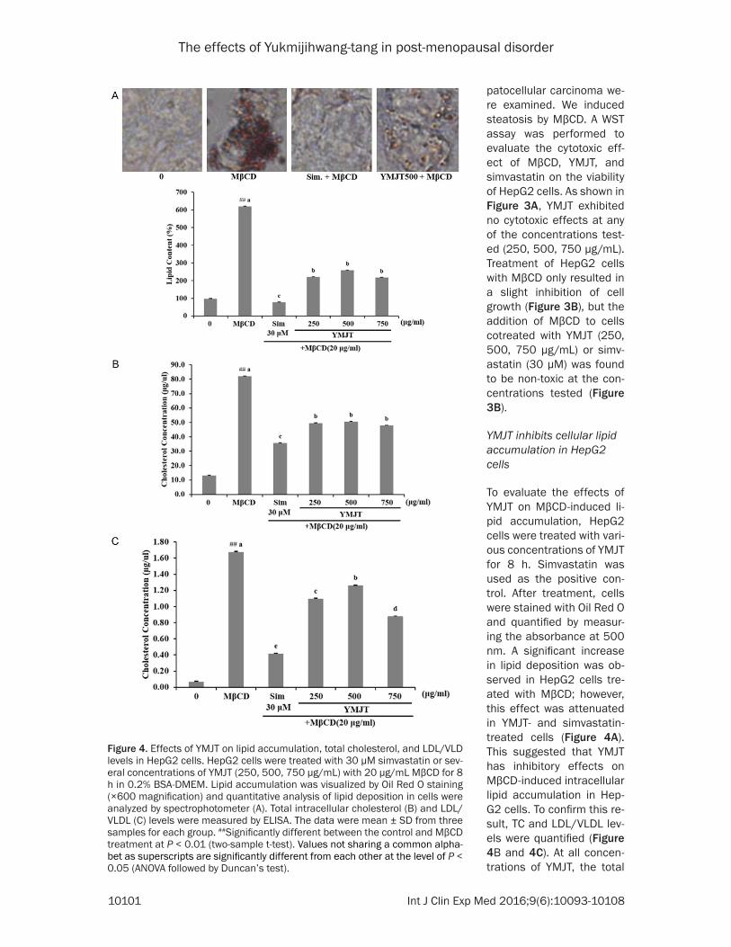
patocellular carcinoma we- re examined. We induced steatosis by MβCD. A WST assay was performed to evaluate the cytotoxic eff- ect of MβCD, YMJT, and simvastatin on the viability of HepG2 cells. As shown in Figure 3A, YMJT exhibited no cytotoxic effects at any of the concentrations test-ed (250, 500, 750 μg/mL). Treatment of HepG2 cells with MβCD only resulted in a slight inhibition of cell growth (Figure 3B), but the addition of MβCD to cells cotreated with YMJT (250, 500, 750 μg/mL) or simv-astatin (30 μM) was found to be non-toxic at the con-centrations tested (Figure 3B).
YMJT inhibits cellular lipid accumulation in HepG2 cells
To evaluate the effects of YMJT on MβCD-induced li- pid accumulation, HepG2 cells were treated with vari-ous concentrations of YMJT for 8 h. Simvastatin was used as the positive con-trol. After treatment, cells were stained with Oil Red O and quantified by measur-ing the absorbance at 500 nm. A significant increase in lipid deposition was ob- served in HepG2 cells tre- ated with MβCD; however, this effect was attenuated in YMJT- and simvastatin-treated cells (Figure 4A). This suggested that YMJT has inhibitory effects on MβCD-induced intracellular lipid accumulation in Hep- G2 cells. To confirm this re- sult, TC and LDL/VLDL lev-els were quantified (Figure 4B and 4C). At all concen-trations of YMJT, the total
Figure 4. Effects of YMJT on lipid accumulation, total cholesterol, and LDL/VLD levels in HepG2 cells. HepG2 cells were treated with 30 μM simvastatin or sev-eral concentrations of YMJT (250, 500, 750 μg/mL) with 20 μg/mL MβCD for 8 h in 0.2% BSA-DMEM. Lipid accumulation was visualized by Oil Red O staining (×600 magnification) and quantitative analysis of lipid deposition in cells were analyzed by spectrophotometer (A). Total intracellular cholesterol (B) and LDL/VLDL (C) levels were measured by ELISA. The data were mean ± SD from three samples for each group. ##Significantly different between the control and MβCD treatment at P < 0.01 (two-sample t-test). Values not sharing a common alpha-bet as superscripts are significantly different from each other at the level of P < 0.05 (ANOVA followed by Duncan’s test).

The effects of Yukmijihwang-tang in post-menopausal disorder
10102 Int J Clin Exp Med 2016;9(6):10093-10108
cholesterol levels were significantly inhibited. In addition, the LDL/VLDL levels were lower in these cells (Figure 4C).
YMJT regulates cholesterol synthesis in HepG2 cells
In HepG2 cells, we identified the mechanisms and confirmed to regulate cholesterol synthe-sis. Cholesterol metabolism is regulated by the transcription factor SREBP2 [30], which directly regulates the transcription of important genes involved in cholesterol irritation or biosynthe-sis, such as HMGCR and LDLR.
We evaluated the effects of YMJT on the pro-tein levels of the SREBP2, HMGCR, and LDLR in HepG2 cells. In MβCD-treated cells, LDLR and SREBP2 protein levels were diminished. However, they could be rescued to nearly wild-
Menopause is well-known to an increased prev-alence of a metabolic syndrome and were shown to increase the risk of cardiovascular disease and so on [33]. Progression of NASH occurs frequently in postmenopausal females [5, 6], with the accelerated accumulation of vis-ceral fat also being common in these patients [34, 35]. Recent studies have suggested that cholesterol-lowering drugs may slow the pro-gression of NASH. Statins have been shown to slow the progression of NASH in ovariectomized mice fed a high-fat and high-cholesterol diet [5], while atorvastatin was found to be effective in treating NASH [36, 37]. It is reported that the progression of atherosclerosis and dyslipid-emia increases after menopause and estrogen administration can improve these symptoms [38, 39]. Estrogen decreased serum cholester-ol levels and ameliorated steatohepatitis pro-gression in mice [4], and protected women with
Figure 5. Effects of YMJT on cholesterol synthesis in HepG2 cells. HepG2 cells were treated with 30 μM simvastatin or YMJT (250, 500 μg/mL) with 20 μg/mL MβCD for 8 h in 0.2% BSA-DMEM. Cell lysates were then harvested by RIPA buf-fer and subjected to Western blotting analysis for SREBP2, HMGCR, and LDLR protein expression. Quantified data of protein levels indicates in lower panel. The values of density of proteins were all justified with β-actin. The relative den-sity ratios of untreated cells were set at a value of 1.0. Values not sharing a common alphabet as superscripts are significantly different from each other at the level of P < 0.05 (ANOVA followed by Duncan’s test).
type levels following YMJT treatment (Figure 5). In co- ntrast, HMGCR protein lev-els were decreased by tre- atment with MβCD or YMJT (Figure 5). These results show that YMJT decreased lipid accumulation through the regulation of genes in- volved in cholesterol synth- esis.
YMJT stimulates AMPK phosphorylation in HepG2 cells
AMPK is thought to func-tion as a metabolic master switch in response to cha- nges in cellular energy and plays a key role in regulat-ing fat metabolism in the liver [31, 32]. Therefore, as a marker of AMPK activity, the phosphorylation of AM- PK was measured. AMPK phosphorylation was signif-icantly increased by YMJT compared with MβCD alone (Figure 6). This suggests th- at YMJT attenuated hepatic lipid accumulation through AMPK activation.
Discussion

The effects of Yukmijihwang-tang in post-menopausal disorder
10103 Int J Clin Exp Med 2016;9(6):10093-10108
NASH from severe liver fibrosis. An aromatase knockout mouse, which cannot synthesize endogeneous estrogen, developed hepatic ste-atosis [40]. Thus estrogens are important regu-lators of lipid homeostasis and estrogen is often prescribed to treat the NASH that is asso-ciated with menopause. Because estrogens are a fundamental driving force in the develop-ment of breast cancer [41], estrogen adminis-tration could be associated with an increased risk of developing breast cancer as one of its side effects.
The use of traditional herbal medicines to pre-vent and treat a variety of diseases has gener-ated considerable interest. Recently, several effects of YMJT have been demonstrated scien-tifically. For example, YMJT prevents and treats the diseases related to the kidney [42]. YMJT has regenerative effects on injured peripheral nerve fibers [43]. Antioxidative effects of YM- JT in the liver of a senescence-accelerated mouse have been reported [44]. According to Traditional Korean Medicine (TKM), menopaus-al symptoms are associated with a decline in kidney energy [1], and YMJT has been used
In Korea, some traditional prescription or herb-al products are used to manage metabolic syn-drome during menopause. In our previous report, Palmiwon showed therapeutic effects against metabolic syndrome as measured by weight gain and a decrease in the lipid profile in OVX rats [26]. As shown in this report, YMJT will be therapeutic and protective effects against the metabolic syndrome during menopause. We think YMJT has effects not only on one symptom but also on multiple targets involved in lipid related diseases.
This study was done to measure the effects of YMJT on targets associated with lipid factors, including retroperitoneal fat, peri-renal fat, and serum lipid levels. YMJT reduced retroperito-neal and serum TC, TG, and LDL levels, the ath-erogenic index, cardiac risk factors, and the intima-media thickness. YMJT was resulted to improve NASH and increase the lumen diame-ter and HDL levels to those similar to the posi-tive controls. These results show that YMJT can improve symptoms caused by menopausal hyperlipidemia.
Figure 6. Effects of YMJT on AMPK phosphorylation in HepG2 cells. HepG2 cells were treated with 30 μM simvastatin or YMJT (250, 500 μg/mL) with 20 μg/mL MβCD for 8 h in 0.2% BSA-DMEM. Cell lysates were then harvested and sub-jected to Western blotting analysis for AMPK phosphorylation (pThr-172-AMPK). Quantified data of protein levels indicates in lower panel. The values of density of proteins were all justified with β-actin. The relative density ratios of untreated cells were set at a value of 1.0. Values not sharing a common alphabet as superscripts are significantly different from each other at the level of P < 0.05 (ANOVA followed by Duncan’s test).
to improve the function of the kidney. Moreover, YMJT decreased serum choles-terol and triglyceride in rat models of diabetes [25]. YMJT can regulate glucose and lipid metabolism in rat models of diabetes [45]. In obese Zucker rats, Yukmijih- wangtang-Jahage improved the lipid profile [46]. There- fore, we evaluated the ther-apeutic effects of YMJT on lipid related diseases of me- nopausal hyperlipidemia by utilizing at in vitro and in vivo model.
We performed a quantita-tive analysis of 5 reference standards (morroniside, lo- ganin, paeoniflorin, 5-HMF, paeonol) in YMJT using a HPLC. Identifying the major components is of great im- portance because their ori-gin is relevant to the effects of the herbal medicine.

The effects of Yukmijihwang-tang in post-menopausal disorder
10104 Int J Clin Exp Med 2016;9(6):10093-10108
In previous studies, some natural products were identified to manage metabolic syndrome during menopause [47]. YMJT has a therapeu-tic and protective effect against metabolic syn-drome related to cholesterol during meno-pause. Dietary cholesterol in NASH is an important factor both in rodents and humans [5]. In the present study, YMJT ameliorated NASH by reducing serum cholesterol levels.
Some reported studies, menopausal symptoms were reported a relationship between the intake of natural products. Linum Usitatissimum (Linseed) Seed Oil, a prescribed dose from 40 to 50 mg/day, was resulted to have cholesterol-lowering properties in postmenopausal women [48, 49]. An isoflavone(genistein, daidzein, for-mononetin, and biochanin) from red clover increased HDL and decreased apolipoprotein B levels when ingested the prescribed doses of 28.5, 57, or 85.5 mg/day in postmenopausal women [50]. The intake of 90 mg/day of Isoflavone (1:1:0.2 genistein: daidzein: glycit-ein) improved in postmenopausal women with hypercholesterolemia [51]. Dehydroepiandrost- erone at a dose of 25 mg/day showed improved lipid patterns [52]. Determining whether YMJT could improve cholesterol related disease in postmenopausal females will be a subject addressed by our future research.
In addition, we performed an experiment using the MβCD-induced hepatic steatosis model in HepG2 cells in order to examine the cholester-ol-decreasing effect of YMJT. We determined that MβCD treatment induced a significant increase in lipid accumulation, TC and LDL/VLDL levels, all of which were reduced following treatment with YMJT (Figure 4). These results show that YMJT exerts cholesterol-lowering effects on HepG2 cells.
The SREBP-2 regulates the expression of many genes involved in cholesterol synthesis and uptake. Activation of SREBP2 is dependent on the cholesterol status of the cell [14, 53]. Mβ- CD increased cholesterol levels and caused of the down-regulation of the effector proteins SREBP2, LDLR, and HMG-CoA. Co-treatment with YMJT and MβCD decreased cholesterol levels, bring in increased expression of SREBP2 and LDLR (Figure 5). There are some reports proposing that the up-regulation of either SREBP2 or LDLR is responsible for appreciated cholesterol levels. The green alga Haematoco-
ccus pluvialis shows a hypocholesterolemic result through the upregulation of LDLR expres-sion [54]. Also, Resveratrol has anti-atherogen-ic effects and increases the activity of LDLR in hepatocytes through the activation of SREBPs [55]. Our result, in contrast with the normal regulatory response, the expression of HMGCR was decreased (Figure 5).
The pattern of protein expression of hepatic LDLR and SREBP2 was very similar to our previ-ous study [26]. This study demonstrated that YMJT could reduce body weight and improve the blood lipid profile. In HepG2 cells, YMJT regulated the expression of SREBP2, suggest-ing that the effects of YMJT on the lipid profile might also be linked to the SREBP2 pathway. We used simvastatin as a positive control. This drug is known to suppress HMGCR activity [56]. The data shown here suggest that the response to YMJT was very similar to that of simvastatin in terms of decreasing cholesterol synthesis in HepG2 cells exposed to MβCD.
Increased AMPK phosphorylation was also observed following YMJT treatment (Figure 6). AMPK has become the focus of many recent studies as a therapeutic target of metabolic disease and as a central regulator of lipid metabolism pathways [57-59]. Based on its role, AMPK has appeared as a good target for the treatment of fatty liver disease. In this study, YMJT might also regulate cholesterol synthesis via AMPK phosphorylation [32, 58, 60, 61]. Overall, the present in vitro data sug-gest that inhibition of lipid synthesis on MβCD-induced HepG2 cells by YMJT blocks the pro-gression of hepatocyte steatosis. Therefore, YMJT might negatively regulate the lipid metab-olism by activating AMPK phosphorylation.
YMJT is a TKM formula that is utilized when a patient has been determined as having inade-quate kidney and liver yin. Patients exhibiting kidney and liver Yin insufficiency have been used to supply energy. YMJT has been support-ed from: flushed or red face, headaches, hot flashes, night sweats, nocturnal emission, un- easiness or mental unrest. This is the aspect of the symptoms of menopause which we must consider. In this viewpoint, we examined the effects of cholesterol related disease. YMJT was showed almost the same effect as Palmi- won without relation on yin or yang [26].
The effects of Yukmijihwang-tang in post-menopausal disorder
10105 Int J Clin Exp Med 2016;9(6):10093-10108
We think that YMJT is a novel prescription medi-cine that can improve multiple lipid-associated factors in menopause. Further studies are required to determine the significance of the up-regulation of AMPK phosphorylation by YMJT.
Conclusions
In conclusion, we have shown that YMJT may be used as a novel agent to treat or prevent high-cholesterol and high-fat-induced cholesterol related disease during menopause. Our results also support the view that the anti-steatosis effects of YMJT may be attributed to the regula-tion of the cholesterol synthesis pathway and activation of AMPK.
Acknowledgements
This research was supported by a grant from the Korea Institute of Oriental Medicine (Grant No. K16291) and by Korea Research Fellowship program funded by the Ministry of Science, ICT and Future Planning through the National Research Foundation of Korea (Grant No. 2015H1D3A10662410).
Disclosure of conflict of interest
None.
Address correspondence to: Byoung Seob Ko. Kore- an Medicine Convergence Research Division, Korea Institute of Oriental Medicine, Republic of Korea. Tel: +82-42-868-9542; Fax: +82-42-868-9293; E-mail: [email protected]
References
[1] Jun H. Donguibogam (Treasured Mirror of East-ern Medicine, Sang Woo A. and Ohmin K., edi-tor), Ministry of Health & Welfare and Korea Institute of Oriental Medicine, Seoul, 2013; part 1, pp. 82, 425.
[2] Erling F, Prediman KS, Valentin F. Coronary plaque disruption. Circulation 1995; 92: 657-671.
[3] Das K and Kar P. Non alcoholic steatohepatitis. J Assoc Physicians India 2005; 53: 195-199.
[4] Kamada Y, Kiso S, Yoshida Y, Chatani N, Kizu T, Hamano M, Tsubakio M, Takemura T, Ezaki H and Hayashi N. Estrogen deficiency worsens steatohepatitis in mice fed high-fat and high-cholesterol diet. Am J Physiol Gastrointest Liv-er Physiol 2011; 301: G1031-G1043.
[5] Kamada Y, Kiso S, Yoshida Y, Chatani N, Kizu T, Hamano M, Egawa M, Takemura T, Ezaki H and Furuta K. Pitavastatin ameliorated the progres-sion of steatohepatitis in ovariectomized mice fed a high fat and high cholesterol diet. Hepa-tol Res 2013; 43: 401-412.
[6] Yang JD, Abdelmalek MF, Pang H, Guy CD, Smith AD, Diehl AM and Suzuki A. Gender and menopause impact severity of fibrosis among patients with nonalcoholic steatohepatitis. Hepatology 2014; 59: 1406-1414.
[7] Amadou A, Fabre A, Torres-Mejía G, Ortega-Ol-vera C, Angeles-Llerenas A, McKenzie F, Biessy C, Hainaut P and Romieu I. Hormonal Therapy and Risk of Breast Cancer in Mexican Women. PLoS One 2013; 8: e79695.
[8] Nakamoto K, Takayama F, Mankura M, Hidaka Y, Egashira T, Ogino T, Kawasaki H and Mori A. Beneficial effects of fermented green tea ex-tract in a rat model of non-alcoholic steatohep-atitis. J Clin Biochem Nutr 2009; 44: 239.
[9] Van Rooyen DM and Farrell GC. SREBP-2: a link between insulin resistance, hepatic cho-lesterol, and inflammation in NASH. J Gastro-enterol Hepatol 2011; 26: 789-792.
[10] Sakai J and Rawson RB. The sterol regulatory element-binding protein pathway: control of lipid homeostasis through regulated intracel-lular transport. Curr Opin Lipidol 2001; 12: 261-266.
[11] Zhao L, Chen Y, Tang R, Chen Y, Li Q, Gong J, Huang A, Varghese Z, Moorhead JF and Ruan XZ. Inflammatory stress exacerbates hepatic cholesterol accumulation via increasing cho-lesterol uptake and de novo synthesis. J Gas-troenterol Hepatol 2011; 26: 875-883.
[12] Hennessy MC, Devender JL and Wimberly M. A Receptor-Mediated Pathway for Cholesterol Homeostasis. 1986.
[13] Vallett SM, Sanchez HB, Rosenfeld JM and Os-borne TF. A direct role for sterol regulatory ele-ment binding protein in activation of 3-hydroxy-3-methylglutaryl coenzyme A reductase gene. J Biol Chem 1996; 271: 12247-12253.
[14] Horton JD, Goldstein JL and Brown MS. SREB-Ps: activators of the complete program of cholesterol and fatty acid synthesis in the liver. J Clin Invest 2002; 109: 1125-1131.
[15] Horton JD, Shimomura I, Brown MS, Hammer RE, Goldstein JL and Shimano H. Activation of cholesterol synthesis in preference to fatty acid synthesis in liver and adipose tissue of transgenic mice overproducing sterol regula-tory element-binding protein-2. J Clin Invest 1998; 101: 2331.
[16] Kang Q and Chen A. Curcumin suppresses ex-pression of low-density lipoprotein (LDL) recep-tor, leading to the inhibition of LDL-induced

The effects of Yukmijihwang-tang in post-menopausal disorder
10106 Int J Clin Exp Med 2016;9(6):10093-10108
activation of hepatic stellate cells. Br J Phar-macol 2009; 157: 1354-1367.
[17] Bilheimer DW, Goldstein JL, Grundy SM, Starzl TE and Brown MS. Liver transplantation to pro-vide low-density-lipoprotein receptors and low-er plasma cholesterol in a child with homozy-gous familial hypercholesterolemia. N Engl J Med 1984; 311: 1658-1664.
[18] Xin P, Han H, Gao D, Cui W, Yang X, Ying C, Sun X and Hao L. Alleviative effects of resveratrol on nonalcoholic fatty liver disease are associ-ated with up regulation of hepatic low density lipoprotein receptor and scavenger receptor class B type I gene expressions in rats. Food Chem Toxicol 2013; 52: 12-18.
[19] Sharpe LJ and Brown AJ. Controlling cholester-ol synthesis beyond 3-hydroxy-3-methylgluta-ryl-CoA reductase (HMGCR). J Biol Chem 2013; 288: 18707-18715.
[20] Viollet B, Foretz M, Guigas B, Horman S, Dentin R, Bertrand L, Hue L and Andreelli F. Activation of AMP-activated protein kinase in the liver: a new strategy for the management of metabolic hepatic disorders. J Physiol 2006; 574: 41-53.
[21] Lee S, Lee M-S, Kim C-T, Kim I-H and Kim Y. Ginsenoside Rg3 reduces lipid accumulation with AMP-activated protein kinase (AMPK) acti-vation in HepG2 cells. Int J Mol Sci 2012; 13: 5729-5739.
[22] Schimmack G, DeFronzo RA and Musi N. AMP-activated protein kinase: role in metabolism and therapeutic implications. Diabetes Obes Metab 2006; 8: 591-602.
[23] Hwang YH, Kim T, Cho WK, Jang D, Ha JH, Ma JY. Food-and gender-dependent pharmacoki-netics of paeoniflorin after oral administration with Samul-tang in rats. J Ethnopharmacol 2012; 142: 161-167.
[24] Han MK, Won CW, Kim CH and Kwon YD. Ef-fects of Yukmijiwhang-tang(Liuweidihuang-tang) with Puerariae Radix on Ovariectomized Osteoporotic Rats. Journal of Oriental Rehabili-tation Medicine 2007; 17: 1-21.
[25] Kwack KH, Kim SH and Song HJ. The Effects of Yukmijihwangtang & Discoreae Radix on the Changes of Blood Glucoese & Serum in Dia-betic Rats induced by Alloxan. Journal of Orien-tal Medical Pathology 1993; 8: 137-156.
[26] Go H, Ryuk JA, Lee HW, Ko BS. Palmiwon at-tenuates hepatic lipid accumulation and hy-perlipidemia in a menopausal rat model. Menopause 2015; 22: 872-884.
[27] Fantuzzi G and Mazzone T. Adipose tissue and atherosclerosis: exploring the connection. Ar-terioscler Thromb Vasc Biol 2007; 27: 996-1003.
[28] Sniderman A, Shapiro S, Marpole D, Skinner B, Teng B and Kwiterovich PO Jr. Association of
coronary atherosclerosis with hyperapobetali-poproteinemia [increased protein but normal cholesterol levels in human plasma low densi-ty (beta) lipoproteins]. Proc Natl Acad Sci U S A 1980; 77: 604-608.
[29] Brunt EM, Kleiner DE, Wilson LA, Belt P and Neuschwander-Tetri BA. Nonalcoholic fatty liv-er disease (NAFLD) activity score and the his-topathologic diagnosis in NAFLD: distinct clini-copathologic meanings. Hepatology 2011; 53: 810-820.
[30] Zhao L, Chen Y, Tang R, Chen Y, Li Q, Gong J, Huang A, Varghese Z, Moorhead JF and Ruan XZ. Inflammatory stress exacerbates hepatic cholesterol accumulation via increas-ing cholesterol uptake and de novo synthesis. J Gastroenterol Hepatol 2011; 26: 875-883.
[31] Hardie DG. AMP-activated/SNF1 protein kinas-es: conserved guardians of cellular energy. Nat Rev Mol Cell Biol 2007; 8: 774-785.
[32] Liu JF, Ma Y, Wang Y, Du ZY, Shen JK and Peng HL. Reduction of lipid accumulation in HepG2 cells by luteolin is associated with activation of AMPK and mitigation of oxidative stress. Phyto-ther Res 2011; 25: 588-596.
[33] Jouyandeh Z, Nayebzadeh F, Qorbani M and Asadi M. Metabolic syndrome and menopause. J Diabetes Metab Disord 2013; 12: 1.
[34] Ley CJ, Lees B and Stevenson JC. Sex-and menopause-associated changes in body-fat distribution. Am J Clin Nutr 1992; 55: 950-954.
[35] Kotani K, Tokunaga K, Fujioka S, Kobatake T, Keno Y, Yoshida S, Shimomura I, Tarui S and Matsuzawa Y. Sexual dimorphism of age-relat-ed changes in whole-body fat distribution in the obese. Int J Obes Relat Metab Disord 1994; 18: 207-202.
[36] Kiyici M, Gulten M, Gurel S, Nak SG, Dolar E, Savci G, Adim SB, Yerci O and Memik F. Ursode-oxycholic acid and atorvastatin in the treat-ment of nonalcoholic steatohepatitis. Can J Gastroenterol 2003; 17: 713-718.
[37] Hyogo H, Tazuma S, Arihiro K, Iwamoto K, Na-beshima Y, Inoue M, Ishitobi T, Nonaka M and Chayama K. Efficacy of atorvastatin for the treatment of nonalcoholic steatohepatitis with dyslipidemia. Metabolism 2008; 57: 1711-1718.
[38] Hong MK, Romm PA, Reagan K, Green CE and Rackley CE. Effects of estrogen replacement therapy on serum lipid values and angiograph-ically defined coronary artery disease in post-menopausal women. Am J Cardiol 1992; 69: 176-178.
[39] Mikkola TS and Clarkson TB. Estrogen re- placement therapy, atherosclerosis, and vas-

The effects of Yukmijihwang-tang in post-menopausal disorder
10107 Int J Clin Exp Med 2016;9(6):10093-10108
cular function. Cardiovasc Res 2002; 53: 605-619.
[40] Nemoto Y, Toda K, Ono M, Fujikawa-Adachi K, Saibara T, Onishi S, Enzan H, Okada T and Shi-zuta Y. Altered expression of fatty acid-metabo-lizing enzymes in aromatase-deficient mice. J Clin Invest 2000; 105: 1819-1825.
[41] Wright PK, Jones SB, Ardern N, Ward R, Clarke RB, Sotgia F, Lisanti MP, Landberg G and Lamb R. 17β-estradiol regulates giant vesicle forma-tion via estrogen receptor-alpha in human breast cancer cells. Oncotarget 2014; 5: 3055-65.
[42] Lee MH and Son IC. Effects of Aqua-Acupunc-ture of Yukmijihwangtang and Palmijihwang-tang Water Extracts on the Renal Function. The Journal of Korean Acupuncture & Moxibustion Society 1998; 15: 255-277.
[43] Han GS, Yu BC, An JJ, Jo HK, Ryu HR, Seol Ic and Kim YS. A Prior Study on the Effect of Yuk-mijihwang-tang to Regeneration of Injured Pe-ripheral Nerve Fiber. The Journal of Daejeon Oriental Medicine 2006; 15: 181-186.
[44] Park SM, Lim MH, Lee JH and Park JH. Antioxi-dative effects of Bojungikgi-tang and Yukmiji-whang-tang in the Liver of senescence-accel-erated mouse (SAM). The Korea Association of Herbology 2003; 18: 175-191.
[45] Byun SH. Immunohistochemical Study for the Effects of Yukmizihwangtang and Yukmizih-wangtang-Deer Antler on the Diabetic Rats. The Journal of Jeahan Oriental Medical Acade-my 1995; 1: 1-16.
[46] Seo EK, Kang DH, Seo JW, Kim KS, Lee TK, Lee YC, Kyung-Soo N and Kim CH. Anti-diabetic ef-fect of Yukmijihwangtang-Jahage in obese Zucker rats. Korean Journal of Life Science 2000; 10: 388-396.
[47] Parhizkar S, Latiff LA, Rahman SA and Dollah MA. Preventive effect of Nigella sativa on met-abolic syndrome in menopause induced rats. Journal of Medicinal Plants Research 2011; 5: 1478-1484.
[48] Cunnane SC, Hamadeh MJ, Liede AC, Thomp-son LU, Wolever TM and Jenkins DJ. Nutritional attributes of traditional flaxseed in healthy young adults. Am J Clin Nutr 1995; 61: 62-68.
[49] Jenkins DJ, Kendall CW, Vidgen E, Agarwal S, Rao AV, Rosenberg RS, Diamandis EP, Novok-met R, Mehling CC, Perera T, Griffin LC and Cunnane SC. Health aspects of partially defat-ted flaxseed, including effects on serum lipids, oxidative measures, and ex vivo androgen and progestin activity: a controlled crossover trial. Am J Clin Nutr 1999; 69: 395-402.
[50] Clifton-Bligh PB, Baber RJ, Fulcher GR, Nery ML and Moreton T. The effect of isoflavones extracted from red clover (Rimostil) on lipid
and bone metabolism. Menopause 2001; 8: 259-265.
[51] Lissin LW, Oka R, Lakshmi S and Cooke JP. Iso-flavones improve vascular reactivity in post-menopausal women with hypercholesterol-emia. Vasc Med 2004; 9: 26-30.
[52] Lasco A, Frisina N, Morabito N, Gaudio A, Mori-ni E, Trifiletti A, Basile G, Nicita-Mauro V and Cucinotta D. Metabolic effects of dehydroepi-androsterone replacement therapy in post-menopausal women. Eur J Endocrinol 2001; 145: 457-461.
[53] Pal S, Ho N, Santos C, Dubois P, Mamo J, Croft K and Allister E. Red wine polyphenolics in-crease LDL receptor expression and activity and suppress the secretion of ApoB100 from human HepG2 cells. J Nutr 2003; 133: 700-706.
[54] Yang Y, Seo JM, Nguyen A, Pham TX, Park HJ, Park Y, Kim B, Bruno RS and Lee J. Astaxan-thin-rich extract from the green alga Haemato-coccus pluvialis lowers plasma lipid concentra-tions and enhances antioxidant defense in apolipoprotein E knockout mice. J Nutr 2011; 141: 1611-1617.
[55] Yashiro T, Nanmoku M, Shimizu M, Inoue J and Sato R. Resveratrol increases the expression and activity of the low density lipoprotein re-ceptor in hepatocytes by the proteolytic activa-tion of the sterol regulatory element-binding proteins. Atherosclerosis 2012; 220: 369-374.
[56] Kureishi Y, Luo Z, Shiojima I, Bialik A, Fulton D, Lefer DJ, Sessa WC and Walsh K. The HMG-CoA reductase inhibitor simvastatin activates the protein kinase Akt and promotes angiogen-esis in normocholesterolemic animals. Nat Med 2000; 6: 1004-1010.
[57] Do Yeon Kim JSP, Yuan HD and Chung SH. Fer-mented ginseng attenuates hepatic lipid accu-mulation and hyperglycemia through AMPK activation. Food Sci. Biotechnol 2009; 18: 172-178.
[58] Yuan HD, Yuan HY, Chung SH, Jin GZ and Piao GC. An active part of Artemisia sacrorum Ledeb. attenuates hepatic lipid accumulation through activating AMP-activated protein ki-nase in human HepG2 cells. Biosci Biotechnol Biochem 2010; 74: 322-328.
[59] Feng Q, Gou XJ, Meng SX, Huang C, Zhang YQ, Tang YJ, Wang WJ, Xu L, Peng JH and Hu YY. Qushi Huayu Decoction Inhibits Hepatic Lipid Accumulation by Activating AMP-Activated Pro-tein Kinase In Vivo and In Vitro. Evid Based Complement Alternat Med 2013; 2013: 184358.
[60] Kang OH, Kim SB, Seo YS, Joung DK, Mun SH, Choi JG, Lee YM, Kang DG, Lee HS and Kwon DY. Curcumin decreases oleic acid-induced lipid accumulation via AMPK phosphorylation

The effects of Yukmijihwang-tang in post-menopausal disorder
10108 Int J Clin Exp Med 2016;9(6):10093-10108
in hepatocarcinoma cells. Eur Rev Med Phar-macol Sci 2013; 17: 2578-2586.
[61] Ou TT, Hsu MJ, Chan KC, Huang CN, Ho HH and Wang CJ. Mulberry extract inhibits oleic acid-
induced lipid accumulation via reduction of li-pogenesis and promotion of hepatic lipid clear-ance. J Sci Food Agric 2011; 91: 2740-2748.