organ specific gene expression in the regenerating tail of
TRANSCRIPT

Contents lists available at ScienceDirect
Developmental Biology
journal homepage: www.elsevier.com/locate/developmentalbiology
Original research article
Organ specific gene expression in the regenerating tail of Macrostomumlignano
Birgit Lengerera, Julia Wunderera, Robert Pjetaa, Giada Cartac, Damian Kaob, Aziz Aboobakerb,Christian Beiseld, Eugene Berezikove, Willi Salvenmosera, Peter Ladurnera,⁎
a Institute of Zoology and Center of Molecular Bioscience Innsbruck, University of Innsbruck, Technikerstr. 25, A-6020 Innsbruck, Austriab Department of Zoology, University of Oxford, South Parks Road, Oxford OX1 3PS, United Kingdomc Division of Physiology, Medical University of Innsbruck, Schöpfstraße 41/EG, A-6020 Innsbruck, Austriad Department of Biosystems Science and Engineering, ETH Zürich, Mattenstrasse 26, 4058 Basel, Switzerlande European Research Institute for the Biology of Ageing, University of Groningen, University Medical Center Groningen, A. Deusinglaan 1, NL-9713 AVGroningen, The Netherlands
A R T I C L E I N F O
Keywords:Differentiationin situ hybridizationStyletMale copulatory apparatus
A B S T R A C T
Temporal and spatial characterization of gene expression is a prerequisite for the understanding of cell-, tissue-,and organ-differentiation. In a multifaceted approach to investigate gene expression in the tail plate of the free-living marine flatworm Macrostomum lignano, we performed a posterior-region-specific in situ hybridizationscreen, RNA sequencing (RNA-seq) of regenerating animals, and functional analyses of selected tail-specificgenes. The in situ screen revealed transcripts expressed in the antrum, cement glands, adhesive organs, prostateglands, rhabdite glands, and other tissues. Next we used RNA-seq to characterize temporal expression in theregenerating tail plate revealing a time restricted onset of both adhesive organs and copulatory apparatusregeneration. In addition, we identified three novel previously unannotated genes solely expressed in theregenerating stylet. RNA interference showed that these genes are required for the formation of not only thestylet but the whole male copulatory apparatus. RNAi treated animals lacked the stylet, vesicula granulorum,seminal vesicle, false seminal vesicle, and prostate glands, while the other tissues of the tail plate, such asadhesive organs regenerated normally. In summary, our findings provide a large resource of expression dataduring homeostasis and regeneration of the morphologically complex tail regeneration and pave the way for abetter understanding of organogenesis in M. lignano.
1. Introduction
Regeneration and organ formation rely on restricted spatial andtemporal gene expression. In recent years, several studies wereaimed at the characterization of post-embryonic organogenesis inPlatyhelminthes. Members of this phylum are known for theirastonishing regeneration abilities, with some species being ableto regrow complete animals from small tissue pieces (Morgan,1901; Reddien and Sanchez Alvarado, 2004). Most regenerationstudies were performed in the asexual strains of the freshwaterspecies Schmidtea mediterranea and Dugesia japonica (reviewedin (Aboobaker, 2011; Adler and Sanchez Alvarado, 2015; Reddien,2013; Rink, 2013)). Therefore, the molecular program required forthe regeneration of most organs of these animals is relatively well-
characterized (reviewed in (Roberts-Galbraith and Newmark,2015)). A common approach to identifying regulatory genes fororgan regeneration is to screen annotated transcription factors andgenes involved in signaling pathways. In S. mediterranea, forexample, the epidermal growth factor (EGF) receptor pathwaywas found to regulate regeneration and homeostasis in the pharynxand eye pigment cells (Fraguas et al., 2011), the gut (Barberanet al., 2016a), and the protonephridia (Barberan et al., 2016b; Rinket al., 2011). Although this approach is often successful, it may leadto a biased selection of candidate genes. Another, unbiased strategyis to characterize the expression profile of defined tissues and tofunctionally test upregulated transcripts. One elegant way to obtaintissue-specific expression is to purify organs, which has beensuccessfully done with the intestines (Forsthoefel et al., 2012),
http://dx.doi.org/10.1016/j.ydbio.2017.07.021Received 28 April 2017; Received in revised form 21 July 2017; Accepted 27 July 2017
⁎ Corresponding author.E-mail addresses: [email protected] (B. Lengerer), [email protected] (J. Wunderer), [email protected] (R. Pjeta), [email protected] (G. Carta),
[email protected] (D. Kao), [email protected] (A. Aboobaker), [email protected] (C. Beisel), [email protected] (E. Berezikov),[email protected] (W. Salvenmoser), [email protected] (P. Ladurner).
Developmental Biology 433 (2018) 448–460
Available online 28 July 20170012-1606/ © 2017 The Authors. Published by Elsevier Inc. This is an open access article under the CC BY-NC-ND license (http://creativecommons.org/licenses/BY-NC-ND/4.0/).
MARK
cephalic ganglia (Wang et al., 2016), and eyes (Lapan and Reddien,2012) of S. mediterranea. Another practicable method is toamputate the area of interest and to characterize the expressionof the regenerating tissue (Adler et al., 2014; Roberts-Galbraithet al., 2016).
In contrast to triclads, few molecular studies have been performedin other taxa of Platyhelminthes. Over recent years, the marine,obligatorily cross-fertilizing hermaphrodite Macrostomum lignanohas been successfully developed as a model organism (Ladurner et al.,2005b). M. lignano belongs to the Macrostomorpha, the most basalgroup of Rhabditophora (Egger et al., 2015; Laumer et al., 2015). Itssmall size of around one millimetre and fast generation time of threeweeks enables easy culturing within laboratory conditions (Ladurneret al., 2005b). Several methods, including in situ hybridization (ISH)(Pfister et al., 2007), RNA interference (Pfister et al., 2008), BrdU(Ladurner et al., 2000), antibody staining (Ladurner et al., 2005a),and transgenesis (Wudarski et al., 2017) have been established. M.lignano is able to regenerate its anterior-most region (Egger et al.,2006; Verdoodt et al., 2012) as well as any tissue posterior to thepharynx (Egger et al., 2006). A study focused on the regenerating tailplate demonstrated that the posterior blastema is an accumulation ofproliferating neoblasts (Egger et al., 2009). After amputation of thetail plate adhesive organs can be observed in squeeze preparationsand stained specimens after just 48 h of regeneration (Egger et al.,2009; Lengerer et al., 2016). Three days after amputation the malecopulatory apparatus has begun to rebuild, and the vesicula granu-lorum and a small stylet are visible. Within the next two to three daysthe stylet grows to full size and the male copulatory apparatus regainsfunctionality (Egger et al., 2009). Depending on the individualanimal, a full set of adhesive organs are regenerated between six toten days, marking the completion of tail plate regeneration (Eggeret al., 2009; Lengerer et al., 2016).
Recently, literature on transcriptome and genome assemblies ofM. lignano (Grudniewska et al., 2016; Wasik et al., 2015) has beenpublished. A positional RNA sequencing (RNA-seq) analysis wasperformed by Arbore et al., to identify transcripts specificallyexpressed in the head-, testis-, ovary-, and tail region. Thereby, acollection of 366 tail-region-specific transcripts were identified(Arbore et al., 2015). Interestingly, a recent study by Ramm et al.has shown that 150 of these transcripts exhibited plasticity ofmRNA expression levels depending on their social environment. Aspart of this study animals were kept alone or in groups of eight. Adifferential gene expression analysis revealed transcripts up- ordownregulated in the larger group size (Ramm et al.). Based on thedata of these studies we aimed to identify transcripts involved inregeneration of the tail region.
Here, we present a region-specific in situ hybridization screen of111 transcripts predominately expressed in the posterior region ofMacrostomum lignano. Using RNA-seq we characterized temporalexpression in the regenerating tail plate. The expression of selectedtranscripts in the regenerating tissues were confirmed with in situhybridization and analysed with RNA interference. Three novelgenes were found to be expressed in the regenerating stylet, andtheir knock-down resulted in animals specifically lacking the malecopulatory apparatus.
2. Material and methods
2.1. Animal culture
Macrostomum lignano (Ladurner et al., 2005b) cultures of theinbred line DV1 (Janicke et al., 2013) were kept in petri dishes withnutrient enriched artificial seawater (Guillard's f/2 medium)(Anderson, 2005) and were fed ad libitum with the diatom Nitzschiacurvilineata. Animals were maintained in a climate chamber with20 °C, 60% humidity and a 14:10 day-night cycle.
2.2. Whole mount in situ hybridization
Whole mount in situ hybridization was performed as previouslydescribed (Lengerer et al., 2014). Briefly, primers were designed withPrimer3 (Untergasser et al., 2012) and a T7 promoter region was addedat the 5`end of the reverse primers. Primer sequences are listed inSuppl. Table 1. Template DNA was produced using standard PCRreactions. To synthesize single stranded digoxigenin-labelled RNAprobes, T7 polymerase (Promega or Thermo Scientific) and DIGlabelling mix (Roche) were used. Anti-digoxigenin-AP Fab fragments(Roche) were diluted 1:2000 and the signal was developed using theNBT/BCIP system (Roche) at 37 °C. Specimen were mounted inMowiol® 4–88 (Roth, Germany), prepared according to the manufac-turer's protocol and images were taken using a Leica DM5000microscope.
2.3. Sample preparation for RNA-seq
For RNA-seq, tissue samples were collected from 60 worms persample. The “A” sample included whole regenerating animals (includ-ing the regenerating tail). In the “B” sample the regenerating tail wasamputated and only the anterior part of the animals was collected forRNA isolation. For the first amputation, 120 adult worms were cutbehind developing eggs at the level of cement glands, using a razorblade under a binocular microscope. Afterwards, the worms weretransferred to petri dishes containing culture medium and algea. Toavoid algea contamination in the RNA samples, animals were starved16 h prior to the second amputation and fixation. In case of 12 h ofregeneration, the amputated animals were not fed after the firstamputation. After the given times of regeneration, 60 of the 120worms were transferred to TRI reagent® (“A”) and 60 were amputateda second time to remove the regenerating tail. The anterior fragmentsof the twice amputated worms (“B”) were immediately transferred toTRI reagent® (Sigma) and the regenerating tails were discarded. Thetissue samples was stored at −80 °C until total RNA extraction, done byTRI reagent/Chloroform extraction. All regeneration experiments wererepeated in three replicates.
2.4. Differential gene expression
For the identification of differentially expressed transcripts, sixTruSeq Stranded mRNA libraries (Illumina) for every regeneration timepoint (three biological replicates each of RNA-seq “A” and “B”) weregenerated. The libraries were sequenced with 50 bp Illumina singlereads. However, the quality of the reads of one biological replicate fromthe timepoints 12 h until day four could not be used for the analysis. Thereads of the regeneration time course were mapped to the referencetranscriptome (version MLRNA131024, http://www.macgenome.org/download/MLRNA131024/) with Bowtie2 (Langmead and Salzberg,2012). The data have been deposited with links to BioProjectaccession number PRJNA381865 in the NCBI BioProject database(https://www.ncbi.nlm.nih.gov/bioproject/). Differentially expressedtranscripts were identified using DESeq. 2 (Love et al., 2014). Wedefined transcripts as differentially expressed between RNA-seq “A” and“B”, if they show a 2-fold difference in the number of mapped reads anda cut off p-value of < 0.01. The list of the transcripts and thecorresponding fold change throughout regeneration time course areprovided in (Suppl. Table 2).
2.5. RNA interference
RNAi was performed as previously described (Kuales et al., 2011).Briefly, a double-stranded RNA (dsRNA) probe was generated by an invitro transcription system using primer pairs with Sp6 and T7promoter regions (T7 and SP6 Ribomax™ large scale RNA kit,Promega). DsRNA was diluted in artificial sea water (ASW) to a final
B. Lengerer et al. Developmental Biology 433 (2018) 448–460
449
concentration of 15 ng/μl (400 μl per well). The solution was supple-mented with algae and with antibiotics (antibiotic concentration:50 µg/ml). Streptomycin, Kanamycin, and Ampicillin were alternatedevery day to prevent the selection of resistant bacterial strains. Adultanimals were amputated at the level of cement glands and treated withdsRNA during the entire regeneration process. 25 animals were kept ineach well of a 12-well plate. Every 24 h, animals were transferred to aclean well plate and the dsRNA solution was changed. Throughout thewhole experiment, animals were fed ad libitum and were maintained atnormal culture conditions (in a climate chamber with 20 °C, 60%humidity and a 14:10 day-night cycle). Here we use luciferase dsRNA(Suppl. Fig. 10), or the omission of dsRNA (Fig. 3, Fig. 4, Fig. 5, Suppl.Fig. 8) as controls. As also shown in earlier studies, RNAi treatmenthad no mock effect on tissue and organ morphology on light- andelectron microscopic level (Kuales et al., 2011; Pfister et al., 2008; Sekiiet al., 2009). The efficacy of the knockdown was verified by performingwhole mount in situ hybridization. Phenotypes were documented invivo, after carefully squeezing the animals between a microscope slideand a cover slip. Images were taken using a Leica DM5000 microscope.Selected phenotypes were additionally documented with phalloidin,lectin, and antibody labelling (see next sections).
2.6. Phalloidin and lectin PNA labelling
Animals were relaxed with 7.14% MgCl2 hexahydrate and then fixedin 4% formaldehyde (made from paraformaldehyde) in PBS (PFA) for1 h. Afterwards the specimen were washed six times 10 min in Tris-buffered saline (pH 8.0) supplemented with 5 mM CaCl2 and 0.1%Triton (TBS-T). Unspecific background staining was blocked by pre-incubation in TBS-T containing 3% (w/v) bovine serum albumin (BSA-T) for 1 h at RT. Biotinylated PNA lectin (Vector Laboratories) wasdiluted 1:200 and applied to the specimen for 2 h at RT. After sixwashes of 10 min each in TBS-T, the specimen were incubated for 1 hin Dylight488-conjugated-streptavidin (Vector Laboratories) andPhalloidin-Rhodamine both diluted 1:300 in BSA-T at RT (in dark-ness). After several washing steps in TBS-T, the specimen weremounted in Vectashield and analysed using a Leica DM5000 or LeicaSP5 II confocal scanning microscope.
2.7. Macif1 antibody staining
Animals were relaxed with 7.14% MgCl2 hexahydrate and then fixedin 4% formaldehyde (made from paraformaldehyde) in PBS (PFA) for1 h. Afterwards the specimen were washed several times in PBS and0.1% Triton (PBS-T) and heated overnight in a 1:10 diluted epitoperetrieval solution (DakoCytomation K5336) at 80 °C. After severalwashing steps with PBS-T, the specimen were blocked in PBS-Tcontaining 1% (w/v) bovine serum albumin (BSA-T) for 4 h at 4 °C.Then they were incubated with 1:1000 diluted polyclonal Rabbit-α-macif1 antibody (Lengerer et al., 2016) in 1% BSA-T overnight at 4 °C.After several washes with PBS-T, the specimen were incubated for 1 hin a swine- α-rabbit-FITC antibody diluted 1:500 in BSA-T at roomtemperature. After several washing steps in PBS-T, the specimen weremounted in Vectashield and analysed using a Leica DM5000 or LeicaSP5 II confocal scanning microscope.
2.8. EdU labelling
Intact adults were soaked in the thymidine analogue 5-ethynyl-2′-deoxyuridine (EdU; Invitrogen) at a concentration of 100 µM inartificial seawater for seven days continuously. Afterwards, the animalswere relaxed with 7.14% MgCl2 hexahydrate and fixed in 4% PFA for30 min. The specimen were washed several times with PBS-Triton andblocked with 3% BSA in PBS-T. After several washes in PBS-T, thespecimen were incubated in Click-iT® EdU reaction cocktail (concen-trations according to manufacturer's instructions – Invitrogen). DNA
was visualized with an addition of DAPI (1 µg/ml in PBS-T) for 30 minat room temperature. After several washes with PBS-T, the specimenwere mounted in Vectashield (Vector) and analysed using a Leica SP5II confocal scanning microscope.
3. Results
3.1. Posterior-region-specific in situ hybridization screen
To identify organ-specific expression in the posterior region ofMacrostomum lignano we performed a medium-scale ISH screenbased on the positional transcriptome of Arbore et al. (2015). Briefly,Arbore et al. performed a positional RNA-seq analysis and defined fourregions along the anterior-posterior body axis of M. lignano: the head-,testis-, ovary-, and tail-region. To obtain region-specific samples,animals were cut at different body regions, and the anterior part wasused for RNA-seq. Expression differences between samples werecalculated to determine region-specific transcripts (Arbore et al.,2015). For example, transcripts with an at least 4-fold higher expres-sion in the whole animal compared to the region anterior of thedeveloping eggs were considered as potentially tail-region-specific. Theso defined tail region contains all tissues posterior to the ovaries,including developing eggs, the female and male genitalia, and adhesiveorgans (Fig. 1A). To avoid confusion between the terms “tail region”and “tail plate”, we termed this body area “posterior region” within thisstudy. Candidates for the posterior-region-specific ISH were chosenindependently of any sequence annotations. The transcripts wereselected according to following criteria: (1) transcripts with ≥ 4-foldhigher mapped reads in whole animals compared to the region anteriorof the developing eggs (putative posterior-region-specific); (2) tran-scripts with ≥ 50 mapped reads either absolute, or calculated RPK =reads per kilobase in whole animals (to exclude very low expressedtranscripts); (3) a length of at least 200 bp, to enable labelling with ISHprobes. Overall, 316 transcripts fulfilled these criteria. 150 transcriptschanged their expression level due to changes in the social environ-ment, 140 of which were upregulated in larger groups (Ramm et al.).These transcripts were predicted to represent seminal fluid candidatesand will be reported elsewhere as part of a project on seminal fluiddiversity and function in M. lignano (Weber et al.). Finally, 166transcripts were selected for the present study.
Of the 166 screened transcripts (transcriptome versionMLRNA110815), 111 had an expression pattern in whole mount ISH ofadult animals (Suppl. Table 1). We classified the spatial expression patterninto seven categories: antrum, cement glands, male copulatory apparatus,adhesive organs, cells enriched in posterior region, posterior region andother tissues, and other pattern (Fig. 1B). Of the transcripts, 34 wereexpressed in female tissues, including 19 antrum-specific (Fig. 1C, Suppl.Fig. 1) and 15 cement gland-specific (Fig. 1D, Suppl. Fig. 2A-O). The 13transcripts expressed in the male copulatory apparatus included 11 prostategland cell-specific (Fig. 1E, Suppl. Fig. 2P-Z) and two with expression in thestylet and the false seminal vesicle (Suppl. Fig. 2AA-AB). 20 transcriptswere expressed in the cells of the adhesive organs (Fig. 1F). By thelocalization of their expression, they were categorized into secretory glandcell-specific (16) (Fig. 1F, Suppl. Fig. 3A-P), anchor cell-specific (3) (Suppl.Fig. 3Q-S), or expression in both secretory gland cells and anchor cells (1)(Suppl. Fig. 3T). The category “cells enriched in posterior region” was usedto summarize four different expression patterns. Two transcripts of thiscategory were localized in rhabdite glands, which are distributed over thewhole animal but are more numerous in the tail plate (Fig. 1G, Suppl.Fig. 3U,V). One transcript labelled paired cells on the ventral side of theposterior region (Suppl. Fig. 3W). Two transcripts were expressed in asubset of epidermal cells surrounding the whole tail plate (Suppl. Fig. 3X-Y). Beside the expression of the transcripts, these epidermal cells were notdistinguishable from other epidermal cells. One staining revealed singlecells in the dorsal part of the tail plate (Suppl. Fig. 3Z). The correspondingcell type is currently unknown. The term “posterior region and other
B. Lengerer et al. Developmental Biology 433 (2018) 448–460
450

tissues” was used to describe transcripts with an expression in tissuesspecific for the posterior region and an additional expression in anteriorbody parts, mostly in the gonads (14) (Fig. 1H, Suppl. Fig. 4). Another 24transcripts showed non-posterior-region-specific expression pattern (Suppl.Fig. 5), with the majority being expressed in testes, ovaries, or both gonads.For 55 transcripts either no specific primers could be designed (2), the PCRfailed (15), or the ISH did not result in a specific staining (38).
3.2. Expression analysis during tail plate regeneration
Next, we aimed to define the expression profile during tail plateregeneration using RNA-seq. In contrast to the positional transcriptome(Arbore et al., 2015), we amputated the animals posterior of the developing
eggs at the level of the cement glands. This cutting level was chosen toexclude developing eggs from the amputated tissue and thereby reduce thecomplexity of expressed mRNA. Furthermore, the same amputation levelwas used in previous studies on tail plate- (De Mulder et al., 2009; Eggeret al., 2009; Nimeth et al., 2007) and adhesive organ- regeneration(Lengerer et al., 2016), which facilitated the comparability of the results.We observed that tail amputation resulted in morphological changes in theanterior part of the animal, especially in the testes and ovaries. Tail-amputated animals stopped sperm production, reduced testes size (perso-nal observation), and altered overall gene expression in the regeneratinganimal (Wasik et al., 2015). A comparison of regenerating animals withintact ones would represent all expressional changes throughout the wholeanimal. However, we were interested in the genes exclusively expressed in
Fig. 1. Overview expression pattern of the posterior region specific in situ hybridization screen. (A) Schematic drawing of an adult Macrostomum lignano. (B) Pie cart indicating theexpression categories and number of transcripts with corresponding in situ pattern. (C-H) Exemplary in situ hybridizations of the posterior region specific expression categories. Scalebar: 100 µm.
B. Lengerer et al. Developmental Biology 433 (2018) 448–460
451

the regenerating tail. Due to the small size of the amputated tail fragments,it was not possible to collect these fragments directly, as they tended todisintegrate after amputation. To overcome these limitations, two sampleswere taken at every regeneration time point (Fig. 2A). Sample A includedwhole regenerating animals of the respective time point, including theregenerating tail. In sample B, the regenerating tail was amputated, andonly the anterior part of the animals was collected for RNA isolation. Thisexperimental setup allowed focus on the expression dynamics, especiallywithin the regenerating area. We defined seven regeneration time points –12, 24, 48, 72, 96, 120, and 168 h post-amputation - to cover the timerequired for the formation of all organs specific for the tail plate.
For differential gene expression, RNA was isolated in triplicates ofsamples A and B for every time point. Illumina libraries weregenerated, and 50 bp sequencing was performed (see Material andMethods; raw reads available under BioProject accession numberPRJNA381865). The generated Illumina reads were mapped to thereference transcriptome available at that time (versionMLRNA131024). Because of our interest in organ regeneration, weselected 220 transcripts that exhibited a differential expression (≥ 2-fold, p-value 0.01) in at least two time points between 48 and 168 h ofregeneration (Suppl. Table 2, Sheet1). From these, the spatial expres-
sion was already known for 186 transcripts based on the posterior-region-specific and seminal fluid ISH screen (Weber et al.).Additionally, 34 novel, differentially expressed transcripts were foundin the RNA-seq data (Suppl. Table 2, Sheet1). Notably, 164 transcriptsshowed expression in the posterior-region-specific and seminal fluidISH screen (Weber et al.), but they did not show differential expressionin the RNA-seq data (Suppl. Table 2, Sheet2). We assume that thisresulted from the presence of multiple isoforms in the transcriptome(see discussion).
In previous studies it was shown that after tail plate amputation,adhesive organs start to differentiate after 48 h (Egger et al., 2009;Lengerer et al., 2016). The RNA-seq data corroborated this time courseof differentiation and adhesive organ-specific transcripts(MLRNA131024 transcriptome) had increased expression in the tailfrom 48 h onwards (Fig. 2B). The male copulatory apparatus starts toregenerate at 72 h. At this point, the tip of the stylet is already visible,and in the following two to three days the stylet grows to full size. Atthe same time, the attached vesicula granulorum, true and falseseminal vesicle, and the prostate gland cells are rebuilt (Egger et al.,2009). In our RNA-seq dataset, transcripts of the male copulatoryapparatus were upregulated in the tail from 96 h onward (Fig. 2C,Suppl. Fig. 6A). Three transcripts (RNA1310_25676,RNA1310_28866, RNA1310_36278) were also expressed at earliertime points in the RNA-seq dataset. For prostate-specific transcriptRNA1310_25676, the temporal and spatial expression during regen-eration confirmed the early appearance of the transcript (Suppl.Fig. 6B).
3.3. Spatial and temporal co-expression groups in adhesive organs
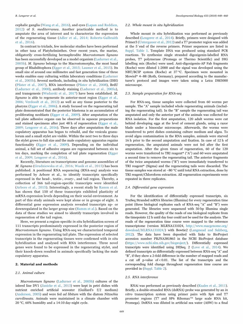
The adhesive organs in M. lignano consist of three cell types: asupportive anchor cell and two secretory glands - one adhesive and onereleasing gland cell (Lengerer et al., 2014; Tyler, 1976). After tail plateamputation, the first adhesive organs differentiate after 48 h (Egger et al.,2009; Lengerer et al., 2016). Over the following days their numbersincrease, until they reach their full number of about 130 organs after ninedays of regeneration (Egger et al., 2009; Lengerer et al., 2016). Accordingly,all tested secretory gland-specific transcripts (RNA815_13121.1,RNA815_21583, RNA815_23142, RNA815_27695.2, RNA815_48402,RNA1310_81421) and anchor cell-specific transcripts (RNA1310_4919,macif1) where expressed in regenerating animals from 48 h onwards afteramputation (Fig. 3A-B, Suppl. Fig. 7). Likewise, the expression levels of thetranscripts from the macif1 gene (RNA1310_7834/ RNA1310_9642/RNA815_3251.1) in the differential RNA-seq data increased after 48 h(Suppl. Table 2, Sheet 1). In addition, we identified one novel anchor cellspecific transcript (RNA1310_30724) that encodes for a 144 amino acidlong protein containing two EF-hand domains. This transcript showed acharacteristic anchor-cell-specific expression profile during regeneration(Fig. 3C).
The transcript RNA1310_47545 showed an unexpected mode ofexpression with respect to RNA-seq data and ISH patterns. Initiallyclassified as posterior-region-specific in Arbore et al. (Arbore et al.,2015), here we show an expression limited to the ovaries in intactanimals (Suppl. Fig. 5M, Fig. 3D1). However, during regeneration thetranscript was strongly expressed in the rostrum and in single cellsthroughout the body (Fig. 3D2-D4). After 48 h of regeneration, anadditional expression in the anchor cells was visible (Fig. 3D3-D4).
3.4. Characterization of a new gene required for microvilli formationin anchor cells
To determine their function, all identified anchor cell-specifictranscripts (RNA1310_4919, RNA815_51776, RNA815_8153,RNA1310_30724, RNA1310_47545) were analysed with RNA inter-ference (RNAi) during regeneration. As positive control, RNAi of theformer described anchor cell-specific macif1 (Lengerer et al., 2014)
Fig. 2. Amputation scheme used for RNA-seq experiments and exemplary expression ofadhesive organ and copulatory apparatus specific transcripts over regeneration timecourse. (A) Schematic drawing of the amputation scheme used for RNA-seq experiments.(B) Adhesive organs specific transcripts and (C) copulatory apparatus specific transcriptswith a differential expression between sample A and B (log2 scale).
B. Lengerer et al. Developmental Biology 433 (2018) 448–460
452
was performed. The ISH after nine days of treatment revealed that allfive knock-downs were efficient (data not shown). However, only theknock-down of RNA1310_4919 led to a detectable phenotype(Fig. 3E-N). The RNA1310_4919 RNAi-treated animals showed anon-adhesive phenotype. Squeezing preparations revealed a short-ening of their anchor cell specific microvilli (Fig. 3G, H).Furthermore, the microvilli were only weakly stained with phalloidin(Fig. 3I, J), demonstrating a reduction of actin filaments. As thesemorphological changes strongly resembled the previously describedmacif1 (RNAi) phenotype (Lengerer et al., 2014), we tested ifRNA1310_4919 had an effect on the expression of macif1. Thestaining with the available Macif1 antibody (Lengerer et al., 2016)showed no differences between control and RNAi-treated animals(Fig. 3K, L), indicating that RNA1310_4919 does not influence theexpression of macif1. Also, the labelling of the adhesive vesicles usingthe lectin PNA (Lengerer et al., 2016) showed no alterations in theadhesive gland cells of RNAi treated animals (Fig. 3M, N).
3.5. Identification of novel genes required for the formation of themale copulatory apparatus
The RNA-seq data revealed six transcripts with a differentialexpression higher in the tail restricted to 96 and 120 h of regeneration.Two of these transcripts (RNA1310_39915.2 and RNA1310_80800)were socially sensitive expressed and will be reported elsewhere(Weber et al.). For one transcript, no specific primers could be designed(RNA1310_126882). ISH experiments revealed that the expression ofthe other three transcripts (RNA1310_72446, RNA1310_45118, andRNA1310_51713) was limited to the regenerating stylet (Fig. 4A-C).Stylet formation is completed after post-embryonic development andno expression of the corresponding genes was observed in intactanimals (Fig. 4A1, B1, C1). This might be due to a slow homeostaticcell turnover in the stylet. To gain further insight to stylet-related cellrenewal in intact adults, we performed seven days continuous EdUexposure. Notably, we have not discovered pronounced accumulation
Fig. 3. Expression pattern of adhesive organs specific transcripts in regenerating animals and RNAi phenotype of RNA1310_4919. (A-D) Representative ISH pattern of (A1-4) thesecretory gland cell specific transcript RNA815_23142, (B1-4) the anchor cell specific transcripts RNA1310_4919, (C1-4) RNA1310_30724, and (D1-4) RNA1310_47545. (E-N) Tailplate of control- and RNA1310_4919 RNAi treated animals after nine days of regeneration: (E, F) ISH of RNA1310_4919, (G, H) squeeze preparation, (I, J) phalloidin staining, (K, L)Macif1 antibody staining, and (M, N) adhesive gland cell labelling with lectin PNA. Arrowheads highlight the microvilli of anchor cells. Note the shorter microvilli after RNAi treatment.Scale bars: (A) 100 µm, (E-N) 20 µm.
B. Lengerer et al. Developmental Biology 433 (2018) 448–460
453

of EdU-positive cells in the stylet region (in 38 out of 39 animals)(Suppl. Fig. 8A1–4), indicating that the cells of the stylet were notrenewed within this time. This is in contrast to a newly formingcopulatory apparatus (seen in 1 out of 39 animals) (Suppl. Fig. 8B1–4),which can occur when animals lose their stylet. From these results weconclude that there is very slow cell turnover in the stylet. Thisobservation was also supported by long term RNAi experiments inintact animals (see next sections).
No homologues proteins were found using BLAST search (blastx)(Altschul et al., 1997) against available protein databases for all threesequences (Suppl. Table 3). Based on their expression pattern, we referto the three transcripts as Mlig-stylet1 (RNA1310_72446), Mlig-stylet2 (RNA1310_45118), and Mlig-stylet3 (RNA1310_51713). Toevaluate their function, we soaked tail-amputated animals in dsRNAuntil the regeneration was completed after nine days. To confirm the
knock-down, a subset of 10 animals were fixed after four days oftreatment and whole mount ISH was performed. All transcripts wereefficiently knocked down after four days of regeneration (Fig. 4D1-F2).The single knock-down of Mlig-stylet1 and Mlig-stylet3 led to knock-down phenotypes in the male copulatory apparatus of varying shapeand frequency (Table 1). The strongest phenotype resulted in indivi-duals missing all parts of the male copulatory apparatus (Fig. 5A). Thissevere morphological phenotype was observed in squeezing prepara-tions of living animals (Fig. 5B, E). In contrast to control animals(Fig. 5B), treated animals had no stylet, vesicula granulorum, true andfalse seminal vesicle, or prostate gland cells (Fig. 5E). Phalloidinstaining confirmed the absence of the muscular seminal vesicle,vesicula granulorum, and the muscles associated with the stylet(Fig. 5C, F). According to the loss of prostate gland cells, the expressionof prostate genes was also diminished (Fig. 5D, G). Moreover, the
Fig. 4. Whole mount ISH pattern of stylet-specific transcripts in intact, regenerating, and RNAi treated animals. (A-C) Representative ISH pattern of (A1-5) Mlig-stylet1(RNA1310_72446), (B1-5) Mlig-stylet2 (RNA1310_45118), and (C1-5) Mlig-stylet3 (RNA1310_51713). (D-F) ISH pattern of control and RNAi treated specimen after four days ofregeneration of (D1-2) Mlig-stylet1, (E1-2) Mlig-stylet2, and (F1-2) Mlig-stylet3. Arrowheads highlight the regenerating stylet. Scale bars: (A) 100 µm; (D-F) 20 µm.
B. Lengerer et al. Developmental Biology 433 (2018) 448–460
454
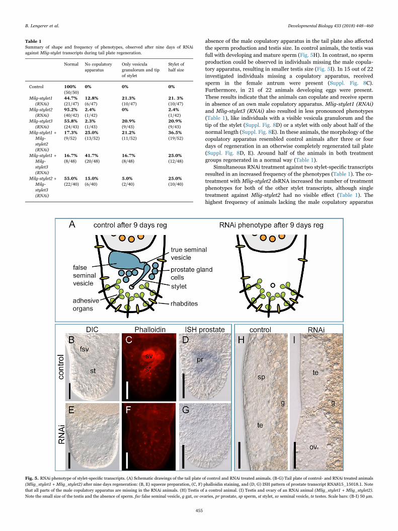
absence of the male copulatory apparatus in the tail plate also affectedthe sperm production and testis size. In control animals, the testis wasfull with developing and mature sperm (Fig. 5H). In contrast, no spermproduction could be observed in individuals missing the male copula-tory apparatus, resulting in smaller testis size (Fig. 5I). In 15 out of 22investigated individuals missing a copulatory apparatus, receivedsperm in the female antrum were present (Suppl. Fig. 8C).Furthermore, in 21 of 22 animals developing eggs were present.These results indicate that the animals can copulate and receive spermin absence of an own male copulatory apparatus. Mlig-stylet1 (RNAi)and Mlig-stylet3 (RNAi) also resulted in less pronounced phenotypes(Table 1), like individuals with a visible vesicula granulorum and thetip of the stylet (Suppl. Fig. 8D) or a stylet with only about half of thenormal length (Suppl. Fig. 8E). In these animals, the morphology of thecopulatory apparatus resembled control animals after three or fourdays of regeneration in an otherwise completely regenerated tail plate(Suppl. Fig. 8D, E). Around half of the animals in both treatmentgroups regenerated in a normal way (Table 1).
Simultaneous RNAi treatment against two stylet-specific transcriptsresulted in an increased frequency of the phenotypes (Table 1). The co-treatment with Mlig-stylet2 dsRNA increased the number of treatmentphenotypes for both of the other stylet transcripts, although singletreatment against Mlig-stylet2 had no visible effect (Table 1). Thehighest frequency of animals lacking the male copulatory apparatus
Table 1Summary of shape and frequency of phenotypes, observed after nine days of RNAiagainst Mlig-stylet transcripts during tail plate regeneration.
Normal No copulatoryapparatus
Only vesiculagranulorum and tipof stylet
Stylet ofhalf size
Control 100% 0% 0% 0%(50/50)
Milg-stylet1(RNAi)
44.7% 12.8% 21.3% 21. 3%(21/47) (6/47) (10/47) (10/47)
Milg-stylet2(RNAi)
95.2% 2.4% 0% 2.4%(40/42) (1/42) (1/42)
Milg-stylet3(RNAi)
55.8% 2.3% 20.9% 20.9%(24/43) (1/43) (9/43) (9/43)
Milg-stylet1 +Milg-stylet2(RNAi)
17.3% 25.0% 21.2% 36.5%(9/52) (13/52) (11/52) (19/52)
Milg-stylet1 +Milg-stylet3(RNAi)
16.7% 41.7% 16.7% 25.0%(8/48) (20/48) (8/48) (12/48)
Milg-stylet2 +Milg-stylet3(RNAi)
55.0% 15.0% 5.0% 25.0%(22/40) (6/40) (2/40) (10/40)
Fig. 5. RNAi phenotype of stylet-specific transcripts. (A) Schematic drawings of the tail plate of control and RNAi treated animals. (B-G) Tail plate of control- and RNAi treated animals(Mlig_stylet1 + Mlig_stylet2) after nine days regeneration: (B, E) squeeze preparation, (C, F) phalloidin staining, and (D, G) ISH pattern of prostate transcript RNA815_15018.1. Notethat all parts of the male copulatory apparatus are missing in the RNAi animals. (H) Testis of a control animal. (I) Testis and ovary of an RNAi animal (Mlig_stylet1 + Mlig_stylet2).Note the small size of the testis and the absence of sperm. fsv false seminal vesicle, g gut, ov ovaries, pr prostate, sp sperm, st stylet, sv seminal vesicle, te testes. Scale bars: (B-I) 50 µm.
B. Lengerer et al. Developmental Biology 433 (2018) 448–460
455

(41.7%) after nine days was achieved by the combined treatment ofMlig-stylet1 and Mlig-stylet3 dsRNA (Table 1). Next, we explored ifthe lack of the male copulatory apparatus is due to the absence of anorgan primordium or if a primordium is formed and breaks down later.Therefore, we performed simultaneous RNAi treatment of Mlig-stylet1and Mlig-stylet3 and quantified the presence of a visible malecopulatory primordia after three or four days in live squeeze prepara-tions (Suppl. Fig. 8F). In 64 out of 65 animals a primordium waspresent. However, after nine days 13 out of 55 animals (10 animalswere lost during manipulation) completely lacked the male copulatoryapparatus. From this finding we conclude that the expression of theMlig-stylet genes is not required for primordium formation, but for thedifferentiation of the male copulatory apparatus. Further, we wereasking whether the expression of the Mlig-stylet genes influenced eachother. For that reason, we performed RNAi and ISH of all three Mlig-stylet genes and found that their expression is independent (Suppl.Fig. 9). Next, we evaluated if animals lacking the copulatory apparatuscan recover from the RNAi treatment. Therefore, we transferredanimals lacking the copulatory apparatus after nine days of regenera-tion and RNAi treatment (Mlig-stylet1 + Mlig-stylet2) to normalculture medium. Five days after the end of the treatment, all animalshad regenerated a stylet, and the testes produced sperm (5 out of 5).This indicated that immediately after the end of the dsRNA treatment,the missing structures started to regenerate. Therefore, the copulatoryapparatus can be regenerated in the otherwise fully developed tailplate. To evaluate if RNAi also affected an existing stylet in an intactadult worm, we treated adults for 22 days with Mlig-stylet1 and Mlig-stylet3 dsRNA. All animals (44 out of 44) showed a normal styletphenotype at the end of the treatment period.
4. Discussion
4.1. Selection of transcripts for the posterior-region-specific ISHscreen
The knowledge of spatial and temporal expression pattern isessential to understanding the molecular mechanism controlling tissuemaintenance and regeneration. Therefore, large-scale ISH screens havebeen performed in most model organisms, such as mouse (Eichele andDiez-Roux, 2011; Lein et al., 2007; Neidhardt et al., 2000), chicken(Bell et al., 2004; Darnell et al., 2007), zebrafish (Kudoh et al., 2001;Thisse and Thisse, 2008), Xenopus (Pollet et al., 2005; Pollet andNiehrs, 2001), Drosophila (Frise et al., 2010; Molnar et al., 2012;Tomancak et al., 2002; Weiszmann et al., 2009), and Ciona intestinalis(Imai et al., 2006; Miwata et al., 2006). In Platyhelminthes, largeexpressional datasets are available for Schmidtea mediterranea(Forsthoefel et al., 2012; Lapan and Reddien, 2012; Roberts-Galbraith et al., 2016; Sanchez Alvarado et al., 2002; Thi-Kim Vuet al., 2015). In most members of this phylum, expression analysis iseither missing or limited to a low number of genes, such as inSchistosoma mansoni (Rofatto et al., 2012; Wang and Collins, 2016;Wilson et al., 2015), Dugesia japonica (Hwang et al., 2015; Pang et al.,2016; Shibata et al., 2012), or Macrostomum lignano (Grudniewskaet al., 2016; Kuales et al., 2011; Pfister et al., 2007). The study onpositional RNA-seq in M. lignano by Arbore et al. provided a resourcefor further analysis of spatially restricted cell-, tissue-, and organ-specific-genes (Arbore et al., 2015). However, only exemplary tran-scripts were shown for the different regions. Out of the 366 posterior-region-specific transcripts, three were confirmed to show a tail-specificexpression (Arbore et al., 2015). Yet the posterior region of M. lignanoholds a variety of tissues and organs involved in reproduction andadhesion. Regarding our interest in reproductive organs and adhesionof M. lignano, we aimed to screen the expression of all posterior-region-specific transcripts by ISH. In order to distribute the screeningworkload between our lab and the Ramm lab (University of Bielefeld), afurther selection of transcripts was made. The Ramm lab is focused on
seminal fluid-related transcripts, while our lab is interested in devel-opmental genes and genes involved in egg and animal adhesion. In arecent study, Ramm et al. showed that 150 posterior-region-specifictranscripts exhibited increased expression levels with respect to groupsize (Ramm et al.). The majority of these transcripts are upregulated ingroups of eight animals, compared to solitary animals. As prostategland cells are present in the posterior region, it was predicted thatthese social-sensitive transcripts are involved in seminal fluid produc-tion (Ramm et al.). Therefore, all 150 differentially expressed tran-scripts in the posterior region were screened in a study targeted at theidentification of seminal fluid proteins by the Ramm lab (Weber et al.).Many indeed showed expression in the prostate glands (Weber et al.),proving the adequacy of the seminal-fluid protein selection. For theISH screen we selected posterior-region-specific transcripts that didnot show plastic expression according to the social environment (seefirst section of results), which encompass expression in the reproduc-tive organs, cement glands, adhesive organs, and rhabdite glands.
4.2. Methodological considerations: transcriptomes, strategy ofamputation, RNA-seq analysis
The positional RNA-seq approach by Arbore et al. (Arbore et al.,2015) was based on the transcriptome "MLRNA110815" available atthat time and containing 76,437 contigs. Likewise, the ISH screenpresented here is based on this transcriptome version. The RNA-seqregeneration time course dataset was generated when a new version ofa M. lignano transcriptome was available (MLRNA131024; 174,922contigs). This version had improvements regarding assembly strategy,contig length, CEGMA coverage, and isoform content (Simanov, 2014).Therefore, we decided to map the RNA-seq reads against the newtranscriptome. However, one has to take into account that the twotranscriptomes do not show a 1:1 transcript correlation. Rather, theincreased number of contig isoforms led to the fact that multipletranscripts of the MLRNA131024 transcriptome often correspond toone transcript of the MLRNA110815 transcriptome. Thus, for example,transcript RNA815_31710 from the MLRNA110815 transcriptomerelates to transcripts RNA1310_20970, RNA1310_22602, andRNA1310_23356 from the MLRNA131024 transcriptome (Suppl.Table 2). In order to avoid any confusion, both transcript names areincluded in the figures and tables.
Upon tail plate amputation, the circular and longitudinalmuscles contract to close the wound, and the epidermal cells flattento cover the wound surface. Due to the muscular contractions, theearly formed blastema is bent to the ventral side and remains inthis position until about 24 h post amputation (Egger et al., 2009).This early blastema is very fragile and tends to disintegrate when itis amputated. Therefore, we did not collect the regenerating taildirectly but decided to perform a differential approach, comparingregenerating animals with and without the regenerating tail(Fig. 2). However, this approach might not allow the identificationof tail plate expressed genes with additional expression in theanterior part of the animals. In such cases, the increase ofexpression during regeneration in the tail plate does not compen-sate for the high expression in the anterior part. For this reason,stem cell-specific genes like piwi and vasa did not show up in thedifferential RNA-seq. Additionally, the reads were mapped to theunclustered transcriptome version that was available at the time(MLRNA131024), which contains a high number of transcripts(174,922). This high number is due to multiple variants of onetranscript. In such cases, mapping of reads results in a dilution inthe number of reads across the transcript variants. This can lead toa false-negative differential expression in the RNA-seq data.Recently, an improved transcriptome version ML150904 waspublished containing less than half that number of transcripts(60,180) (Grudniewska et al., 2016). Even in this improvedtranscriptome, more than half of the genes were found to be
B. Lengerer et al. Developmental Biology 433 (2018) 448–460
456
duplicated (Grudniewska et al., 2016), reflecting chromosomeduplication events in the used M lignano DV1 line (Zadesenetset al., 2016).
4.3. Tissue and organ specific expression in the posterior-region
In our posterior-region-specific ISH screen, a variety of molecularmarkers for different tissues, cells, and organs were identified. Theposterior region contains the structures of the male and femalegenitalia, as well as the adhesive organs. Out of the 13 transcriptsspecific for the male copulatory apparatus, 11 were expressed inprostate gland cells (Suppl. Fig. 2P-Z), and those transcripts were notupregulated in the social RNA-seq screen (Ramm et al.). Our attempt toknock-down one of the non-differentially expressed transcripts(RNA1310_25676), which is one of the earliest expressed prostate-specific transcripts in regenerating animals, did not lead to a reductionof the mRNA level. Therefore, future studies are necessary to evaluatethe function of the identified prostate-specific-transcripts.
We identified 19 transcripts specific for the antrum (Suppl. Fig. 1)and 15 transcripts expressed in the cement gland cells (Suppl. Fig. 2A-O). When an egg matures, it migrates into the female antrum. Thefemale opening is surrounded by the cement gland cells. It wasproposed that the secretions of cement gland cells form the outer layerof the egg shell and provide the permanent glue that attaches the egg tothe substratum (Ladurner et al., 2005b). A similar shell formation wasdescribed in polyclad flatworms (Ishida, 1989, 1986). Our list oftranscripts expressed in the antrum and cement gland cells now allowscharacterization of the genes required for egg shell formation and theproteinaceous components of the permanent adhesive. Future investi-gations based on this data will help to describe the mode of egg shellformation and to identify novel adhesive proteins.
Currently we are investigating the adhesive secretions of the duo-gland adhesive system in a multidisciplinary project aimed at thecharacterization of temporary marine adhesives. A comprehensivefunctional analysis of secreted adhesive proteins identified here willbe presented elsewhere. In the current study, we therefore concen-trated on the function of anchor-cell-specific transcripts. Overall, sixtranscripts with an expression in the anchor cells during homeostasisand/or regeneration were identified (Suppl. Fig. 3Q-T, Fig. 3B-D). Theknock-down of RNA1310_4919 led to severe morphological changes ofthe anchor cells and resulted in a non-adhesive phenotype (Fig. 3E-N).The sequence of RNA1310_4919 encodes for a 953 amino acid longformin-like protein, with a FH2 domain at its C-terminal end(Interpro) (Finn et al., 2017). Formins are known to regulate actinfilament elongation and to catalyse the assembly of long filaments. TheFH2 domains dimerize and form a donut-shaped ring that binds tobarbed ends of actin filaments. Formin-dimers stay attached to thebarbed ends during the elongation and generate long, unbranchedbundles of actin filaments (reviewed in (Carlier et al., 2015; Grikscheitand Grosse, 2016; Shekhar et al., 2016)). The expression of the formin-like RNA1310_4919 was restricted to the anchor cells of adhesiveorgans (Suppl. Fig. 3Q, Fig. 3B,E). RNAi-mediated knock-down led toshortened anchor-cell-specific microvilli with reduced actin filamentbundles (Fig. 3G-J). The phenotype depicted the formerly describedRNAi phenotype of the intermediate filament macif1 (Lengerer et al.,2014), but it did not affect the expression of the latter (Fig. 3K-L). Dueto the cell-type specific expression and the RNAi phenotype, we assumethat the identified formin-like protein RNA1310_4919 is required forthe elongation of actin bundles in the anchor-cell-specific microvilli.The phenotype corroborated the relevance of the structural integrity ofthe anchor-cell-specific microvilli during the adhesion process(Lengerer et al., 2014).
Additionally, the posterior-region-specific ISH screen revealed so farundescribed cell types (Suppl. Fig. 3W-Z). Based on their shape andlocation, the paired cells in the posterior region could represent nervecell bodies (Suppl. Fig. 3W) (Ladurner et al., 2005a; Morris et al., 2007).
Two transcripts were expressed in a subset of epidermal cells in the tailplate (Suppl. Fig. 3X-Y), which were morphologically undistinguishablefrom other epidermal cells. Also, the cell types corresponding to thelabelled cells in the tail plate and single cells posterolateral to thepharynx could not be identified (Suppl. Fig. 3Z). In summary, the largenumber of expression patterns pave the way for future studies of thedifferent organs and cells.
4.4. Identification and characterization of stylet-specific genes
Previous studies in freshwater flatworms showed that the expres-sion of genes required for organogenesis is often maintained in fullydeveloped organs (Adler et al., 2014; Forsthoefel et al., 2012; Fraguaset al., 2011; Lapan and Reddien, 2011, 2012; Rink et al., 2011). In ourregeneration RNA-seq dataset, the majority of transcripts upregulatedin regenerating tails were also expressed in the tail plate of homeostaticanimals (Suppl. Fig. 2, Sheet 1). Only a small proportion of transcriptswere exclusively expressed during tail regeneration (Suppl. Fig. 2,Sheet 1). Among those, we identified three novel stylet-specific geneswith a restricted expression in the forming stylet (Fig. 4A1-C5). Thedown-regulation of these transcripts led to regeneration defects in themale copulatory apparatus. The most severe phenotype was thecomplete absence of any tissues of the male copulatory apparatus,including stylet, true and false seminal vesicle, and prostate glands(Fig. 5). RNAi and ISH revealed that the knockdown of one Mlig-styletgene had no visible effect on the expression of the other two Mlig-styletgenes (Suppl. Fig. 9). The variation of phenotype shapes and occur-rence could be the result of incomplete knock-down and/or otherredundant genes that were not identified (Table 1, Fig. 5).Interestingly, animals treated with dsRNA of Mlig-stylet1 and Mlig-stylet3 formed a visible male copulatory apparatus primordium afterthree and four days (Suppl. Fig. 8F). After prolonged RNAi treatment(nine days), animals lacking the male copulatory apparatus showed nosigns of a primordium anymore (Fig. 5). This may indicate that theMlig-stylet genes are not initially required for the formation of theprimordium, but for the differentiation of the cells of the malecopulatory apparatus. When the male copulatory apparatus fails to beformed, the primordium seems to disintegrate. Nevertheless, as noconserved domains or any homology to other proteins was identified,the specific function of the stylet-specific genes remains elusive.
In Macrostomum lignano approximately one third of all cells arerenewed within two weeks (Nimeth et al., 2002). After seven dayscontinuous EdU treatment of intact animals, no EdU-positive cells inthe area of the stylet were present (Suppl. Fig. 8A). Accordingly, theknockdown of Mlig-stylet1 and Mlig-stylet 3 for three weeks in intactadults led to no stylet phenotype. Both indicates that once the malecopulatory apparatus is formed, the cell turnover in the stylet happensvery slowly. Previous findings showed, that in mass culture occasionallyadults without a functional stylet can be found (Schärer and Vizoso,2007). It was observed that adult animals can lose their stylet, which isrebuild after the loss (L. Schärer pers. comm.). In accordance to thisobservation, we identified one individual that was rebuilding the styletat the time of the fixation, resulting in an accumulation of EdU-positivcells at the area (Suppl. Fig. 8B).
Reparative regeneration is thought to be initiated as a response to atraumatic injury (reviewed in (Erler and Monaghan, 2015)). An interestingaspect of the copulatory apparatus is that upon the stop of the RNAitreatment, the missing copulatory apparatus regenerated within the other-wise complete tail plate. LoCascio et al. proposed a model for passive tissueregeneration through constant progenitor production that could explainthis phenomena (LoCascio et al., 2017). However, the proposed modelrequires a constant rate of homeostatic cell turnover, which seems not to bethe case in the stylet of M. lignano. At the moment it remains unclear howthe absence of the stylet triggers its formation in an intact tail plate. It maybe that the restoration of the copulatory apparatus does not require aregenerative trigger but recapitulates regular post-embryonic development.
B. Lengerer et al. Developmental Biology 433 (2018) 448–460
457
Several studies in M. lignano were aimed at the investigation of sexallocation and resulting phenotypic plasticity (Janicke et al., 2013;Schärer, 2009; Schärer and Ladurner, 2003). The RNAi phenotype ofanimals lacking the male copulatory apparatus provides a new tool tostudy behaviour and transcriptomic consequences of hermaphroditesartificially depleted of their male function. Additional to the lack of themale copulatory apparatus, the RNAi-treated animals consequently didnot restore their sperm production during regeneration. This resultedin smaller testes without any mature sperm. In future studies, thissevere morphological phenotype could be used to distinguish mRNAsexpressed specifically within the male reproductive system. For exam-ple, in S. mediterranea, RNAi of Six1/2-2 and POU2/3 was used todeplete the protonephridia and to compare the expression betweenRNAi-treated animals and controls (Scimone et al., 2011), by which thekey regulatory genes for protonephridia regeneration and essentialproteins for their function in excretion and osmoregulation wereidentified. A similar approach in M. lignano using Mlig-stylet (RNAi)would allow the identification of genes responsible for organogenesis ofthe male copulatory apparatus, sperm production, and prostate semi-nal fluid proteins.
5. Conclusions
Altogether, our expression analysis of posterior-region-specifictranscripts during homeostasis and regeneration provide a valuableresource for future studies on flatworm biology. The described mole-cular markers for various organs will pave the way for investigations ongenes involved in permanent and reversible adhesion, copulation,reproduction, and egg formation. Furthermore, we identified threenovel genes required for organogenesis of the male copulatory appa-ratus. With the advent of transgenesis in M. lignano, the expressionpatterns described here will support the molecular characterization ofcell-, tissue-, and organ-specific differentiation. The data provided hereenables comparative analysis of regeneration between flatworm speciesand beyond the Platyhelminthes.
Competing interests
The authors declare that they have no competing interests.
Contributions
BL, JW, RP, and GC performed ISH and RNAi experiments andinterpreted data. DK and AA analysed sequencing data and performedthe differential gene expression analysis. CB conceived sequencingexperiments and contributed sequencing data. EB contributed pre-viously unpublished transcriptome data. WS helped interpreting ISHand RNAi data. PL and BL conceived and designed the study and wrotethe paper. All authors read and approved the final manuscript.
Funding
The project is supported by Austrian Science Fund (FWF): [P25404-B25]. BL was a recipient of a DOC Fellowship of the AustrianAcademy of Sciences and is supported by a PhD Fellowship of theUniversity of Innsbruck (24020). RP was supported by a PhDFellowship of the University of Innsbruck. DK and AAA were fundedby BBSRC grant BB/K007564/1.
Acknowledgements
We thank L. Schärer for his help in designing the RNA-Seq samplingstrategy, which was motivated by his desire to learn about genes involved incopulatory stylet formation. We thank the anonymous reviewers for theircomments and suggestions that helped to improve the manuscript.
Appendix A. Supporting information
Supplementary data associated with this article can be found in theonline version at doi:10.1016/j.ydbio.2017.07.021.
References
Aboobaker, A.A., 2011. Planarian stem cells: a simple paradigm for regeneration. TrendsCell Biol. 21, 304–311.
Adler, C.E., Sanchez Alvarado, A., 2015. Types or states? Cellular dynamics andregenerative potential. Trends Cell Biol. 25, 687–696.
Adler, C.E., Seidel, C.W., McKinney, S.A., Sanchez Alvarado, A., 2014. Selectiveamputation of the pharynx identifies a FoxA-dependent regeneration program inplanaria. Elife 3, e02238.
Altschul, S.F., Madden, T.L., Schaffer, A.A., Zhang, J., Zhang, Z., Miller, W., Lipman,D.J., 1997. Gapped BLAST and PSI-BLAST: a new generation of protein databasesearch programs. Nucleic Acids Res. 25, 3389–3402.
Anderson, R.A., 2005. Algal Culturing Techniques. Elsevier Academic Press, Burlington,San Diego, London.
Arbore, R., Sekii, K., Beisel, C., Ladurner, P., Berezikov, E., Schärer, L., 2015. PositionalRNA-Seq identifies candidate genes for phenotypic engineering of sexual traits.Front. Zool. 12, 14.
Barberan, S., Fraguas, S., Cebria, F., 2016a. The EGFR signaling pathway controls gutprogenitor differentiation during planarian regeneration and homeostasis.Development 143, 2089–2102.
Barberan, S., Martin-Duran, J.M., Cebria, F., 2016b. Evolution of the EGFR pathway inMetazoa and its diversification in the planarian Schmidtea mediterranea. Sci. Rep. 6,28071.
Bell, G.W., Yatskievych, T.A., Antin, P.B., 2004. GEISHA, a whole-mount in situhybridization gene expression screen in chicken embryos. Dev. Dyn.: Off. Publ. Am.Assoc. Anat. 229, 677–687.
Carlier, M.F., Pernier, J., Montaville, P., Shekhar, S., Kuhn, S., Cytoskeleton, D., Motility,g., 2015. Control of polarized assembly of actin filaments in cell motility. Cell Mol.Life Sci. 72, 3051–3067.
Darnell, D.K., Kaur, S., Stanislaw, S., Davey, S., Konieczka, J.H., Yatskievych, T.A., Antin,P.B., 2007. GEISHA: an in situ hybridization gene expression resource for thechicken embryo. Cytogenet. Genome Res. 117, 30–35.
De Mulder, K., Pfister, D., Kuales, G., Egger, B., Salvenmoser, W., Willems, M., Steger, J.,Fauster, K., Micura, R., Borgonie, G., Ladurner, P., 2009. Stem cells are differentiallyregulated during development, regeneration and homeostasis in flatworms. Dev.Biol. 334, 198–212.
Egger, B., Ladurner, P., Nimeth, K., Gschwentner, R., Rieger, R., 2006. The regenerationcapacity of the flatworm Macrostomum lignano -on repeated regeneration,rejuvenation, and the minimal size needed for regeneration. Dev. Genes Evol. 216,565–577.
Egger, B., Gschwentner, R., Hess, M.W., Nimeth, K.T., Adamski, Z., Willems, M., Rieger,R., Salvenmoser, W., 2009. The caudal regeneration blastema is an accumulation ofrapidly proliferating stem cells in the flatworm Macrostomum lignano. BMC Dev.Biol. 9, 41.
Egger, B., Lapraz, F., Tomiczek, B., Muller, S., Dessimoz, C., Girstmair, J., Skunca, N.,Rawlinson, K.A., Cameron, C.B., Beli, E., Todaro, M.A., Gammoudi, M., Norena, C.,Telford, M.J., 2015. A transcriptomic-phylogenomic analysis of the evolutionaryrelationships of flatworms. Curr. Biol. 25, 1347–1353.
Eichele, G., Diez-Roux, G., 2011. High-throughput analysis of gene expression on tissuesections by in situ hybridization. Methods 53, 417–423.
Erler, P., Monaghan, J.R., 2015. The link between injury-induced stress and regenerativephenomena: a cellular and genetic synopsis. Biochim. Biophys. Acta 1849, 454–461.
Finn, R.D., Attwood, T.K., Babbitt, P.C., Bateman, A., Bork, P., Bridge, A.J., Chang, H.Y.,Dosztanyi, Z., El-Gebali, S., Fraser, M., Gough, J., Haft, D., Holliday, G.L., Huang,H., Huang, X., Letunic, I., Lopez, R., Lu, S., Marchler-Bauer, A., Mi, H., Mistry, J.,Natale, D.A., Necci, M., Nuka, G., Orengo, C.A., Park, Y., Pesseat, S., Piovesan, D.,Potter, S.C., Rawlings, N.D., Redaschi, N., Richardson, L., Rivoire, C., Sangrador-Vegas, A., Sigrist, C., Sillitoe, I., Smithers, B., Squizzato, S., Sutton, G., Thanki, N.,Thomas, P.D., Tosatto, S.C., Wu, C.H., Xenarios, I., Yeh, L.S., Young, S.Y., Mitchell,A.L., 2017. InterPro in 2017-beyond protein family and domain annotations. NucleicAcids Res. 45, D190–D199.
Forsthoefel, D.J., James, N.P., Escobar, D.J., Stary, J.M., Vieira, A.P., Waters, F.A.,Newmark, P.A., 2012. An RNAi screen reveals intestinal regulators of branchingmorphogenesis, differentiation, and stem cell proliferation in planarians. Dev. Cell23, 691–704.
Fraguas, S., Barberan, S., Cebria, F., 2011. EGFR signaling regulates cell proliferation,differentiation and morphogenesis during planarian regeneration and homeostasis.Dev. Biol. 354, 87–101.
Frise, E., Hammonds, A.S., Celniker, S.E., 2010. Systematic image-driven analysis of thespatial Drosophila embryonic expression landscape. Mol. Syst. Biol. 6, 345.
Grikscheit, K., Grosse, R., 2016. Formins at the junction. Trends Biochem. Sci. 41,148–159.
Grudniewska, M., Mouton, S., Simanov, D., Beltman, F., Grelling, M., de Mulder, K.,Arindrarto, W., Weissert, P.M., van der Elst, S., Berezikov, E., 2016. Transcriptionalsignatures of somatic neoblasts and germline cells in Macrostomum lignano. Elife 5.
Hwang, B., An, Y., Agata, K., Umesono, Y., 2015. Two distinct roles of the yorkie/yapgene during homeostasis in the planarian Dugesia japonica. Dev. Growth Differ. 57,209–217.
Imai, K.S., Levine, M., Satoh, N., Satou, Y., 2006. Regulatory blueprint for a chordate
B. Lengerer et al. Developmental Biology 433 (2018) 448–460
458

embryo. Science 312, 1183–1187.Ishida, S., 1989. Further studies on the shell-forming granules and eggshell formation in
polyclads (Turbellaria, Platyhelminthes). Sci. Rep. Hirosaki Univ. 36, 55–72.Ishida, S.T., W., 1986. Eggshell formation in polyclads (turbellaria). Hydrobiologia 132,
127–135.Janicke, T., Marie-Orleach, L., De Mulder, K., Berezikov, E., Ladurner, P., Vizoso, D.B.,
Scharer, L., 2013. Sex allocation adjustment to mating group size in a simultaneoushermaphrodite. Evolution 67, 3233–3242.
Kuales, G., De Mulder, K., Glashauser, J., Salvenmoser, W., Takashima, S., Hartenstein,V., Berezikov, E., Salzburger, W., Ladurner, P., 2011. Boule-like genes regulate maleand female gametogenesis in the flatworm Macrostomum lignano. Dev. Biol. 357,117–132.
Kudoh, T., Tsang, M., Hukriede, N.A., Chen, X., Dedekian, M., Clarke, C.J., Kiang, A.,Schultz, S., Epstein, J.A., Toyama, R., Dawid, I.B., 2001. A gene expression screen inzebrafish embryogenesis. Genome Res. 11, 1979–1987.
Ladurner, P., Rieger, R., Baguna, J., 2000. Spatial distribution and differentiationpotential of stem cells in hatchlings and adults in the marine platyhelminthMacrostomum sp.: a bromodeoxyuridine analysis. Dev. Biol. 226, 231–241.
Ladurner, P., Pfister, D., Seifarth, C., Schärer, L., Mahlknecht, M., Salvenmoser, W.,Gerth, R., Marx, F., Rieger, R., 2005a. Production and characterisation of cell- andtissue-specific monoclonal antibodies for the flatworm Macrostomum sp.Histochem. Cell Biol. 123, 89–104.
Ladurner, P., Schärer, L., Salvenmoser, W., Rieger, R.M., 2005b. A new model organismamong the lower Bilateria and the use of digital microscopy in taxonomy ofmeiobenthic platyhelminthes: Macrostomum lignano, n. sp. (Rhabditophora,Macrostomorpha). J. Zool. Syst. Evol. Res. 43, 114–126.
Langmead, B., Salzberg, S.L., 2012. Fast gapped-read alignment with Bowtie 2. Nat.Methods 9, 357–359.
Lapan, S.W., Reddien, P.W., 2011. dlx and sp6-9 Control optic cup regeneration in aprototypic eye. PLoS Genet. 7, e1002226.
Lapan, S.W., Reddien, P.W., 2012. Transcriptome analysis of the planarian eye identifiesovo as a specific regulator of eye regeneration. Cell Rep. 2, 294–307.
Laumer, C.E., Hejnol, A., Giribet, G., 2015. Nuclear genomic signals of the'microturbellarian' roots of platyhelminth evolutionary innovation. Elife 4.
Lein, E.S., Hawrylycz, M.J., Ao, N., Ayres, M., Bensinger, A., Bernard, A., Boe, A.F.,Boguski, M.S., Brockway, K.S., Byrnes, E.J., Chen, L., Chen, L., Chen, T.M., Chin,M.C., Chong, J., Crook, B.E., Czaplinska, A., Dang, C.N., Datta, S., Dee, N.R., Desaki,A.L., Desta, T., Diep, E., Dolbeare, T.A., Donelan, M.J., Dong, H.W., Dougherty, J.G.,Duncan, B.J., Ebbert, A.J., Eichele, G., Estin, L.K., Faber, C., Facer, B.A., Fields, R.,Fischer, S.R., Fliss, T.P., Frensley, C., Gates, S.N., Glattfelder, K.J., Halverson, K.R.,Hart, M.R., Hohmann, J.G., Howell, M.P., Jeung, D.P., Johnson, R.A., Karr, P.T.,Kawal, R., Kidney, J.M., Knapik, R.H., Kuan, C.L., Lake, J.H., Laramee, A.R., Larsen,K.D., Lau, C., Lemon, T.A., Liang, A.J., Liu, Y., Luong, L.T., Michaels, J., Morgan,J.J., Morgan, R.J., Mortrud, M.T., Mosqueda, N.F., Ng, L.L., Ng, R., Orta, G.J.,Overly, C.C., Pak, T.H., Parry, S.E., Pathak, S.D., Pearson, O.C., Puchalski, R.B.,Riley, Z.L., Rockett, H.R., Rowland, S.A., Royall, J.J., Ruiz, M.J., Sarno, N.R.,Schaffnit, K., Shapovalova, N.V., Sivisay, T., Slaughterbeck, C.R., Smith, S.C., Smith,K.A., Smith, B.I., Sodt, A.J., Stewart, N.N., Stumpf, K.R., Sunkin, S.M., Sutram, M.,Tam, A., Teemer, C.D., Thaller, C., Thompson, C.L., Varnam, L.R., Visel, A.,Whitlock, R.M., Wohnoutka, P.E., Wolkey, C.K., Wong, V.Y., Wood, M., Yaylaoglu,M.B., Young, R.C., Youngstrom, B.L., Yuan, X.F., Zhang, B., Zwingman, T.A., Jones,A.R., 2007. Genome-wide atlas of gene expression in the adult mouse brain. Nature445, 168–176.
Lengerer, B., Pjeta, R., Wunderer, J., Rodrigues, M., Arbore, R., Schärer, L., Berezikov,E., Hess, M.W., Pfaller, K., Egger, B., Obwegeser, S., Salvenmoser, W., Ladurner, P.,2014. Biological adhesion of the flatworm Macrostomum lignano relies on a duo-gland system and is mediated by a cell type-specific intermediate filament protein.Front. Zool. 11, 12.
Lengerer, B., Hennebert, E., Flammang, P., Salvenmoser, W., Ladurner, P., 2016.Adhesive organ regeneration in Macrostomum lignano. BMC Dev. Biol. 16.
LoCascio, S.A., Lapan, S.W., Reddien, P.W., 2017. Eye absence does not regulateplanarian stem cells during eye regeneration. Dev. Cell 40, 381–391, (e383).
Love, M.I., Huber, W., Anders, S., 2014. Moderated estimation of fold change anddispersion for RNA-seq data with DESeq. 2. Genome Biol. 15, 550.
Miwata, K., Chiba, T., Horii, R., Yamada, L., Kubo, A., Miyamura, D., Satoh, N., Satou, Y.,2006. Systematic analysis of embryonic expression profiles of zinc finger genes inCiona intestinalis. Dev. Biol. 292, 546–554.
Molnar, C., Casado, M., Lopez-Varea, A., Cruz, C., de Celis, J.F., 2012. Geneticannotation of gain-of-function screens using RNA interference and in situhybridization of candidate genes in the Drosophila wing. Genetics 192, 741–752.
Morgan, T.H., 1901. Growth and regeneration in Planaria lugubris. Arch. Entwickl. Org.13, 179–212.
Morris, J., Cardona, A., De Miguel-Bonet Mdel, M., Hartenstein, V., 2007. Neurobiologyof the basal platyhelminth Macrostomum lignano: map and digital 3D model of thejuvenile brain neuropile. Dev. Genes Evol. 217, 569–584.
Neidhardt, L., Gasca, S., Wertz, K., Obermayr, F., Worpenberg, S., Lehrach, H.,Herrmann, B.G., 2000. Large-scale screen for genes controlling mammalianembryogenesis, using high-throughput gene expression analysis in mouse embryos.Mech. Dev. 98, 77–94.
Nimeth, K., Ladurner, P., Gschwentner, R., Salvenmoser, W., Rieger, R., 2002. Cellrenewal and apoptosis in Macrostomum sp. [Lignano]. Cell Biol. Int. 26, 801–815.
Nimeth, K.T., Egger, B., Rieger, R., Salvenmoser, W., Peter, R., Gschwentner, R., 2007.Regeneration in Macrostomum lignano (Platyhelminthes): cellular dynamics in theneoblast stem cell system. Cell Tissue Res. 327, 637–646.
Pang, Q., Gao, L., Hu, W., An, Y., Deng, H., Zhang, Y., Sun, X., Zhu, G., Liu, B., Zhao, B.,2016. De novo transcriptome analysis provides insights into immune related genes
and the RIG-I-Like receptor signaling pathway in the freshwater planarian (Dugesiajaponica). PLoS One 11, e0151597.
Pfister, D., De Mulder, K., Philipp, I., Kuales, G., Hrouda, M., Eichberger, P., Borgonie,G., Hartenstein, V., Ladurner, P., 2007. The exceptional stem cell system ofMacrostomum lignano: screening for gene expression and studying cell proliferationby hydroxyurea treatment and irradiation. Front. Zool. 4, 9.
Pfister, D., De Mulder, K., Hartenstein, V., Kuales, G., Borgonie, G., Marx, F., Morris, J.,Ladurner, P., 2008. Flatworm stem cells and the germ line: developmental andevolutionary implications of macvasa expression in Macrostomum lignano. Dev.Biol. 319, 146–159.
Pollet, N., Niehrs, C., 2001. Expression profiling by systematic high-throughput in situhybridization to whole-mount embryos. Methods Mol. Biol. 175, 309–321.
Pollet, N., Muncke, N., Verbeek, B., Li, Y., Fenger, U., Delius, H., Niehrs, C., 2005. Anatlas of differential gene expression during early Xenopus embryogenesis. Mech.Dev. 122, 365–439.
Ramm, S.A., Lengerer, B., Arbore, R., Pjeta, R., Wunderer, J., Giannakara, A., Berezikov,E., Ladurner, P., Schärer, L., The Transcriptional Landscape of Sex AllocationPlasticity: Socially-Sensitive Gene Expression in the Hermaphroditic FlatwormMacrostomum lignano. Unpublished results.
Reddien, P.W., 2013. Specialized progenitors and regeneration. Development 140,951–957.
Reddien, P.W., Sanchez Alvarado, A., 2004. Fundamentals of planarian regeneration.Annu. Rev. Cell Dev. Biol. 20, 725–757.
Rink, J.C., 2013. Stem cell systems and regeneration in planaria. Dev. Genes Evol. 223,67–84.
Rink, J.C., Vu, H.T., Sanchez Alvarado, A., 2011. The maintenance and regeneration ofthe planarian excretory system are regulated by EGFR signaling. Development 138,3769–3780.
Roberts-Galbraith, R.H., Newmark, P.A., 2015. On the organ trail: insights into organregeneration in the planarian. Curr. Opin. Genet. Dev. 32, 37–46.
Roberts-Galbraith, R.H., Brubacher, J.L., Newmark, P.A., 2016. A functional genomicsscreen in planarians reveals regulators of whole-brain regeneration. Elife 5.
Rofatto, H.K., Parker-Manuel, S.J., Barbosa, T.C., Tararam, C.A., Alan Wilson, R., Leite,L.C., Farias, L.P., 2012. Tissue expression patterns of Schistosoma mansoni venomallergen-like proteins 6 and 7. Int. J. Parasitol. 42, 613–620.
Sanchez Alvarado, A., Newmark, P.A., Robb, S.M., Juste, R., 2002. The Schmidteamediterranea database as a molecular resource for studying platyhelminthes, stemcells and regeneration. Development 129, 5659–5665.
Schärer, L., 2009. Tests of sex allocation theory in simultaneously hermaphroditicanimals. Evolution 63, 1377–1405.
Schärer, L., Ladurner, P., 2003. Phenotypically plastic adjustment of sex allocation in asimultaneous hermaphrodite. Proc. Biol. Sci. 270, 935–941.
Schärer, L., Vizoso, D.B., 2007. Phenotypic plasticity in sperm production rate: there'smore to it than testis size. Evol. Ecol. 21, 295–306.
Scimone, M.L., Srivastava, M., Bell, G.W., Reddien, P.W., 2011. A regulatory program forexcretory system regeneration in planarians. Development 138, 4387–4398.
Sekii, K., Salvenmoser, W., De Mulder, K., Schärer, L., Ladurner, P., 2009. Melav2, anelav-like gene, is essential for spermatid differentiation in the flatwormMacrostomum lignano. BMC Dev. Biol. 9, 62.
Shekhar, S., Pernier, J., Carlier, M.F., 2016. Regulators of actin filament barbed ends at aglance. J. Cell Sci. 129, 1085–1091.
Shibata, N., Hayashi, T., Fukumura, R., Fujii, J., Kudome-Takamatsu, T., Nishimura, O.,Sano, S., Son, F., Suzuki, N., Araki, R., Abe, M., Agata, K., 2012. Comprehensive geneexpression analyses in pluripotent stem cells of a planarian, Dugesia japonica. Int. J.Dev. Biol. 56, 93–102.
Simanov, D., 2014. Genomic Resources for the flAtworm Model OrganismMacrostomum lignano, Hubrecht Institute of The Royal Acedemy of Arts andSciences. University Medical Center Utrecht.
Thi-Kim Vu, H., Rink, J.C., McKinney, S.A., McClain, M., Lakshmanaperumal, N.,Alexander, R., Sanchez Alvarado, A., 2015. Stem cells and fluid flow drive cystformation in an invertebrate excretory organ. Elife 4.
Thisse, C., Thisse, B., 2008. High-resolution in situ hybridization to whole-mountzebrafish embryos. Nat. Protoc. 3, 59–69.
Tomancak, P., Beaton, A., Weiszmann, R., Kwan, E., Shu, S., Lewis, S.E., Richards, S.,Ashburner, M., Hartenstein, V., Celniker, S.E., Rubin, G.M., 2002. Systematicdetermination of patterns of gene expression during Drosophila embryogenesis.Genome Biol. 3, (RESEARCH0088).
Tyler, S., 1976. Comparative ultrastructure of adhesive systems in the turbellaria.Zoomorphology 84, 1–76.
Untergasser, A., Cutcutache, I., Koressaar, T., Ye, J., Faircloth, B.C., Remm, M., Rozen,S.G., 2012. Primer3–new capabilities and interfaces. Nucleic Acids Res. 40, e115.
Verdoodt, F., Bert, W., Couvreur, M., De Mulder, K., Willems, M., 2012. Proliferativeresponse of the stem cell system during regeneration of the rostrum inMacrostomum lignano (Platyhelminthes). Cell Tissue Res. 347, 397–406.
Wang, I.E., Lapan, S.W., Scimone, M.L., Clandinin, T.R., Reddien, P.W., 2016. Hedgehogsignaling regulates gene expression in planarian glia. Elife 5.
Wang, J., Collins, J.J., 3rd, 2016. Identification of new markers for the Schistosomamansoni vitelline lineage. Int. J. Parasitol. 46, 405–410.
Wasik, K., Gurtowski, J., Zhou, X., Ramos, O.M., Delas, M.J., Battistoni, G., ElDemerdash, O., Falciatori, I., Vizoso, D.B., Smith, A.D., Ladurner, P., Scharer, L.,McCombie, W.R., Hannon, G.J., Schatz, M., 2015. Genome and transcriptome of theregeneration-competent flatworm, Macrostomum lignano. Proc. Natl. Acad. Sci.USA 112, 12462–12467.
Weber, M., Wunderer, J., Lengerer, B., Pjeta, R., Rodrigues, M., Schärer, L., Ladurner, P.,Ramm, S., A Targeted In Situ Hybridization Screen Identifies Putative Seminal FluidProteins in a Simultaneously Hermaphroditic Flatworm. Unpublished results.
B. Lengerer et al. Developmental Biology 433 (2018) 448–460
459

Weiszmann, R., Hammonds, A.S., Celniker, S.E., 2009. Determination of gene expressionpatterns using high-throughput RNA in situ hybridization to whole-mountDrosophila embryos. Nat. Protoc. 4, 605–618.
Wilson, R.A., Li, X.H., MacDonald, S., Neves, L.X., Vitoriano-Souza, J., Leite, L.C.,Farias, L.P., James, S., Ashton, P.D., DeMarco, R., Castro Borges, W., 2015. Theschistosome esophagus is a 'hotspot' for microexon and lysosomal hydrolase geneexpression: implications for blood processing. PLoS Negl. Trop. Dis. 9, e0004272.
Wudarski, J., Simanov, D., Ustyantsev, K., de Mulder, K., Grelling, M., Grudniewska, M.,
Beltman, F., Glazenburg, L., Demircan, T., Wunderer, J., Qi, W., Vizoso, D.B.,Weissert, P.M., Olivieri, D., Mouton, S., Guryev, V., Aboobaker, A., Scharer, L.,Ladurner, P., Berezikov, E., 2017. A platform for efficient transgenesis inMacrostomum lignano, a flatworm model organism for stem cell research. bioRxiv.
Zadesenets, K.S., Vizoso, D.B., Schlatter, A., Konopatskaia, I.D., Berezikov, E., Scharer,L., Rubtsov, N.B., 2016. Evidence for karyotype polymorphism in the free-livingflatworm, Macrostomum lignano, a model organism for evolutionary anddevelopmental biology. PLoS One 11, e0164915.
B. Lengerer et al. Developmental Biology 433 (2018) 448–460
460