oral microbiology - masarykova univerzita · oral microbiology vladana woznicová dept. of medical...
TRANSCRIPT
Oral microbiology
Vladana Woznicová Dept. of Medical Microbiology, Faculty of Medicine, MU Brno
Lectures - Dentistry / spring 2014
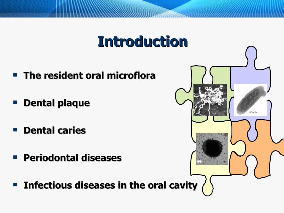
IntroductionIntroduction
The resident oral microfloraThe resident oral microflora
Dental plaDental plaqueque
Dental cariesDental caries
Periodontal diseasesPeriodontal diseases
Infectious diseases in the oral cavityInfectious diseases in the oral cavity
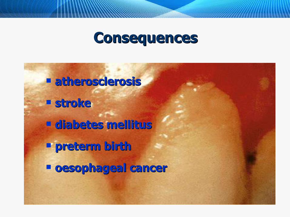
ConsequencesConsequences
atherosclerosisatherosclerosis
strokestroke
diabetes mellitusdiabetes mellitus
preterm birth preterm birth
oesophageal canceroesophageal cancer
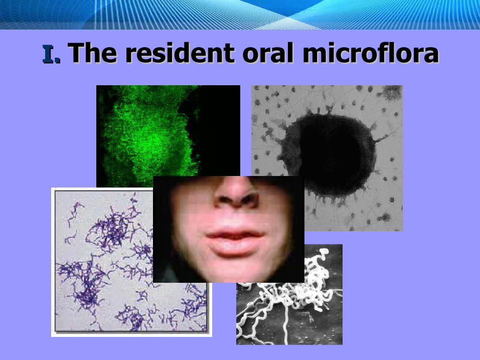
I. I. The resident oral microfloraThe resident oral microflora
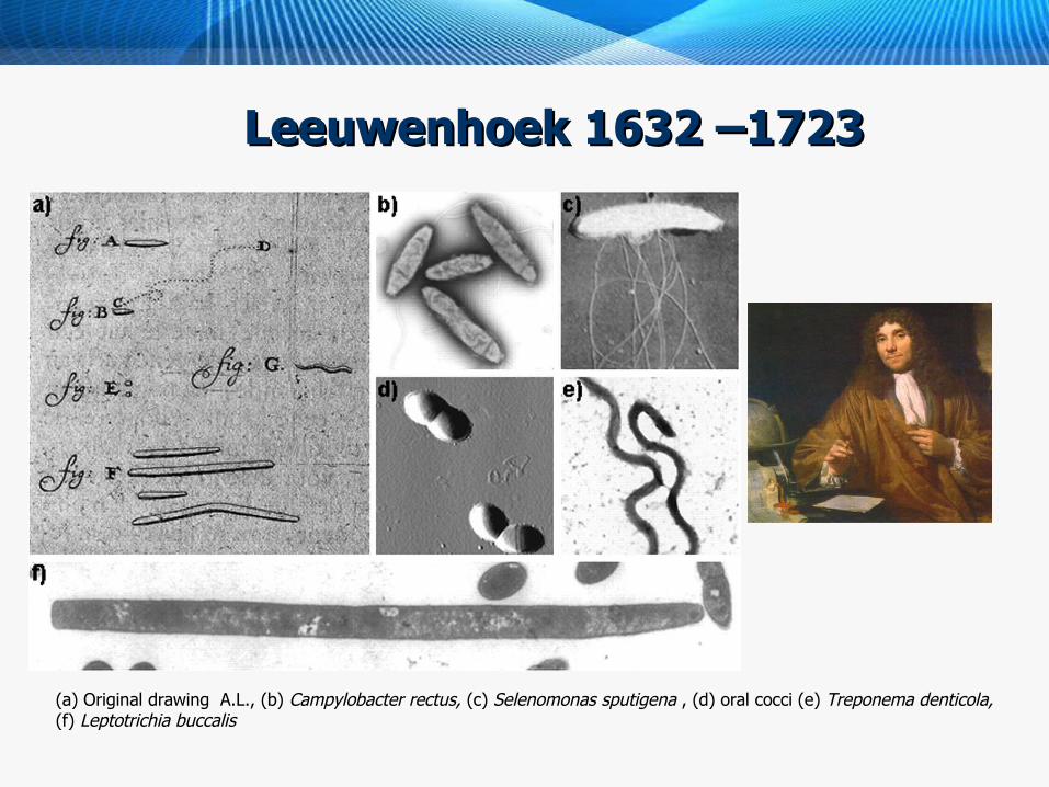
Leeuwenhoek 1632 –1723Leeuwenhoek 1632 –1723
(a) Original drawing A.L., (b) Campylobacter rectus, (c) Selenomonas sputigena , (d) oral cocci (e) Treponema denticola, (f) Leptotrichia buccalis
The resident oral microfloraThe resident oral microflora
One of the broadest microbial communities, over 700 700 generagenera, some were not still described
Resident – commensal, or transient
Ecological system
Biofilm formationBiofilm formation
Influential factor of human health (both local and in general)
Etiology of dental caries and parodontitisdental caries and parodontitis
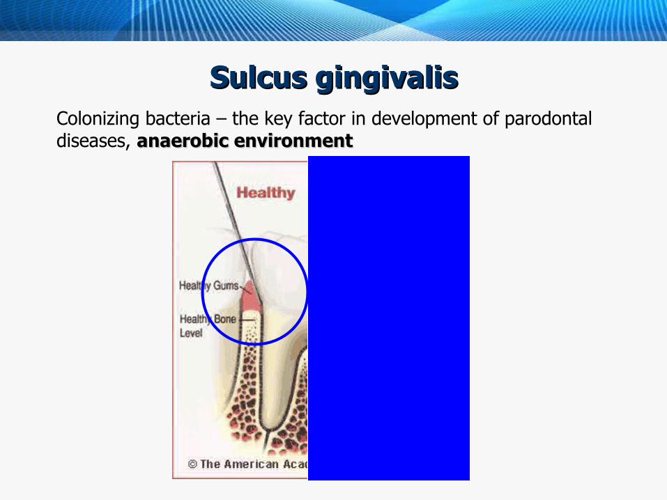
Sulcus gingivalisSulcus gingivalisColonizing bacteria – the key factor in development of parodontal diseases, anaerobic environmentanaerobic environment
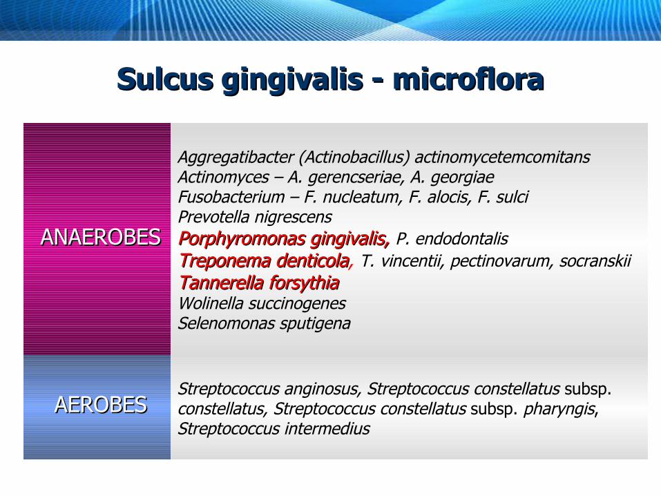
ANAEROBESANAEROBES
Aggregatibacter (Actinobacillus) actinomycetemcomitans Actinomyces – A. gerencseriae, A. georgiae Fusobacterium – F. nucleatum, F. alocis, F. sulci Prevotella nigrescens Porphyromonas gingivalis, Porphyromonas gingivalis, P. endodontalis Treponema denticolaTreponema denticola, T. vincentii, pectinovarum, socranskii Tannerella forsythiaTannerella forsythiaWolinella succinogenes Selenomonas sputigena
AEROBESAEROBESStreptococcus anginosus, Streptococcus constellatus subsp. constellatus, Streptococcus constellatus subsp. pharyngis, Streptococcus intermedius
Sulcus gingivalis - microfloraSulcus gingivalis - microflora

StreptococcusStreptococcus
α-hemolytic streptococci, divided into the following groups:
S. mutans group: S. mutans - the MOST FREQUENT, less often S. sobrinus, S.
cricetus, and S. rattus (rare), make acids from saccharides
S. salivarius group: S. salivarius, S. vestibularis - in saliva and on the tongue
surface, growth in mucous colonies, can cause endocarditis.
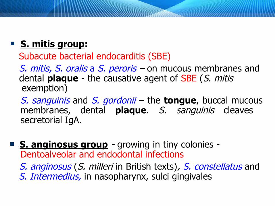
S. mitis group: Subacute bacterial endocarditis (SBE) S. mitis, S. oralis a S. peroris – on mucous membranes and dental plaque - the causative agent of SBE (S. mitis exemption) S. sanguinis and S. gordonii – the tongue, buccal mucous
membranes, dental plaque. S. sanguinis cleaves secretorial IgA.
S. anginosus group - growing in tiny colonies - Dentoalveolar and endodontal infections
S. anginosus (S. milleri in British texts), S. constellatus and S. Intermedius, in nasopharynx, sulci gingivales

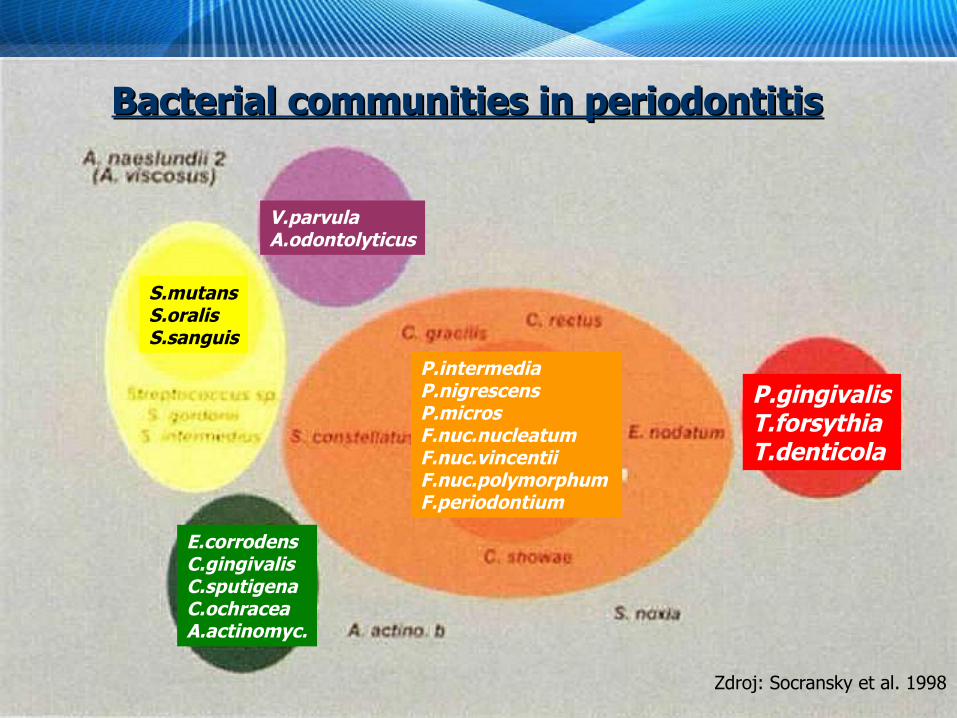
Zdroj: Socransky et al. 1998
P.gingivalisT.forsythiaT.denticola
P.intermediaP.nigrescensP.microsF.nuc.nucleatumF.nuc.vincentiiF.nuc.polymorphumF.periodontium
S.mutansS.oralisS.sanguis
E.corrodensC.gingivalisC.sputigenaC.ochraceaA.actinomyc.
V.parvulaA.odontolyticus
Bacterial communities in periodontitisBacterial communities in periodontitis
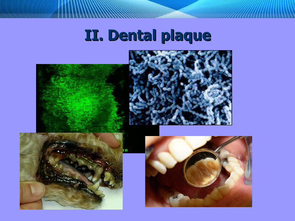
II. Dental plaqueII. Dental plaque
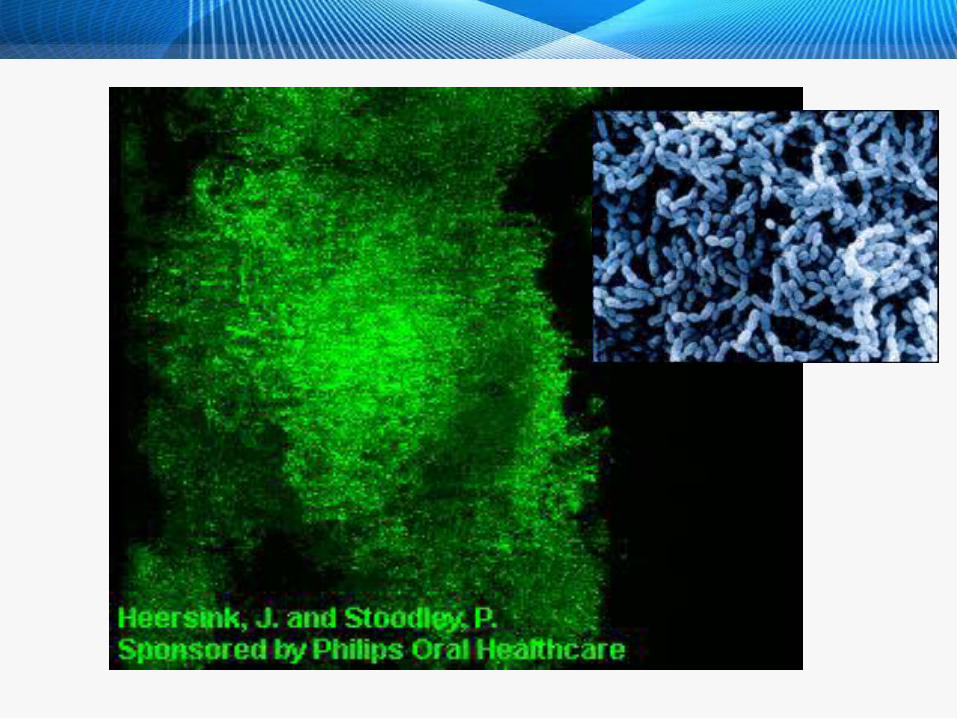
Dental plaque - biofilmDental plaque - biofilm
AdherentAdherent microbial layer on the tooth surface = microbial layer on the tooth surface = live live andand dead bacteria + their products + host compounds dead bacteria + their products + host compounds (from saliva)(from saliva)
It can NOT be washed, can be removed only mechanically (tooth brushing)
Composition dependent on its location and age
Location: Supragingival plaqueSupragingival plaque Subgingival plaqueSubgingival plaque
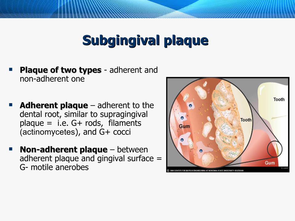
SSubgingival plaqueubgingival plaque Plaque of two typesPlaque of two types - adherent and
non-adherent one
Adherent plaqueAdherent plaque – adherent to the dental root, similar to supragingival plaque = i.e. G+ rods, filaments (actinomycetes), and G+ cocci
Non-adherent plaqueNon-adherent plaque – between adherent plaque and gingival surface = G- motile anerobes
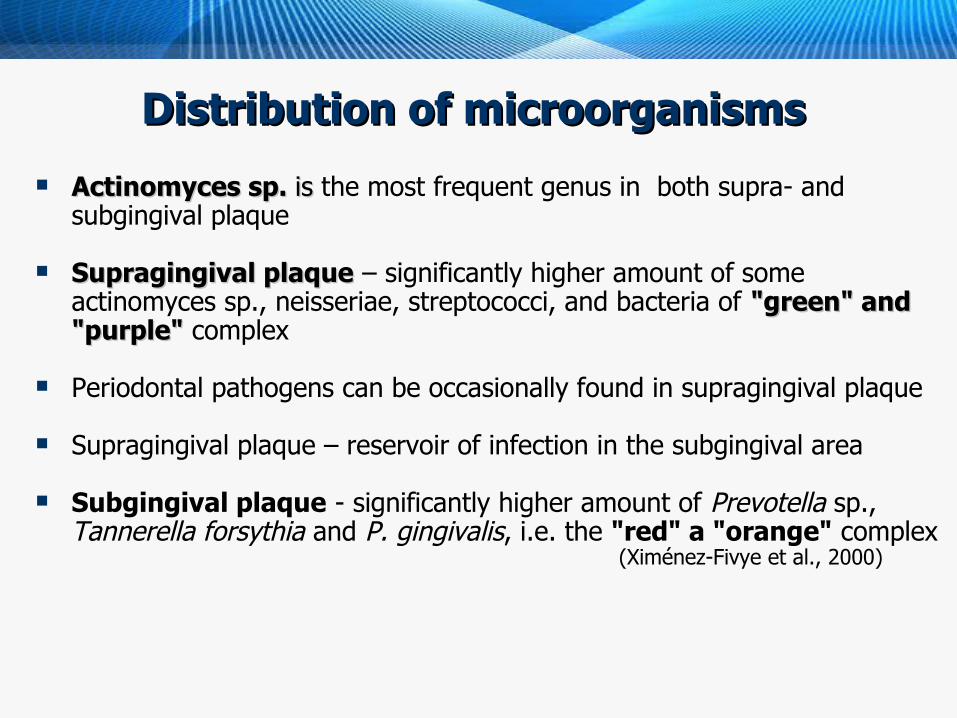
Distribution of microorganisms Distribution of microorganisms
Actinomyces sp. Actinomyces sp. isis the most frequent genus in both supra- and subgingival plaque
Supragingival plaqueSupragingival plaque – significantly higher amount of some actinomyces sp., neisseriae, streptococci, and bacteria of "green" and "green" and "purple""purple" complex
Periodontal pathogens can be occasionally found in supragingival plaque
Supragingival plaque – reservoir of infection in the subgingival area
Subgingival plaque - significantly higher amount of Prevotella sp., Tannerella forsythia and P. gingivalis, i.e. the "red" a "orange" complex
(Ximénez-Fivye et al., 2000)
Zdroj: Socransky et al. 1998
P.gingivalisT.forsythiaT.denticola
P.intermediaP.nigrescensP.microsF.nuc.nucleatumF.nuc.vincentiiF.nuc.polymorphumF.periodontium
S.mutansS.oralisS.sanguis
E.corrodensC.gingivalisC.sputigenaC.ochraceaA.actinomyc.
V.parvulaA.odontolyticus
Bacterial communities in periodontitisBacterial communities in periodontitis

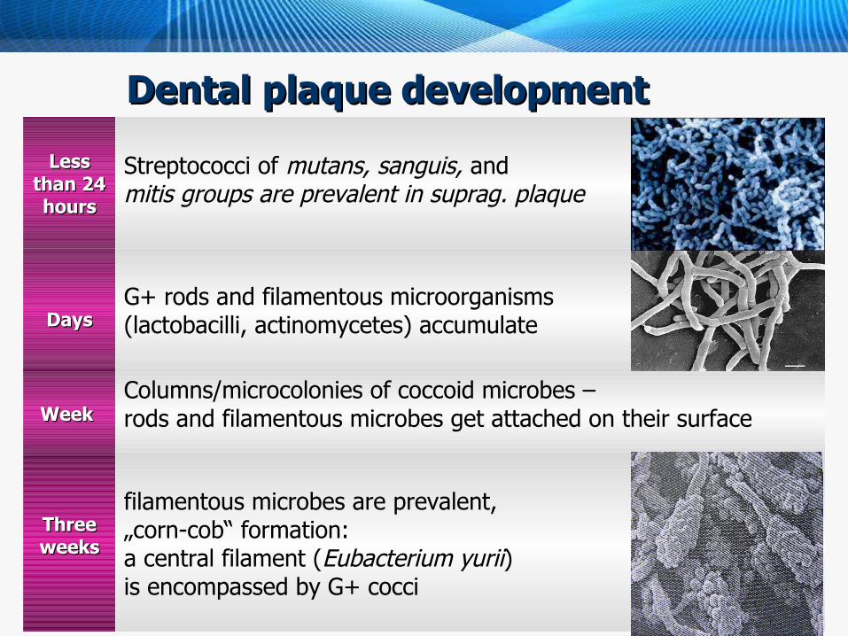
Development of dental plaque Development of dental plaque
Less Less than 24 than 24 hourshours
Streptococci of mutans, sanguis, and mitis groups are prevalent in suprag. plaque
DaysDaysG+ rods and filamentous microorganisms (lactobacilli, actinomycetes) accumulate
Week Week Columns/microcolonies of coccoid microbes – rods and filamentous microbes get attached on their surface
Three Three weeksweeks
filamentous microbes are prevalent, „corn-cob“ formation: a central filament (Eubacterium yurii) is encompassed by G+ cocci
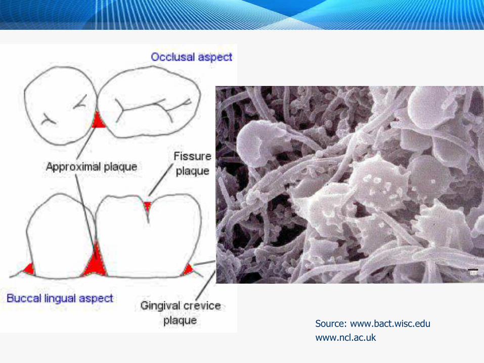
Source: www.bact.wisc.eduwww.ncl.ac.uk
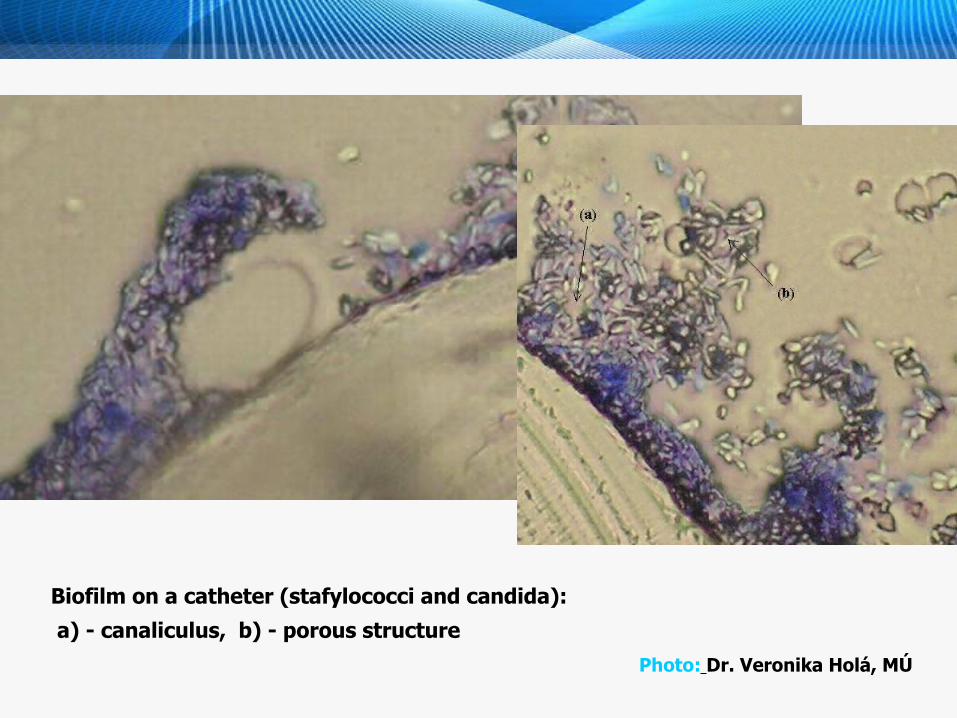
Biofilm on a catheter (stafylococci and candida):
a) - canaliculus, b) - porous structure Photo: Dr. Veronika Holá, MÚ
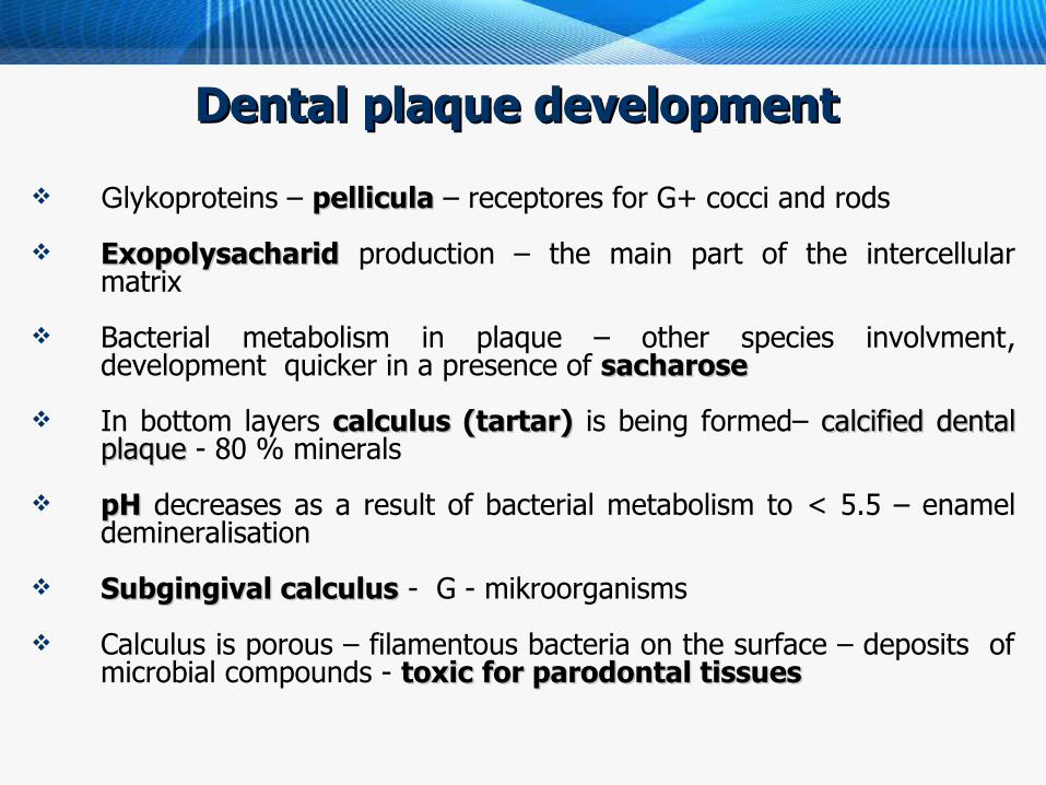
Dental plaque developmentDental plaque development
Glykoproteins – pelliculapellicula – receptores for G+ cocci and rods
ExopolysacharidExopolysacharid production – the main part of the intercellular matrix
Bacterial metabolism in plaque – other species involvment, development quicker in a presence of sacharsacharoseose
In bottom layers calculus (tartar)calculus (tartar) is being formed– calcified dental calcified dental plaqueplaque - 80 % minerals
pHpH decreases as a result of bacterial metabolism to < 5.5 – enamel demineralisation
Subgingival calculusSubgingival calculus - G - mikroorganisms
Calculus is porous – filamentous bacteria on the surface – deposits of microbial compounds - toxictoxic for parodontal tissuesfor parodontal tissues
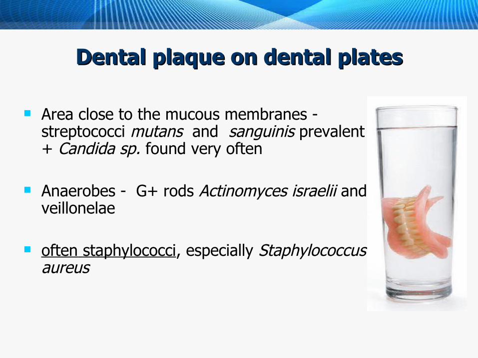
Dental plaque on dental platesDental plaque on dental plates
Area close to the mucous membranes - streptococci mutans and sanguinis prevalent + Candida sp. found very often
Anaerobes - G+ rods Actinomyces israelii and veillonelae
often staphylococci, especially Staphylococcus aureus
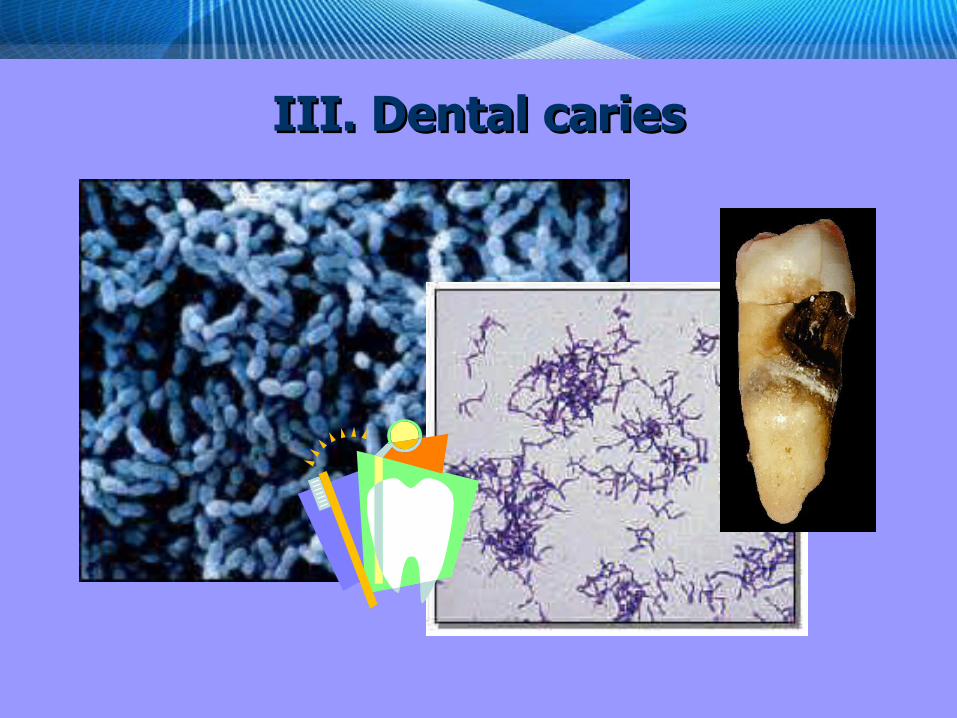
III. Dental cariesIII. Dental caries
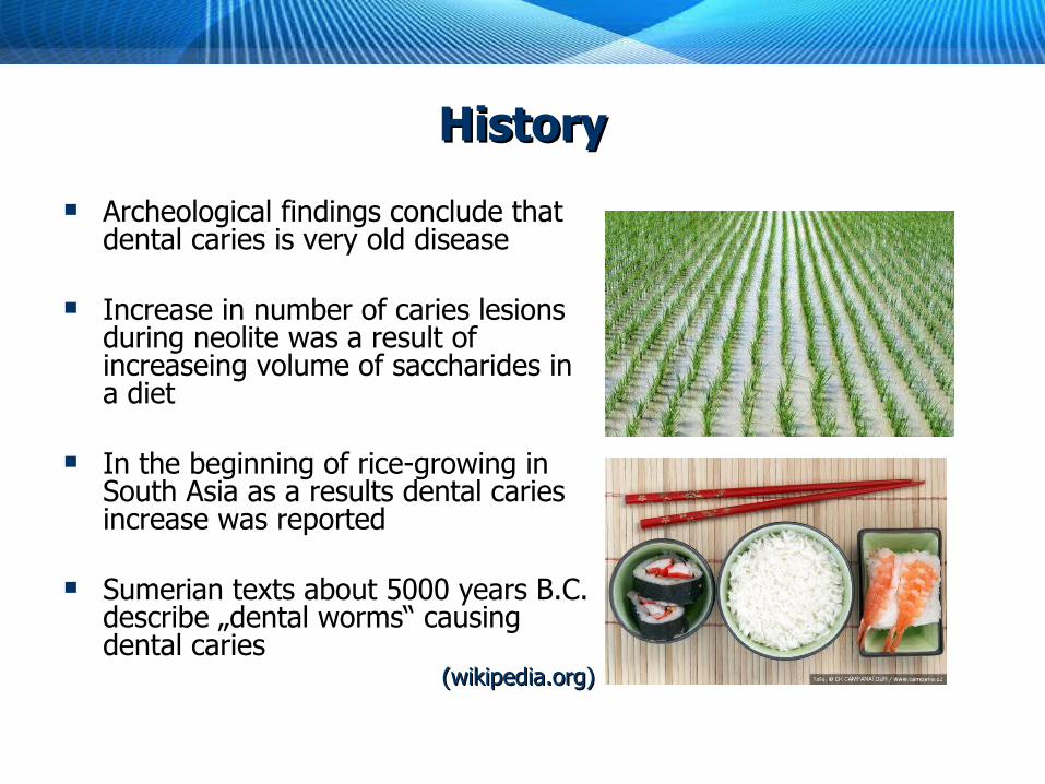
HistoryHistory
Archeological findings conclude that dental caries is very old disease
Increase in number of caries lesions during neolite was a result of increaseing volume of saccharides in a diet
In the beginning of rice-growing in South Asia as a results dental caries increase was reported
Sumerian texts about 5000 years B.C. describe „dental worms“ causing dental caries
(wikipedia.org)(wikipedia.org)
Microbiology of cariesMicrobiology of caries
Dental caries – the most frequent current disease
Definition - local destruction of the tooth tissuelocal destruction of the tooth tissue
Microbiological point of view – chronic infection caused by chronic infection caused by normal oral floranormal oral flora
Destruction is a result of demineralisationdemineralisation of the tooth caused by acids producted by microorganisms in the dental plaque during metabolism of saccharides from food saccharides from food
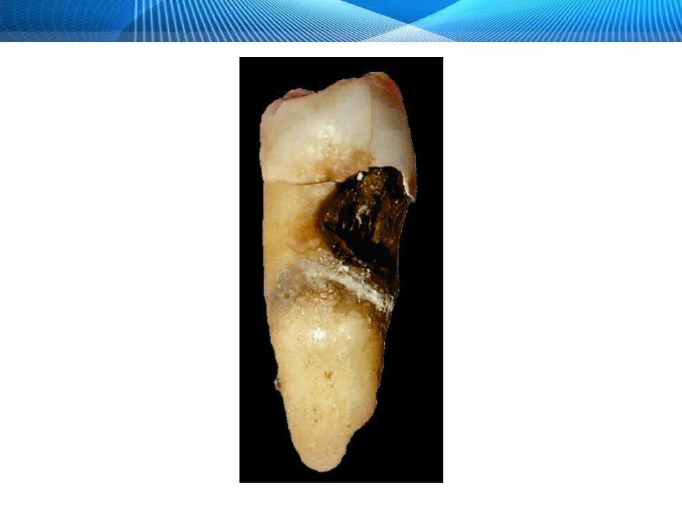
Course of cariesCourse of caries
Primary lesion of enamel (whitish spot) is reversible, it can remineralise
After destruction of enamel, the process spreads to dentin
and causes inflammation and necrosis
Also development of periapical acute or chronic inflammation
Dental caries = multifactorial diseaseDental caries = multifactorial disease
1.1. endogenous factors endogenous factors 2.2. foodfood3.3. microbes in the dental plaguemicrobes in the dental plague
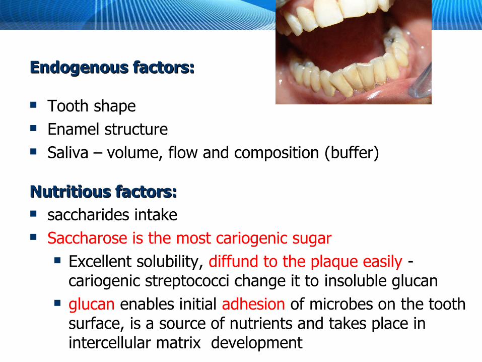
Endogenous factors:Endogenous factors:
Tooth shape Enamel structure Saliva – volume, flow and composition (buffer)
Nutritious factors:Nutritious factors: saccharides intake Saccharose is the most cariogenic sugar
Excellent solubility, diffund to the plaque easily - cariogenic streptococci change it to insoluble glucan
glucan enables initial adhesion of microbes on the tooth surface, is a source of nutrients and takes place in intercellular matrix development
Role of microbesRole of microbes
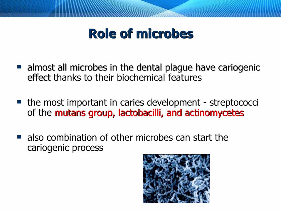
almost allalmost all microbes in the dental plague have cariogenic microbes in the dental plague have cariogenic effecteffect thanks to their biochemical features
the most important in caries development - streptococci of the mutans group, lactobacilli, and actinomycetesmutans group, lactobacilli, and actinomycetes
also combination of other microbes can start the cariogenic process
StreptococcusStreptococcus
α-hemolytic streptococci, divided into the following groups:
S. mutans group: S. mutans - the MOST FREQUENT, less often S. sobrinus,
S. cricetus, and S. rattus rarely, produce acids from saccharides
S. salivarius group: S. salivarius, S. vestibularis - in saliva and on the tongue,
growth in mucous colonies, can cause endocarditis
S. mitis group: S. mitis, S. oralis a S. peroris – on mucous membranes and in the dental plaque - the causative agent of sepsis lenta (S. mitis is an exemption)
S. sanguinis and S. gordonii – on the tongue, buccal musous membranes, dental plaque. S. sanguinis cleaves secretorial IgA
Both species are important cause of subacute bacterial subacute bacterial endocarditis (endocarditis (sepsis lentasepsis lenta) )
S. anginosus group - tiny colonies - S. anginosus (S. milleri in British texts), S. constellatus with two subspecies, constellatus and pharyngis, and S. intermedius.
In nasopharynx, sulci gingivales, dentoalveolar and endodontal infections
Caries and Caries and mutansmutans group streptococci I group streptococci I
In man usually: S. mutans (serotypes c, e, and f) S. sobrinus (serotypes d and g)
Some strains seems to be more cariogenic.
Ethiological role - facts:
Numberes in the plaque and saliva correlates with caries prevalence and incidence
Isolated from the tooth surface immediately before caries
Immunisation of animals with S. mutans specif. serotypes decreases caries incidence
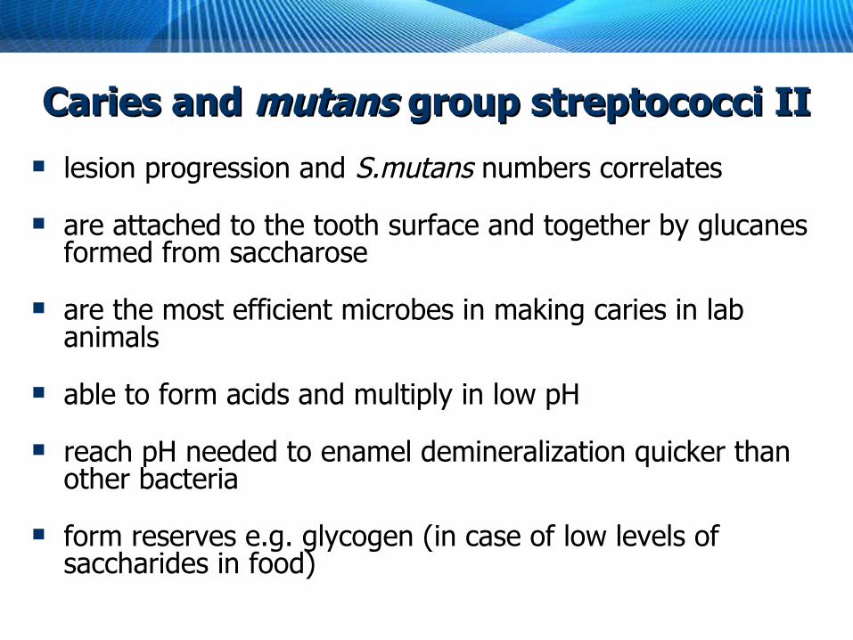
lesion progression and S.mutans numbers correlates
are attached to the tooth surface and together by glucanes formed from saccharose
are the most efficient microbes in making caries in lab animals
able to form acids and multiply in low pH
reach pH needed to enamel demineralization quicker than other bacteria
form reserves e.g. glycogen (in case of low levels of saccharides in food)
Caries and Caries and mutansmutans group streptococci II group streptococci II
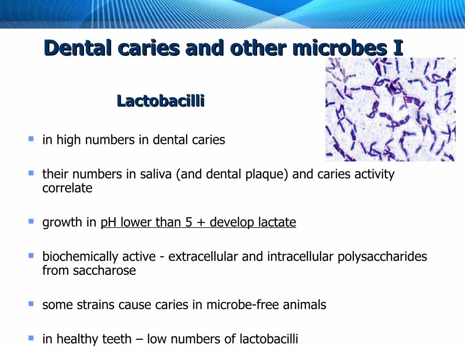
Dental cDental caries and other microbesaries and other microbes I I
LactobacilliLactobacilli
in high numbers in dental caries
their numbers in saliva (and dental plaque) and caries activity correlate
growth in pH lower than 5 + develop lactate
biochemically active - extracellular and intracellular polysaccharides from saccharose
some strains cause caries in microbe-free animals
in healthy teeth – low numbers of lactobacilli
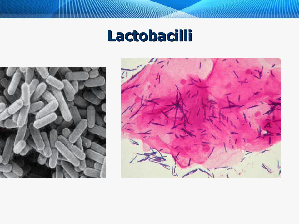
LactobacilliLactobacilli
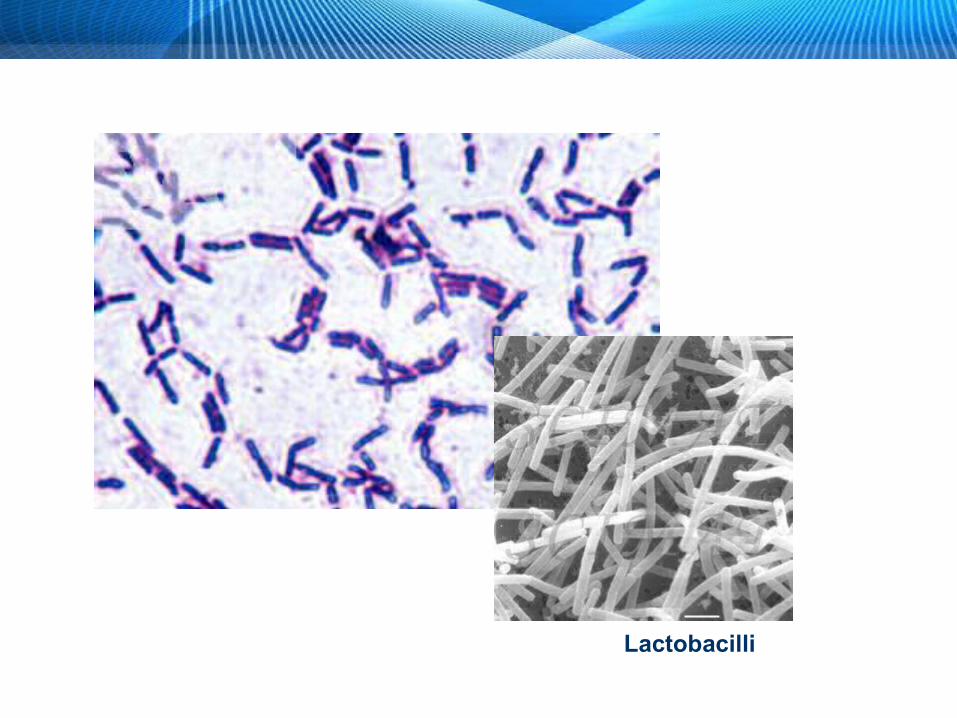
Lactobacilli
Dental cDental caries and other microbesaries and other microbes II II
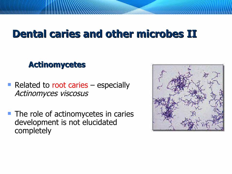
Actinomycetes Actinomycetes
Related to root caries – especially Actinomyces viscosus
The role of actinomycetes in caries
development is not elucidated completely
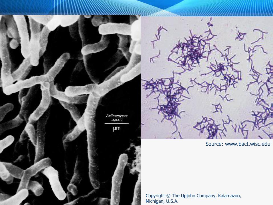
Copyright © The Upjohn Company, Kalamazoo, Michigan, U.S.A.
Source: www.bact.wisc.edu
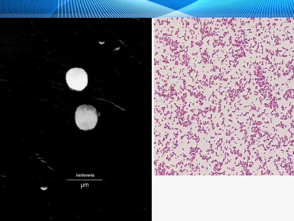
Caries and other microbesCaries and other microbes
Veillonela sp.Veillonela sp.
in high numbers in supragingival plague of most people
need lactatelactate, are NOT able to use saccharides and use lactate developed by other microbes – transform it to less cariogenic organic acids
……………… positive outcomes………? positive outcomes………?
Ecological plaque hypothesisEcological plaque hypothesis
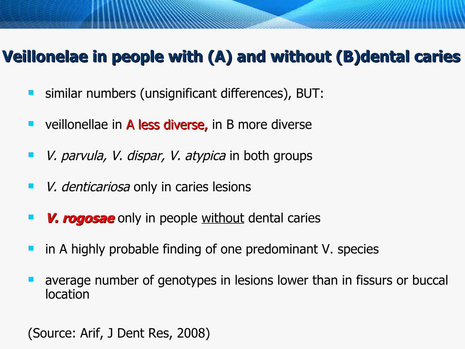
Veillonelae in people with (A) and without (B)dental cariesVeillonelae in people with (A) and without (B)dental caries
similar numbers (unsignificant differences), BUT:
veillonellae in A less diverse, A less diverse, in B more diverse
V. parvula, V. dispar, V. atypica in both groups
V. denticariosa only in caries lesions
V. rogosaeV. rogosae only in people without dental caries
in A highly probable finding of one predominant V. species
average number of genotypes in lesions lower than in fissurs or buccal location
(Source: Arif, J Dent Res, 2008)
Dental plaque developmentDental plaque development
Less Less than 24 than 24 hourshours
Streptococci of mutans, sanguis, and mitis groups are prevalent in suprag. plaque
DaysDaysG+ rods and filamentous microorganisms (lactobacilli, actinomycetes) accumulate
Week Week Columns/microcolonies of coccoid microbes – rods and filamentous microbes get attached on their surface
Three Three weeksweeks
filamentous microbes are prevalent, „corn-cob“ formation: a central filament (Eubacterium yurii) is encompassed by G+ cocci
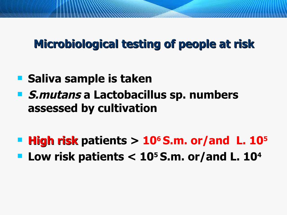
Microbiological testing of people at riskMicrobiological testing of people at risk
Saliva sample is taken S.mutans a Lactobacillus sp. numbers
assessed by cultivation
High riskHigh risk patients > 106 S.m. or/and L. 105
Low risk patients < 105 S.m. or/and L. 104
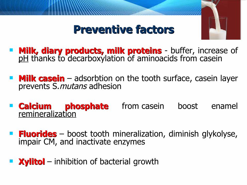
Preventive factorsPreventive factors
Milk, diary products, milk proteinsMilk, diary products, milk proteins - buffer, increase of pH thanks to decarboxylation of aminoacids from casein
Milk caseinMilk casein – adsorbtion on the tooth surface, casein layer prevents S.mutans adhesion
Calcium phosphateCalcium phosphate from casein boost enamel remineralization
Fluorides Fluorides – boost tooth mineralization, diminish glykolyse, impair CM, and inactivate enzymes
Xylitol Xylitol – inhibition of bacterial growth
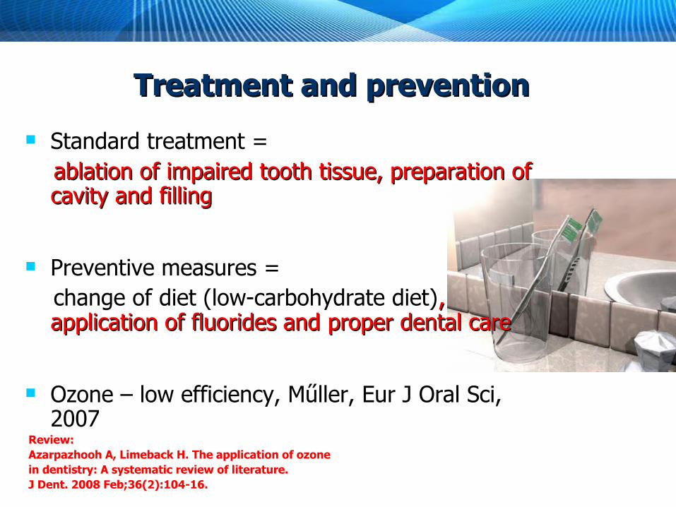
Treatment and preventionTreatment and prevention Standard treatment = ablation of ablation of impairedimpaired tooth tissue, prepara tooth tissue, preparation oftion of
ccavity aavity and fillingnd filling
Preventive measures = change of diet (low-carbohydrate diet), ,
aappplipliccation of fluorides and ation of fluorides and proper proper dental caredental care
Ozone – low efficiency, Műller, Eur J Oral Sci, 2007
Review: Azarpazhooh A, Limeback H. The application of ozone in dentistry: A systematic review of literature. J Dent. 2008 Feb;36(2):104-16.
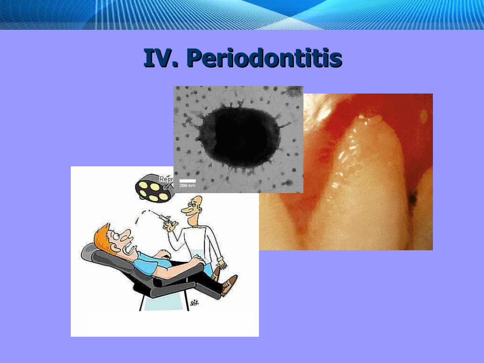
IV. PeriodontitisIV. Periodontitis
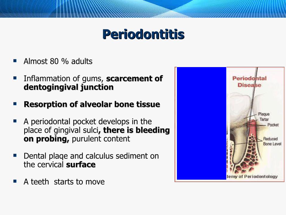
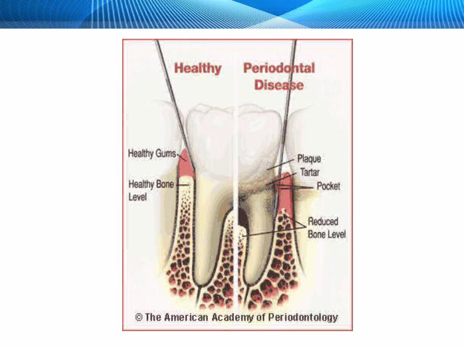
PeriodontitisPeriodontitis
Almost 80 % adults
Inflammation of gums, scarcement of scarcement of dentogingival junctiondentogingival junction
Resorption of alveolar bone tissueResorption of alveolar bone tissue
A periodontal pocket develops in the place of gingival sulci, there is bleeding , there is bleeding on probing,on probing, purulent content
Dental plaqe and calculus sediment on the cervical surface surface
A teeth starts to move
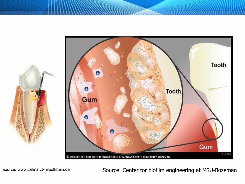
Source: Center for biofilm engineering at MSU-BozemanSource: www.zahnarzt-hilpoltstein.de
Gum reactionGum reaction Dental plaque in the gum margine - chronic
inflammation of the tissue around sulcus gingivalis = marginal gingivitismarginal gingivitis
Exsudation – chemotaxis of anaerobic and proteolytic bacteria
Increasing migration of leucocytes
Inflammation breaks function of the junctional epithel, plaque spreads apically to subgingival areaplaque spreads apically to subgingival area
Symptoms much more intensive with older and thicker plaque
Microbiology of chronic marginal Microbiology of chronic marginal gingivitisgingivitis
Clinical symptoms - ocasional gum bleeding - inflamed, hurtfullness is minimal
Early stageEarly stage – after a one week course - number of capnofile number of capnofile and strictly anaerobic microbesand strictly anaerobic microbes is growing (especially Actinomyces sp. and anaerobic G- rods)
Late stageLate stage – more microbes, anaerobes areanaerobes are prevalent (in black colonies growing e.g. Porphyromonas gingivalis and Prevotella intermedia, oral spirochetes)
Bleeding from gumsBleeding from gums lead to multiplication of black-pigmented anaerobic rods, blood is a source of haemin haemin
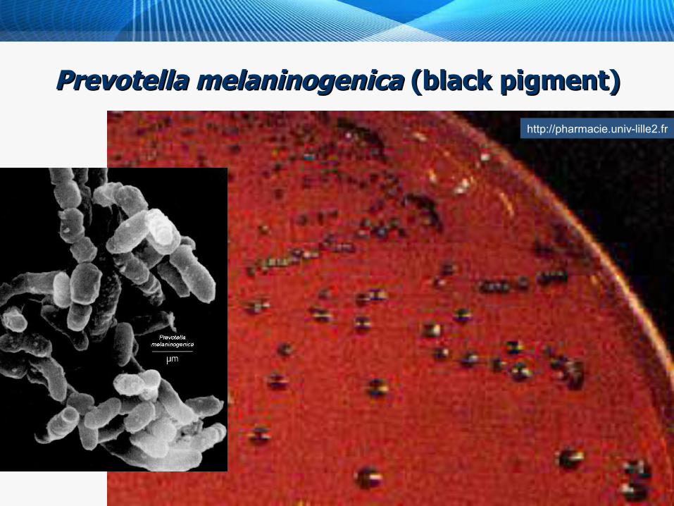
Prevotella melaninogenicaPrevotella melaninogenica (black pigment) (black pigment)
http://pharmacie.univ-lille2.fr
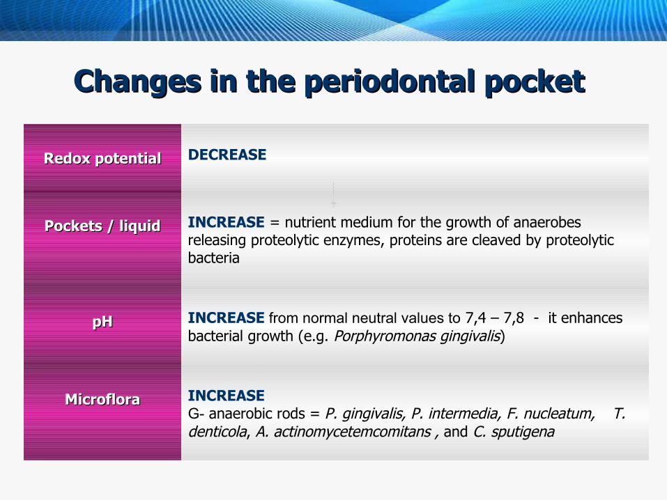
Changes in the periodontal pocketChanges in the periodontal pocket
Redox potentialRedox potential DECREASE
Pockets / liquidPockets / liquid INCREASE = nutrient medium for the growth of anaerobes releasing proteolytic enzymes, proteins are cleaved by proteolytic bacteria
pHpH INCREASE from normal neutral values to 7,4 – 7,8 - it enhances bacterial growth (e.g. Porphyromonas gingivalis)
MicrofloraMicroflora INCREASEG- anaerobic rods = P. gingivalis, P. intermedia, F. nucleatum, T. denticola, A. actinomycetemcomitans , and C. sputigena

Infuence of subgingival plaque - studiesInfuence of subgingival plaque - studies
Strong correlation between plaque volume and prevalence and severity of periodontal diseses
Volunteers studiesVolunteers studies – poor dental hygiene = plaq growth and gingivitis – after plaq removal gingivitis heals
Local application e.g. chlorhexidine diminish plaq and prevent gingivitis
Microbe-free animal modelsMicrobe-free animal models - bacteria of „red complex“ from human plaq lead to parodontal infection and immunoinflammatory bone resorption (Kesavalu 2007)
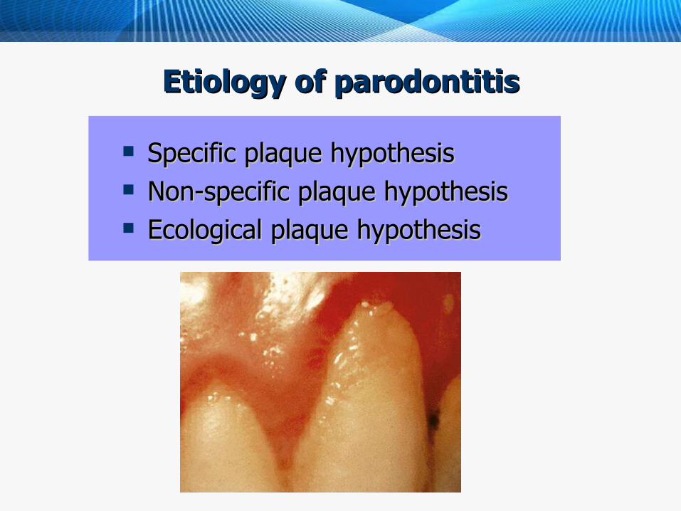
Etiology of parodontitisEtiology of parodontitis
Specific plaque hypothesisSpecific plaque hypothesis Non-specific plaque hypothesisNon-specific plaque hypothesis Ecological plaque hypothesisEcological plaque hypothesis
Specific plaque hypothesisSpecific plaque hypothesis
Etiology of parodontitis = specific microorganismsEtiology of parodontitis = specific microorganisms
Necrotizing ulcerative gingivitis – key agens fusobacteria and spirochetes
Terapeutic success with antimicrobials inhibiting anaerobes – e.g. metronidazole
Rapidly progreding juvenile parodontitis - Aggregatibacter actinomycetemcomitans – sensitive to tetracycline – treatment
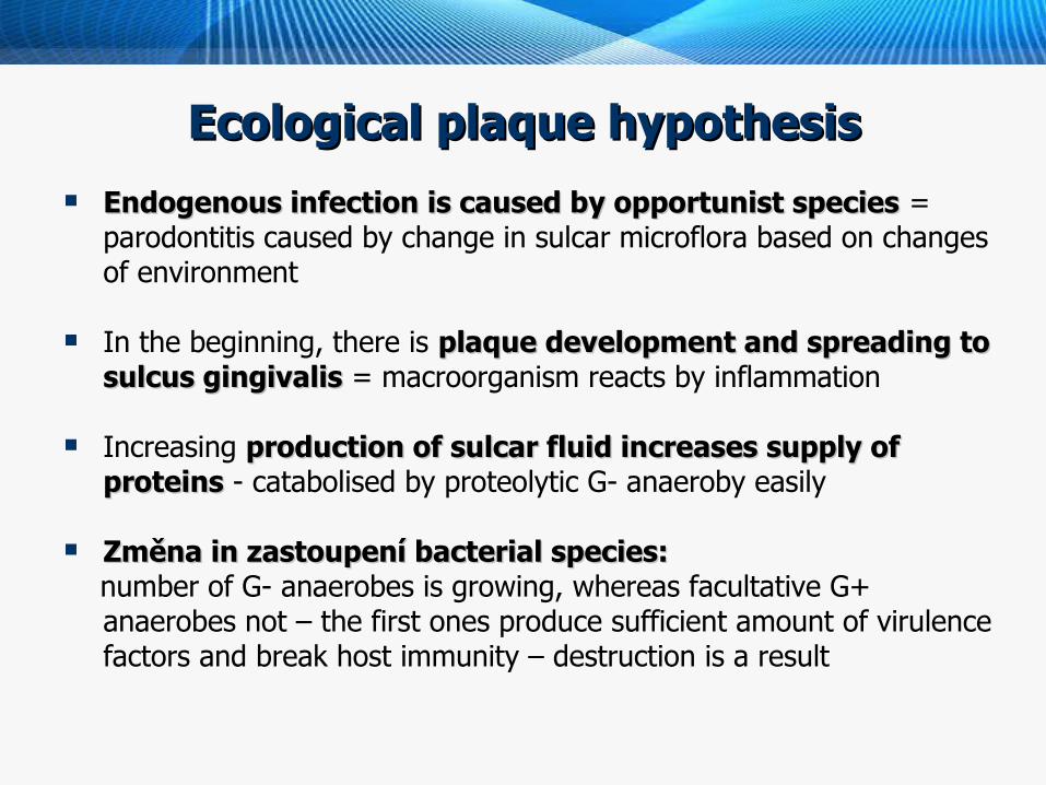
Ecological plaque hypothesisEcological plaque hypothesis Endogenous infection is caused by opportunist speciesEndogenous infection is caused by opportunist species =
parodontitis caused by change in sulcar microflora based on changes of environment
In the beginning, there is plaque development and spreading to plaque development and spreading to sulcus gingivalissulcus gingivalis = macroorganism reacts by inflammation
Increasing production of sulcar fluid increases supply of production of sulcar fluid increases supply of proteinsproteins - catabolised by proteolytic G- anaeroby easily
Změna in zastoupení bacterial species: Změna in zastoupení bacterial species: number of G- anaerobes is growing, whereas facultative G+
anaerobes not – the first ones produce sufficient amount of virulence factors and break host immunity – destruction is a result
Therapeutic strategiesTherapeutic strategies
Specific plaque hypothesisSpecific plaque hypothesis – therapy focused on specific pathogen removal, e.g. antibiotics administrationantibiotics administration
Non- specific and ecological hypothesesNon- specific and ecological hypotheses - parodontal disease can be treated by measurments aimed at reduction of plaque reduction of plaque volume volume
PreventionPrevention
Regular removal of dental plaque Regular removal of dental plaque byby proper cleaning of the teeth
Perfect removal of calculusPerfect removal of calculus
Improvement of exogenous factorsImprovement of exogenous factors (… impaired prothetic devices etc.)
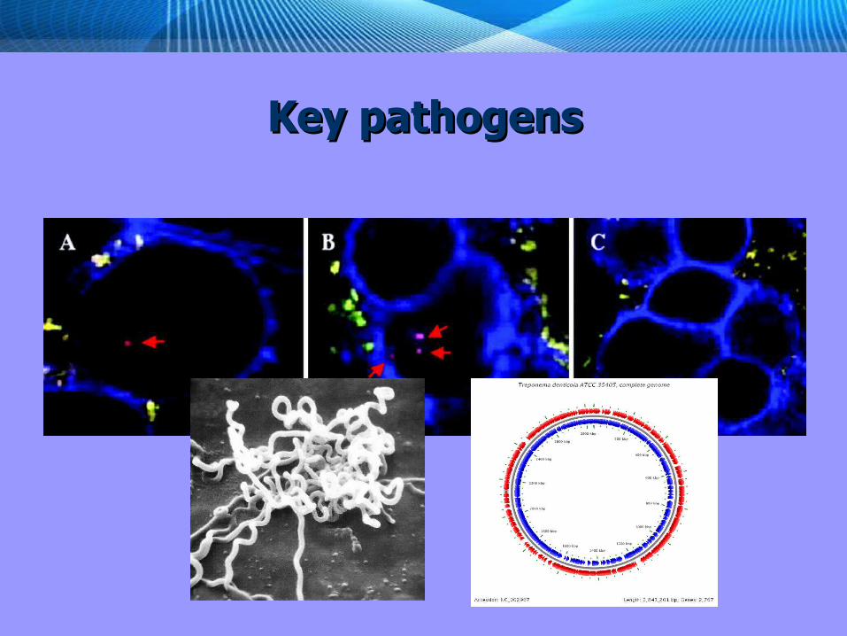
Key pathogensKey pathogens
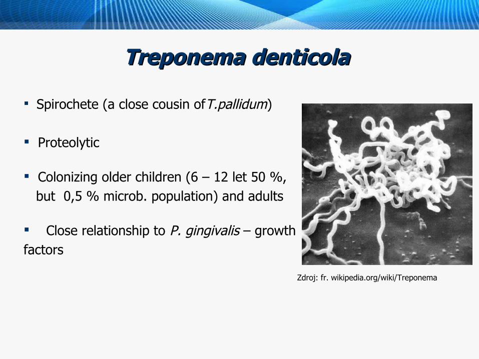
Treponema denticola Treponema denticola
Zdroj: fr. wikipedia.org/wiki/Treponema
Spirochete (a close cousin ofT.pallidum)
Proteolytic
Colonizing older children (6 – 12 let 50 %, but 0,5 % microb. population) and adults
Close relationship to P. gingivalis – growth factors

Porphyromonas gingivalisPorphyromonas gingivalis
Highly proteolyticHighly proteolytic
Fimbriae Fimbriae – adhesioin and colonisation
Releases vesiclesvesicles containing parts of outer membranes - proteins, LPS, capsule etc.
Vesicles - transport of toxins and enzymes, bacterial adherention and aggregation, adherention of thrombcytes
Black pigment = acummulated hemin – a source of iron (a growth factor) Zdroj: www.pgingivalis.org
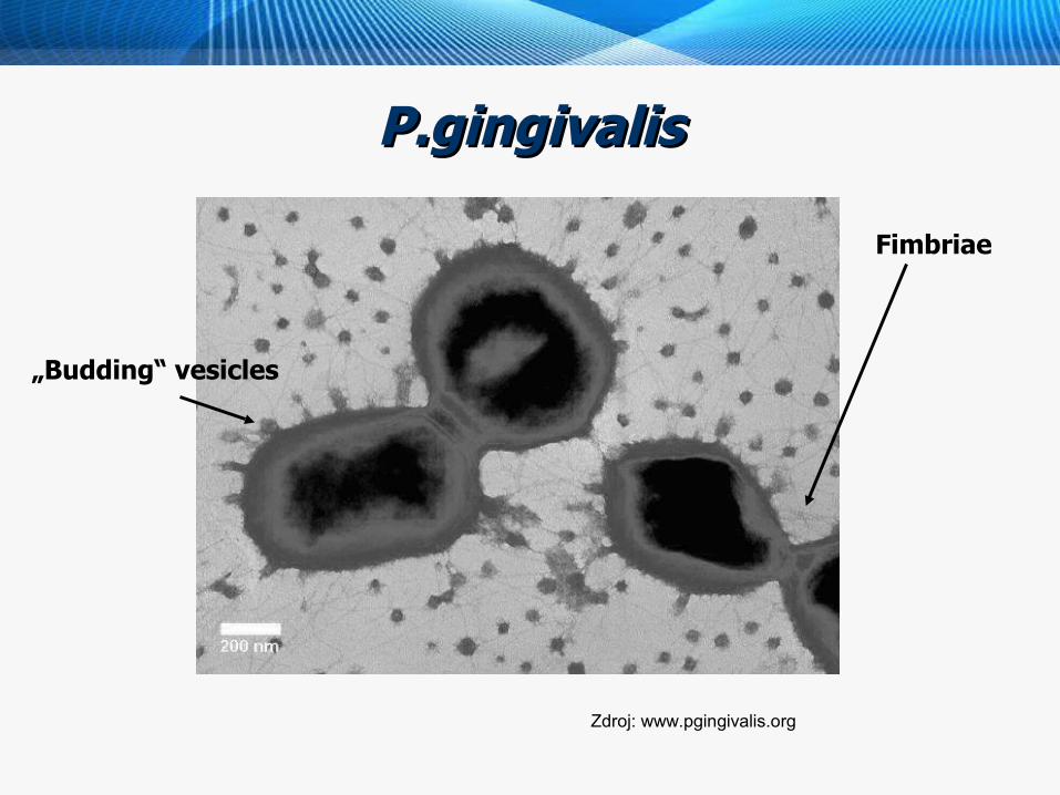
P.gingivalisP.gingivalis
Zdroj: www.pgingivalis.org
„Budding“ vesicles
Fimbriae
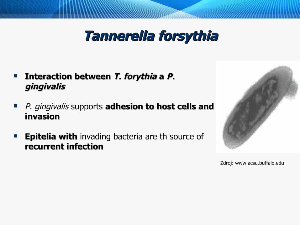
Tannerella forsythiaTannerella forsythia
Interaction between Interaction between T. forythiaT. forythia a a P. P. gingivalisgingivalis
P. gingivalis supports adhesion to host cells and adhesion to host cells and invasioninvasion
Epitelia withEpitelia with invading bacteria are th source of recurrent infection recurrent infection
Zdroj: www.acsu.buffalo.edu
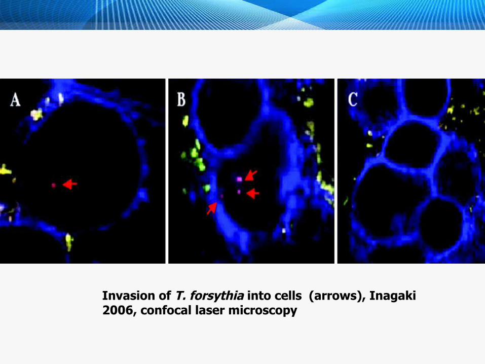
Invasion of T. forsythia into cells (arrows), Inagaki 2006, confocal laser microscopy
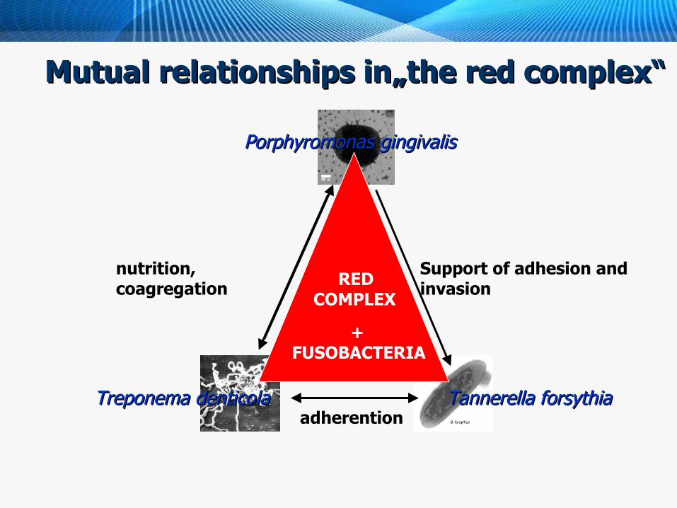
Mutual relationships in„the red complex“Mutual relationships in„the red complex“
Tannerella forsythiaTannerella forsythia
Porphyromonas gingivalisPorphyromonas gingivalis
Treponema denticolaTreponema denticola
nutrition, coagregation
Support of adhesion and invasion RED
COMPLEX
adherention
+ FUSOBACTERIA
Oral microflora in systemic diseasesOral microflora in systemic diseases
Cardiovascular diseases - bacterial endocarditis, aterosclerosis - esp. coronary arterias (Gotsman et al. 2007)
Strokes (Pussinen et al. 2004)
Pneumonias
Diabetes mellitus (Mealey, Rethman 2003)
Preterm births and low birth weight (Lin et al. 2007)
Oesophagal carcinoma (Narikiyo et al. 2004)
MMechanismsechanisms
Microbes from the mouthMicrobes from the mouth = metastatic infections (bacteremia after tooth extraction - bacterial endocarditis)
Bacterial enzymes and toxines fromBacterial enzymes and toxines from parodontal focuses = metastatic damage (e.g. endotoxin G- bacteria from subgingival biofilm)
Antigens of oral bacteria and pro-inflammatory cytokinesAntigens of oral bacteria and pro-inflammatory cytokines
from inflamed parodont = metastatic inflammation (reaction Ag-Ab where immunocomplexes)
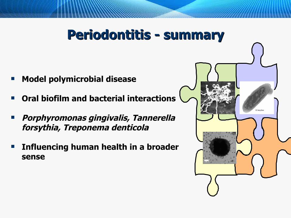
Periodontitis - summaryPeriodontitis - summary
Model polymicrobial disease Oral biofilm and bacterial interactions
Porphyromonas gingivalis, Tannerella forsythia, Treponema denticola
Influencing human health in a broader sense
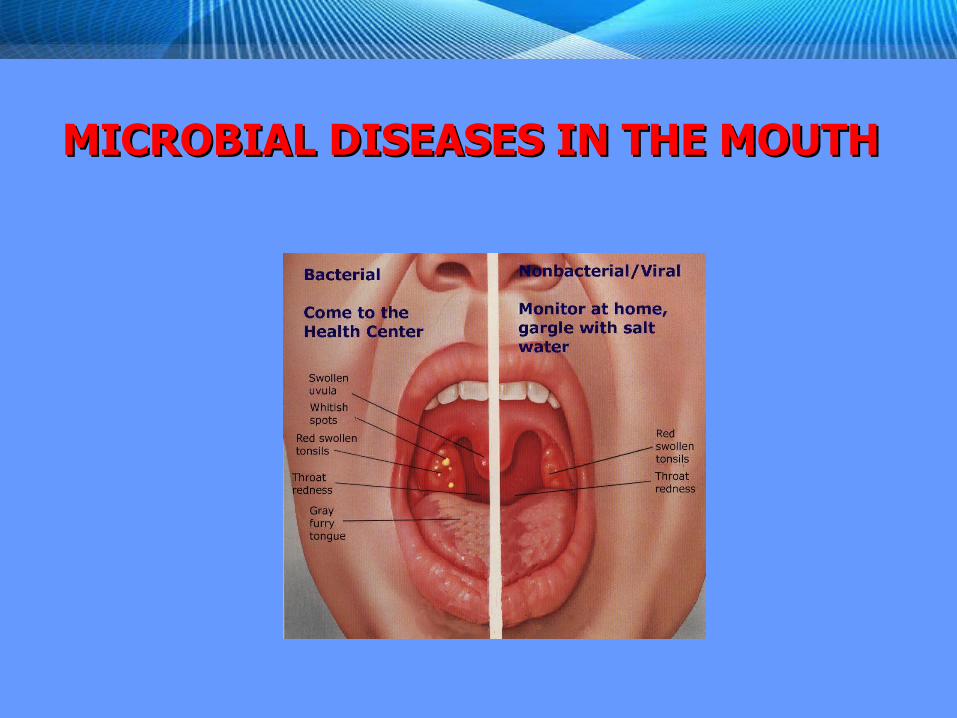
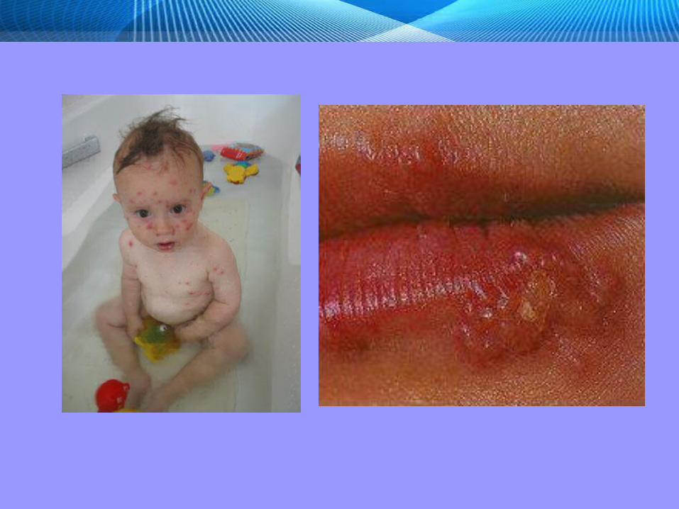
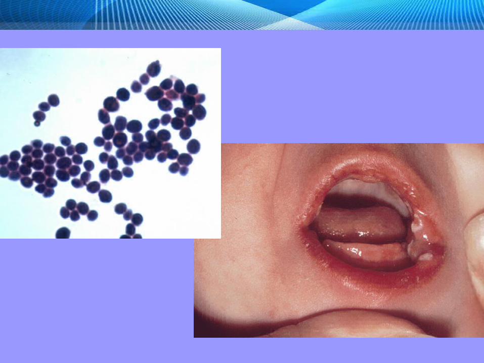
MICROBIAL DISEASES IN THE MOUTHMICROBIAL DISEASES IN THE MOUTH
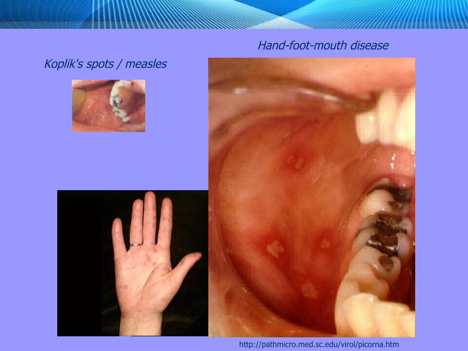
Koplik's spots / measles
Hand-foot-mouth disease
http://pathmicro.med.sc.edu/virol/picorna.htm
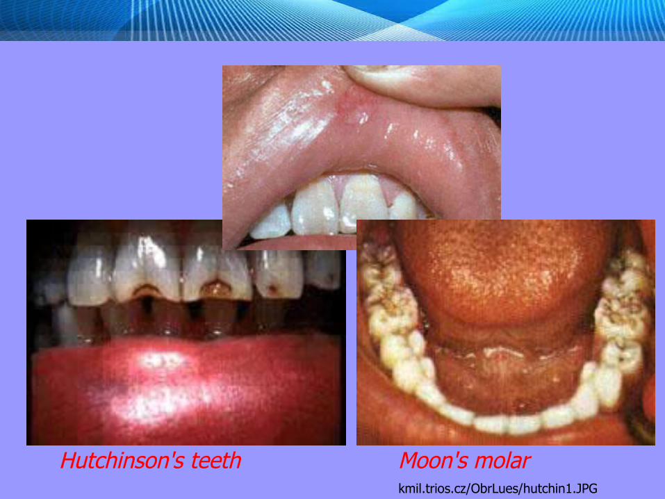
kmil.trios.cz/ObrLues/hutchin1.JPG
Hutchinson's teeth Moon's molar
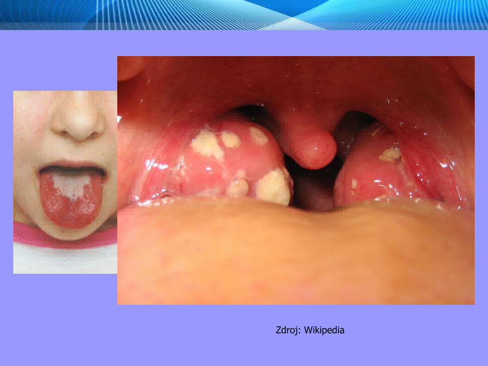
Zdroj: Wikipedia
Thank you