oral medicine case final presentations rasha ahmed badahdah 0931010
TRANSCRIPT

Oral Medicine Case
final Presentations
Rasha Ahmed Badahdah 0931010

DEMOGRAPHIC DATA:
Age : 19 years old. Nationality : Chadian. Gender : female.

CHIEF COMPLAINT: The patient wants to restore her upper
anterior teeth.
HISTORY OF PRESENT ILLNESS:
The caries started about one year ago.
She did not seek any treatment for her teeth .

DENTAL HISTORY : Not significant
FAMILY HISTORY :
Not significant
MEDICAL HISTORY: Not significant
SOCIAL HISTORY and HABITS:
Marital status : Single . Occupation : student. There are no significant habits The patient brushes her teeth once per
day , and she does not floss her teeth.

CLINICAL EXAMINATIONS:
A-Extra-Oral Examination. B-Intra-Oral Examination.
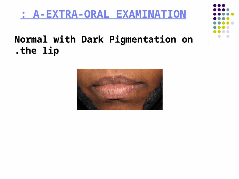
A-EXTRA-ORAL EXAMINATION:
Normal with Dark Pigmentation on the lip.
B-INTRA-ORAL EXAMINATION:
•*Pigmentation all around in the baccal and labial mucosa , gingiva and tongue.
*Maxillary Lateral retained Deciduous teeth .


*Two lesions near the mucogingival
junction related to upper central incisors
#11 #21.•Badly decayed teeth:
• #11,#21,#36,#46#47 .


RADIOGRAPHIC FINDING:
Periapical radiolucency related to #11,#21,#46,#47
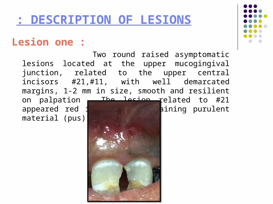
DESCRIPTION OF LESIONS:
Lesion one : Two round raised asymptomatic lesions located at the
upper mucogingival junction, related to the upper central incisors #21,#11, with well demarcated margins, 1-2 mm in size, smooth and resilient on palpation . The lesion related to #21 appeared red in color , containing purulent material (pus).

Teeth Examination:
Vitality test
Thermal examination ( cold and hot and EPT)
was done & showed that teeth #11,#21 did not respond
Checking for periapical lesions:
Done by percussion, showed there no response at teeth #11,#21
Periodontal examination
Done with a graduated periodontal probe for pocket detection , no pockets were revealed

For lesion one : To diagnose the location of the problematic tooth, tracing done with a size #25 Gutta -Percha cone, threaded into the Opening
of the sinus tract.

Lesion two :
Generalized dark (brownish/bluish) pigmentation in the buccal and labial mucosae , upper and lower gingivae and tongue.


DIFFERENTIAL DIAGNOSIS: For lesion one:
1)Fistula 2)Periapical abscess.
3)Periodontal abscess 4)Periapical cyst.
For lesion two: 1)Physiological pigmentation
2)Systemic disease :Addison's disease.
FINAL DIAGNOSIS: For lesion one:
Fistulas

Management
For lesion One : Consultation with the endodontic department
for Root Canal Treatment.

FINAL DIAGNOSIS: For lesion two:
Physiological pigmentation

FOLLOW UP:

THANK YOU FOR YOUR ATTENTION,,,