oral biology, oral pathology, and oral...
TRANSCRIPT

BioMed Research International
Oral Biology, Oral Pathology, and Oral Treatments
Guest Editors: Samir Nammour, Toni Zeinoun, Kenji Yoshida, and Aldo Brugnera Junior

Oral Biology, Oral Pathology,and Oral Treatments
BioMed Research International
Oral Biology, Oral Pathology,and Oral Treatments
Guest Editors: SamirNammour, Toni Zeinoun,Kenji Yoshida,and Aldo Brugnera Junior

Copyright © 2016 Hindawi Publishing Corporation. All rights reserved.
This is a special issue published in “BioMed Research International.” All articles are open access articles distributed under the CreativeCommons Attribution License, which permits unrestricted use, distribution, and reproduction in any medium, provided the originalwork is properly cited.

Contents
Oral Biology, Oral Pathology, and Oral TreatmentsSamir Nammour, Toni Zeinoun, Kenji Yoshida, and Aldo Brugnera JuniorVolume 2016, Article ID 2849795, 1 page
A Preliminary In Vitro Study on the Efficacy of High-Power PhotodynamicTherapy (HLLT):Comparison between Pulsed Diode Lasers and Superpulsed Diode Lasers and Impact of HydrogenPeroxide with Controlled StabilizationGianluigi Caccianiga, Marco Baldoni, Carlo Angelo Ghisalberti, and Alessio PaiuscoVolume 2016, Article ID 1386158, 6 pages
Laser Application in Dentistry: Irradiation Effects of Nd:YAG 1064nm and Diode 810nm and 980nmin Infected Root Canals—A Literature OverviewYves Saydjari, Thorsten Kuypers, and Norbert GutknechtVolume 2016, Article ID 8421656, 10 pages
MicroRNAs as SalivaryMarkers for Periodontal Diseases: A New Diagnostic Approach?Gerhard Schmalz, Simin Li, Ralph Burkhardt, Sven Rinke, Felix Krause, Rainer Haak, and Dirk ZiebolzVolume 2016, Article ID 1027525, 14 pages
A Comparative Study of Microleakage on Dental Surfaces Bonded withThree Self-Etch AdhesiveSystems Treated with the Er:YAG Laser and BurYoussef Sanhadji El Haddar, Sibel Cetik, Babak Bahrami, and Ramin AtashVolume 2016, Article ID 2509757, 6 pages
Clinical, Radiographic andMicrobiological Evaluation of High Level LaserTherapy, a NewPhotodynamicTherapy Protocol, in Peri-Implantitis Treatment; a Pilot ExperienceGianluigi Caccianiga, Gerard Rey, Marco Baldoni, and Alessio PaiuscoVolume 2016, Article ID 6321906, 8 pages
Safety Irradiation Parameters of Nd:YAP Laser Beam for Endodontic Treatments: An In Vitro StudyA. Namour, S. Geerts, T. Zeinoun, R. De Moor, and S. NammourVolume 2016, Article ID 4741516, 5 pages
Pilot Study of Laser Doppler Measurement of Flow Variability in the Microcirculation ofthe Palatal MucosaPierre Le Bars, Gaston Niagha, Ayepa Alain Kouadio, Julien Demoersman, Elisabeth Roy, Valérie Armengol,and Assem SoueidanVolume 2016, Article ID 5749150, 7 pages

EditorialOral Biology, Oral Pathology, and Oral Treatments
Samir Nammour,1 Toni Zeinoun,2 Kenji Yoshida,3 and Aldo Brugnera Junior4
1Department of Dental Science, Faculty of Medicine, University of Liege, Quai Godfroid Kurth 45, 4020 Liege, Belgium2Department of Oral and Maxillofacial Surgery, Faculty of Dentistry, Lebanese University, Beirut, Lebanon3Department of Oral and Maxillofacial Surgery, School of Dentistry, Aichi-Gakuin University, 2-11 Suemori-dori,Chikusa-ku, Nagoya, Aichi-ken 464-8651, Japan4CITS-Center of Health Technological Innovation and Biomedical Engineering Institute, Unicastelo, Sao Jose dos Campos, SP, Brazil
Correspondence should be addressed to Samir Nammour; [email protected]
Received 6 June 2016; Accepted 6 June 2016
Copyright © 2016 Samir Nammour et al. This is an open access article distributed under the Creative Commons AttributionLicense, which permits unrestricted use, distribution, and reproduction in any medium, provided the original work is properlycited.
Oral biology, oral pathology, and oral treatments are inter-esting fields in dentistry. The rapid evolution of technologiesand the continuous apparition of newmaterials and productsavailable for practitioners oblige searchers to evaluate theirimpact on oral tissues and teeth. The evaluation of thebiocompatibility of new products is essential to avoid anytissues damage caused by an eventual toxicity or side effectsof therapeutic products or materials.
This special issue is a compendium of different studiesand fundamental and clinical researches. Some papers arefocused on the microbiological evaluation of the effect oflow level laser therapy (LLLT) in peri-implantitis treatment,a new diagnostic approach using microRNAs as salivarymarkers for periodontal diseases, evaluation of safety irra-diation parameters of Nd:YAP laser beam during an invitro endodontic treatments, a literature overview about theeffects of Nd:YAG 1064 nm and diode 810 nm and 980 nm ininfected root canals, efficacy of ultrasonic andEr:YAG laser inremoving bacteria from the root canal system, a comparativestudy of microleakage on dental surfaces bonded with threeself-etch adhesive systems treated with the Er:YAG laser andbur, and the study of laser Doppler measurement of flowvariability in the microcirculation of the palatal mucosa.
We hope that the content of this special issue allowsreaders to understand the interaction of materials with
oral tissues and provides to practitioners new therapeuticmethods for their daily practices.
Samir NammourToni ZeinounKenji Yoshida
Aldo Brugnera Junior
Hindawi Publishing CorporationBioMed Research InternationalVolume 2016, Article ID 2849795, 1 pagehttp://dx.doi.org/10.1155/2016/2849795

Research ArticleA Preliminary In Vitro Study on the Efficacy of High-PowerPhotodynamic Therapy (HLLT): Comparison between PulsedDiode Lasers and Superpulsed Diode Lasers and Impact ofHydrogen Peroxide with Controlled Stabilization
Gianluigi Caccianiga,1,2 Marco Baldoni,1 Carlo Angelo Ghisalberti,3 and Alessio Paiusco1,2
1School of Medicine and Surgery, University of Milano-Bicocca, Milan, Italy2Faculty of Medical Sciences, LUDES HEI Foundation, Malta3Department of Biomedical Sciences for Health, University of Milan, Milan, Italy
Correspondence should be addressed to Gianluigi Caccianiga; [email protected]
Received 5 January 2016; Revised 10 June 2016; Accepted 4 July 2016
Academic Editor: Toni Zeinoun
Copyright © 2016 Gianluigi Caccianiga et al. This is an open access article distributed under the Creative Commons AttributionLicense, which permits unrestricted use, distribution, and reproduction in any medium, provided the original work is properlycited.
Aim. In periodontology lasers have been suggested for the photodynamic therapy (PDT): such therapy can be defined as theinactivation of cells, microorganisms, or molecules induced by light and not by heat. The aim of this study was to evaluate resultsof PDT using a 980 nm diode laser (Wiser Doctor Smile, Lambda SPA, Italy) combined with hydrogen peroxide, comparing apulsed diode laser (LI) activity to a high-frequency superpulsed diode laser (LII). Materials and Methods. Primary fibroblastsand keratinocytes cell lines, isolated from human dermis, were irradiated every 48 h for 10 days using LI and LII combined withSiOxyL+� Solution (hydrogen peroxide (HP) stabilized with a glycerol phosphate complex). Two days after the last irradiation,the treated cultures were analyzed by flow cytofluorometry (FACS) and western blotting to quantify keratin 5 and keratin 8with monoclonal antibodies reactive to cytokeratin 5 and cytokeratin 8. Antimicrobial activity was also evaluated. Results. Bothexperimental models show the superiority of LII against LI. In parallel, stabilized HP provided better results in the regeneration testin respect to common HP, while the biocidal activity remains comparable. Conclusion. The use of high-frequency lasers combinedwith stabilized hydrogen peroxide can provide optimal results for a substantial decrease of bacterial count combinedwith amaximalbiostimulation induction of soft tissues and osteogenesis.
1. Introduction
Laser versatility in dentistry, alternatively to or combinedwith scalpels, rotary instruments, and other surgical proto-cols, ensures less painful and invasive treatments, being alsomore precise and efficient and showing a high hemostaticcontrol.
The benefit of this approach has been underlined formore than a decade [1]. Combined with traditional instru-ments, lasers can be used in all dentistry areas: oral surgery,implantology, periodontology, conservative dentistry, dentalaesthetics, and endodontics, provided proper integrations ofthe application protocols are foreseen [2].
In periodontology lasers have been suggested for thephotodynamic therapy (PDT): such therapy can be definedas the inactivation of cells, microorganisms, or moleculesinduced by light and not by heat.
PDT requires a light source (laser), a photosensitizer(a substance containing oxygen), and oxygenated tissues.Oxygen in fact is the crucial molecule for performing PDT.“Photodynamic” implies the application of luminous pho-tonic dynamics on biological molecules [2–5].
The mechanism of action of PDT foresees the interactionof light with the dye the target tissues have been imbibedwith. The dyed molecules adapt to the bacterial membraneof microorganisms [6, 7]. The laser light activates the dye
Hindawi Publishing CorporationBioMed Research InternationalVolume 2016, Article ID 1386158, 6 pageshttp://dx.doi.org/10.1155/2016/1386158

2 BioMed Research International
molecule or photosensitizer, while the resulting reaction withoxygen releases triplet oxygen with 2 unpaired, parallel-spinelectrons [8, 9]. Given the coupling of 2 unpaired, opposite-spin electrons, the interaction between triplet oxygen andlaser energy results in the formation of singlet oxygen, whichdetermines the oxidation of the lipid membrane of bacteriaand their cell death [2, 10–13].
To date only lasers with high penetration depth (600 to1100 nm) have been taken into consideration for PDT, sincethey are scarcely absorbed by water and hydroxyapatite andin particular diode lasers. Thanks to such low absorptionlevel, wavelengths comprised within this range can penetratein tissues up to 2 cm. This can be especially suitable forthe treatment of pathologies characterized by high bacterialdissemination, like periodontal diseases, whereasmechanicaltreatment protocols can only act on the directly treatedsurfaces, such as the hard tissues of the tooth (cement anddentine) and the hard and soft tissues of the periodontiumcomprised within the treatment site. The possibility of adeeper penetration could be useful to eradicate those bacteriathat are involved in the pathology but that are not necessarilycontiguous with the sick tooth.
Under normal setting conditions, diode lasers with powerbeyond 2 Watts (HLLT: High Level Laser Therapy) showa high thermal effect [14]; that is the reason why researchhas basically tested low-power diode lasers (LLLT: LowLevel Laser Therapy) with energy pulses comprised withinmilliseconds (pulsed lasers) or continuously emitted energypulses that cannot produce a significant temperature rise(above 45 centigrade degrees) and that are managed togetherwith dyed photosensitizers, with typical absorption ranges inlong wavelength bands.
However, it has been noticed that classic PDT is only par-tially effective in diseases showing deep bacterial infiltration[15]. This can be ascribed to the scarce peak power applied,below 2 Watts, as well as to the scarce penetration capacityof the laser light in tissues imbibed with photosensitizer,with a biocidal effect that can only be limited to the externaland/or superficial areas in nonsurgical or open surgeries, forexample, in the surgical treatment of peri-implantitis.
Although it does not show significant advantages inrespect to surgery, classic PDT with pulsed or continuousLLLT and blue photoactivators seems to have a positive effecton inflammatory indexes [16].
The various photosensitizing chromophoric agents havebeen compared on S. mutans strains as an oral biofilmmodel. Toluidine blue ortho (TBO) was the only one able tosubstantially reduce a bacterial load of 3 Log, while others,such as methylene blue (MB), malachite green (MG), eosin(EOS), erythrosine (ERI), and rose Bengal (RB), proved to beless efficient [17].
Thenonsurgical periodontal therapy combining a 980 nmlaser with hydrogen peroxide is gaining more and moreconsensus in clinical dental practice as shown in Rey protocol[18, 19].
The benefits of hydrogen peroxide as opposed to classicphotodynamic therapy (PDT) performed with photoactivat-ing agents with absorption within the visible band consistin a higher bioavailability and deeper penetration in the
Photo-activatingenergy
Dental PDTPhotoactivating
energy
Dental plaque Interstitial bacteria
Transparent solution Deepactivity
Epithelium
Phot
osen
sibili
zer
Impregnatedsuperficial plaque
Superficialaction
Hard tissueConnectivetissue (bone or tooth)
(SiOxyL+ solution)
SiOxyL+
Figure 1: Benefits of the transparent photosensitizer.
biofilms as well as in the scarce interference in respect to theirradiation performed (Figure 1) [18].
The aim of this study was to evaluate the efficacy oflow-frequency diode lasers (LI) compared to high-frequencylasers (LII), related also to effects of stabilizers contributionon hydrogen peroxide properties. The advantage to use LIIcould be to have more efficacy than LI in order to deliversinglet oxygen when laser meets hydrogen peroxide (morethan 7000 times per second compared to 50/500 impacts).High frequency could improve the activity of the impactsinside the soft tissues and the efficacy of decontaminatingeffects of HLLT.
2. Materials and Methods
All tests were made by the same investigator.
2.1. Reagents. The 3% hydrogen peroxide stabilized with200 ppm acetanilide (catalogue number 323381, HP-C), the30% nonstabilized hydrogen peroxide, the sodium phos-phate monobasic hexahydrate, and the glycerol phosphatedisodium salt hydrated were purchased from Sigma-Aldrich(Milan, Italy).
The hydrogen peroxide solution with physiologic sta-bilization (HP-GC) is prepared by diluting with bidistilledwater the nonstabilized H
2O2solution in 1 : 10 v/v and by dis-
solving the triad glycerin/monosodium phosphate/glycerolphosphate disodium with a 50/7/1 molar ratio in a quantityequivalent to 3.7% p/p of the solution.
2.2. Irradiation Sources. The irradiation sources were asfollows:
(i) (LI) 980 nmdiode laser (WiserDoctor Smile, LambdaSpA, Italy) with 400 micron fiber, set to 2.5 Watts,(mean energy 0.625W), and 𝑇ON 5 milliseconds and𝑇OFF 15 milliseconds, with 50 Hertz frequency andapplication time 50 seconds. Operator used the sameway to irradiate all samples.
(ii) (LII) High-frequency laser (Wiser Doctor Smile,Lambda SpA, Italy), set to “decontamination,” with400-micron fiber, with characteristics as shown under

BioMed Research International 3
Table 1: High-frequency laser technical specifications.
Laser source SemiconductorWavelength 980 nmMax power 7WPowerresolution Digital 0.1W to 7.0W, resolution 0.1W
Available pulses Peak power > 2W, mean power < 0.8W,frequency > 8KHz
Settings allowedDecontamination, regeneration,peri-implantitis, light biostimulation, mediumbiostimulation
Table 1 and application time 50 seconds (mean energy0.5W, frequency > 7000Hz).
2.3. Evaluation of the Biostimulating Effect. Primary fibrob-lasts and keratinocytes cell lines (Matched Set-CryopreservedDermal Fibroblasts and Keratinocytes, Tebu-Bio�) isolatedfrom human dermis are placed, respectively, in the culturemedia Euroclone� andTebu-bio (HumanAdultKeratinocyteGrowth Medium KM-2).
The culture of the lines is confluent-type (70–80%min)in a 1 : 1 mix of the two culture media (final FBS + 5%,named “A”). In racks equippedwith 12 1 cmwells, single-layerfibroblasts (0.5 × 10⁵ cells/well) and keratinocytes (1 × 10⁵cells/well) are seeded or grown in an “organotypic” coculture.
0.3mL of HP-GP solution, HP-C solution, or distilledwater is added, respectively, to the cultures (control). Culturemedia are changed every 48 hours matching the irradiationtreatment that is performed with LI and LII at 48 h intervalsfor 10 days.
The interval of 48 h in vitro is the minimum but alsosufficient to allow the fibroblasts and keratinocytes to doubletheir population. In fact, the average time of cell-doublingfor keratinocytes is 40.5 h, something more for fibroblasts.In this way, the new generation of the cells has the timeto express and produce keratin 5 and keratin 8, proteins ofreinforcement of the junctional epithelium, stimulated bymoderate stress factors.
According to common protocols in an vitro study, twodays after the last irradiation, the treated cultures are analyzedby flow cytofluorometry (FACS) and western blotting toquantify keratin 5 and keratin 8 with monoclonal antibodiesreactive to (cyto)keratin 5 and (cyto)keratin 8 (KRT 5/8,Antibodies-Online�).
2.4. Verification of the Biocidal Activity. The test is performedwith current methods [18] on cultures of typical strainscausing infections in the oral cavity.
Selection of the pathogenic strains is as follows:
(i) Haemophilus actinomycetemcomitans CIP 52103T(“HA”).
(ii) Bacteroides forsythus CIP 105219T (“BF”).(iii) Porphyromonas gingivalis CIP 103683T (“PG”).
Kera
tin 5
and8
(k5
8an
d k
) exp
ress
ion
1.4
1.2
1.0
0.8
0.6
0.4
0.2
0.0
Co k5 Ker k5 Fibr k5 Co k8 Ker k8 Fibr k8
HP-GP + Laser IHP-C + Laser ILaser I
HP-GP + Laser IIHP-C + Laser IILaser II
Figure 2: Cytostimulating activity. Western blot expression ofkeratin 5 (k5) and keratin 8 (k8) from organotypic coculture (Co),keratinocytes (Ker), and fibroblasts (Fibr) after 6x irradiation with(blue bars) Laser I alone; (red bar) Laser I with hydrogen peroxide-glycerol phosphate complex (HP-GP); (green bar) Laser I withcommon hydrogen peroxide (HP-C); (violet bar) Laser II alone;(light blue bar) Laser II with hydrogen peroxide-glycerol phosphatecomplex (HP-GP); and (orange bar) Laser I with common hydrogenperoxide (HP-C) Laser I. Experiments were performed 3 times. Dataare given as mean ± standard deviation (s.d.).
(iv) Micromonas micros CIP 105294T (“MM”).(v) Fusobacterium nucleatum CIP 101130T (“FN”).
A 30 𝜇L suspension for each strain is placed in 1.5mLEppendorf tubes with 5% of culture medium and is treatedwith LI and LII with 10 s irradiations along the test tube,specifically 5 s of vertical motion and 5 s of rotary motion.Washing is performed with 1 part of hydrogen peroxidesolution in 2 parts of culture solution with a 3min contacttime, checking for any temperature increase. At the end thepopulation density is measured in CFUs (colony-formingunits).
2.5. Statistical Analysis. All experiments were performed3 times. Differences between groups were determined byANOVA. p values of less than 0.05∗ are considered significant.Data are given as mean ± standard deviation (s.d.). Allstatistical analyses were performed employing the statisticalalgorithms in Microsoft� Excel� per Mac, release 14.6.5.
3. Results
The comparative evaluation of biostimulation data (Figure 2)and biocidal efficiency data (Figure 3) indicates a higherefficiency of high-frequency lasers (LII) in respect to diodelasers (LI).The same experimental kit shows the effects of thestabilizers contribution on hydrogen peroxide properties.Theuse of hydrogen peroxide in a glycerol phosphate complex(HP-GP) provides a substantial decrease of the bacterial load

4 BioMed Research International
10
9
8
7
6
5
4
3
2
1
0
HA BF PG MM FN
HP-GP + Laser IHP-C + Laser ILaser I
HP-GP + Laser IIHP-C + Laser IILaser II
−lo
g UFC
Figure 3: Sanitizing activity on the main bacteria involved in peri-odontal diseases. Decrease of pathogenic bacteria expressed as UnitForming Colonies (UFC) of Actinobacillus actinomycetemcomitans(HA), Bacteroides forsythus (BF) or Tannerella forsythensis, Porphy-romonas gingivalis (PG), Micromonas micros or Peptostreptococcusmicros (MM), and Fusobacterium nucleatum (FN) upon treatmentwith Laser I + HP-GP; Laser I + HP-C; Laser I; Laser II + HP-GP;Laser II + HP-C; and Laser II. Experiments were performed 3 times.Data are given as mean ± standard deviation (s.d.).
that can be compared to that of the common hydrogenperoxide (HP-C) and to the minimization of the cytotoxicimpact thanks to the particular physiologic-like composition(Figure 4).
In more detail, the expression of keratin 5–8 uponrepeated LI exposure was slightly higher in the presenceof hydrogen peroxide-glycerol phosphate complex (HP-GP) compared to common hydrogen peroxide (HP-C) inkeratinocytes and fibroblast cultures and cocultures. Theabsence of HP as in control group (Laser I) slightly improvedbiostimulation, although the differences were not statisticallysignificant (𝑝 = 0.3646). Overall higher biostimulation wasattained from the data with LII, yet the same pattern isobserved. HP-GP performed better than HP-C, while LaserII produces a higher expression of keratin 5 to keratin 8.Again, the difference between tests and control group wasnot statistically significant (𝑝 = 0.0415). Conversely, highsanitizing efficiency in the in vitro model was attained byeither glycerol phosphate-stabilized hydrogen peroxide orcommon hydrogen peroxide in conjunction with Laser I/II,which afforded almost negligible decrease of pathogeniccontamination (Figure 3).
4. Discussion
PDT performed with pulsed or continuous LLLT seems toshow clear efficacy limits due to the following reasons:
(a) The very low power (below 1 Watt) cannot ensure aproper bactericidal efficacy on microorganisms thatare responsible for periodontal diseases.
(b) The laser penetration capacity is limited, due tothe energy absorbed in tissues imbibed with dyedphotosensitizer [18, 20, 21].
Nonetheless, LLLT shows a good biostimulation effect: thepurpose of laser-assisted biostimulation is to stimulate theactivity of the cells designated to the regeneration of tissues[18, 20, 21] lost because of the aggression of oral pathogens.Moreover, laser biostimulation significantly activates theproliferation and differentiation of adult mesenchymal stemcells in the line required in the defect area caused by theperiodontal disease [17, 22–24].
With HLLT pulsed (LI) the very long pulse time (withinthe milliseconds range) can emit frequencies that do notexceed 7000Hz; this reduces the activation capacity of thehydrogen peroxide’s derivate (SiOxyL+ solution) and thecorrespondent release of singlet oxygen, which is crucial toensure a decontaminating effect on microorganisms.
The use of HLLT with the “SiOxyL+ HLL Technology,”a superpulsed laser, goes beyond the limits of conventionalPDT, since it allows combining the high peak power requiredto eliminate pathogens in the oral cavity (higher than 2W)with a low mean power (below 0.8W) that is suitable topromote laser-assisted biostimulation, whereas temperaturedoes not exceed 45∘C and remains inside the range of tissuevasodilation.
Moreover, a frequency higher than 7KHz as determinedby the pulse length in microseconds (superpulsed laser)triggers thousands of activation events per second of theSiOxyL+ solution, resulting in continuous production ofsinglet oxygen that causes the cell death of the pathogenicbacteria that are responsible for infection diseases in themouth [18, 20–22].
The use of diluted solutions of hydrogen peroxide com-bined with a 980 nm laser seems able to provide for a deepsanitization [18, 20]. Hydrogen peroxide is characterized bya moderate antibacterial capacity, and the laser increasesits efficiency thanks to the photodynamic action due to theactivation of peroxide. In fact, the transfer of energy from thelaser to the H
2O2molecule results in its homolytic scission
to OH− (hydroxyl-radical) or its decomposition to H2O and
1O2(singlet oxygen).The limits of this method, if any, are to be ascribed to
the hydrogen peroxide quality, specifically to the type ofstabilizers that are required to avoid the decomposition of theaqueous solution of H
2O2. When irradiated, stabilizers such
as colloidal tin, silver nitrate, organophosphates, nitrates, andacetonitrile may generate free radicals and have thereforeirritating effects.
It seemed appropriate to further increase the balancebetween antiseptic and regenerating properties. Laboratorymethods were employed in order to evaluate a hydrogenperoxide composition with the best ratio between stability,antibacterial action, and low impact (nonnegative contribu-tion) to laser biostimulation.

BioMed Research International 5
Laserradiation
Laserradiation
GlycerolHO
HO
HO
OH
OH
OH
+
GlycerophosphatePhosphate
Peroxyglycerophosphate
1O2Singletoxygen
H2OWater
DNA and RNAdamage/deactivation
Biostimulation
Biocidal action
+
Cytotoxicity
Hydroxy-
Hydrogenperoxide
radical
OH∙
OH∙
complex (SiOxyL+ solution)
Figure 4: Mechanism of action of SiOxyL+ HLL Technology (High-Frequency Diode Laser Wiser and SiOxyL+ solution).
Hydrogen peroxide at 10 volumes 3% has no cytotoxiceffect on human cells, as can instead occur with peroxide at20 volumes; however, biostimulation implementation can bean important aim in therapies using HLLT Technology.
The addition of a complex containing peroxide-glycerolphosphate is based on the fact that this component pro-motes fibroblasts cellular vitality. So composition of commonhydrogen peroxide was modified evaluating the adjunctivebenefits of this complex, creating SiOxyL+ solution.
Tests of cell viability, made on fibroblasts and ker-atinocytes, effectively showed an activity implementationof these cells compared to the use of common hydrogenperoxide at 10 volumes 3%. Nowadays there are no similarstudies published in literature, but some in vivo studiesperformed on periodontal disease and bone regenerationshowed the excellent tissue response toHLLTperformedwithSiOxyL+ solution.
5. Conclusions
A solution to optimize such therapies seems to be theuse of high-frequency lasers (LII) combined with hydrogenperoxide stabilized with glycerol phosphate complex (HP-GP) that provides optimal results for a substantial decreaseof the bacterial load combinedwith amaximal biostimulationinduction of soft tissues and osteogenesis.
Competing Interests
The authors declare that they have no competing interests.
References
[1] G. Rey, J. Bruneteaud, G. Bourg-Heckley, J. Levy, and G.Quentel, “Applications and compared Lasers in dental surg-eries,” Implantology, pp. 56–72, 2004.
[2] H. Gursoy, C. Ozcakir-Tomruk, J. Tanalp, and S. Yilmaz, “Pho-todynamic therapy in dentistry: a literature review,” ClinicalOral Investigations, vol. 17, no. 4, pp. 1113–1125, 2013.
[3] P.Meisel andT. Kocher, “Photodynamic therapy for periodontaldiseases: state of the art,” Journal of Photochemistry and Photo-biology B: Biology, vol. 79, no. 2, pp. 159–170, 2005.
[4] B. W. Sigusch, A. Pfitzner, V. Albrecht, and E. Glockmann,“Efficacy of photodynamic therapy on inflammatory signs andtwo selected periodontopathogenic species in a beagle dogmodel,” Journal of Periodontology, vol. 76, no. 7, pp. 1100–1105,2005.
[5] A. Pfitzner, B. W. Sigusch, V. Albrecht, and E. Glockmann,“Killing of periodontopathogenic bacteria by photodynamictherapy,” Journal of Periodontology, vol. 75, no. 10, pp. 1343–1349,2004.
[6] R. R. A. Hayek, N. S. Araujo, M. A. Gioso et al., “Comparativestudy between the effects of photodynamic therapy and conven-tional therapy on microbial reduction in ligature-induced peri-implantitis in dogs,” Journal of Periodontology, vol. 76, no. 8, pp.1275–1281, 2005.
[7] A. A. Takasaki, A. Aoki, K. Mizutani et al., “Application ofantimicrobial photodynamic therapy in periodontal and peri-implant diseases,” Periodontology 2000, vol. 51, no. 1, pp. 109–140, 2009.
[8] M. Wilson, “Lethal photosensitisation of oral bacteria andits potential application in the photodynamic therapy of oralinfections,” Photochemical & Photobiological Sciences, vol. 3, no.5, pp. 412–418, 2004.
[9] J. M. De Almeida, L. H. Theodoro, A. F. Bosco, M. J. HitomiNagata, M. Oshiiwa, and V. G. Garcia, “In vivo effect ofphotodynamic therapy on periodontal bone loss in dentalfurcations,” Journal of Periodontology, vol. 79, no. 6, pp. 1081–1088, 2008.
[10] K. Konopka and T. Goslinski, “Photodynamic therapy in den-tistry,” Journal of Dental Research, vol. 86, no. 8, pp. 694–707,2007.
[11] A. Azarpazhooh, P. S. Shah, H. C. Tenenbaum, andM. B. Gold-berg, “The effect of photodynamic therapy for periodontitis: asystematic review andmeta-analysis,” Journal of Periodontology,vol. 81, no. 1, pp. 4–14, 2010.
[12] T.Maisch, “Anti-microbial photodynamic therapy: useful in thefuture?” Lasers inMedical Science, vol. 22, no. 2, pp. 83–91, 2007.

6 BioMed Research International
[13] R. Polansky, M. Haas, A. Heschl, and G. Wimmer, “Clinicaleffectiveness of photodynamic therapy in the treatment ofperiodontitis,” Journal of Clinical Periodontology, vol. 36, no. 7,pp. 575–580, 2009.
[14] M. Umana, D. Heysselaer, M. Tielemans, P. Compere, T.Zeinoun, and S. Nammour, “Dentinal tubules sealing by meansof diode lasers (810 and 980 nm): a preliminary in vitro study,”Photomedicine and Laser Surgery, vol. 31, no. 7, pp. 307–314, 2013.
[15] J. D. Carroll, M. R. Milward, P. R. Cooper, M. Hadis, and W.M. Palin, “Developments in low level light therapy (LLLT) fordentistry,” Dental Materials, vol. 30, no. 5, pp. 465–475, 2014.
[16] G. P. Bombeccari, G. Guzzi, F. Gualini, S. Gualini, F. Santoro,and F. Spadari, “Photodynamic therapy to treat periimplantitis,”Implant Dentistry, vol. 22, no. 6, pp. 631–638, 2013.
[17] G. Caccianiga, G. Cordasco, A. Leonida et al., “Periodontaleffects with self ligating appliances and laser biostimulation,”Dental Research Journal, vol. 9, supplement 2, pp. S186–S191,2012.
[18] G. Caccianiga, A. Cambini, G. Rey, A. Paiusco, T. Fumagalli,and M. S. Giacomello, “The use of Laser diodes superpulses inImplantology,” European Journal of Inflammation, vol. 10, no. 2,pp. 97–100, 2012.
[19] G. Rey, “L’apport du laser dans le traitement des pochesparadontales,” Implantodontie, vol. 38, pp. 27–34, 2000.
[20] G. Caccianiga, E. Urso, R. Monguzzi, K. Gallo, and G. Rey,“Efecto bactericida del laser de diodo en periodoncia,” Avancesen Odontoestomatologia, vol. 24, no. 2, pp. 157–166, 2008.
[21] G. Caccianiga, E. Urso, K. Gallo, andG. Rey, “Efecto bactericidadel laser Nd:YAP. Estudio in vitro,” Avances en Odontoestoma-tologia, vol. 23, no. 3, pp. 127–133, 2007.
[22] A. Leonida, A. Paiusco, G. Rossi, F. Carini, M. Baldoni, and G.Caccianiga, “Effects of low-level laser irradiation on prolifer-ation and osteoblastic differentiation of human mesenchymalstem cells seeded on a three-dimensional biomatrix: in vitropilot study,” Lasers in Medical Science, vol. 28, no. 1, pp. 125–132,2013.
[23] J. T. Hashmi, Y.-Y. Huang, S. K. Sharma et al., “Effect of pulsingin low-level light therapy,” Lasers in Surgery and Medicine, vol.42, no. 6, pp. 450–466, 2010.
[24] A. Pejcic, D. Kojovic, L. Kesic, and R. Obradovic, “The effectsof low level laser irradiation on gingival inflammation,” Pho-tomedicine and Laser Surgery, vol. 28, no. 1, pp. 69–74, 2010.

Review ArticleLaser Application in Dentistry: Irradiation Effects ofNd:YAG 1064 nm and Diode 810 nm and 980 nm in InfectedRoot Canals—A Literature Overview
Yves Saydjari,1,2 Thorsten Kuypers,2 and Norbert Gutknecht1
1Department of Conservative Dentistry, Periodontology and Preventive Dentistry, RWTH Aachen University, Pauwelsstraße 30,52074 Aachen, Germany2Praxis fur Laserzahnheilkunde Dres. Jung & Kuypers, Neusser Strasse 600, 50737 Koln, Germany
Correspondence should be addressed to Yves Saydjari; [email protected]
Received 4 January 2016; Revised 18 April 2016; Accepted 15 May 2016
Academic Editor: Toni Zeinoun
Copyright © 2016 Yves Saydjari et al. This is an open access article distributed under the Creative Commons Attribution License,which permits unrestricted use, distribution, and reproduction in any medium, provided the original work is properly cited.
Objective. In endodontics, Nd:YAG laser (1064 nm) and diode laser (810 nm and 980 nm) devices are used to remove bacteria ininfected teeth. A literature review was elaborated to compare and evaluate the advantages and disadvantages of using these lasers.Methods. Using combined search terms, eligible articles were retrieved fromPubMed and printed journals.The initial search yielded40 titles and 27 articles were assigned to full-text analysis. The studies were classified based upon laser source, laser energy level,duration/similarity of application, and initial and final bacterial count at a minimum of 20 prepared root canals. Part of the analysiswas only reducedmicroorganisms andmechanically treated root canals upon preparation size of ISO 30. All studies were comparedto evaluate the most favorable laser device for best results in endodontic therapy. Results. A total of 22 eligible studies were foundregarding Nd:YAG laser 1064 nm. Four studies fulfilled all demanded criteria. Seven studies referring to the diode laser 980 nmwere examined, although only one fulfilled all criteria. Eleven studies were found regarding the diode laser 810 nm, although onlyone study fulfilled all necessary criteria. Conclusions. Laser therapy is effective in endodontics, although a comparison of efficiencybetween the laser devices is not possible at present due to different study designs, materials, and equipment.
1. Introduction
The bacterial contamination of the root canal system in atooth is the main factor of pulpal and periapical lesions[1]. The polymicrobial flora comprises an almost equal pro-portion of gram-negative and gram-positive bacteria [2, 3].Those that are highly pathogenic like Escherichia coli producetoxic substances such as proteolytic enzymes or endotoxins,which affect and damage the surrounding periodontics [4, 5].Endodontic therapy in dentistry involves decimating thesebacteria.
In the past, the removal of an infected tooth was the onlymethod of therapy success. In the 1940s, penicillin was usedto treat infected teeth and periodontal tissues [6]. However,this intervention eliminated the symptoms rather than themain cause, while unnecessary antibiotic resistances werealso created.
For decontamination, the infected teeth were treatedby chemical-mechanical preparation to achieve a completeremoval of the entire pulp tissue [7]. In addition to themechanical treatment of the root canals, antibacterial rinsingsolutions and drugs like calcium hydroxide applied into theroot canal were used for the supportive decontamination.Sodium hypochlorite (NaOCl) proved to be an efficientrinsing solution [8]. A direct contact between chemical agentsandmicroorganisms is required to gain its bactericidal effect.
None of the known chemical agents are currently ableto satisfy all demanded requirements of root canal rinsingsolutions [9].
A lege artis primary root canal treatment lies—accordingto the published success—between 70 and 85% [10]. Theaccessory side channels leaving the main canal in the areaof apex occur approximately 70% in all teeth, primarilycomplicating success, as shown in Figure 1.
Hindawi Publishing CorporationBioMed Research InternationalVolume 2016, Article ID 8421656, 10 pageshttp://dx.doi.org/10.1155/2016/8421656

2 BioMed Research International
Figure 1: Schematic representation of the canal ramifications ofteeth 13 and 24 by Blechschmidt and Meyer. A portion leads to theperiodontal ligament, while another ends blindly in the dentin [20].
If certain sections of a tooth are insufficiently prepared,infected tissue remains, which can lead to an exacerbation ofthe inflammatory process. Moreover, an effective antisepticrinsing is not possible if the preparation size is too low [9].
The limited penetration depth (approx. 100 𝜇m) of chem-ical substances restricts the bacterial reduction in deeperdentin layers [11].
To remove the smear layer formed by the mechanicalpreparation to prevent a recolonization of the root canalsystem [12, 13], an extra rinsing fluid like chelate ethylene-diaminetetraacetate (EDTA) or citric acid is necessary. Inthis case, a laser-supported root canal treatment could bean added value. Michiels et al. were able to demonstratea significant higher reduction of reinfection of root canalsafter smear layer modification by the Nd-YAG laser versusan EDTA solution [14]. This result shows that the laser canalso reduce the risk of leakage after root canal filling and itsconsequences.
In addition, adverse effects like toxicity, bad taste, andunpleasant odor of irrigation solutions have been shown inseveral clinical trials [15].
Spratt et al. proved in 2001 that the rinsing solution is onlyable to have an adequate bactericidal effect in reducing thebiofilm through long exposure time [16].
Another important factor is that root canals are noncircu-larly sectioned yet have an oval cross section, which restricts amechanical treatmentwith round instruments. A disinfectingrinsing solution combined with a laser could also providevaluable assistance to effectively remove any remaining tissueand bacteria.
Samiei et al. showed statistical differences in their invitro study about mechanical stepback technique and lasercleaning of the root canals in teeth. The cleaning efficacy ofcombined laser and rotary was better than the single stepbacktechnique [17].
Calcium hydroxide has also been proven particularlyeffective in root canals. This antibacterial product shouldremain in the root canal for at least seven days to achieve thebest effect. In this context, Archilla et al. demonstrated thatonly a single Nd:YAG laser session is necessary to eliminatethe same amount of endotoxin as calcium hydroxide is able toachieve in seven days [18].The laser as adjunct in endodontictherapy could offer new possibilities regarding the problems
described above, preventing a reinfection with its followingconsequences.
The laser development occurred in the 1950s, shortly afterwhich it was used in medicine and primarily in the field ofophthalmology and dermatology. In 1971, the first CO
2laser
was used in endodontics to seal the apical foramen [19].The term laser (English for light amplification by stim-
ulated emission of radiation) is an acronym describingits operating principle indeed. It acts as a light amplifierand promotes the exponential reproduction of photons dueto induced emission. Each laser has various purposes indentistry, depending upon different wavelengths.
The effects of laser irradiation in biological tissue dependon various factors [20].
(1) Laser
(i) wavelength and absorption in tissue,(ii) mode of operation CW (clocked, pulsed, and Q-
switched),(iii) energy or power output (single-pulse energy/power
Watt per cm2),(iv) active time (e.g., pulse duration),(v) repetition rate (Hz),(vi) application method of the laser (contact/noncontact,
focused/defocused, and rapid movements/at onepoint),
(vii) time of application.
(2) Surrounding Media
(i) air,(ii) water,(iii) blood.
(3) Tissue
(i) absorption coefficient corresponding to laser wave-length,
(ii) thermal conduction coefficient.
The laser light can be reflected on the surface (reflexion) oremerge after penetrating the tissue (transmission). There alsomay be remissions and diffusions in the irradiated tissue.
The higher the absorption, the lesser the penetrationdepth and thermal side effects, since the energy is absorbedby the tissue absorption and its associated processes.
Laser energy can be delivered in various forms, wherebythe operating mode depends on the kind of power output:
(i) continuous power output = continuous wave = CW,(ii) chopped mode,(iii) free running pulse,(iv) Q-switch mode.

BioMed Research International 3
Table 1: Lasers in dentistry.
Laser device Use in dentistry WavelengthNeodymium:YAG laser (Nd:YAG laser) Surgery, endodontics, and periodontics 1064 nmErbium:YAG laser (Er:YAG laser), erbium, andchromium:YSGG laser (Er, CR:YSGG laser)
Surgery, endodontics, and cavitypreparation 2940 nm, 2780 nm
Diode laser Surgery, endodontics, and periodontics 810–980 nmCO2laser Surgery 10600 nm
Three possible theories exist for bactericidal effects of NIRlaser light in the literature [21–23]:
(i) direct heat absorption through the bacterium itself,(ii) heating by absorption of the substrate in which the
bacterium is located,(iii) photodamage effect.
The commonly used lasers in dentistry are the neodym-ium:YAG laser with 1064 nm, the diode laser with 810–980 nm, erbium lasers with 2940 nm/2780 nm, and the CO
2
laser with 10600 nm. Table 1 shows their typical fields indentistry.
Many attempts have been made to investigate the antimi-crobial potential of lasers, with numerous studies showingthat the emission of laser light has a bactericidal effect in aroot canal [24–29].
This literature overview provides the current state ofscience about Nd:YAG and diode lasers (1064 nm, 810 nm,and 980 nm) in endodontics and their action spectra in peri-odontal tissuewith determined power settings. A comparisonof these effects should evaluate a preferable laser device assupport for the best results in endodontic treatments.
The Department of Restorative Dentistry at RWTHAachen University in Germany—headed by Professor Dr.Gutknecht—has already developed a treatment protocol thatcould support the classic endodontic therapy concept due tothe laser-specific bactericidal effect.
The proper use of the laser as an adjunct in endodontictherapy with known standards is recommended for the bestclinical benefits for the patient.
2. Materials and Methods
To compare the variety of studies, the following criteria wereselected for an adequate comparison:
(i) comparable operational settings of the laser device(200/300/400 microns fiber, 1.5W, 15 pps/cw),
(ii) similar experimental design,(iii) at least 20 treated root canals,(iv) prepared root canals to minimum ISO 30.
These parameters were chosen on account of the ability forreproduction and the actual state of knowledge by researchresults of the Conservative Dentistry Department, RWTHAachen. The operational setting of 1.5W and 15 pps showedacceptable clinical results. In these studies, the risk of possibledamaging side effects was also clarified.
Table 2: Keywords used to research and their number of results onthe website http://www.ncbi.nlm.nih.gov/pubmed.
Search keyword ResultsLaser in dentistry 6688Laser, endodontics 795Diode laser, in dentistry 614Nd:YAG-laser, in dentistry 532Nd:YAG-laser, root canal 160Nd:YAG-laser, endodontics 143Diode laser, root canal 100Diode laser, endodontics 98Laser, root canal 37
Some studies did not operate with contaminated teethbut rather with dentin cuts, inoculated agar plates, or animalteeth. Since these studies used at least similar parameterscompared to what is mentioned above, they were alsoincluded in the general evaluation owing to the impact of thelaser light on different microorganisms.
Furthermore, different variables such as the effect of thelaser with respect to apical reinfection after successful rootfilling are listed separately or edited in Section 4, as long asthey can contribute relevant information to the purpose ofthis review.
First, a PubMed online search was performed usingspecific keywords, which are listed in Table 2.
A manual search in the library of Conservative Dentistryof the RWTH Aachen was progressed, whereby the listedmagazines were evaluated.
English
(i) Journal of Clinical Laser Medicine & Surgery,(ii) Photomedicine and Laser Surgery,(iii) The Journal of Oral Laser Applications,(iv) Lasers in Medical Science.
German
(i) Zeitschrift fur Laserzahnheilkunde,(ii) Laserzahnmedizin Jahrbuch ’11.
Most of the studies encountered in print media were alsoavailable online. The search was conducted from April 2011until April 2016.

4 BioMed Research International
Results
Screening
Identification
Non-full-textversionsn = 0
Fullyapplicableparameters
n = 4
Full-textversions n = 4
Non-full-textversionsn = 7
Full-textversionsn = 11
Dueapplicableparameters
n = 18
Studies with similarstudy design
n = 22
Figure 2: Presentation of search strategy for Nd:YAG laser 1064 nm.
Table 3: Overview of comparable studies for Nd:YAG 1064 nm.
Year of publication First author Study design Title
1999 Moritz [30] In vitro The bactericidal effect of Nd:YAG, Ho:YAG, and Er:YAG laser irradiation in theroot canal: an in vitro comparison
1997 Moritz [26] In vivo Nd:YAG laser irradiation of infected root canals in combination withmicrobiological examinations
1996 Gutknecht [31] In vivo Long-term clinical evaluation of endodontically treated teeth by Nd:YAG lasers1996 Gutknecht [32] In vitro Bactericidal effect of the Nd:YAG laser in in vitro root canals
For Nd:YAG laser, a total of 22 studies fulfilled theinclusion criteria for the most part and researched withrateable scientific evidence plotted in Figure 2. Four studiesprovide the desired requirement, while eighteen studiespartly fulfilled the criteria and are listed separately.
Figure 3 shows that seven studies were evaluated for980 nmdiode laser, of which only one study fully provides thedesired requirements. Six studies partly fulfilled the criteriaand are listed separately.
Proceeding strictly according to the required laser set-tings, only one study was found for diode laser 810 nm thatfully complies with the requirements detailed in Figure 4.Excluded studies contain different laser settings, and lackof information regarding the laser fiber used or a substratewas irradiated rather than teeth, but listed in Section 4 forinformation value.
3. Results
3.1. Studies on Nd:YAG Laser. For the Nd:YAG laser, a total offour comparable studies were found, as shown in Table 3.
Moritz et al. showed that a setting of 1.5W for Nd:YAGlaser has the best results in terms of bactericidity with less
risk of thermal damage to tissue [26, 30]. They reached abacterial reduction of 99.16% forE. coli andE. faecalis. In spiteof itsmassive cell wall, the highly heat-resistant E. faecaliswassufficiently reduced [30].
Moritz et al. achieved an almost complete eliminationof bacteria in their in vivo study in 1997 with the Nd:YAGlaser after two radiation treatments. In 50% of cases, theyreached this result after the first radiation. The maximumlog kill amounted to 4.22 for Streptococcus and 3.33 forStaphylococcus. In the control group, an antibacterial solution(H2O2) was used and only one log kill of a logarithm could be
achieved. In this instance, the kind of the irrigation solutionshould also be considered. NaOCl leads to better results incombination with H
2O2. Furthermore, they also noted that
a sufficient elimination of bacteria in the entire root canalcan be achieved by sufficiently long exposure and adequatemanagement of the light fiber [26].
Gutknecht et al. showed a success rate in their longitudi-nal study of 82% and reached a germ reduction of 84% withNd:YAG laser up to a depth of 1000 𝜇m still [31]. In theirstudy in 1996, Gutknecht et al. showed that between 97.91%and 99.9997%of bacteria (E. faecalis)were eliminated by laserradiation [32].

BioMed Research International 5
Results
Screening
Identification
Dueapplicableparameters
n = 6
Full-textversionsn = 1
Non-full-textversionsn = 5
Studies with similarstudy design
n = 7
Non-full-textversionsn = 0
Full-textversionsn = 1
Fullyapplicableparameters
n = 1
Figure 3: Presentation of search strategy for diode laser 980 nm.
Results
Screening
Identification Studies with similarstudy design
n = 11
Fullyapplicableparameters
n = 1
Dueapplicableparameters
n = 10
Full-textversionsn = 9
Non-full-textversionsn = 0
Non-full-textversionsn = 1
Full-textversionsn = 1
Figure 4: Presentation of search strategy for diode laser 810 nm.
Table 4:One study for the diode laser 810 nmmatches all demandedcriteria.
Year ofpublication First author Study design Title
2012 Beer [33] Extracted teeth
Comparison of twodiode lasers onbactericidity in
root canals—an invitro study
3.1.1. Studies on Diode Laser. For each diode laser device(810 nm and 980 nm), only one study fulfilled the demandedparameters, as shown in Tables 4 and 5.
Table 5:One study for the diode laser 980 nmmatches all demandedcriteria.
Year ofpublication First author Study design Title
2006 Schoop [34] Dentin cuts Innovative wavelengthsin endodontic treatment
3.2. Diode Laser 810 nm. Beer et al. could achieve a bacterialreduction of 98.8% with the 810 nm diode laser in 2012,describing “the laser as modern state-of-the-art instrumentfor endodontics” [33]. Irradiation of the input cavity showedsignificantly better results.
6 BioMed Research International
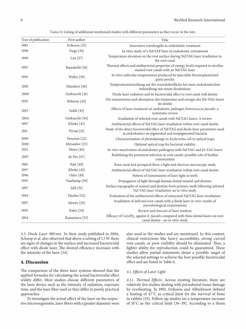
Table 6: Listing of additional mentioned studies with different parameters as they occur in the text.
Year of publication First author Title1983 Eriksson [35] Innovative wavelengths in endodontic treatment1998 Farge [36] In vitro study of a Nd:YAP laser in endodontic retreatment
1999 Lan [37] Temperature elevation on the root surface during Nd:YAG laser irradiation inthe root canal
1997 Ramskold [38] Thermal effects and antibacterial properties of energy levels required to sterilizestained root canals with an Nd:YAG laser
1995 Weller [39] In vitro radicular temperatures produced by injectable thermoplasticizedgutta-percha
2001 Mazaheri [40] Temperaturentwicklung auf der wurzeloberflache bei einer endodontischenbehandlung mit einem diodenlaser
2000 Gutknecht [41] Diode laser radiation and its bactericidal effect in root canal wall dentin
1993 Behrens [42] Die transmission und absorption der temperatur und energie des Nd-YAG-lasersim dentin
2013 Sadik [43] Effects of laser treatment on endodontic pathogen Enterococcus faecalis: asystematic review
2004 Gutknecht [44] Irradiation of infected root canals with Nd:YAG lasers. A review1997 Klinke [45] Antibacterial effects of Nd:YAG laser irradiation within root canal dentin
2011 Pirnat [21] Study of the direct bactericidal effect of Nd:YAG and diode laser parameters usedin endodontics on pigmented and nonpigmented bacteria
1999 Neuman [22] Characterization of photodamage to Escherichia coli in optical traps2008 Mirsaidov [23] Optimal optical trap for bacterial viability2012 Meire [46] In vitro inactivation of endodontic pathogens with Nd:YAG and Er:YAG lasers
2007 de Paz [47] Redefining the persistent infection in root canals: possible role of biofilmcommunities
1985 Nair [48] Root canal and periapical flora: a light and electron microscopy study1997 Klinke [45] Antibacterial effects of Nd:YAG laser irradiation within root canal dentin1996 Odor [49] Pattern of transmission of laser light in teeth1995 Vaarkamp [50] Propagation of light through human dental enamel and dentine
1997 Jalil [51] Surface topography of enamel and dentine from primary teeth following infraredNd-YAG laser irradiation: an in vitro study
1994 Hardee [52] Evaluation of the antibacterial effects of intracanal Nd:YAG laser irradiation
1997 Moritz [29] Irradiation of infected root canals with a diode laser in vivo: results ofmicrobiological examinations
1993 Kales [54] Review and forecast of laser markets
2014 Kanumuru [53] Efficacy of Ca(oH)2against E. faecalis compared with three dental lasers on root
canal dentin—an in vitro study
3.3. Diode Laser 980 nm. In their study published in 2006,Schoop et al. also observed that above a setting of 1.5W thereare signs of changes in the surface and increased bactericidaleffect with diode laser. The desired efficiency increases withthe intensity of the laser [34].
4. Discussion
The comparison of the three laser systems showed that theapplied formulas for calculating the actual bactericidal effectwidely differ. Most studies choose different parameters ofthe laser device such as the intensity of radiation, exposuretime, and the laser fiber used or they differ in purely practicalapproaches.
To investigate the actual effect of the laser on the respec-tivemicroorganisms, laser fibers with a greater diameter were
also used in the studies and are mentioned. In this context,clinical restrictions like heavy accessibility, strong curvedroot canals, or poor visibility should be eliminated. Thus, alighter ability for reproduction could be guaranteed. Thesestudies allow partial statements about a possible target ofthe selected settings to achieve the best possible bactericidaleffect and are listed in Table 6.
4.1. Effects of Laser Light
4.1.1. Thermal Effects. Across existing literature, there arerelatively few studies dealing with periodontal tissue damageby overheating. In 1983, Eriksson and Albrektsson defineda heating of 47∘C as critical limit for the survival of bonein rabbits [35]. Follow-up studies set a temperature increaseof 10∘C as the critical limit [36–39]. According to a thesis

BioMed Research International 7
by Mazaheri in 2001 at RWTH Aachen, the maximum aver-age temperature (10ms interval pause, 10ms pulse length)remains in the irradiation of root canals with the diode laserwith a setting of 3W still below the critical limit when theoptical fiber is performed permanently moving coronal andapical in a circularmotion in the root canal [40]. Gutknecht etal. observed a bacterial reduction in a depth of 500microns inthe teeth of cattle at a setting of 3Wcw [41].The temperaturelimit is exceeded at 4W and prolonged irradiation for 15seconds, resulting in thermal damage.
4.1.2. Power Settings. In this research, a value of 1.5W forthe diode and Nd:YAG laser has been set as an inclusioncriterion. With this setting, a thermal damage is excludedwithin recommended handling for both laser devices andthe bactericidal effects are acceptable [42]. A temperatureon the root surface was observed after 45 sec. of 37∘C at therecommended setting 15 pps and 1.5W and after 90 sec. of38∘C.
In a systematic review of the current literature aboutthe effectiveness of Nd:YAG laser on the pathogenic gram-positive bacteria E. faecalis, Sadik et al. showed that 1.5Wcould allow an effective bacteria reduction [43].
4.1.3. Effects of Laser Irradiated Root Surfaces. Gutknechtdescribed that an application of the laser below 1W is lessimportant in endodontics because neither is the smear layercompletely removed nor are the dentinal tubules sealed.With settings of 1.25W–1.5W significant changes on theroot canal surface were determined. The organic materialwas completely removed and the surface of the inorganicsubstance was merged, resulting in a partial or completeocclusion of dentinal tubules [44]. This fact is to be valuedpositively because a reinfection is less possible with closecanals.
In 2008, Klinke et al. discussed the angle between theoptical fiber of the laser and the dentinal wall [45]. The laserbeam hits the wall primarily at a very acute angle, dependingon the mobility of the fiber in the canal, the root canalcurvature, and the exit windowof the laser beam from the endof the fiber. In their study, the angle between the glass fiberand dentin surface was defined as 5∘. The lesser eliminationof bacteria compared to other studies could result fromthis aspect. Further studies in terms of this angle would beinteresting.The actual surface of the dentin also plays a role interms of bactericidal effect. Darker areas cause carbonizationand require a higher absorption of laser energy. The result isa local temperature increase with a bactericidal effect, albeitwithin no transmission of laser energy into deeper layers ofdentin.
Beer et al. investigated irradiating the opening cavity ofa tooth before irradiating the root canal itself, resulting ina significant higher bactericidal effect [33]. Further studieswould be interesting to explore this issue in greater depth.
4.1.4. Effects on Microorganisms. Pirnat et al. examined thedirect effect of Nd:YAG (1064 nm) and diode laser (810 nm)on P. gingivalis, E. coli, and E. faecalis in 2011.They postulated
two possible theories for the bactericidal effect of NIR laserlight: the first refers to heating by absorption of the substratein which the bacterium is located and the second refersto the direct absorption through the bacterium itself. Intheir attempt, external factors such as surrounding tissue orblood should not have an influence on the results. For thisreason, they irradiated a sapphire substrate that is opticallytransparent for the NIR spectrum and concluded that bothlaser systems have a minor direct bactericidal effect onnonpigmented bacteria such as E. coli and E. faecalis [21].However, such substrates significantly differ from the in vivosituation; for example, there is no oxygen in the bacterialmicroenvironment. This is necessary for the bacteria photo-damage effect, although the mechanism of this degradationwas not further understood [22, 23]. Future studies in thisdirection would be useful.
The gram-positive bacterium E. faecalis is more resistantin this study according to its cell wall structure comparedwith the gram-negative bacterium E. coli. The Nd:YAG lasercould reduce 57% of the pigmented bacterium P. gingivalisand 37% could be ascertained for the diode laser. The mostdetermining factor is believed to be the presence of the blackpigment protoporphyrin IX in P. gingivalis, which absorbs theenergy of the NIR light. Likewise, no growth was ascertainedon the agar plates used. This fact shows that not onlythe bacterium itself but also its environment plays a keyrole for an effective endodontic laser therapy. Meire et al.irradiated bacteria inoculated agar plates (Candida albicans,Enterococcus faecalis, and Propionibacterium acnes) in a studypublished in 2012. The Er:YAG laser was predominant in thisexperiment compared to Nd:YAG laser [46]. However, thepresent thickness of the Er:YAG laser fiber limits an efficienttransference of the light in the root canal.
The agar plates and the bacterial suspensions used in thisstudy absorbed the laser light to a small extent. Furthermore,nonpigmented bacteria were used, which could explain thelesser effect of the Nd:YAG laser in this experiment. Thedifferent absorption of wavelengths in dentin has an effect onthe depth of penetration.The Er:YAG laser had a lesser effecton the bacteria found in deeper dentinal tubules, whereas theNd:YAG laser was significantly superior.
Meire et al. supported the statement made by Pirnatet al. that the Nd:YAG laser kills the bacteria probably byheating their environment. A comparison of studies coveringthe antimicrobial effect of laser light is not easy to realizebecause the statements about energy density or experimentalconditions are often lacking. In a natural environment suchas root canal wall dentin bacteria occur in a biofilm [47,48], making them more responsive to laser light by highcell density and the presence of extracellular matrix. Thisfact could explain the poor action of the Nd:YAG laser onagar plates and bacterial suspensions. Different studies haveshown that the bactericidal effect in the tooth is strengthenedthrough enamel prisms and dentinal tubules as these act as alight guide [45, 49, 50]. However, additional in vivo studiesare needed.
Meire et al. suppose that blood or blood products ina natural environment could lead to a raised number ofporphyrins and melanin pigments in the bacteria in which
8 BioMed Research International
the bactericidal effect is improved by Nd:YAG laser. Anotherinteresting aspect is the dentin, which was examined moreclosely in a study in 1997 [51]. Carious dentin absorbs 1064 nmmore wavelength in comparison to healthy dentin, whichincreases the desired bactericidal effect.
Hardee et al. achieved a bacterial reduction of 99% ofthe test bacterium Bacillus stearothermophilus with Nd:YAGlaser, in conjunction with a log kill of 2 in comparison to a log6 population before irradiation. Usually this bacterium is notfound in infected root canals. It was selected due to its highheat resistance because the bactericidal effect ofNd:YAG laseris assumed by heat [52].
The Department of Restorative Dentistry, RWTHAachen, currently deals with the effect of ring-firing laserfibers in the root canals, which allows the laser light to notonly emit in vertical direction. New possibilities concerningthe bactericidal depth effect of diode lasers and Nd:YAGlasers could be achieved.
4.2. Nd:YAG versus Diode Laser. A direct comparison of theselected devices is currently not feasible in relation to exactsimilar demanded experimental setups.
In 1997, Moritz et al. described the diode laser (810 nm)and the Nd:YAG laser (1064 nm) in endodontic treatment asequally effective and they recommended further studies toevaluate the anaerobic bacteria [26].
In a study by Kanumuru and Subbaiah in 2014, theNd:YAG laser was most effective in the elimination of E.faecalis compared to 980 nm and followed 810 nm diode laser[53].
Due to the accumulation of different aggressive and resis-tant bacteria in an infected root canal, the additional use ofNd:YAG and diode lasers in combination with conventionalmethods such as mechanical conditioning or rinsing fluidsseems to hold a positive value, as can be demonstrated by thisliterature review.
4.3. Nd:YAG Laser
Advantages. The Nd:YAG laser has clear advantages in thedepth effect compared with 810 nm and 980 nm diode laser.Farmore studies about Nd:YAG can be found in the literaturecompared to both diode lasers in endodontics. It is effectiveagainst pigmented microorganisms.
Furthermore, it removes the smear layer in a root canal,which interferes with adequate disinfection using additionalrinsing fluids. It also has a simultaneous additional bacterici-dal effect.
Disadvantages. Drawbacks include the relatively high costand its size in comparison to the two diode lasers. They areeasy to handle due to their small size and the device can beused without power supply in battery mode, which Nd:YAGlaser is incapable of at present.
4.4. Diode Lasers 810 nm and 980 nm. Comparing the 810 nmwith the 980 nm diode laser, both are equally favorable. Bothare adequate funds in endodontic therapy and should beinvestigated in further detail. For 810 nm diode lasers, the
majority of studies can be found in the literature, althoughthe parameters are not exactly comparable.
According to a study of Kales in 1993, the diode laserdetermines 99% of the turnover on the whole market and isestimated at 25%by the buyers in comparison to all other laserdevices [54].
4.4.1. Variability of Reported Results. Sadik et al. postulatedthat the various investigated laser systems of the past 30 yearscould not be compared with a meta-analysis since the resultsof the studies were not presented in a standardized manner.From this perspective, it would be desirable if future studiesuse a solid study design with the same basic parameters, suchas the diameter of laser fiber, the same practical approach tothe irradiation of the teeth (number of repetitions, pauses),pulse frequency (pps), and power (W) [43].
This statement is the final testimony and prime causebecause this present literature review also does not lead toany clear result in terms of effectiveness brought against thebacteria in an infected root canal compared to the three lasers.There are too many different variable facts in the studies tomake a statement about the more effective wavelength orthe preferable device and the data situation is contradictory.The Nd:YAG laser is more frequently evaluated, although thecomparability of the different study designs is also lacking.The various studies are difficult to measure, given thatdifferent parameters, fiber strengths, or handlingmethods areused.
At present, a statement based upon recommended guide-lines is not really possible. When properly used, it emergesthat disinfection by laser can increase the endodontic successwith a very low risk of damaging side effects and withacceptable durability.
A recommended standardized procedure for the indi-vidual wavelengths is suggested, although further scientificstudies would be desirable. Additional in vivo studies withNd:YAG and diode lasers in endodontics are necessary. Itshould be considered internationally with the same proce-dure including a clear treatment outline. Generally estab-lished criteria such as the same fibers (diameter), the samesettings of the laser parameters (power, pulse frequency),the same trace of radiation in practical implementation, andduration are essential to conduct a comparison about theantibacterial effects of endodontic treatment between thethree laser devices. This would be desirable to define anevidence-based “gold standard.”
5. Conclusions
In endodontics, Nd:YAG laser (1064 nm) and diode laser(810 nm and 980 nm) devices are used to remove bacteria ininfected teeth.This literature overview aimed to compare andevaluate the advantages anddisadvantages in using these laserdevices with standardized settings.
The PubMed database was searched using precise key-words between April 2011 and April 2016. Likewise, printmedia from the Library of RWTH Aachen University wereexamined.

BioMed Research International 9
A total of 22 eligible studies were found regardingNd:YAG laser 1064 nm. Four studies fulfilled all demandedcriteria in this review for this laser device. Seven studiesreferring to the diode laser 980 nm were examined, althoughonly one fulfilled all criteria. Eleven studies were foundregarding the diode laser 810 nm, but also only one studycould fulfill all necessary criteria.
The analysis of the selected studies showed that allthree laser systems are able to successfully decimate bacteriathat are present in infected teeth. Pigmented bacteria areefficiently better removed by the Nd:YAG laser. Moreover,in deeper dentin layers, Nd:YAG laser showed better results.Concerning handiness, size, and purchase price, the diodelaser is preferable.
In summary, a direct comparison cannot be madebetween the selected laser devices due to different studydesigns, materials, and equipment. Prospective randomizedtrials are needed to further verify which laser system is tobe preferred for the best results in endodontic therapy andevaluate an evidence-based and international guideline.
Competing Interests
The authors declare that they have no conflict of interests.
References
[1] S. Kakehashi, H. R. Stanley, and R. J. Fitzgerald, “The effectsof surgical exposures of dental pulps in germ-free and con-ventional laboratory rats,” Oral Surgery, Oral Medicine, OralPathology, vol. 20, no. 3, pp. 340–349, 1965.
[2] G. Sundqvist, “Taxonomy, ecology, and pathogenicity of theroot canal flora,” Oral Surgery, Oral Medicine, Oral Pathology,vol. 78, no. 4, pp. 522–530, 1994.
[3] B. Tsatsas, A. Tzamouranis, and F. Mitsis, “A bacteriologicalexamination of root canals before filling,” Journal of the BritishEndodontic Society, vol. 7, no. 2, pp. 78–80, 1974.
[4] S. E. Gharbia and H. N. Shah, “Hydrolytic enzymes liberated byblack-pigmented gram-negative anaerobes,” FEMS Immunologyand Medical Microbiology, vol. 6, no. 2-3, pp. 139–145, 1993.
[5] P. N. R. Nair, U. Sjogren, G. Krey, K.-E. Kahnberg, and G.Sundqvist, “Intraradicular bacteria and fungi in root-filled,asymptomatic human teeth with therapy-resistant periapicallesions: a long-term light and electron microscopic follow-upstudy,” Journal of Endodontics, vol. 16, no. 12, pp. 580–588, 1990.
[6] F. D. Ostrander, M. C. Crowley, and J. Dowson, “A clinical studyof the treatment of root canal and periapical infections withpenicillin,” Journal of Dental Research, vol. 26, no. 6, pp. 403–407, 1947.
[7] R.W. Hession, “Long-term evaluation of endodontic treatment:anatomy, instrumentation, obturation—the endodontic prac-tice triad,” International Endodontic Journal, vol. 14, no. 3, pp.179–184, 1981.
[8] M. Shih, F. J.Marshall, and S. Rosen, “The bactericidal efficiencyof sodiumhypochlorite as an endodontic irrigant,”Oral Surgery,Oral Medicine, Oral Pathology, vol. 29, no. 4, pp. 613–619, 1970.
[9] N. Gutknecht, “Lasereinsatz in der Endodontie—Vorausset-zungen fur denTherapieerfolg,” in Laserzahnmedizin Jahrbuch,vol. 11, pp. 57–61, Oemus, Aachen, Germany, 2011.
[10] M. Hulsmann and E. Schafer, “Good clinical practice,” in DieWurzelkanalbehandlung. Stellungnahme des Endodontie-Beiratsder DGZ, DGZMK-Stellungnahme, 2007.
[11] A. Vahdaty, T. R. Pitt Ford, and R. F. Wilson, “Efficacyof chlorhexidine in disinfecting dentinal tubules in vitro,”Endodontics & Dental Traumatology, vol. 9, no. 6, pp. 243–248,1993.
[12] D. R. Drake, A. H. Wiemann, E. M. Rivera, and R. E. Walton,“Bacterial retention in canal walls in vitro: effect of smear layer,”Journal of Endodontics, vol. 20, no. 2, pp. 78–82, 1994.
[13] D. Clark-Holke, D. Drake, R.Walton, E. Rivera, and J. M. Guth-miller, “Bacterial penetration through canals of endodonticallytreated teeth in the presence or absence of the smear layer,”Journal of Dentistry, vol. 31, no. 4, pp. 275–281, 2003.
[14] R. Michiels, T. E. M. Vergauwen, A.Mavridou, M.Meire, M. DeBruyne, and R. J. G. De Moor, “Investigation of coronal leakageof root fillings after smear-layer removal with EDTAorNd:YAGlasing through capillary-flow porometry,” Photomedicine andLaser Surgery, vol. 28, supplement 2, pp. S43–S50, 2010.
[15] C. Yesilsoy, E.Whitaker, D. Cleveland, E. Phillips, andM. Trope,“Antimicrobial and toxic effects of established andpotential rootcanal irrigants,” Journal of Endodontics, vol. 21, no. 10, pp. 513–515, 1995.
[16] D. A. Spratt, J. Pratten, M. Wilson, and K. Gulabivala, “Anin vitro evaluation of the antimicrobial efficacy of irrigantson biofilms of root canal isolates,” International EndodonticJournal, vol. 34, no. 4, pp. 300–307, 2001.
[17] M. Samiei, S. M. V. Pakdel, S. Rikhtegaran, S. Shakoei, D.Ebrahimpour, and P. Taghavi, “Scanning electron microscopycomparison of the cleaning efficacy of a root canal systemby Nd:YAG laser and rotary instruments,” Microscopy andMicroanalysis, vol. 20, no. 4, pp. 1240–1245, 2014.
[18] J. R. F. Archilla, M. S. N. A. Moreira, S. P. H. Miyagi, A. C.Bombana, N. Gutknecht, and M. M. Marques, “Single sessionof Nd:YAG laser intracanal irradiation neutralizes endotoxin indental root dentin,” Journal of Biomedical Optics, vol. 17, no. 11,Article ID 118002, 2012.
[19] J. A. Weichman and F. M. Johnson, “Laser use in endodontics.A preliminary investigation,” Oral Surgery, Oral Medicine, OralPathology, vol. 31, no. 3, pp. 416–420, 1971.
[20] N. Gutknecht, Lasertherapie in der Zahnarztlichen Praxis,Quintessenz, Berlin, Germany, 1999.
[21] S. Pirnat, M. Lukac, and A. Ihan, “Study of the direct bacte-ricidal effect of Nd:YAG and diode laser parameters used inendodontics on pigmented and nonpigmented bacteria,” Lasersin Medical Science, vol. 26, no. 6, pp. 755–761, 2011.
[22] K. C. Neuman, E. H. Chadd, G. F. Liou, K. Bergman, and S. M.Block, “Characterization of photodamage to Escherichia coli inoptical traps,” Biophysical Journal, vol. 77, no. 5, pp. 2856–2863,1999.
[23] U. Mirsaidov, W. Timp, K. Timp, M. Mir, P. Matsudaira, andG. Timp, “Optimal optical trap for bacterial viability,” PhysicalReview E—Statistical, Nonlinear, and SoftMatter Physics, vol. 78,no. 2, Article ID 021910, 2008.
[24] R. Franzen, N. Gutknecht, S. Falken, N. Heussen, and J. Meister,“Bactericidal effect of a Nd:YAG laser on Enterococcus faecalisat pulse durations of 15 and 25ms in dentine depths of 500 and1,000 𝜇m,” Lasers in Medical Science, vol. 26, no. 1, pp. 95–101,2011.
[25] N. Gutknecht, R. Franzen, M. Schippers, and F. Lampert,“Bactericidal effect of a 980-nm diode laser in the root canal

10 BioMed Research International
wall dentin of bovine teeth,” Journal of Clinical Laser Medicineand Surgery, vol. 22, no. 1, pp. 9–13, 2004.
[26] A. Moritz, O. Doertbudak, N. Gutknecht, K. Goharkhay, U.Schoop, and W. Sperr, “Nd:YAG laser irradiation of infectedroot canals in combination withmicrobiological examinations,”Journal of the American Dental Association, vol. 128, no. 11, pp.1525–1530, 1997.
[27] U. Schoop,W. Kluger, A. Moritz, N. Nedjelik, A. Georgopoulos,and W. Sperr, “Bactericidal effect of different laser systems inthe deep layers of dentin,” Lasers in Surgery and Medicine, vol.35, no. 2, pp. 111–116, 2004.
[28] Y. Yasuda, T. Kawamorita, H. Yamaguchi, and T. Saito, “Bacte-ricidal effect of Nd:YAG and Er:YAG lasers in experimentallyinfected curved root canals,” Photomedicine and Laser Surgery,vol. 28, supplement 2, pp. S75–S78, 2010.
[29] A. Moritz, N. Gutknecht, U. Schoop, K. Goharkhay, O. Doert-budak, and W. Sperr, “Irradiation of infected root canals witha diode laser in vivo: results of microbiological examinations,”Lasers in Surgery and Medicine, vol. 21, no. 3, pp. 221–226, 1997.
[30] A. Moritz, U. Schoop, K. Goharkhay et al., “The bactericidaleffect of Nd:YAG, Ho:YAG, and Er:YAG laser irradiation in theroot canal: an in vitro comparison,” Journal of Clinical LaserMedicine and Surgery, vol. 17, no. 4, pp. 161–164, 1999.
[31] N. Gutknecht, F. Kaiser, A. Hassan, and F. Lampert, “Long-termclinical evaluation of endodontically treated teeth by Nd:YAGlasers,” Journal of Clinical Laser Medicine and Surgery, vol. 14,no. 1, pp. 7–11, 1996.
[32] N.Gutknecht, A.Moritz, G. Conrads, T. Sievert, and F. Lampert,“Bactericidal effect of the Nd:YAG laser in in vitro root canals,”Journal of Clinical Laser Medicine and Surgery, vol. 14, no. 2, pp.77–80, 1996.
[33] F. Beer, A. Buchmair, J. Wernisch, A. Georgopoulos, and A.Moritz, “Comparison of twodiode lasers on bactericidity in rootcanals-an in vitro study,” Lasers in Medical Science, vol. 27, no.2, pp. 361–364, 2012.
[34] U. Schoop, W. Kluger, S. Dervisbegovic et al., “Innovativewavelengths in endodontic treatment,” Lasers in Surgery andMedicine, vol. 38, no. 6, pp. 624–630, 2006.
[35] A. R. Eriksson and T. Albrektsson, “Temperature thresholdlevels for heat-induced bone tissue injury: a vital-microscopicstudy in the rabbit,” The Journal of Prosthetic Dentistry, vol. 50,no. 1, pp. 101–107, 1983.
[36] P. Farge, P.Nahas, andP. Bonin, “In vitro study of aNd:YAP laserin endodontic retreatment,” Journal of Endodontics, vol. 24, no.5, pp. 359–363, 1998.
[37] W.-H. Lan, “Temperature elevation on the root surface duringNd:YAG laser irradiation in the root canal,” Journal of Endodon-tics, vol. 25, no. 3, pp. 155–156, 1999.
[38] L. O. Ramskold, C. D. Fong, and T. Stromberg, “Thermaleffects and antibacterial properties of energy levels required tosterilize stained root canals with an Nd:YAG laser,” Journal ofEndodontics, vol. 23, no. 2, pp. 96–100, 1997.
[39] R. N. Weller and K. A. Koch, “In vitro radicular temperaturesproduced by injectable thermoplasticized gutta-percha,” Inter-national Endodontic Journal, vol. 28, no. 2, pp. 86–90, 1995.
[40] P. Mazaheri, Temperaturentwicklung auf der Wurzeloberflachebei einer endodonitischen Behandlung mit einem Diodenlaser,Mainz, 2001.
[41] N. Gutknecht, D. Van Gogswaardt, G. Conrads, C. Apel,C. Schubert, and F. Lampert, “Diode laser radiation and itsbactericidal effect in root canal wall dentin,” Journal of ClinicalLaser Medicine and Surgery, vol. 18, no. 2, pp. 57–60, 2000.
[42] V. G. Behrens, N. Gutknecht, and R. Renziehausen, “Dietransmission und absorption der temperatur und energie desNd-YAG-lasers im dentin,” ZWR, vol. 102, no. 9, pp. 629–634,1993.
[43] B. Sadik, S. Arikan, N. Belduz, Y. Yasa, D. Karasoy, and M.Cehreli, “Effects of laser treatment on endodontic pathogenEnterococcus faecalis: a systematic review,” Photomedicine andLaser Surgery, vol. 31, no. 5, pp. 192–200, 2013.
[44] N. Gutknecht, “Irradiation of infected root canals with Nd:YAGlasers. A review,” LaserZahnheilkunde, vol. 4, no. 4, pp. 219–226,2004.
[45] T. Klinke, W. Klimm, and N. Gutknecht, “Antibacterial effectsof Nd:YAG laser irradiation within root canal dentin,” Journalof Clinical Laser Medicine and Surgery, vol. 15, no. 1, pp. 29–31,1997.
[46] M. A. Meire, T. Coenye, H. J. Nelis, and R. J. G. De Moor, “Invitro inactivation of endodontic pathogens with Nd:YAG andEr:YAG lasers,” Lasers inMedical Science, vol. 27, no. 4, pp. 695–701, 2012.
[47] L. C. de Paz, “Redefining the persistent infection in root canals:possible role of biofilm communities,” Journal of Endodontics,vol. 33, no. 6, pp. 652–662, 2007.
[48] P. N. Nair and H. U. Luder, “Root canal and periapical flora: alight and electron microscopy study,” Schweizerische Monatss-chrift fur Zahnmedizin, vol. 95, no. 10, pp. 992–1003, 1985.
[49] T. M. Odor, T. F. Watson, T. R. Pitt Ford, and F. Mcdonald,“Pattern of transmission of laser light in teeth,” InternationalEndodontic Journal, vol. 29, no. 4, pp. 228–234, 1996.
[50] J. Vaarkamp, J. J. ten Bosch, and E. H. Verdonschot, “Propaga-tion of light through human dental enamel and dentine,” CariesResearch, vol. 29, no. 1, pp. 8–13, 1995.
[51] L. A. Jalil, R. Labella, and G. J. Pearson, “Surface topographyof enamel and dentine from primary teeth following infraredNd-YAG laser irradiation: an in vitro study,” Lasers in MedicalScience, vol. 12, no. 1, pp. 61–67, 1997.
[52] M. W. Hardee, L. J. Miserendino, W. Kos, and H. Walia,“Evaluation of the antibacterial effects of intracanal Nd:YAGlaser irradiation,” Journal of Endodontics, vol. 20, no. 8, pp. 377–380, 1994.
[53] N. R. Kanumuru and R. Subbaiah, “Bacterial efficacy ofCa(oH)
2against E. faecalis compared with three dental lasers
on root canal dentin—an in vitro study,” Journal of Clinical andDiagnostic Research, vol. 8, no. 11, pp. ZC135–ZC137, 2014.
[54] D. Kales, “Review and forecast of laser markets,” Laser FocusWorld, vol. 29, pp. 72–73, 1993.

Review ArticleMicroRNAs as Salivary Markers for Periodontal Diseases:A New Diagnostic Approach?
Gerhard Schmalz,1 Simin Li,1 Ralph Burkhardt,2 Sven Rinke,3 Felix Krause,1
Rainer Haak,1 and Dirk Ziebolz1
1Department of Cariology, Endodontology and Periodontology, University of Leipzig, 04109 Leipzig, Germany2Institute of Laboratory Medicine, Clinical Chemistry, and Molecular Diagnostics, University of Leipzig, 04109 Leipzig, Germany3Department of Prosthodontics, University Medical Center Goettingen, Goettingen, Germany
Correspondence should be addressed to Dirk Ziebolz; [email protected]
Received 19 January 2016; Accepted 20 March 2016
Academic Editor: Kenji Yoshida
Copyright © 2016 Gerhard Schmalz et al. This is an open access article distributed under the Creative Commons AttributionLicense, which permits unrestricted use, distribution, and reproduction in any medium, provided the original work is properlycited.
The aim of this review is to discuss current findings regarding the roles of miRNAs in periodontal diseases and the potential use ofsaliva as a diagnosticmedium for correspondingmiRNA investigations. For periodontal disease, investigations have been restrictedto tissue samples and five miRNAs, that is, miR-142-3p, miR-146a, miR-155, miR-203, and miR-223, were repeatedly validated invivo and in vitro by different validation methods. Particularly noticeable are the small sample sizes, different internal controls, anddifferent case definitions of periodontitis in in vivo studies. Beside of that, the validated miRNAs are associated with inflammationand therefore with various diseases. Furthermore, several studies successfully explored the use of salivary miRNA species for thediagnosis of oral cancer. Different cancer types were investigated and heterogeneous methodology was used; moreover, no overlapof results was found. In conclusion, fivemiRNAs have consistently been reported for periodontitis; however, their disease specificity,detectability, and expression in saliva and their importance as noninvasivemarkers are questionable. In principle, a salivarymiRNAdiagnosticmethod seems feasible.However, standardized criteria and protocols for preanalytics,measurements, and analysis shouldbe established to obtain comparable results across different studies.
1. Introduction
MicroRNAs (miRNAs) are endogenous ∼22 nt RNAs thatplay important regulatory roles in animals and plants bytargeting mRNAs for cleavage or translational repression [1].Many multifarious investigations regarding miRNAs havedemonstrated their essential roles in physiological and patho-logical processes in the immune response [2]. Additionally,miRNAs likely contribute to development and progression ofsystemic diseases, such as cancer [3] and vascular disease [4].Consequently, there is considerable interest in use ofmiRNAsas not only diagnostic markers but also potential therapeutictargets for various diseases [5]. In particular, the roles ofmiRNAs in oral cancer and precancer have been investigated.MiRNA expression appears to differ between healthy tissueand squamous cell carcinoma tissues of the oral cavity, which
is discussed in an existing review [6]. Furthermore, someinvestigations have linked precancerous lesions and their riskof becoming malignant to changes in miRNA expression [7].
Furthermore,miRNAs are associatedwith bacterial infec-tions [8] and, thus, are most likely associated with infectiousdiseases of oral cavity, for example, dental caries, endodonticinfections, and periodontitis. Kim et al. (2015), however,already give an overview of current findings regarding dentaldisease, including periodontal disease and oral cancer. Kimet al. (2015) also mentioned limitations of current diagnosticand potential benefit of salivarymiRNAdiagnostic [9].This isthe reason to discuss the roles of miRNAs in periodontal dis-eases. As of now, only a few in vitro and in vivo investigationsregarding these roles are available. A recent review articledemonstrated potential roles of miRNAs during periodontalinflammation, showing the considerable role of them in
Hindawi Publishing CorporationBioMed Research InternationalVolume 2016, Article ID 1027525, 14 pageshttp://dx.doi.org/10.1155/2016/1027525

2 BioMed Research International
periodontitis [10]. Thereby, different roles and mechanismsare shown and a very comprehensive overview on molecularpathways is given; however, methodology was only discussedbetween selected studies.
As mentioned above, an interesting approach is theanalysis of miRNA species in saliva. High quality miRNAswere shown to be detectable in saliva [11], and the possibilityof their usage in oral cancer detection appears to have ahigh potential for future diagnostics [12]. This noninvasiveapproach could be relevant for further diseases of the oralcavity. Nevertheless, the potential usage of miRNA as salivarymarkers for periodontal diseases was already not discussedyet. As shown for oral cancer, using miRNA for noninvasivediagnostics seems feasible [12] and could therefore serve asa salivary marker for periodontitis. The potential is high,so maybe recent indispensable invasive diagnostics or X-raycould become avoided by salivary miRNA diagnostic in thefuture and an early diagnosis might help to prevent bone and,in conclusion, tooth loss.
Nevertheless, despite of the huge potential,many differentresults are available, both for salivary oral cancer diagnosticsand for analysis of periodontal tissue. Accordingly, it isquestionable if the reasons are methodological differencesbetween the studies or maybe if the usage of miRNAs ingeneral is unsuitable.
Therefore, this review article shows current results of invitro and in vivo studies regarding periodontal diseases aswell as results of selected current studies dealing with salivarymiRNAs for oral cancer detection. The aim was to detectpromising future perspectives, but also current challengesregarding this issue. In particular, methodological aspectsare focused to detect potential limitations of miRNAs asnoninvasive markers for periodontitis.
2. Methods
2.1. Search Strategy. Our literature research was performedusing the online database PubMed (http://www.ncbi.nlm.nih.gov/pubmed). To obtain a broad overview of the availabletheme-specific publications, we used different keywords forour search. We used “miRNA AND periodontitis” as key-words to obtain results for periodontal diseases. We used“miRNA AND saliva AND oral disease” and “miRNA ANDsaliva AND cancer” as keywords to obtain relevant articlesfor noninvasive possibilities, that is, the use of saliva toinvestigate miRNAs in diseases of the oral cavity. Then, thereference lists of relevant articles were searched for furtherresults. To interpret the results, relevant publications weresearched and analyzed critically. Neither a meta-analysis norother statistical comparisons of existing data were performed.
2.2. Selection Criteria. Only current full-length articles inEnglish language were included. The results were verifiedfor their relevance. The following selection criteria weredefined: miRNA investigations for in vitro and in vivo studiesexamining periodontal disease. Additionally, in vivo humaninvestigations regarding salivary miRNA analysis in oralcancer and precancer were included.
2.3. Selection Process. Following the literature search, all ref-erences were limited to relevant publications. In vivo peri-odontal disease investigations that did not address peri-odontal tissue were excluded. Salivary diagnostic studies thatdid not explicitly concentrate on miRNAs were excluded,and only investigations for oral cancer and precancer wereincluded.
3. Results
3.1. Periodontal Diseases
3.1.1. In Vitro. Eight in vitro studies, which detected differentmiRNAs, were included for periodontal disease (Table 1).Nahid et al. (2011) examined the expression of cytokinesassociated with inflammation and differences in expressionof cytokines and miRNA during infection with live and heat-killed bacteria in THP-1 monocytes. They demonstrated thatexpression of miR-146a is associated with infections causedby periodontal pathogenic bacteria in vitro [13]. Anotherstudy indicated that miR-146a is significantly overexpressedin THP-1 cells after stimulation with LPS from P.g. [14].Furthermore, miR-146a was shown to be a negative regulatorof TLR-NF𝜅B pathway in human periodontal ligament cellsafter P. gingivalis LPS stimulation, therefore being involvedin inflammatory response [15]. Moreover, expression of miR-146a and miR-146b-5p in human gingival fibroblasts afterstimulation with P.g. LPS was also investigated in a study byXie et al. (2013). They demonstrated that expression of thesemiRNAs significantly increased upon LPS stimulation andconcluded that miR-146 could work as a negative regulatorof inflammation in periodontal disease [16]. Differentialexpression of miR-146a and miR-155 in dental pulp and ingingival and periodontal fibroblasts after stimulation withLPS from Escherichia coli was also reported. The increasedexpression of miR-146a was the highest in gingival fibroblastsand decreased expression of miR-155 was only significantin gingival fibroblasts [17]. Naqvi et al. (2014) showed dif-ferent expressions of miR-146a. In this in vitro study THP-1 macrophages that were stimulated with LPS from Aggre-gatibacter actinomycetemcomitans, P.g., and also P.g. grownon cigarette smoke extract were examined. The differentLPS caused both some identical and also varied expressionsof miRNAs in human macrophages. Moreover, LPS fromP.g. grown on cigarette smoke extract compared to LPSfrom unaffected P.g. caused differential miRNA expression,particularly that of miR-29b in human macrophages [18].
Results reported in other two studies weremore heteroge-nous: Ouhara et al. (2014) demonstrated upregulated miR-584 in human gingival epithelial cells after stimulation withP.g. [19]. Furthermore, Moffat and Lamont (2011) focusedon miR-203 expression in gingival epithelial cells stimulatedwith P.g. In this study, 14 miRNAs displayed significant chan-ges in expression after exposure to P.g.; of these miRNAs,miR-203 was examined in detail and showed higher expres-sion in tissues after P.g. stimulation [20].
3.1.2. In Vivo Animal. Nahid et al. (2011) investigated expres-sion of miR-146a, miR-132, and miR-155 in spleens and

BioMed Research International 3
Table1:Com
paris
onof
metho
dsfro
mcurrentinvestig
ations
regardingmiRNAs
inperio
dontaldisease.
Author
andyear
Objecto
finvestigation
RNAextractio
nmetho
dDetectio
nmetho
dfor
miRNAprofi
leVa
lidationof
specific
miRNAs
Num
bero
fstudy
participants
Internal
control
Statisticalanalysis
Investigated
and/or
deregu
lated
miRNAs∗
Invitro
human
Moff
attand
Lamon
t2011[20]
Gingiva
epith
elial
cells
TRIzol(In
vitro
gen),
miRNeasy
kit
(Qiagen)
Microarray(LC
Sciences)
qRT-PC
RTaqM
an(App
liedBiosystems)
—RN
U-48
ANOVA
,𝑡-te
stmiR-203
Nahid
etal.2011[13]
Hum
anTH
P-1
mon
ocytes
mirV
anam
iRNA
isolatio
nkit
(Ambion
)—
TaqM
anmicroRN
Aassay(App
lied
Biosystems)
—RN
U44
One-w
ayanalysis
ofvaria
nce
follo
wed
bythe
two-sid
ed,
unpaire
dStud
ent’s𝑡-te
st
miR-146
amiR-155
miR-132
Hon
daetal.2012[14
]TH
P-1cellsand
THP-1-d
erived
macroph
ages
TRIzolreagent
(Invitro
gen)
Agilent
human
miRNAmicroarrays
(Release
12.0)
(Agilent
techno
logies)
TaqM
anmicroRN
AAs
say(App
lied
Biosystems)
—RN
U44
Unp
aired𝑡-te
st,ANOVA
-Williams
test
miR-146
amiR-155
Xiee
tal.2013
[16]
Hum
anging
ival
fibroblasts
TRIzolreagent
(Invitro
gen)
miRNAmicroarray
analyses
(Kangchen
Bio-Tech)
qRT-PC
Ranalysis
(SYB
RGreen
qPCR
MasterM
ixPA
-112,
SABiosciences,Qiagen)
—U6sm
all
nucle
arRN
AStud
ent’s𝑡-te
stmiR-146
amiR-146
b-5p
Sipertetal.2014[17]
Hum
anfib
roblasts
from
dentalpu
lps,
ging
ivae,and
perio
dontal
ligam
ents
TRIzol(In
vitro
gen)
NCod
emiRNArapid
labelin
gsyste
m(C
at.
#MIRLS
RPD-20)
(Invitro
gen)
Ncode
Multi-Speciesm
iRNA
microarraykitV
2,(In
vitro
gen)
Taqm
anmiRNAassays,
(App
liedBiosystems)
—U6B
Two-way
ANOVA
follo
wed
byBo
nferroni
post
hoctest
miR-146
amiR-155
Naqvietal.2014[18]
Hum
anTH
P-1-d
ifferentia
ted
macroph
ages
miRNeasy
kit
(Qiagen)
NanoStringnC
ounter
miRNAassay
(NanoString
Techno
logies)
Quantitativ
ereal-tim
ePC
REv
aGreen
Master
Mix(Biotiu
m)
—RN
U6B
Stud
ent’s𝑡-te
st(tw
o-tailed)
miR-29b
miR-32
miR-146
amiR-891
Ouh
arae
tal.2014
[19]
Simianvirus4
0antig
enim
mortalized
ging
ivalepith
elial
celllin
e,OBA
-9
mirV
anam
iRNA
IsolationKit
(App
liedBiosystem)
miRCU
RYLN
AmicroRN
AArray,
v.16.0(Exiqo
)
TaqM
anMicroRN
AAssaysS
ystem
(App
lied
Biosystems)
—RN
U48
Stud
ent’s𝑡-te
st,Tu
keyho
nestly
significant
difference
miR-584
Jiang
etal.2015[15]
Hum
anperio
dontal
ligam
entcells
TRIzolreagent
(Invitro
gen)
—
Quantitativ
eRT-PC
Ranalysis(RTSY
BR®
Green
qPCR
Master
Mixes
PA-112,
SABiosciences,Qiagen)
—U6sm
all
nucle
arRN
A
ANOVA
and
Stud
ent-
New
man-K
euls
test
miR-146
a
4 BioMed Research International
Table1:Con
tinued.
Author
andyear
Objecto
finvestigation
RNAextractio
nmetho
dDetectio
nmetho
dfor
miRNAprofi
leVa
lidationof
specific
miRNAs
Num
bero
fstudy
participants
Internal
control
Statisticalanalysis
Investigated
and/or
deregu
lated
miRNAs∗
Invivo
anim
al
Nahid
etal.2011[13]
Maxillae
andspleens
from
ApoE−/−
mice
mirV
anam
iRNA
isolatio
nkit
(Ambion
)—
TaqM
anmicroRN
Aassay(App
lied
Biosystems)
—snoR
NU202
One-w
ayanalysis
ofvaria
nce
follo
wed
bythe
two-sid
ed,
unpaire
dStud
ent’s𝑡-te
st
miR-146
amiR-155
miR-132
Invivo
human
Leee
tal.2011[21]
Gingivaltissue
mirV
ana™
miRNA
Isolationkit
(Ambion
)
RT2
miRNAPC
Rarraysyste
m(SABiosciences)
TaqM
anmiRNAassays
(App
liedBiosystems)
n.i.
RNU44
Stud
ent’s𝑡-te
st
miR-181b
miR-19b
miR-23a
miR-30a
miR-le
t7a
miR-301a
Xiee
tal.2011[22]
Gingivaltissue
TRIzolreagent
(Invitro
gen)
miRNAmicroarray
(KangchenBio-Tech)
Quantitativ
eRT-PC
Ranalysis(RTSY
BRGreen
qPCR
Master
Mixes
PA-112,
SABiosciences,Qiagen)
10perio
dontitis
patie
nts
10healthy
subjects
U6sm
all
nucle
arRN
A
Unp
aired
Stud
ent’s𝑡-te
st
miR-126
miR-20a
miR-142-3p
miR-19a
let-7
fmiR-203
miR-17
miR-223
miR-146
bmiR-146
amiR-155
miR-205
Stoecklin
-Wasmer
etal.
2012
[24]
ging
ivaltissue
TRIzol(In
vitro
gen)
RNeasy
(Qiagen,)∗
Microarray∗∗
qRT-PC
R
198ging
ivaltissue
samples,158
diseased
and40
healthysamples
from
86patie
nts
with
perio
dontitis
n.i.
R(th
eRDevelo
pment
CoreT
eam,2005)
andbiocon
ductor
statistic
alfram
eworks
miR-451
miR-223
miR-486-5p
miR-1246
miR-1260
miR-141

BioMed Research International 5
Table1:Con
tinued.
Author
andyear
Objecto
finvestigation
RNAextractio
nmetho
dDetectio
nmetho
dfor
miRNAprofi
leVa
lidationof
specific
miRNAs
Num
bero
fstudy
participants
Internal
control
Statisticalanalysis
Investigated
and/or
deregu
lated
miRNAs∗
Perrietal.2012
[26]
Gingivaltiss
ueTissueLyserL
Tand
miRNeasy
MiniK
it(Q
iagen)
Quantitativ
emicroRN
APC
Rarray
miRNAPC
RArray
(SABiosciences)
—
10no
nobese
patie
ntsa
nd10
obesep
atients
each
grou
pwith
5perio
dontally
healthysites
and
5chronic
perio
dontitissites
SNORD
48and
RNU6-2
ANOVA
and
nonp
aired
Stud
ent’s𝑡-te
st
miR-142
-3p
miR-15a
miR-30e
miR-30d
miR-22
miR-130a
miR-106b
miR-103
miR-185
miR-210
miR-18a
Ogataetal.2014[23]
Gingivaltissue
miRNeasy
MiniK
it(Q
iagen)
Hum
anmiRNA
microarray8×15Kkit
(Agilent
Techno
logies)
Real-timeP
CRSY
BRAd
vantageq
PCR
Prem
ix(C
lontec)
3chronic
perio
dontitis
patie
ntsa
nd3
edentulous
resid
ualridges
U6
One-w
ayANOVA
miR-150
miR-200b
miR-223
miR-144
miR-379
miR-222
Kaleae
tal.2015
[27]
Gingivaltissue
mirV
anam
iRNA
IsolationKit
(Ambion
)
Affy
metrix
GeneC
hip
miRNA3.0arrays
(Affy
metrix
)
TaqM
anMicroRN
Aexpressio
nassays
(App
liedBiosystems)
36eligible
individu
als
U6sno
RNA
andRN
U6B
Partek
6.6and
1-way
analysisof
varia
nce,
R-sta
tistic
alenvironm
ent
miR-4721
miR-557
miR-196a
miR-323a-3p
miR-200b-5p
miR-188-5p
Motedayyenetal.2015
[25]
Gingivaltissue
mirV
anam
iRNA
isolatio
nkit
(Ambion
)—
Real-timeP
CR(TaqMan
Universal
MasterM
ixII,no
UNG,and
hsa-miRNA146a
kits,
AppliedBiosystems)
10healthy
controls,
20chronic
perio
dontitis
patie
nts
n.i.
Stud
ent’s𝑡-te
stor
Mann-Whitney𝑈
test;
Pearsontest
orSpearm
antest
miR-146
a
RNAextractio
nandiso
latio
nmetho
dsarelisted.Th
eproofofqu
ality
andqu
antitya
ndqu
ality
assurancea
reshow
nasim
portantqualityc
riteriatodraw
conclusio
nsregardingthe
reprod
ucibilityandsta
ndardizatio
nof
theinvestig
ations.Furthermore,metho
dsform
iRNAprofi
lingandmiRNAvalid
ationarelisted.Inadditio
n,then
umbero
fstudy
participantsisshow
nto
draw
conclusio
nsregardingthev
alidity
ofther
esults.
n.i.:no
inform
ation,∗
onlymiRNAsw
hich
werev
alidated
byRT
-PCR
werec
onsid
ered,and∗∗
Stoecklin
-Wasmer
etal.refer
toan
earlier
repo
rtby
Dem
mer
etal.2008[62].M
iRNAsw
hich
werer
eportedin
more
than
ones
tudy
areh
ighlighted
inbo
ldtype.

6 BioMed Research International
Table 2: Criteria for patient selection.
Author and year PD CAL BOP Radiographic bone lossHealthy Periodontitis Healthy Periodontitis Healthy Periodontitis Healthy Periodontitis
Stoecklin-Wasmer et al. 2012 [24] ≤4mm >4mm ≤2mm ≥3mm negative positive n.i. n.i.Xie et al. 2011 [22] <3mm ≥5mm <1mm ≥3mm n.i. ∗ No YesLee et al. 2011 [21] ≤3mm >5mm none >3mm ∗∗ n.i. No YesPerri et al. 2012 [26] ≤4mm >5mm n.i. n.i. Negative Positive No YesOgata et al. 2014 [23] n.i. ≥6mm n.i. >6mm n.i. Positive n.i. n.i.Motedayyen et al. 2015 [25] <3mm n.i. <3mm n.i. n.i. n.i. No n.i.Kalea et al. 2015 [27] n.i. >5mm n.i. ∗ ∗ ∗ n.i. n.i. n.i. ∗ ∗ ∗
PD: pocket depth; CAL: clinical attachment loss; BOP: bleeding on probing; n.i.: no information. ∗GI > 1; ∗∗BOP in whole gingiva < 10%, ∗∗∗bone loss > 30%.
maxillae of ApoE−/− mice after infection with periodontalpathogenic bacteria P.g., Treponema denticola and Tannerellaforsythia. MiR-146a primarily showed a significant increasein both maxillae and spleens, while miR-132 and miR-155showed only minor changes [13].
3.1.3. In Vivo Human. Seven in vivo studies investigatingdifferential expression of miRNAs in human tissue sampleswere included (Table 1). In particular, Lee et al. (2011) com-pared the expression of inflammatorymiRNAs in healthy andinflamed periodontal tissue. Microarray analysis indicatedthat expression of six miRNAs was upregulated more thaneightfold and that expression of 22 miRNAs increased morethan fourfold in periodontal disease tissue compared withhealthy tissue. Of these miRNAs, six could be validated byqRT-PCR [21]. A similar investigation considered miRNAprofiles of healthy and inflamed gingival tissues. Microarrayanalysis of healthy and inflamed gingival tissues indicatedthat expression of five miRNAs was upregulated more thanfivefold, 85 miRNAs, twofold to fivefold. Moreover, expres-sion of 34 miRNAs was downregulated twofold to fivefold ininflamed tissue. 12 miRNAs were validated by qRT-PCR [22].A study of Japanese patients identified 17 upregulated and22 downregulated miRNAs; six were selected for validation[23]. A further investigation also detected differentialmiRNAexpression by microarray analysis, wherein 91 upregulatedand 68 downregulated miRNAs were found. Six miRNAswere validated, and potential target genes were investigated[24]. Furthermore, Motedayyen et al. (2015) showed a posi-tive relationship betweenmiR-146a and clinical parameters inpatients with chronic periodontitis [25].Thus,manymiRNAswith differential expression as detected by microarray analy-sis have not been validated by qRT-PCR; these miRNAs willnot be discussed (Table 1).
Additionally, two studies investigated the roles of miR-NAs in periodontal disease in association with systemicfactors and disease; thus, differential expression of miRNAsbetween obese periodontitis patients and nonobese perio-dontitis patients was examined. However, different, nonover-lappingmiRNAs were reported in each investigation [26, 27].
Notably, criteria for patient selection and for classificationof healthy and diseased patients were similar across allstudies. However, differences exist between investigationsin defining periodontal health and periodontitis (Table 2).
Additionally the number of study participants for in vivoinvestigations varied enormously between studies with tenpatients or less for each group in most cases (Table 1). Like-wise, methodological approach was similar across studies:microarray based technology was often used to screen andidentify miRNAs with differential expression, and qRT-PCRwas used for subsequent validation of top-hits. However,detailed methods were different; thus, for example, differentmiRNA isolation methods were used. Also differences in sta-tistical analysis are conspicuous, especially internal controlsthat distinguish between the studies (Table 1).
3.2. MiRNA Detection in Saliva as a Noninvasive DiagnosticTool. Nine investigations were included that illustrate thepotential of saliva as noninvasive diagnostic tool for miRNAanalyzes. We focused on salivary miRNA species analysis as adiagnostic method for oral cancer and precancer detection,because no results of appropriate dental and periodontaldisease investigations could be found. Six of included studiesexamined oral squamous cell carcinoma, one precancerouslesions, one esophageal cancer, and one tumor of parotidgland. Park et al. (2009) showed presence of miRNA speciesin whole saliva and in saliva supernatants and describeddifferential expression of miR-125a and miR-200a betweencases and controls. Besides that stability of exogenous (miR-124a) and endogenous (miR-191) miRNA in saliva wasalso examined in this study, and endogenous miRNA wasshown to have higher stability than exogenous miRNA[28]. Another investigation found that miR-9, miR-191, andmiR-134 appeared to be potential markers for noninvasivediagnosis of oral squamous cell carcinoma using saliva.Additionally, this study describes a reliable isolation methodfor miRNAs from even small volumes of saliva [29]. In astudy by Hung et al. (2016) salivary miRNAs, specificallymiR-31 and miR-21 were investigated in oral premalignantdisorders, particularly showing miR-31 to be a sufficientmarker for high risk disorders and malignant transformation[30]. Also Liu et al. (2012) examined miR-31, and its expres-sion in saliva was compared with its expression in plasma;thereby, a correlation could be shown. These authors alsoobserved a decrease in expression of miR-31 after surgicalresection of primary tumor [31]. Similarly, Duz et al. (2016)reduced miR-139-5p expression in patients with tonguecarcinoma and normalization after surgical treatment [32].

BioMed Research International 7
In a study investigating expression of salivary miRNAs inpremalignant lesions of oral cavity, a correlation was foundbetween deregulated miRNA expression in tissue and saliva,although miRNA concentrations in saliva were lower [33].A further investigation compared expression of miRNA insaliva between patients with oral squamous cell carcinoma,patients with oral squamous cell carcinoma in remission,patients with oral lichen planus, and a healthy control group.In addition to differences in miRNA expression between thegroups, miR-27b was significantly overexpressed in salivaof patients with oral squamous cell carcinoma. Further-more, completely different profiles of miRNAs could befound in cancerous tissues compared to biological fluids[34]. Additionally, another study found miRNA-184 to havethe potential to distinguish between OSCC and potentiallymalignant disorders [35].
In addition, altered miRNA expression in saliva was alsoverified for esophageal cancer [36] and parotid gland tumors[37]. Their methodologies are shown in Table 3. In prin-ciple, methods of measurement (microarray, qRT-PCR) aresimilar between the studies. Remarkable differences couldbe found regarding form of saliva, extraction methods,statistical analysis, and internal controls. The number ofstudy participants was already higher than for periodontitis.However, heterogeneous groups sizes with partly seven oreight patients each group [33, 34] and partly 50 patients andmore each group [28, 29] were investigated (Table 3).
4. Discussion
Several studies investigating putative role of miRNAs inperiodontal diseases have been performed; however, spec-trum of miRNAs identified was substantially heterogeneous.Both in vitro and in vivo investigations were performed forperiodontal disease, and five miRNAs, that is, miR-142-3p,miR-146a, miR-155, miR-203, and miR-223, were validatedin more than one study. Accordingly, we can conclude thatthese miRNAs may play important role and thus couldbecome potential markers for periodontal disease. However,majority of identified miRNA species differed significantlyacross studies (Table 1).
MiR-146a and miR-155 were dominant in in vitro studies.Otherwise, these miRNAs have also been identified in otherinflammatory diseases and various cancers. MiR-146a seemsto play a role in multifarious diseases and its function inbacterial infections has already been discussed [38]. MiR-155has also been mentioned [39]. LPS may have a substantialeffect on the expression of miR-146a and miR-155 [40].Consequently, their altered expression during periodontaldisease is not surprising because bacterial virulence factorssuch as LPS are relevant in pathogenesis of periodontitis [41].These miRNAs are associated with many other inflammatorydiseases, which might be indicative of a limited specificity.Additionally, miR-142-3p is associated with inflammationand LPS exposure [42, 43]. MiR-203 may also play an impor-tant role in inflammation and correlate with LPS exposure[44]. Interestingly, miR-203 expression is associated withimmune reaction of skin [45, 46]. MiR-203 expression in
keratinocytes is important; therefore, this miRNA may alsobe involved in gingivitis and periodontitis as determined byits effect on gingival keratinocytes. Finally,miR-223 also playsan important role in inflammation and LPS exposure [47].Accordingly, correlations between these five miRNAs andperiodontal disease are possible. Interestingly, these miRNAsappear to play a role in oral cancer. Thus, their roles inperiodontal disease should be further examined (Table 4)as it might indicate a link between periodontitis and headand neck squamous cell carcinoma (HNSCC). Figure 1 showsa summary of the interactions between periodontitis andHNSCC [48], and some potentially involvedmiRNAs. In thiscontext, miR-29b, which showed an in vitro correlation withLPS from P.g. grown on cigarette smoke extract [18], has alsobeen examined for oral cancer [49]. These findings raise thequestion if these miRNAs could serve as diagnostic markersfor periodontal diseases or simply reflect an increased inflam-matory state associated with various diseases.
Another problem is the great diversity of results. Al-though many miRNAs were found, convergence betweenthe investigations, particularly in vivo, is small, resultingin conflicting conclusions. Reasons for differences betweeninvestigations of periodontal disease may differ. On the onehand, small variety in patient selection and different clinicalprocedures potentially causes these differences (Table 2). Forfuture studies an evident graduation, for example, Page andEke 2007, should be used for standardization [50]. On theother hand, variations in methods used for miRNA detectioncould play a crucial role (Table 3). A comparison of methodsused suggests that miRNA detection procedures were quitesimilar. However, many differences in detailed proceduresare evident. Using microarray and quantitative RT-PCR foranalyzing and validating miRNAs are essential componentsof most investigations. However, detailed procedures formethods primarily differ, so several arrays from differentmanufacturing companies were performed. Importantly, dif-ferences in all of chosen criteria can be found between theinvestigations. Besides variations in statistical analyses it isimportant that internal controls differed in most cases.
Another aspect that could be crucial for the varyingresults is the small number of study participants. One studyeven investigated only three healthy and three diseasedindividuals [23]. A large group of 86 patients was selectedonly once [24]. The use of small sample sizes increasesvulnerability to a range of errors and biases and may be amajor reason for heterogeneous study results and potentialfalse-positive findings. Similar issues were faced in earlyhuman genetic association studies, where many of reportedgenotype-phenotype associations could not be replicated insubsequent studies [51, 52]. Today, replication of initial find-ings in second cohort is considered essential for establishingthe credibility of a genotype-phenotype association [53] andsimilar approaches could be adopted for studies reportingassociations between miRNAs and diseases. Clearly, complexreason for differences in results exists. Different methods,clinical criteria, small groups of patients, and small specificityof miRNAs for periodontal disease could be causal complex.
In summary, comparability of results for the roles ofmiRNAs in periodontal disease is questionable. Potential

8 BioMed Research International
Table3:Com
paris
onof
metho
dsforsalivarymiRNAdiagno
sis.
Author
and
year
Num
bero
fstudy
participants
Form
ofsaliva
miRNAextractio
nmetho
dDetectio
nof
miRNA
profi
leVa
lidation
Internal
control
Statisticalanalysis
Potentialm
arker
forc
arcino
ma
Park
etal.200
9[28]
50oralsquamou
scellcarcinom
a(O
SCC)
patie
nts
and50
healthy
matched
control
subjects
Unstim
ulated
who
lesalivaa
ndsaliva
supernatant
mirV
ana™
miRNA
IsolationKit
(Ambion
)DNA-
free™
(Ambion
)
RT-pream
p-qP
CR(App
liedBiosystems)
(i)RT
-pream
p-qP
CR(App
liedBiosystems)
(ii)F
our-plex
RT-pream
p-qP
CRfor
miR-142-3p,miR-200a,
miR-125a,andmiR-93.
U6snRN
AMann-Whitney𝑈test
miR-125a
miR-200a
Liuetal.2012
[31]
45patie
ntsw
ithOSC
Cand10
patie
ntwith
oral
verrucou
sleuk
oplakia
24healthy
individu
als
Unstim
ulated
saliva
supernatant
mirV
anaP
ARIS
Isolationkit
(Ambion
)DNased
igestio
n
TaqM
anmiRNA
assaysyste
m(App
lied
Biosystems)
TaqM
anmiRNAassay
syste
m(App
lied
Biosystems)
miR-16
Mann-Whitney
test,
Wilcoxon
matched
pairs
test,
andlin
ear
regressio
nanalysis,
receiver-operatin
gcharacteris
ticsR
OC
analysis
miR-31
Yang
etal.2013
[33]
8progressingLG
Dleuk
oplakias
7no
nprogressin
gLG
Dleuk
oplakias
7healthy
volunteers
Unstim
ulated
saliva
RNeasy
Micro
Kit
(Qiagen)
TaqM
an®low
density
array(TLD
A)
qRT-PC
Rsyste
m(App
liedBiosystems)
were
used
forg
lobal
miR
expressio
nanalysisin
tissue
samples
TaqM
anMicroRN
AAs
say(App
lied
Biosystems)
RNU6
Rand
omvaria
nce
𝑡-te
st,Be
njam
ini-H
ochb
erg
false
discoveryrate
(FDR)
metho
d,Mann-Whitney𝑈
test,
Stud
ent’s𝑡-te
sts(tw
o-sid
ed)
miR-10b,m
iR-145,
miR-99b,m
iR-708,
miR-181c,miR-30e,
miR-660,m
iR-197
Xiee
tal.2013
[36]
39patie
ntsw
ithesop
hagealcancer
and19
healthy
controls
Stim
ulated
who
lesalivaa
ndsupernatant
mirV
anaP
ARIS
Kit(Ambion
)
7who
lesalivas
amples
forE
Cgrou
pand3
forh
ealth
ygrou
pfor
Agilent
microarray
11.0(A
gilent
Techno
logies)
RT-qPC
RSY
BRPrem
ixEx
Taq(TaK
aRa)
miR-16
Mann-Whitney𝑈test
ortheK
ruskall-W
allis
𝐻test,𝜒
2
test,
receiver-operatin
gcharacteris
ticsR
OC
curves,Spearman’s
correlationtest
miR-10b,m
iR-144
,miR-451
(inwho
lesaliva)
miR-10b∗
,miR-144
,miR-21,
miR-451
(insaliva
supernatant)
Matse
etal.
2013
[37]
38patie
ntsw
ithmalignant
and29
with
benign
parotid
gland
tumors
Who
lesaliva
andsupernatant
mirV
anaP
ariskit
(Ambion
)DNaseI
(Qiagen)
TaqM
anHum
anMicroRN
ACa
rds
(App
liedBiosystems)
TaqM
anMicroRN
Aassays
(App
lied
Biosystems)
TaqM
anmicroRN
Aassays
(App
lied
Biosystems)
U6snRN
AWilcoxon
rank
-sum
test,
ROCcurves
hsa-miR-374,
hsa-miR-222,
hsa-miR-15b,
hsa-let-7
g,hsa-miR-132,
mmu-miR-140
-5p

BioMed Research International 9
Table3:Con
tinued.
Author
and
year
Num
bero
fstudy
participants
Form
ofsaliva
miRNAextractio
nmetho
dDetectio
nof
miRNA
profi
leVa
lidation
Internal
control
Statisticalanalysis
Potentialm
arker
forc
arcino
ma
Mom
en-H
eravi
etal.2014[34]
9OSC
Cpatie
nts
before
treatment,8
patie
ntsw
ithOSC
Cin
remission,8
patie
ntsw
ithOLP,
and9healthy
controls
Unstim
ulated
who
lesaliva
RNeasy
kit
(Qiagen)
multip
lexed
NanoStringnC
ounter
miRNAexpressio
nassay(N
anoString
Techno
logies)
TaqM
anMicroRN
Aassay(App
lied
Biosystems)
miRNA-
191
1-way
analysisof
varia
nce,follo
wed
bya2
-tailed
Mann-Whitney𝑈test
orStud
ent’s𝑡-te
st,RO
Ccurves
analysis
miR-27b
Salazare
tal.
2014
[29]
5HNSC
Cpatie
nts,
5healthycontrols
fore
xpression
analysis
56HNSC
Cpatie
nts,56
healthy
controlsfor
valid
ation
Unstim
ulated
who
lesaliva
QIAzollysis
reagent(Qiagen)
NucleoSpin
miRNAkit
(Macherey-Nagel)
miScript™miRNA
PCRarrays
(Qiagen)
RT-qPC
RmiScript
SYBR
greenPC
Rmaster
mix(Q
iagen)
SNORD
96A
MannWith
ney
𝑈-te
st,RO
Ccurves,
Wilcoxon
rank
sum
test,
Bonferroni
metho
d
miR-9,m
iR-134,
miR-191
Zahran
etal.
2015
[35]
20healthyc
ontro
ls,40
potentially
malignant
disorders,20
OSC
C,20
recurrentaph
thou
ssto
matitis
Unstim
ulated
saliva
supernatant
miRNeasy
serum/plasm
aextractio
nkit
(Qiagen)
—RT
-qPC
RSY
BRgreen
PCRkit(Qiagen)
SNORD
68
One-w
ayANOVA
,𝐹-te
st,Dun
nett𝑡-te
st,Scheffe’smultip
lecomparis
on,
two-tailedtests
miR-21,miR-184,
miR-145
Duz
etal.2016
[32]
50salivas
amples
from
25TS
CCpatie
nts(on
cepriortoandon
ceaft
ersurgical
treatment)
Unstim
ulated
saliva
supernatant
mirV
anaP
ARIS
kit(Ambion
)
8samples
(4TS
CCpatie
nts4
healthy
control)usingAgilent
8×60Khu
man
V19
miRNAmicroarrays
TaqM
anMicroRN
Aassay(App
lied
Biosystems)
RNU6b
Two-sid
edStud
ent’s
𝑡-te
st,Re
ceiver
operating
characteris
tic(ROC)
curves,95%
confi
denceinterval
(CI)
miR-139-5p
Hun
getal.
2016
[30]
20patie
ntsw
ithoralpo
tentially
malignant
disorders,24
healthyindividu
als
Unstim
ulated
saliva
supernatant
mirV
anaP
ARIS
isolatio
nkit
(Ambion
)—
TaqM
anmicroRN
Aassay(App
lied
Biosystems)
miR-16
Unp
airedtest,
ROC
curves,K
aplan-Meier
metho
d,log-rank
test,
Cox
prop
ortio
nal
hazard
mod
el
miR-21
miR-31
RNAextractio
nandiso
latio
nmetho
dsarelisted.Th
eproofofqu
ality
andqu
antitya
ndqu
ality
assurancea
reshow
nasim
portantqualityc
riteriatodraw
conclusio
nsregardingthe
reprod
ucibilityandsta
ndardizatio
noftheinvestig
ations.Furthermore,metho
dsform
iRNAprofi
linga
ndmiRNAvalid
ationarelisted.Inadditio
n,then
umbero
fstudy
participantsisprovided
todraw
conclusio
nsregardingthe
valid
ityoftheresults.
Theform
ofsalivaisa
lsolisted.

10 BioMed Research International
Table 4: Comparison of miRNas frequently mentioned in connection with periodontal disease with results from selected oral cancer andprecancer investigations.
Kozaki et al.2008 [63]
Park et al.2009 [28]
Cervigne et al.2009 [64]
Scapoli et al.2010 [65]
Lajer et al.2011 [66]
Lundegard et al.2015 [56]
miR-146a X X X XmiR-155 X X XmiR-203 X XmiR-142-3p X XmiR-223 X XEach miRNA that was validated in the context of periodontal disease was also mentioned in investigations for cancer and precancer of the oral cavity. Thistable provides a small exemplary overview. Consequently, even more studies with similar results are not shown here.
Chronicperiodontitis
Keratinocytes
Bacteria (i) Endotoxin (LPS)
(ii) Enzymes (proteases, collagenases, fibrinolysin,phospholipase A)
(iii) Metabolic by-products(H2S, NH3, fatty acids)
HNSCC
miR-203 miR-203
miR-142-3p
miR-146a
miR-155
miR-223
miR-142-3p
miR-146a
miR-155
miR-223
miR-29b miR-29b
Inflammation(i) Cytokines,
(ii) Growth factors(iii) Prostaglandins(iv) Enzymes (iNOS,
COX-2, MMP)(v) Reactive oxygen
species (hydrogen peroxide, oxy
(vi) Reactive nitrogen species (nitric oxides)
(vii) Reactive lipids and metabolites (malondialdehyde, 4-hydroxy-2-nonenal)
radicals)
chemokines
Mucosal injury
(i) Susceptibility to HPV and other viral infections
(ii) Penetration of othercarcinogens (tobacco,alcohol, diet . . . )
Figure 1: Model for the relationship among chronic periodontitis, HNSCC (modif. Han et al. 2014 [48]) and potential miRNAs that may beinvolved in the corresponding processes.
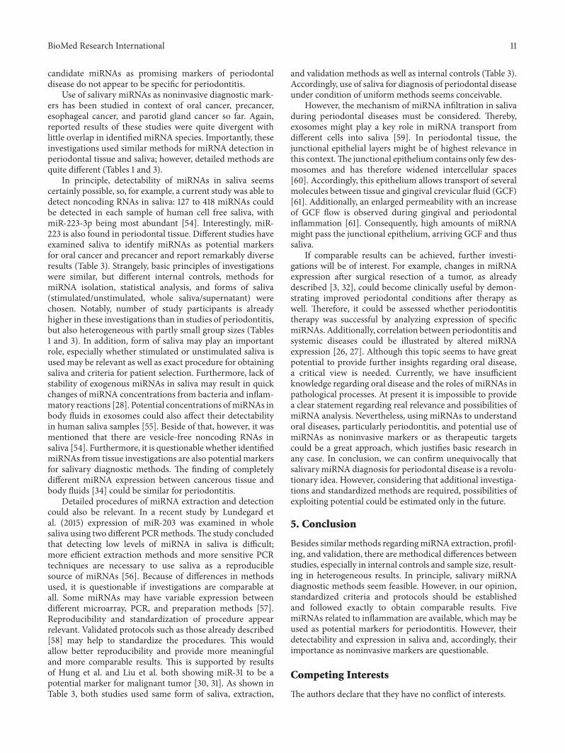
BioMed Research International 11
candidate miRNAs as promising markers of periodontaldisease do not appear to be specific for periodontitis.
Use of salivary miRNAs as noninvasive diagnostic mark-ers has been studied in context of oral cancer, precancer,esophageal cancer, and parotid gland cancer so far. Again,reported results of these studies were quite divergent withlittle overlap in identified miRNA species. Importantly, theseinvestigations used similar methods for miRNA detection inperiodontal tissue and saliva; however, detailed methods arequite different (Tables 1 and 3).
In principle, detectability of miRNAs in saliva seemscertainly possible, so, for example, a current study was able todetect noncoding RNAs in saliva: 127 to 418 miRNAs couldbe detected in each sample of human cell free saliva, withmiR-223-3p being most abundant [54]. Interestingly, miR-223 is also found in periodontal tissue. Different studies haveexamined saliva to identify miRNAs as potential markersfor oral cancer and precancer and report remarkably diverseresults (Table 3). Strangely, basic principles of investigationswere similar, but different internal controls, methods formiRNA isolation, statistical analysis, and forms of saliva(stimulated/unstimulated, whole saliva/supernatant) werechosen. Notably, number of study participants is alreadyhigher in these investigations than in studies of periodontitis,but also heterogeneous with partly small group sizes (Tables1 and 3). In addition, form of saliva may play an importantrole, especially whether stimulated or unstimulated saliva isused may be relevant as well as exact procedure for obtainingsaliva and criteria for patient selection. Furthermore, lack ofstability of exogenous miRNAs in saliva may result in quickchanges of miRNA concentrations from bacteria and inflam-matory reactions [28]. Potential concentrations ofmiRNAs inbody fluids in exosomes could also affect their detectabilityin human saliva samples [55]. Beside of that, however, it wasmentioned that there are vesicle-free noncoding RNAs insaliva [54]. Furthermore, it is questionable whether identifiedmiRNAs from tissue investigations are also potential markersfor salivary diagnostic methods. The finding of completelydifferent miRNA expression between cancerous tissue andbody fluids [34] could be similar for periodontitis.
Detailed procedures of miRNA extraction and detectioncould also be relevant. In a recent study by Lundegard etal. (2015) expression of miR-203 was examined in wholesaliva using two different PCRmethods.The study concludedthat detecting low levels of miRNA in saliva is difficult;more efficient extraction methods and more sensitive PCRtechniques are necessary to use saliva as a reproduciblesource of miRNAs [56]. Because of differences in methodsused, it is questionable if investigations are comparable atall. Some miRNAs may have variable expression betweendifferent microarray, PCR, and preparation methods [57].Reproducibility and standardization of procedure appearrelevant. Validated protocols such as those already described[58] may help to standardize the procedures. This wouldallow better reproducibility and provide more meaningfuland more comparable results. This is supported by resultsof Hung et al. and Liu et al. both showing miR-31 to be apotential marker for malignant tumor [30, 31]. As shown inTable 3, both studies used same form of saliva, extraction,
and validation methods as well as internal controls (Table 3).Accordingly, use of saliva for diagnosis of periodontal diseaseunder condition of uniform methods seems conceivable.
However, the mechanism of miRNA infiltration in salivaduring periodontal diseases must be considered. Thereby,exosomes might play a key role in miRNA transport fromdifferent cells into saliva [59]. In periodontal tissue, thejunctional epithelial layers might be of highest relevance inthis context.The junctional epithelium contains only few des-mosomes and has therefore widened intercellular spaces[60]. Accordingly, this epithelium allows transport of severalmolecules between tissue and gingival crevicular fluid (GCF)[61]. Additionally, an enlarged permeability with an increaseof GCF flow is observed during gingival and periodontalinflammation [61]. Consequently, high amounts of miRNAmight pass the junctional epithelium, arriving GCF and thussaliva.
If comparable results can be achieved, further investi-gations will be of interest. For example, changes in miRNAexpression after surgical resection of a tumor, as alreadydescribed [3, 32], could become clinically useful by demon-strating improved periodontal conditions after therapy aswell. Therefore, it could be assessed whether periodontitistherapy was successful by analyzing expression of specificmiRNAs. Additionally, correlation between periodontitis andsystemic diseases could be illustrated by altered miRNAexpression [26, 27]. Although this topic seems to have greatpotential to provide further insights regarding oral disease,a critical view is needed. Currently, we have insufficientknowledge regarding oral disease and the roles of miRNAs inpathological processes. At present it is impossible to providea clear statement regarding real relevance and possibilities ofmiRNA analysis. Nevertheless, using miRNAs to understandoral diseases, particularly periodontitis, and potential use ofmiRNAs as noninvasive markers or as therapeutic targetscould be a great approach, which justifies basic research inany case. In conclusion, we can confirm unequivocally thatsalivary miRNA diagnosis for periodontal disease is a revolu-tionary idea. However, considering that additional investiga-tions and standardized methods are required, possibilities ofexploiting potential could be estimated only in the future.
5. Conclusion
Besides similarmethods regardingmiRNA extraction, profil-ing, and validation, there are methodical differences betweenstudies, especially in internal controls and sample size, result-ing in heterogeneous results. In principle, salivary miRNAdiagnostic methods seem feasible. However, in our opinion,standardized criteria and protocols should be establishedand followed exactly to obtain comparable results. FivemiRNAs related to inflammation are available, which may beused as potential markers for periodontitis. However, theirdetectability and expression in saliva and, accordingly, theirimportance as noninvasive markers are questionable.
Competing Interests
The authors declare that they have no conflict of interests.

12 BioMed Research International
Acknowledgments
Theauthors acknowledge support from theGermanResearchFoundation (DFG) and Universitat Leipzig within the pro-gram of Open Access Publishing.
References
[1] D. P. Bartel, “MicroRNAs: genomics, biogenesis, mechanism,and function,” Cell, vol. 116, no. 2, pp. 281–297, 2004.
[2] R. M. O’Connell, D. S. Rao, A. A. Chaudhuri, and D. Baltimore,“Physiological and pathological roles for microRNAs in theimmune system,”Nature Reviews Immunology, vol. 10, no. 2, pp.111–122, 2010.
[3] J. Hao, S. Zhao, Y. Zhang et al., “Emerging role ofMicroRNAs incancer and cancer stem cells,” Journal of Cellular Biochemistry,vol. 115, no. 4, pp. 605–610, 2014.
[4] S. Qin and C. Zhang, “MicroRNAs in vascular disease,” Journalof Cardiovascular Pharmacology, vol. 57, no. 1, pp. 8–12, 2011.
[5] R. Maqbool and M. U. Hussain, “MicroRNAs and humandiseases: diagnostic and therapeutic potential,” Cell and TissueResearch, vol. 358, no. 1, pp. 1–15, 2014.
[6] D. Chen, R. J. Cabay, Y. Jin et al., “MicroRNA deregulations inhead and neck squamous cell carcinomas,” Journal of Oral &Maxillofacial Research, vol. 4, no. 1, article e2, 2013.
[7] R. Roy,N.De Sarkar, S. Ghose et al., “Association between risk oforal precancer and genetic variations in microRNA and relatedprocessing genes,” Journal of Biomedical Science, vol. 21, article48, 2014.
[8] C. Staedel and F. Darfeuille, “MicroRNAs and bacterial infec-tion,” Cellular Microbiology, vol. 15, no. 9, pp. 1496–1507, 2013.
[9] S. H. Kim, S. Y. Lee, Y. M. Lee, and Y. K. Lee, “MicroRNAs asbiomarkers for dental diseases,” Singapore Dental Journal, vol.36, pp. 18–22, 2015.
[10] M. Kebschull and P. N. Papapanou, “Mini but mighty: microR-NAs in the pathobiology of periodontal disease,” Periodontology2000, vol. 69, no. 1, pp. 201–220, 2015.
[11] R. S. Patel, A. Jakymiw, B. Yao et al., “High resolution of mic-roRNA signatures in human whole saliva,” Archives of OralBiology, vol. 56, no. 12, pp. 1506–1513, 2011.
[12] X. Lin,H.-C. Lo,D. T.W.Wong, andX. Xiao, “NoncodingRNAsin human saliva as potential disease biomarkers,” Frontiers inGenetics, vol. 6, article 175, 2015.
[13] M.A.Nahid,M.Rivera, A. Lucas, E. K. L. Chan, andL.Kesavalu,“Polymicrobial infection with periodontal pathogens specifi-cally enhances microRNA miR-146a in ApoE-/- mice duringexperimental periodontal disease,” Infection and Immunity, vol.79, no. 4, pp. 1597–1605, 2011.
[14] T. Honda, N. Takahashi, S. Miyauchi, and K. Yamazaki, “Por-phyromonas gingivalis lipopolysaccharide induces miR-146awithout altering the production of inflammatory cytokines,”Biochemical andBiophysical ResearchCommunications, vol. 420,no. 4, pp. 918–925, 2012.
[15] S.-Y. Jiang, D. Xue, Y.-F. Xie et al., “The negative feedbackregulation of microRNA-146a in human periodontal ligamentcells after Porphyromonas gingivalis lipopolysaccharide stimu-lation,” Inflammation Research, vol. 64, no. 6, pp. 441–451, 2015.
[16] Y. F. Xie, R. Shu, S. Y. Jiang, D. L. Liu, J. Ni, and X. L. Zhang,“MicroRNA-146 inhibits pro-inflammatory cytokine secretion
through IL-1 receptor-associated kinase 1 in human gingivalfibroblasts,” Journal of Inflammation, vol. 10, article 20, 2013.
[17] C. R. Sipert, A. C. Morandini, T. J. Dionısio, A. J. Trachtenberg,W. P. Kuo, and C. F. Santos, “MicroRNA-146a and microRNA-155 show tissue-dependent expression in dental pulp, gingivaland periodontal ligament fibroblasts in vitro,” Journal of OralScience, vol. 56, no. 2, pp. 157–164, 2014.
[18] A. R. Naqvi, J. B. Fordham, A. Khan, and S. Nares, “MicroRNAsresponsive to Aggregatibacter actinomycetemcomitans andPorphyromonas gingivalis LPS modulate expression of genesregulating innate immunity in human macrophages,” InnateImmunity, vol. 20, no. 5, pp. 540–551, 2014.
[19] K. Ouhara, I. J. Savitri, T. Fujita et al., “MiR-584 expressed inhuman gingival epithelial cells is induced by porphyromonasgingivalis stimulation and regulates interleukin-8 productionvia lactoferrin receptor,” Journal of Periodontology, vol. 85, no.6, pp. e198–e204, 2014.
[20] C. E. Moffatt and R. J. Lamont, “Porphyromonas gingivalisinduction of microRNA-203 expression controls suppressor ofcytokine signaling 3 in gingival epithelial cells,” Infection andImmunity, vol. 79, no. 7, pp. 2632–2637, 2011.
[21] Y. H. Lee, H. S. Na, S. Y. Jeong, S. H. Jeong, H. R. Park, andJ. Chung, “Comparison of inflammatory microRNA expressionin healthy and periodontitis tissues,” Biocell, vol. 35, no. 2, pp.43–49, 2011.
[22] Y. F. Xie, R. Shu, S. Y. Jiang, D. L. Liu, and X. L. Zhang, “Com-parison of microRNA profiles of human periodontal diseasedand healthy gingival tissues,” International Journal of OralScience, vol. 3, no. 3, pp. 125–134, 2011.
[23] Y.Ogata, S.Matsui, A. Kato, L. Zhou, Y.Nakayama, andH. Taka,“MicroRNA expression in inflamed and noninflamed gingivaltissues from Japanese patients,” Journal of Oral Science, vol. 56,no. 4, pp. 253–260, 2014.
[24] C. Stoecklin-Wasmer, P.Guarnieri, R. Celenti, R. T.Demmer,M.Kebschull, and P. N. Papapanou, “MicroRNAs and their targetgenes in gingival tissues,” Journal of Dental Research, vol. 91, no.10, pp. 934–940, 2012.
[25] H.Motedayyen, S. Ghotloo, M. Saffari, M. Sattari, and R. Amid,“Evaluation ofMicroRNA-146a and its targets in gingival tissuesof patients with chronic periodontitis,” Journal of Periodontol-ogy, vol. 86, no. 12, pp. 1380–1385, 2015.
[26] R. Perri, S. Nares, S. Zhang, S. P. Barros, and S. Offenbacher,“MicroRNA modulation in obesity and periodontitis,” Journalof Dental Research, vol. 91, no. 1, pp. 33–38, 2012.
[27] A. Z. Kalea, R. Hoteit, J. Suvan et al., “Upregulation of gingivaltissue miR-200b in obese periodontitis subjects,” Journal ofDental Research, vol. 94, pp. 59–69, 2015.
[28] N. J. Park, H. Zhou, D. Elashoff et al., “Salivary microRNA:discovery, characterization, and clinical utility for oral cancerdetection,” Clinical Cancer Research, vol. 15, no. 17, pp. 5473–5477, 2009.
[29] C. Salazar, R. Nagadia, P. Pandit et al., “A novel saliva-basedmicroRNA biomarker panel to detect head and neck cancers,”Cellular Oncology, vol. 37, no. 5, pp. 331–338, 2014.
[30] K.-F. Hung, C.-J. Liu, P.-C. Chiu et al., “MicroRNA-31 upregu-lation predicts increased risk of progression of oral potentiallymalignant disorder,” Oral Oncology, vol. 53, pp. 42–47, 2016.
[31] C.-J. Liu, S.-C. Lin, C.-C. Yang, H.-W. Cheng, and K.-W. Chang,“Exploiting salivary miR-31 as a clinical biomarker of oralsquamous cell carcinoma,” Head and Neck, vol. 34, no. 2, pp.219–224, 2012.

BioMed Research International 13
[32] M. B. Duz, O. F. Karatas, E. Guzel et al., “Identification ofmiR-139-5p as a saliva biomarker for tongue squamous cellcarcinoma: A Pilot Study,” Cellular Oncology, vol. 39, no. 2, pp.187–193, 2016.
[33] Y. Yang, Y. X. Li, X. Yang, L. Jiang, Z. J. Zhou, and Y. Q. Zhu,“Progress risk assessment of oral premalignant lesions withsaliva miRNA analysis,” BMC Cancer, vol. 13, article 129, 2013.
[34] F. Momen-Heravi, A. J. Trachtenberg, W. P. Kuo, and Y. S.Cheng, “Genomewide study of salivary MicroRNAs for detec-tion of oral cancer,” Journal of Dental Research, vol. 93, supple-ment 7, pp. 86S–93S, 2014.
[35] F. Zahran, D. Ghalwash, O. Shaker, K. Al-Johani, and C. Scully,“Salivary microRNAs in oral cancer,” Oral Diseases, vol. 21, no.6, pp. 739–747, 2015.
[36] Z. Xie, G. Chen, X. Zhang et al., “Salivary microRNAs aspromising biomarkers for detection of esophageal cancer,” PLoSONE, vol. 8, no. 4, Article ID e57502, 2013.
[37] J. H. Matse, J. Yoshizawa, X. Wang et al., “Discovery and prev-alidation of salivary extracellular microRNA biomarkers panelfor the noninvasive detection of benign and malignant parotidgland tumors,”Clinical Cancer Research, vol. 19, no. 11, pp. 3032–3038, 2013.
[38] Z. Liu, B. Xiao, B. Tang et al., “Up-regulated microRNA-146anegatively modulate Helicobacter pylori-induced inflammatoryresponse in human gastric epithelial cells,” Microbes and Infec-tion, vol. 12, no. 11, pp. 854–863, 2010.
[39] L. N. Schulte, A. J. Westermann, and J. Vogel, “Differentialactivation and functional specialization of miR-146 and miR-155 in innate immune sensing,” Nucleic Acids Research, vol. 41,no. 1, pp. 542–553, 2013.
[40] Y.Cheng,W.Kuang, Y.Hao et al., “Downregulation ofmiR-27a∗and miR-532-5p and upregulation of mir-146a and mir-155 inLPS-induced RAW264.7 Macrophage cells,” Inflammation, vol.35, no. 4, pp. 1308–1313, 2012.
[41] D. Mayrand and S. C. Holt, “Biology of asaccharolytic black-pigmentedBacteroides species,”Microbiological Reviews, vol. 52,no. 1, pp. 134–152, 1988.
[42] S. A. Moschos, A. E. Williams, M. M. Perry, M. A. Birrell, M.G. Belvisi, and M. A. Lindsay, “Expression profiling in vivodemonstrates rapid changes in lung microRNA levels followinglipopolysaccharide-induced inflammation but not in the anti-inflammatory action of glucocorticoids,” BMC Genomics, vol.8, article 240, 2007.
[43] G. Xu, Z. Zhang, J. Wei et al., “MicroR-142-3p down-regulatesIRAK-1 in response to Mycobacterium bovis BCG infection inmacrophages,” Tuberculosis, vol. 93, no. 6, pp. 606–611, 2013.
[44] X.-F. Ke, J. Fang, X.-N. Wu, and C.-H. Yu, “MicroRNA-203accelerates apoptosis in LPS-stimulated alveolar epithelial cellsby targeting PIK3CA,” Biochemical and Biophysical ResearchCommunications, vol. 450, no. 4, pp. 1297–1303, 2014.
[45] M. N. Primo, R. O. Bak, B. Schibler, and J. G. Mikkelsen, “Reg-ulation of pro-inflammatory cytokines TNF𝛼 and IL24 bymicroRNA-203 in primary keratinocytes,” Cytokine, vol. 60, no.3, pp. 741–748, 2012.
[46] T.Wei, N. Xu, F.Meisgen,M. Stahle, E. Sonkoly, and A. Pivarcsi,“Interleukin-8 is regulated by miR-203 at the posttranscrip-tional level in primary human keratinocytes,” European Journalof Dermatology, 2013.
[47] M. Haneklaus, M. Gerlic, L. A. J. O’Neill, and S. L. Masters,“MiR-223: infection, inflammation and cancer,” Journal of Inter-nal Medicine, vol. 274, no. 3, pp. 215–226, 2013.
[48] Y. W. Han, W. Houcken, B. G. Loos, H. A. Schenkein, and M.Tezal, “Periodontal disease, atherosclerosis, adverse pregnancyoutcomes, and head-and-neck cancer,” Advances in DentalResearch, vol. 26, no. 1, pp. 47–55, 2014.
[49] C. N. Yang, Y. T. Deng, J. Y. Tang et al., “MicroRNA-29b reg-ulatesmigration in oral squamous cell carcinoma and its clinicalsignificance,” Oral Oncology, vol. 51, no. 2, pp. 170–177, 2016.
[50] R. C. Page and P. I. Eke, “Case definitions for use in population-based surveillance of periodontitis,” Journal of Periodontology,vol. 78, no. 7, pp. 1387–1399, 2007.
[51] J. N. Hirschhorn, K. Lohmueller, E. Byrne, and K. Hirschhorn,“A comprehensive review of genetic association studies,”Genet-ics in Medicine, vol. 4, no. 2, pp. 45–61, 2002.
[52] A. T. Hattersley and M. I. McCarthy, “What makes a goodgenetic association study?” The Lancet, vol. 366, no. 9493, pp.1315–1323, 2005.
[53] S. J. Chanock, T. Manolio, M. Boehnke et al., “Replicating geno-type-phenotype associations,” Nature, vol. 447, no. 7145, pp.655–660, 2007.
[54] J. H. Bahn, Q. Zhang, F. Li et al., “The landscape of microRNA,piwi-interacting RNA, and circular RNA in human saliva,”Clinical Chemistry, vol. 61, no. 1, pp. 221–230, 2015.
[55] A. Gallo, M. Tandon, I. Alevizos, and G. G. Illei, “The majorityof microRNAs detectable in serum and saliva is concentrated inexosomes,” PLoS ONE, vol. 7, no. 3, article e30679, 2012.
[56] M. Lundegard, K. Nylander, and K. Danielsson, “Difficultiesdetecting miRNA-203 in human whole saliva by the use ofPCR,” Medicina Oral, Patologia Oral y Cirugia Bucal, vol. 20,no. 2, pp. e130–e134, 2015.
[57] R. A. Ach, H. Wang, and B. Curry, “Measuring microRNAs:comparisons of microarray and quantitative PCR measure-ments, and of different total RNA prepmethods,” BMC Biotech-nology, vol. 8, article 69, 2008.
[58] J. M. Yoshizawa and D. T. W. Wong, “Salivary microRNAs andoral cancer detection,” Methods in Molecular Biology, vol. 936,pp. 313–324, 2013.
[59] V. Palanisamy, S. Sharma, A. Deshpande, H. Zhou, J. Gimzew-ski, and D. T. Wong, “Nanostructural and transcriptomic anal-yses of human saliva derived exosomes,” PLoS ONE, vol. 5,article e8577, 2010.
[60] S. Hatakeyama, T. Yaegashi, Y. Oikawa et al., “Expressionpattern of adhesion molecules in junctional epithelium differsfrom that in other gingival epithelia,” Journal of PeriodontalResearch, vol. 41, no. 4, pp. 322–328, 2006.
[61] D. D. Bosshardt and N. P. Lang, “The junctional epithelium:from health to disease,” Journal of Dental Research, vol. 84, no.1, pp. 9–20, 2005.
[62] R. T. Demmer, J. H. Behle, D. L. Wolf et al., “Transcriptomes inhealthy and diseased gingival tissues,” Journal of Periodontology,vol. 79, no. 11, pp. 2112–2124, 2008.
[63] K. Kozaki, I. Imoto, S. Mogi, K. Omura, and J. Inazawa, “Explo-ration of tumor-suppressive microRNAs silenced by DNAhypermethylation in oral cancer,” Cancer Research, vol. 68, no.7, pp. 2094–2105, 2008.
[64] N. K. Cervigne, P. P. Reis, J. Machado et al., “Identification of amicroRNA signature associatedwith progression of leukoplakiato oral carcinoma,” Human Molecular Genetics, vol. 18, no. 24,pp. 4818–4829, 2009.
[65] L. Scapoli, A. Palmieri, L. Lo Muzio et al., “MicroRNA expres-sion profiling of oral carcinoma identifies newmarkers of tumor

14 BioMed Research International
progression,” International Journal of Immunopathology andPharmacology, vol. 23, no. 4, pp. 1229–1234, 2010.
[66] C. B. Lajer, F. C. Nielsen, L. Friis-Hansen et al., “DifferentmiRNA signatures of oral and pharyngeal squamous cell car-cinomas: a prospective translational study,” British Journal ofCancer, vol. 104, no. 5, pp. 830–840, 2011.

Research ArticleA Comparative Study of Microleakage onDental Surfaces Bonded with Three Self-Etch AdhesiveSystems Treated with the Er:YAG Laser and Bur
Youssef Sanhadji El Haddar,1 Sibel Cetik,1,2 Babak Bahrami,1 and Ramin Atash1
1Department of Stomatology and Dentistry, Erasmus Hospital, Universite Libre de Bruxelles, 1070 Brussels, Belgium2Laboratory of Physiology and Pharmaceutics, Faculty of Medicine, Universite Libre de Bruxelles, 1070 Brussels, Belgium
Correspondence should be addressed to Sibel Cetik; [email protected]
Received 26 October 2015; Revised 27 January 2016; Accepted 1 February 2016
Academic Editor: Toni Zeinoun
Copyright © 2016 Youssef Sanhadji El Haddar et al. This is an open access article distributed under the Creative CommonsAttribution License, which permits unrestricted use, distribution, and reproduction in any medium, provided the original work isproperly cited.
Aim. This study sought to compare the microleakage of three adhesive systems in the context of Erbium-YAG laser and diamondbur cavity procedures. Cavities were restored with composite resin. Materials and Methods. Standardized Class V cavities wereperformed in 72 extracted human teeth by means of diamond burs or Er-YAG laser. The samples were randomly divided into sixgroups of 12, testing three adhesive systems (Clearfil s3 Bond Plus, Xeno� Select, and FuturabondU) for eachmethod used. Cavitieswere restored with composite resin before thermocycling (methylene blue 2%, 24 h). The slices were prepared using a microtome.Optical microscope photography was employed to measure the penetration. Results. No statistically significant differences inmicroleakage were found in the use of bur or laser, nor between adhesive systems. Only statistically significant values were observedcomparing enamel with cervical walls (𝑝 < 0.001). Conclusion. It can be concluded that the Er:YAG laser is as efficient as diamondbur concerning microleakage values in adhesive restoration procedures, thus constituting an alternative tool for tooth preparation.
1. Introduction
The Er-YAG is currently the best adapted laser for dentalapplications, due to its wavelength coinciding with the waterand hydroxyapatite peaks of absorption, thus conferring theability to be very well absorbed in the dental tissues ittargets, while also causing only limited penetration [1]. Thetransmitted energy has a thermomechanical effect on thewater contained in the enamel and dentin.
As there is more water contained in decayed dentinaltissue than in healthy dentinal tissue, the treatment is moreefficient on the decayed dentin, enabling selective tissue abla-tion.These observations are in line with the current dentistryapproach of restorative dentistry, which protects the dentalstructure integrity by using the least invasive means possible.In this respect, Er:YAG lasers represent an ideal tool formodern dentistry. However, several parameters must still bestudied, particularly in terms of the efficiency of the adhesivesystems used on the surfaces undergoing these techniques.
The results presented in the literature on this matter are,in fact, highly divergent. Some studies have focused on themorphostructural analysis of the dental tissue following laserablation as demonstrating an architecture in favor of bonding[2], whereas others have argued the contrary [3, 4].
We focused on one of the principal determining parame-ters of bonding quality: microleakage. Studies on this subjectalso present numerous contradictions. Some authors havereported unacceptably high microleakage values [5–8] withlasers, though their results are questionable, due to the use ofexcessive energy values (>300mJ) during treatment. In con-trast, other authors have reported the lack of significant dif-ferences between burs and lasers [9–14], whereas others haveasserted that better waterproof values can be obtained withlasers compared to burs [15, 16].
Our current study thus sought to help clarify this ques-tion of microleakage from adhesive systems used on dentalsurfaces treated with Erbium-YAG laser.
Hindawi Publishing CorporationBioMed Research InternationalVolume 2016, Article ID 2509757, 6 pageshttp://dx.doi.org/10.1155/2016/2509757

2 BioMed Research International
2. Materials and Methods
2.1. Sample Selection. We included 72 extracted human wis-dom teeth, all without crown changes, in this in vitro study.Osseous and gingival tissues as well as any residual calculuswere resected by means of gouge plier and ultrasound. Theywere then cleaned and preserved in a physiological saltsolution (0.9%NaCl) at room temperature prior to the exper-imental phase, in accordance with the recommendationsof the International Organization for Standardization (ISO)[17].
2.2. Cavity Preparations. All samples were prepared by creat-ing class V cavities using either a diamond bur or laser device.
The countersunk cavities were created by means of adiamond bur (Komet; 012 flat-end chuck cylinder) at highspeed with abundant water spraying. These were performedin every 5th cavity in order to maintain an optimal ablationcapacity and limit heating.
The laser-created cavities were prepared by means of anEr:YAG device (Fidelis Plus III, Fotona, Slovenia) with an R14handpiece, on an articulated arm.Thedevice parameterswerechosen according to the manufacturer’s recommendations:300mJ/pulses in 30Hz, with a power of 9 Watts [18] for theenamel; 200mJ/pulse in 20Hz with a power of 4 Watts 20for the dentine; duration of impulse: 100 𝜇s (very short pulse)[19].
The system includes application of a cooling spray andenabled us to perform ablationwithout causing thermal dam-age to surrounding tissues. The radiation was delivered per-pendicularly to the dental surfaces, maintaining a distance ofapproximately 6mm during the operation.
Each cavity was created half in the enamel and half incement.The chosen dimensionswere as follows: 1.5mmdeep,4mm in length in the mesiodistal direction, and 2mm inheight. By using a periodontal probe (PCP UNC 15, Hu-Friedy, Chicago) during the procedures, we were able toscrupulously adhere to the previously defined dimensions.
2.3. Composite Resin Bonding. All the samples prepared in thelaser (𝑛 = 36) and bur (𝑛 = 36) categories were divided intothree groups (𝑛 = 12 in each) in order to test themicroleakageof three different self-etching adhesive systems (Clearfil s3Bond Plus, Kuraray, Japan/Xeno Select, Dentsply, UnitedStates/Futurabond U, VOCO, Germany). Each of these sys-tems was applied following the manufacturer’s instructions.All the cavities were then restored with a composite resin(Filtek� Supreme, 3M, United States), polymerized for 20 secwith an LED lamp (Elipar� S10, 3M,United States,maximumintensity = 1200MW/cm2), andpolished bymeans of abrasivediscs of decreasing size (Sof-Lex�, 3M, United States).
The roots were then partially sectioned, with a colorcode attributed to each of the six groups in order to enabledifferentiation of the samples after 24 h thermocycling. Theapexes were sealed with wax (Cavex Set Up Regular, Cavex,Netherlands). A colored varnish corresponding to each groupwas coated on the teeth in order to cover them completely,preserving the restorations and 1mm of dental tissue around
Table 1: Color code chosen for the different sample groups.
Curettage method Used adhesive system Color of the varnish
Laser Er:YAGClearfil s3 Bond Plus Blue
Xeno Select YellowFuturabond U Red
FraiseClearfil s3 Bond Plus Green
Xeno Select PinkFuturabond U Orange
them.This was to prevent any excessive infiltration of the col-oring agent, whichwould havemade the results unfit for exactinterpretation (see Table 1).
The filling then underwent an ageing process, boostedby thermocycling (2500 cycles of dumping in a bath of 0∘Cthen of 50∘C at 30 sec per bath), and then plunged into 2%methylene blue for 24 hours. Those samples were then abun-dantly rinsed to eliminate any excess of coloring agent.
2.4. Cylinder Shaping. The objective of this step was to blockthe cavities in a suitable position for performing histologicalcuts. For that purpose, the teeth had to be fixed to zinc-platedblind with one eye nut (diameter M6) to be correctly posi-tioned to be screwed to the microtome for making the cuts.The shaping process was as follows:
(i) Sanding the nuts to increase the retention of theenvelopment resin.
(ii) Cutting 20mL plastic irrigation syringes of 20mL(Terumo�, Japan) with a manual saw to serve as amold.
(iii) Sample orientation to obtain a cut of the cavity in thesagittal plan and adhesion of these to the nut with wax(Cavex Set Up Regular, Cavex, Netherlands).
(iv) Positioning of the tooth/nut system in the center ofthe mold on a Vaseline-coated glass plate.
(v) Coating with some transparent polymethyl metha-crylate resin (Orthocryl, Dentaurum, Germany).
(vi) Resin cylinder polymerization in a pressure cooker(𝑇∘ ∼ 50∘C; 𝑝∘ = 2 bars) for 15min.
2.5. Histological Slice Procedure. After demolding, cylinderswere fixed to a microtome (Leitz on 1600, Solms, Germany)through the nut. First, cuts were made at the cavity level inorder to set up the section plane within the zone of interest.After securing one side with a small amount of cyanoacrylateglue, a second cut was made to obtain 700 𝜇m histologicalslices.
Slices were then submitted to microscopic analysis toassess the degree of penetration of the coloring agent in theenamel and cementumwalls. A score of 0–3 was attributed asshown in Table 2.
Figure 1 provides a visual description of the scoring.In order to guarantee assessment objectivity, a double-
blind analysis was performed, in which every blade saw wasassigned a random number (1–72) determined by a random-ization algorithm. All samples could, thus, be studied without
BioMed Research International 3
(C)
(B)
(A)
(a) (b)
(c) (d)
Figure 1: Illustration of the scoring system (a) macroscopic view; score 0: no infiltration. “A” represents the enamel wall, “B” the pulpalwall, and “C” the cement wall. (b) microscopic view; score 1: infiltration (here: enamel infiltration) inferior to the half of the wall length. (c)microscopic view; score 2: infiltration (here: cement infiltration) superior to the half of the wall length, without penetrating the pulpal wall.(d) microscopic view; score 3: infiltration (here: cement infiltration) with pulpal infiltration.
Table 2: Criteria used to score the infiltration.
Score Location of the infiltration0 No infiltration1 Half wall
2 Infiltration from half the wall to the whole wall withoutpenetrating the pulp wall
3 Pulp wall
knowing the group to which they belonged.Three examinersthen analyzed all samples and scored them according to theabove-mentioned methodology.
In the cases where a difference between investigators’observationswas noted, the observationswere discussed until
a consensuswas reached.The results were then listed in a con-tingency table. The collected information was rearranged toallow for comparison of waterproof quality between
(i) enamel and cement walls,(ii) bur curettage and laser,(iii) the various adhesive systems used.
These results were submitted to statistical analysis, using chi-squared test.
3. Results
Tables 3–5 display the results of the microleakage scoresreported in this study. These were submitted to chi-squaredtests in order to estimate whether there was a difference inwaterproof quality between the enamel and cement, between

4 BioMed Research International
Table 3: Scoring for enamel infiltration with laser (a) and bur (b)techniques.
(a)
Score 0 Score 1 Score 2 Score 3Clearfil s3 Bond Plus 3 8 0 1Xeno Select 2 10 0 0Futurabond U 6 4 1 1
(b)
Score 0 Score 1 Score 2 Score 3Clearfil s3 Bond Plus 1 9 2 0Xeno Select 0 10 1 1Futurabond U 1 9 1 1
Table 4: Scoring for the cement infiltration with laser (a) and bur(b) techniques.
(a)
Score 0 Score 1 Score 2 Score 3Clearfil s3 Bond Plus 1 0 0 11Xeno Select 0 3 1 8Futurabond U 1 1 0 10
(b)
Score 0 Score 1 Score 2 Score 3Clearfil s3 Bond Plus 1 2 1 8Xeno Select 1 1 1 9Futurabond U 2 0 0 10
curettage with laser and with a bur, and finally between thedifferent adhesive systems.
(I) A highly significant difference (𝑝 = 0.001) wasfound between the infiltration of coloring agent in theenamel and the dentine, all groups considered.
(II) A statistically significant difference (0.01 < 𝑝 < 0.05)was found concerning the infiltration of the coloringagent in the enamel between the bur and laser tech-niques, regardless of adhesive system type.
(III) Concerning the cement, no statistically significantdifference was noted between the laser and bur (𝑝 >0.05).
(IV) There was no statistically significant differencebetween the various adhesive systems used (𝑝 >0.05).
4. Discussion
In this study, samples from all groups exhibited less microl-eakage at the cervical wall, in line with reports publishedby several authors [8, 20, 21]. This can be accounted for bythe fact that adhesion to the dentin is more technical anddependent upon substract bonding to the enamel. On theother hand, careful observation of the enamel walls revealedinfiltration limited to the enamel-dentin junction in most
Table 5: (a) Values of the difference in waterproof quality betweenenamel and cement methods. “A” represents the enamel wall and“B” the cement wall. (b) Study of waterproof difference betweenlaser (A, C) and bur (B, D) techniques in the enamel and cement.(c) Scores obtained for the adhesive systems.
(a)
Score 0 Score 1 Score 2 Score 3A 13 50 5 4B 6 7 3 56
(b)
Score 0 Score 1 Score 2 Score 3A 11 22 1 2B 2 28 4 2
Score 0 Score 1 Score 2 Score 3C 2 4 1 29D 4 3 2 27
(c)
Score 0 Score 1 Score 2 Score 3Clearfil s3 Bond Plus 6 19 3 20Xeno Select 3 24 3 18Futurabond U 10 14 2 22
samples. This observation aligns with those made by Setienet al., who also observed infiltration in the enamel in sampleswithout previous etching [22]. Ceballos et al. obtained similarresults and described infiltration of 90.7%of the enamelwhenonly lasers were used [7].
The same authors observed infiltration of coloring agentin all groups, which reached the pulp wall in most cases, anobservation also made during our study. Concerning the dif-ference of microleakage between the laser curettage methodand that using a bur, no statistically significant difference wasobserved, in line with numerous conclusions found in litera-ture [7, 9, 23, 24].
However, several authors assert that less microleakageoccurs using lasers if preliminary etching is performed [5,6, 25–27]. In contrast, other recent authors propose betteradhesion and bonding strength with the laser [28–30] andthose are considered even better when the enamel is etched[31].The results of our study demonstrated that, at the enamellevel, the coloring agent penetrated less in the laser groupsamples. One explanation for this was that we obtaineddental surfaces without fragments, with “smear layer” or oilforming a microretentive surface during the laser procedure,as shown in some studies or environmental scanning electronmicroscope (ESEM) analysis [32, 33]. Our results shouldnevertheless be interpreted with caution, being, on the onehand, not highly significant and, on the other hand, referringto surfaces curetted with the bur that underwent no acid pro-cessing. Finally, none of the tested adhesive systems demon-strated a superior performance to any other, irrespective ofsample group. Literature reports that Clearfil s3 Bond pre-sented the highest microtensile bond strength to dentin in

BioMed Research International 5
both laser-irradiated and bur-cut cavity preparationmethodsin studies involving self-etch adhesive systems [34].
5. Conclusion
Based on our results, and within the limits of this study, weconclude that, in terms ofmicroleakage, there is no differencebetween the bur technique of cavity preparation and thatusing an Er:YAG laser. The laser can be used as an alternativeto the bur for the cavity preparation. Furthermore, none of thetested adhesive systems proved superior to any other.
Competing Interests
The authors declare that they have no competing interests.
Acknowledgments
The authors are thankful to the Belgian University Founda-tion for its support regarding the publication of the presentstudy.
References
[1] J. Diaci and B. Gaspirc, “Comparison of Er:YAG andEr,Cr:YSGG lasers used in dentistry,” Journal of Laser HealthAcademy, vol. 1, pp. 1–13, 2002.
[2] M. Curti, J.-P. Rocca, M.-F. Bertrand, and S. Nammour,“Morpho-structural aspects of Er:YAG-prepared class V cavi-ties,” Journal of Clinical Laser Medicine and Surgery, vol. 22, no.2, pp. 119–123, 2004.
[3] M. T. Schein, J. S. Bocangel, G. E. C. Nogueira, and P. A. L.Schein, “SEM evaluation of the interaction pattern betweendentin and resin after cavity preparation using ER:YAG laser,”Journal of Dentistry, vol. 31, no. 2, pp. 127–135, 2003.
[4] C. S. Delfino, W. C. Souza-Zaroni, S. A. M. Corona, J. DjalmaPecora, andR.G. Palma-Dibb, “Effect of Er:YAG laser energy onthe morphology of enamel/adhesive system interface,” AppliedSurface Science, vol. 252, no. 24, pp. 8476–8481, 2006.
[5] V. Armengol, A. Jean, B. Enkel, M. Assoumou, and H. Hamel,“Microleakage of class V composite restorations followingEr:YAG and Nd:YAP laser irradiation compared to acid-etch:an in vitro study,” Lasers in Medical Science, vol. 17, no. 2, pp.93–100, 2002.
[6] M. C. Borsatto, S. A. M. Corona, R. G. P. Dibb, R. P. Ramos, andJ. D. Pecora, “Microleakage of a resin sealant after acid-etching,Er:YAG laser irradiation and air-abrasion of pits and fissures,”Journal of Clinical Laser Medicine and Surgery, vol. 19, no. 2, pp.83–87, 2001.
[7] L. Ceballos, R. Osorio, M. Toledano, and G. W. Marshall,“Microleakage of composite restorations after acid or Er-YAGlaser cavity treatments,”Dental Materials, vol. 17, no. 4, pp. 340–346, 2001.
[8] S. A. M. Corona, M. C. Borsatto, J. D. Pecora, R. A. S. De SaRocha, T. S. Ramos, and R. G. Palma-Dibb, “Assessing microl-eakage of different class V restorations after Er:YAG laser andbur preparation,” Journal of Oral Rehabilitation, vol. 30, no. 10,pp. 1008–1014, 2003.
[9] M. Hossain, Y. Yamada, Y. Nakamura, Y. Murakami, Y.Tamaki, and K. Matsumoto, “A study on surface roughness and
microleakage test in cavities prepared by Er:YAG laser irradia-tion and etched bur cavities,” Lasers in Medical Science, vol. 18,no. 1, pp. 25–31, 2003.
[10] M. F. R. Khan, K. Yonaga, Y. Kimura, A. Funato, and K.Matsumoto, “Study of microleakage at class I cavities preparedby Er:YAG laser using three types of restorative materials,”Journal of Clinical Laser Medicine and Surgery, vol. 16, no. 6, pp.305–308, 1998.
[11] W. Niu, J. N. Eto, Y. Kimura, F. H. Takeda, and K. Matsumoto,“A study on microleakage after resin filling of class V cavitiesprepared by Er:YAG laser,” Journal of Clinical Laser Medicineand Surgery, vol. 16, no. 4, pp. 227–231, 1998.
[12] B. C. Quo, J. L. Drummond, A. Koerber, S. Fadavi, and I.Punwani, “Glass ionomermicroleakage frompreparations by anEr/YAG laser or a high-speed handpiece,” Journal of Dentistry,vol. 30, no. 4, pp. 141–146, 2002.
[13] Y. Shigetani, Y. Tate, A. Okamoto, M. Iwaku, and N. Abu-Bakr,“A study of cavity preparation by Er:YAG laser. Effects on themarginal leakage of composite resin restoration,” Dental Mate-rials Journal, vol. 21, no. 3, pp. 238–249, 2002.
[14] R. F. Z. Lizarelli, P. C. G. Silva, S. T. Porto Neto, and V. S.Bagnato, “Study of microleakage at class V cavities prepared byEr:YAG laser using rewetting surface treatment,” Journal ofClinical Laser Medicine and Surgery, vol. 22, no. 1, pp. 51–55,2004.
[15] E. M. Roebuck, C. J. Whitters, and W. P. Saunders, “Theinfluence of three Erbium:YAG laser energies on the in vitromicroleakage of Class V compomer resin restorations,” Inter-national Journal of Paediatric Dentistry, vol. 11, no. 1, pp. 49–56,2001.
[16] E. K. Kohara, M. Hossain, Y. Kimura, K. Matsumoto, M. Inoue,and R. Sasa, “Morphological and microleakage studies of thecavities prepared by Er:YAG laser irradiation in primary teeth,”Journal of Clinical Laser Medicine and Surgery, vol. 20, no. 3, pp.141–147, 2002.
[17] ISO, Technical Specification. Dental Materials—Testing of Adhe-sion to Tooth Structure, International Organization for Stan-dardization ISO/TS 11405, 2nd edition, 2003.
[18] R. Hibst, “Lasers for caries removal and cavity preparation:state of the art and future directions,” Journal of Oral LaserApplications, vol. 2, pp. 203–212, 2002.
[19] R. S. Navarro, S. Gouw-Soares, A. Cassoni, P. Haypek, D.M. Zezell, and C. de Paula Eduardo, “The influence oferbium:yttrium-aluminum-garnet laser ablation with variablepulse width on morphology and microleakage of compositerestorations,” Lasers in Medical Science, vol. 25, no. 6, pp. 881–889, 2010.
[20] S. A. Corona,M. Borsatto, R. G. Dibb, R. P. Ramos, A. Brugnera,and J. D. Pecora, “Microleakage of class V resin compositerestorations after bur, air-abrasion or Er:YAG laser preparation,”Operative Dentistry, vol. 26, no. 5, pp. 491–497, 2001.
[21] M. F. Bertrand, Preparations cavitaires au laser Er:YAG et collaged’une resine composite [Ph.D. thesis], Universite de Nice-SophiaAntipolis, 2002.
[22] V. J. Setien, D. S. Cobb, G. E. Denehy, andM. A. Vargas, “Cavitypreparation devices: effect on microleakage of Class V resin-based composite restorations,” American Journal of Dentistry,vol. 14, no. 3, pp. 157–162, 2001.
[23] C. R. G. Torres, M. A. Maximo de Araujo, S. C. Teixeira, R.Maximo de Araujo, and M. F. R. Lima Huhtala, “Cavity prepa-ration with Er:YAG laser or rotary instruments: investigation
6 BioMed Research International
of the composite resin restorations interface,” World Journal ofDentistry, vol. 2, p. 83, 2011.
[24] A. C. C. Aranha, M. L. Turbino, G. L. Powell, and C. De PaulaEduardo, “Assessing microleakage of class V resin compositerestorations after Er:YAG laser and bur preparation,” Lasers inSurgery and Medicine, vol. 37, no. 2, pp. 172–177, 2005.
[25] G. R. Palma Dibb, S. A. Milori Corona, M. C. Borsatto, K. C.Ferreira, R. Pereira Ramos, and J. Djalma Pecora, “Assessingmicroleakage on class V composite resin restorations afterEr:YAG laser preparation varying the adhesive systems,” Journalof Clinical LaserMedicine and Surgery, vol. 20, no. 3, pp. 129–133,2002.
[26] M.-F. Bertrand, G. Semez, E. Leforestier, M. Muller-Bolla, S.Nammour, and J.-P. Rocca, “Er:YAG laser cavity preparationand composite resin bonding with a single-component adhe-sive system: Relationship between shear bond strength andmicroleakage,” Lasers in Surgery andMedicine, vol. 38, no. 6, pp.615–623, 2006.
[27] A. Davari, M. Sadeghi, and H. Bakhshi, “Shear bond strength ofan etch-and-rinse adhesive to Er:YAG laser- and/or phosphoricacid-treated dentin,” Journal of Dental Research, Dental Clinics,Dental Prospects, vol. 7, no. 2, pp. 67–73, 2013.
[28] T. M. O. Ramos, T. M. O. Ramos-Oliveira, P. M. O. de Freitas etal., “Effects of Er:YAG and Er,Cr:YSGG laser irradiation on theadhesion to eroded dentin,” Lasers in Medical Science, vol. 30,no. 1, pp. 17–26, 2015.
[29] N. Brulat, C. Fornaini, J.-P. Rocca, and E. Darque-Ceretti, “Roleof surface tension and roughness on the wettability of Er:YAGlaser irradiated dentin: in vitro study,” LaserTherapy, vol. 22, no.3, pp. 187–194, 2013.
[30] B. Bahrami, N. Askari,M. Tielemans et al., “Effect of low fluencydentin conditioning on tensile bond strength of compositebonded to Er:YAG laser-prepared dentin: a preliminary study,”Lasers in Medical Science, vol. 26, no. 2, pp. 187–191, 2011.
[31] F. Oznurhan and A. Olmez, “Morphological analysis of theresin-dentin interface in cavities prepared with Er,Cr:YSGGLaser or bur in primary teeth,”Photomedicine andLaser Surgery,vol. 31, no. 8, pp. 386–391, 2013.
[32] C. Camerlingo, M. Lepore, G. M. Gaeta et al., “Er:YAG lasertreatments on dentine surface: Micro-Raman spectroscopy andSEM analysis,” Journal of Dentistry, vol. 32, no. 5, pp. 399–405,2004.
[33] R. Khatavkar and V. Hegde, “Analyse de surface dumordancageau laser Erbium: YAG compare au mordancage a l’acide Obser-vations ESEM, etude in vitro,” Laser Magazine, vol. 1, pp. 43–45,2012.
[34] G. E. Akin, S. Herguner-Siso, M. Ozcan, O. Ozel-Bektas, and H.Akin, “Bond strengths of one-step self-etch adhesives to laser-irradiated and bur-cut dentin after water storage and thermocy-cling,” Photomedicine and Laser Surgery, vol. 30, no. 4, pp. 214–221, 2012.

Clinical StudyClinical, Radiographic and Microbiological Evaluation ofHigh Level Laser Therapy, a New Photodynamic TherapyProtocol, in Peri-Implantitis Treatment; a Pilot Experience
Gianluigi Caccianiga,1 Gerard Rey,2 Marco Baldoni,1 and Alessio Paiusco1
1Department of Surgery and Translational Medicine, University of Milano-Bicocca, Milan, Italy2University of Paris Diderot, Paris, France
Correspondence should be addressed to Gianluigi Caccianiga; [email protected]
Received 2 December 2015; Accepted 26 January 2016
Academic Editor: Samir Nammour
Copyright © 2016 Gianluigi Caccianiga et al. This is an open access article distributed under the Creative Commons AttributionLicense, which permits unrestricted use, distribution, and reproduction in any medium, provided the original work is properlycited.
Aim. Endosseous implants are widely used to replace missing teeth but mucositis and peri-implantitis are the most frequentlong-term complications related with dental implants. Removing all bacterial deposits on contaminated implant surface is verydifficult due to implant surface morphology. The aim of this study was to evaluate the bactericidal potential of photodynamictherapy by using a new high level laser irradiation protocol associated with hydrogen peroxide in peri-implantitis. Materialsand Methods. 10 patients affected by peri-implantitis were selected for this study. Medical history, photographic documentation,periodontal examination, and periapical radiographs were collected at baseline and 6 months after surgery. Microbiologicalanalysis was performed with PCR Real Time. Each patient underwent nonsurgical periodontal therapy and surgery combined withphotodynamic therapy according to High Level LaserTherapy protocol. Results. All peri-implant pockets were treated successfully,without having any complication and not showing significant differences in results. All clinical parameters showed an improvement,with a decrease of Plaque Index (average decrease of 65%, range 23–86%), bleeding on probing (average decrease of 66%, range26–80%), and probing depth (average decrease of 1,6mm, range 0,46–2,6mm). Periapical radiographs at 6 months after surgeryshowed a complete radiographic filling of peri-implant defect around implants treated. Results showed a decrease of total bacterialcount and of all bacterial species, except for Eikenella corrodens, 6 months after surgery. Conclusion. Photodynamic therapy usingHLLT appears to be a good adjunct to surgical treatment of peri-implantitis.
1. Introduction
Endosseous implants have becomewidely accepted treatmentoptions for the replacement of missing teeth; the increasinguse of implants has led clinicians to observe a higher fre-quency of peri-implant pathologies [1]. Mucositis and peri-implantitis, defined as inflammatory processes in the tissuessurrounding an implant, are the most frequent long-termcomplications related with dental implants [2].
Peri-implantitis is a bacterially induced inflammatoryreaction that results in loss of supporting bone around animplant in function, which may eventually lead to loss ofthe implant fixture (implant failure). Peri-implant mucositisis a reversible inflammatory process in the soft tissuessurrounding a functioning implant, while peri-implantitis is
an inflammation of peri-implant tissues accompanied withchanges in the level of crestal bone and with the presenceof bleeding on probing and/or suppuration, with or withoutconcomitant deepening of peri-implant pockets [3, 4].
A recent study, investigating 1,497 participants and6,283 implants, estimated for the frequency of peri-implantmucositis included 63.4% of participants and 30.7% ofimplants, and those of peri-implantitis were 18.8% of partici-pants and 9.6% of implants [5].
The presence of microorganisms is fundamental for thedevelopment of peri-implant disease [6]. Within weeks afterthe installation of titanium implants, subgingival microfloraassociated with periodontitis is established. Bacterial colo-nization and maturation of biofilms depend on a favourableecological environment and lead to shifts in the composition
Hindawi Publishing CorporationBioMed Research InternationalVolume 2016, Article ID 6321906, 8 pageshttp://dx.doi.org/10.1155/2016/6321906

2 BioMed Research International
and behaviour of the endogenous microbiota that maybecome intolerable for host tissues [7, 8]. A recent studyinvestigated the microbial signatures of the peri-implantmicrobiome in health and disease using 16S pyrosequencing[9]. Peri-implant biofilms demonstrated significantly lowerdiversity than subgingival biofilms in both health and disease;however, several species, including previously unsuspectedand unknown organisms, were unique to this niche.The peri-implant microbiome differs significantly from the periodon-tal community in both health and disease. Peri-implantitisis a microbially heterogeneous infection with predominantlyGram-negative species and is less complex than periodontitis.
Therapies currently recommended for the treatment ofperi-implantitis are primarily based on scientific evidenceresulting from periodontal disease treatment [10]. Biofilmremoval from implant surfaces is the primary goal in thetreatment of peri-implant disease [11, 12].
Therapies such as antibiotics, antiseptics, and laser treat-ments have been proposed as additional therapeutic optionsin nonsurgical treatment of peri-implantitis and mucositis[13]. Also different surgical procedures, sometimes associ-ated with laser irradiation, have been employed to obtainhealing and/or regeneration of defects in patients with peri-implantitis [14].
Cumulative Interceptive SupportiveTherapy (CIST), pro-posed by Lang and Lindhe [15], is a cumulative protocolincluding four subsequent therapeutic phases, which increaseantimicrobial potential depending on lesion extent and sever-ity.
Surgical therapy is first-choice treatment for peri-implantitis because of lesion and compromised implantsurface complexity [16].
Surgery main goal is to create access for debridement anddecontamination of contaminated implant surface. Biofilmand calcified deposits must be removed in order to allowhealing and reduce the risk for disease future progression[17, 18].
Mechanical instrumentation should be followed bychemical decontamination of the implant surface. Differentsolutions have been used, including citric acid, chloramines,tetracycline, chlorhexidine, hydrogen peroxide, and sodiumchloride. No method was superior to the other [19].
Studies from literature show that regenerative surgi-cal therapy of peri-implantitis presents some controversialissues, such as the real possibility to obtain decontaminationof implant surface, regeneration of lost bone tissue, andreosteointegration of implant surface [20, 21].
Lasers were introduced intomedicine in 1964 [22] and arenow successfully widely employed in dentistry for treatmentof different pathologies. Recently, an increasing number ofstudies evaluating the efficacy of photodynamic therapy forperiodontal diseases treatment have been published [23, 24].
Photodynamic therapy (PDT) can be defined as eradi-cation of target cells by reactive oxygen species producedby means of a photosensitizing compound and light of anappropriate wavelength. It could provide an alternative fortargeting microbes directly at the site of infection, thusovercoming the problems associated with antimicrobials.Photodynamic action describes a process in which light, after
being absorbed by dyes, sensitizes organisms for visible lightinduced cell damage [25].
At the beginning of the last century, researchers foundthat microbes became susceptible to visible light mixed witha photosensitizing compound. Raab et al. first showed thekilling of protozoa Paramecium caudatum in the presence ofacridine orangewhen irradiatedwith light in the visible rangeof spectrum.This combination of two nontoxic elements, dyeand light, in an oxygenated environment induces damage andtotal destruction of microorganisms. In 1904, Von Tappeinerand Jodlbauer coined the term photodynamic to describeoxygen-dependent chemical reactions induced by photosen-sitization which could inactivate bacteria [26].
PDT involves three components: photosensitizer, light,and oxygen. When a photosensitizer is irradiated with lightof specific wavelength it undergoes a transition from a low-energy ground state to an excited singlet state. Subsequently,the photosensitizer may decay back to its ground state, withemission of fluorescence, or may undergo a transition to ahigher-energy triplet state. The triplet state can react withendogenous oxygen to produce singlet oxygen and other rad-ical species, causing a rapid and selective destruction of thetarget tissue.
PDT produces cytotoxic effects on subcellular organellesand molecules. Its effects are targeted on mitochondria,lysosomes, cell membranes, and nuclei of tumor cells. Pho-tosensitizer induces apoptosis in mitochondria and necrosisin lysosomes and cell membranes.
The aim of this study was to evaluate the bactericidalpotential of photodynamic therapy by using a new high levellaser irradiation protocol associated with hydrogen peroxidein peri-implantitis.
2. Materials and Methods
2.1. Study Population. We selected 10 patients for this studyaffected by peri-implantitis.
Patient selection was guided by precise inclusion andexclusion criteria:
(i) Age between 35 and 70 years old.(ii) Presence of peri-implantitis which did not undergo
surgical treatment in the last 12 months. At least peri-implant pockets >4mm with bleeding on probing.
(iii) Nonsmoking history.(iv) Absence of allergies.(v) Absence of uncontrolled systemic disease.(vi) Absence of antibiotic therapy in the last 6 months.(vii) Absence of pregnancy or lactating.(viii) Absence of abuse of alcohol or drugs.(ix) Acceptance of the surgical intervention by signing an
informed consensus.
We decide not to impose restriction about the gender of thepatients (male or female).

BioMed Research International 3
Figure 1: Initial radiograph.
2.2. Clinical, Radiographic, and Microbiological Parameters.The initial treatment consisted of a medical history, photo-graphic documentation, periodontal examination, and peri-apical radiographs (Figure 1).
Data were collected at baseline and 6 months aftersurgery.
For each patient periodontal charting was performed,assessing probing depth, Plaque Index, and bleeding onprobing. Microbiological analysis was performed with PCRReal Time, using paper tips to withdraw gingival fluid in peri-implant pockets before and after treatment.
2.3. Presurgical Procedures. One week before surgery eachpatient underwent nonsurgical periodontal therapy com-bined with photodynamic therapy according to High LevelLaser Therapy protocol.
Scaling and root planing of all periodontal and peri-implant pockets was performed using Gracey curettes andultrasonic instruments combined with Betadine (5 : 1 ratio)irrigation and air powder abrasive device with sodium bicar-bonate powder.
2.4. High Level Laser Therapy Protocol. Photodynamic ther-apy was applied using Oxylaser solution (hydrogen peroxidestabilized with glycerophosphoric complex) and high powerdiode laser with the following parameters:
(i) Power: 2.5W.(ii) Frequency: 10.0 kHz.(iii) T-on 20 𝜇s, T-off 80 𝜇s.(iv) Mean power: 0.5W.(v) 60 seconds per site.(vi) Fiber: 400 microns.
Oxylaser solution was irrigated in each periodontal andperi-implant pocket, that emerging from gingival sulcus wasaspirated, and remaining part was left in site for twominutes.
Figure 2: Peri-implant defect.
Figure 3: Bone graft after degranulation and HLLT.
Laser fiber was introduced within the pocket, reachingthe bottom and radiating subgingival tissues with a move-ment back and forth 60 seconds for each single pocket.
2.5. Surgical Procedures. Surgical procedureswere performedunder local anesthesia. Intrasulcular incisions were per-formed and a full thickness mucoperiosteal flap was ele-vated to expose both the labial and palatal aspects of peri-implant defect (Figure 2). Granulation tissue was curettedand removed by using Gracey curettes and ultrasonic instru-ments combined with Betadine (5 : 1 ratio) irrigation and airpowder abrasive device with sodium bicarbonate powder.High level laser irradiation was applied on implant surface 60seconds for each single pocket and debridement procedureswere repeated until complete cleaning of the implant surface.After bone grafting (Figure 3) full thickness buccal andlingual flaps were repositioned and sutured (Figure 4), givinga first internal mattress suture to remove flap tensions.
2.6. Follow-Up. Sutures were removed 15 days after surgeryandHigh Level LaserTherapywas performed to allow furtherdecontamination. Every 20 days for 3months patients under-wentHLLT. 6months after surgery clinical, radiographic, andmicrobiological data were collected (Figures 5–8).
3. Results
Initially 12 patients were considered for this study, but 2were excluded due to the following reasons: 1 patient had
4 BioMed Research International
Figure 4: Sutures.
Figure 5: Radiograph 6 months after surgery.
Figure 6: Reentry surgery for implant placement showing new boneformation on implant treated.
Figure 7: Implant placement in regenerated bone.
Figure 8: Radiographic evaluation after implant placement.
0102030405060708090100
1 2 3 4 5 6 7 8 9 10
(%)
Figure 9: Plaque Index at baseline and 6 months after therapy.
uncontrolled diabetes mellitus and 1 patient did not followhygiene instructions.
All 10 patients included in the study (4 males and 6females; average age 48,6 years; range between 35 and 63years) agreed to undergo surgery and High Level LaserTherapy.
Implants treated in this study were
(i) 4 Nobel implants with TiUnite surface,(ii) 3 Straumann implants with SLA surface (one repre-
sented in the case report),(iii) 1 Straumann implant with SLActive surface,(iv) 2 Zimmer implants with MTX surface.
All peri-implant pockets were treated successfully, withouthaving any complication and not showing significant differ-ences in results.
All clinical parameters showed an improvement, with adecrease of Plaque Index (average decrease of 65%, range23–86%, Figure 9), bleeding on probing (average decrease of66%, range 26–80%, Figure 10), and probing depth (averagedecrease of 1,6mm, range 0,46–2,6mm, Figure 11).
Periapical radiographs at 6 months after surgery showeda complete radiographic filling of peri-implant defect aroundimplants treated.
Microbiological analysis was carried out on different bac-terial species, including Aggregatibacter actinomycetemcomi-tans (Aa), Porphyromonas gingivalis (Pg),Tannerella forsythia(Tf), Treponema denticola (Td), Fusobacterium nucleatum(Fn), Campylobacter rectus (Cr), and Eikenella corrodens (Ec)and on total bacterial count.

BioMed Research International 5
1 2 3 4 5 6 7 8 9 100102030405060708090
(%)
Figure 10: Bleeding on probing at baseline and 6 months aftertherapy.
0
1
2
3
4
5
6
1 2 3 4 5 6 7 8 9 10
Figure 11: Probing depth at baseline and 6 months after therapy.
Results showed a decrease of total bacterial count and ofall bacterial species, except for Ec, 6 months after surgery,with a medium decrease of 98,70% for Aa (Figure 12), 89%for Pg (range 100%–34,55%, Figure 13), 92% for Tf (range100%–34,55%, Figure 14), 88% for Td (range 100%–34,55%,Figure 15), 85,68% for Fn (range 100%–34,55%, Figure 16),89,64% for Cr (range 100%–34,55%, Figure 17), and 85,27%for total bacterial count (range 100%–34,55%, Figure 19). Ecshowed a medium increase of 38,64% (range 100%–491,07%,Figure 18).
4. Discussion
Peri-implant surfaces exposed to peri-implantitis, particu-larly rough ones, promote plaque accumulation and defectevolution both in the dog [2] and in humans [27] but, ifdecontaminated, may regain original osteophilic ability.
The prerequisite for obtaining reosteointegration of arough implant surface exposed by bone loss is deep decon-tamination of bacterial biofilm.
This can be realized with mechanical instrumentation,antiseptics, pharmacological, or photodynamic devices, con-sidering that the primary aim is the removal of toxins andbacteria without permanence of antiseptics or alteration ofimplant morphological and osteophilic characteristics.
Mechanical treatment alone is not able to remove allthe biofilm due to implant morphology and roughness, so
0
5000
10000
15000
20000
25000
1 2 3 4 5 6 7 8 9 10
Figure 12: Aa: microbiological analysis at baseline and 6 monthsafter surgery.
0
200000
400000
600000
800000
1000000
1200000
1400000
1 2 3 4 5 6 7 8 9 10
Figure 13: Pg: microbiological analysis at baseline and 6 monthsafter surgery.
050000100000150000200000250000300000350000400000450000
1 2 3 4 5 6 7 8 9 10
Figure 14: Tf: microbiological analysis at baseline and 6 monthsafter surgery.
it should be integrated with antiseptic or pharmacologicaldevices.
The use of a simple system as the combination of CHXand saline solution at 0.2% could be sufficient to decon-taminate implant surface as shown by Singh [28] in a studyon monkeys in which researchers have achieved 39–46%of reosteointegration with this surface treatment throughregenerative techniques (autogenous bone + ePTFE).
Even Kolonidis et al. [29] have obtained implant surfacereosteointegration in a dog model after treatment with citricacid or H
2O2or saline solution.

6 BioMed Research International
0
100000
200000
300000
400000
500000
600000
700000
800000
900000
1000000
1 2 3 4 5 6 7 8 9 10
Figure 15: Td: microbiological analysis at baseline and 6 monthsafter surgery.
0500000100000015000002000000250000030000003500000400000045000005000000
1 2 3 4 5 6 7 8 9 10
Figure 16: Fn: microbiological analysis at baseline and 6 monthsafter surgery.
0
50000
100000
150000
200000
250000
300000
350000
400000
450000
1 2 3 4 5 6 7 8 9 10
Figure 17: Cr: microbiological analysis at baseline and 6 monthsafter surgery.
However, complete decontamination of a rough implantsurface is very difficult to achieve.
A recent study attempted to assess the cleaning potentialof three different instrumentation methods commonly usedfor implant surface decontamination in vitro, using a bonedefect-simulating model. None of the cleaning proceduresperformed, including Gracey curette, an ultrasonic device,
0
200000
400000
600000
800000
1000000
1200000
1400000
1600000
1 2 3 4 5 6 7 8 9 10
Figure 18: Ec: microbiological analysis at baseline and 6 monthsafter surgery.
0100000200000300000400000500000600000700000800000900000
1 2 3 4 5 6 7 8 9 10
Figure 19: Total microbial count: microbiological analysis at base-line and 6 months after surgery.
and an air powder abrasive device with glycine powder, wasable to perfectly clean implant surface [30].
A treatment option to achieve this fundamental goalcould be represented by photodynamic therapy, in particularby High Level Laser Therapy technology.
The HLLT technology is a therapy based on the combi-nation of a penetrating laser with a modified and stabilizedH2O2solution.
Several in vitro studies showed bactericidal activityof laser irradiation combined with hydrogen peroxide onnumerous bacterial species.
A comparative study on the effects of laser alone andcombined with H
2O2showed these results [31–34]:
(i) Laser used alone produces poor results in the elim-ination of bacterial species involved in periodontaldisease.
(ii) H2O2used alone produces little effects in microor-
ganisms elimination.(iii) Laser combined with hydrogen peroxide shows an
antibacterial action much more effective on most ofthe microorganisms involved in periodontal disease.
Laser energy activates the modified H2O2solution, releas-
ing free radicals and singlet oxygen that have antibacterialactivity on Gram-positive and Gram-negative periodontalpathogens. The photochemical effect of this photodynamictherapy consists of activation of a photosensitizer (in this

BioMed Research International 7
case hydrogen peroxide), with a monochromatic beam, asthe laser beam characterized by a single wavelength. Theinteraction between this photosensitizer and the laser pro-duces photochemical reactions in which the energy acceptoris oxygen.The stabilized hydrogen peroxide contains oxygen,and its presence allows the reactions of photoactivation andproduction of singlet oxygen.The singlet oxygen is an oxygenfree radical that determines bacterial cells death (destructionof bacterial membrane, degradation of lysosomal membrane,alteration of mitochondrial function, and denaturation ofDNA molecules).
Results showed a decrease of total bacterial count and ofall bacterial species, except for Eikenella corrodens. Analyzingmicrobiological results regarding Ec we found that 7 patientshad a medium decrease of 94,42% (range 85,26%–100%) andonly 3 patients had a medium increase of 347,95% (range73,47%–491,07%). In vitro studies we published in the lastyears, evaluating the efficacy of this protocol on differentbacterial species, suggested that HLLT protocol is able todeplete all bacteria examined. Therefore recolonization oftreated peri-implant pockets in these 3 patients by Eikenellacorrodens ismore likely than a persistence in the pocket of thisbacterial species. Recolonization could be related to differentfactors, especially poor oral hygiene (confirmed in these 3patients).
It is important to understand that this laser works at highpower peaks (to kill bacteria), at reduced values of averagepower (below 0.8 watts), and with a very high frequency.All this is allowed by the fact that this laser works inmicroseconds and not in milliseconds, greatly increasing thefrequency. The strong increase in the frequency (in the studyconsisting of 20 microseconds to 80 microseconds of T-onand T-off) allows the use of very high peak power (2.5W)while maintaining an average power below the 0.8 watts,without having any thermal effect.
Summarizing the HLLT it is characterized by
(i) high peak power (2.5 watts): allowing the destructionof microorganisms (decontaminating effect),
(ii) reduced average power (0.5 watts) and timing ofapplication reduced: reducing high thermal effectsthat are harmful to the tissues, resulting in onlymild thermal effects (increased vasodilation), whichincreases blood flow to the site of intervention pro-moting healing and regeneration (increased intakeof growth factors, oxygen, inflammatory, and stemcells),
(iii) high frequency (10,000Hz): important activation andrelease of singlet oxygen (10,000 times per second)that increase the antibacterial activity,
(iv) maximum depth of penetration: with HLLT thephotosensitizer used is oxygen-rich and transparent,increasing laser penetration depth compared to chro-mophores,
(v) elimination of silver compounds by H2O2and stabi-
lization with glycerol-phosphate that has biostimulat-ing effects.
The proposed protocol does not rely only on photodynamictherapy but combines all the chemical and mechanicalactions of the conventional nonsurgical therapy (sonic andcurette instrumentation).
Peri-implant treatment relies on different types of action:
(i) Mechanical action (scaling with sonic instrumentsand/or curettes).
(ii) Chemical action (sonic irrigation with Betadine, insolution 1/5).
(iii) Mechanical and chemical action of air flow with highabrasive bicarbonate powder.
(iv) Physical action (photodynamic therapy): effective ineliminating even the most aggressive bacteria.
The combination of these three phases during therapy allowsa deep disinfection on any implant surface.
In HLLT laser is set so as to avoid significant thermaleffects, which does not modify the implant surface. Thedecontamination is performed with both nonsurgical andsurgical protocol, with the combined use of sonic, chemical,physical, and photodynamic devices.
5. Conclusions
The majority of analyzed studies show modest beneficialeffects of pulsed lasers in comparison to conventional thera-pies (withmanual and/or sonic instrumentation) in the initialtreatment of patients with peri-implantitis. Photodynamictherapy using HLLT, supported by a biological rationale andby preliminary results obtained with this study, appears to bea good adjunct to surgical treatment of peri-implantitis; theefficacy of the proposed protocol highlights the need to act onthe site as less traumatically as possible but in an effective wayin order to improve the bacterial and inflammatory condi-tion.
Reduced periodontal inflammation, with a decrease inprobing depth and bleeding on probing, and the massivereduction of bacteria, particularly aggressive pathogens oftenfound in affected sites, are suggestive of the potential effec-tiveness of this protocol for the treatment of peri-implantdisease.
Competing Interests
The authors declare that they have no competing interests.
References
[1] G. Alsaadi, M. Quirynen, A. Komarek, and D. van Steenberghe,“Impact of local and systemic factors on the incidence of lateoral implant loss,” Clinical Oral Implants Research, vol. 19, no. 7,pp. 670–676, 2008.
[2] T. Berglundh and J. Lindhe, “Dimension of the periimplantmucosa,” Journal of Clinical Periodontology, vol. 23, no. 10, pp.971–973, 1996.
[3] T. Albrektsson and F. Isidor, “Consensus report of session IV,”in Proceedings of the 1st European Workshop on Periodontology,N. P. Lang and T. Karring, Eds., pp. 365–369, 1994.
8 BioMed Research International
[4] N. P. Lang and T. Berglundh, “Periimplant diseases: where arewe now?—consensus of the Seventh European Workshop onPeriodontology,” Journal of Clinical Periodontology, vol. 38, sup-plement 11, pp. 178–181, 2011.
[5] M. A. Atieh, N. H. M. Alsabeeha, C. M. Faggion Jr., and W. J.Duncan, “The frequency of peri-implant diseases: a systematicreview andmeta-analysis,” Journal of Periodontology, vol. 84, no.11, pp. 1586–1598, 2013.
[6] A. Mombelli, M. A. van Oosten, E. Schurch Jr., and N. P. Land,“Themicrobiota associated with successful or failing osseointe-grated titanium implants,” Oral Microbiology and Immunology,vol. 2, no. 4, pp. 145–151, 1987.
[7] S. Mohamed, I. Polyzois, S. Renvert, and N. Claffey, “Effect ofsurface contamination on osseointegration of dental implantssurrounded by circumferential bone defects,” Clinical OralImplants Research, vol. 21, no. 5, pp. 513–519, 2010.
[8] C. Fransson, J. Wennstrom, C. Tomasi, and T. Berglundh,“Extent of peri-implantitis-associated bone loss,” Journal ofClinical Periodontology, vol. 36, no. 4, pp. 357–363, 2009.
[9] P. S. Kumar, M. R. Mason, M. R. Brooker, and K. O’Brien,“Pyrosequencing reveals unique microbial signatures associ-atedwith healthy and failing dental implants,” Journal of ClinicalPeriodontology, vol. 39, no. 5, pp. 425–433, 2012.
[10] I. Ericsson, T. Berglundh, C. Marinello, B. Liljenberg, and J.Lindhe, “Long-standing plaque and gingivitis at implants andteeth in the dog,” Clinical Oral Implants Research, vol. 3, no. 3,pp. 99–103, 1992.
[11] I. K. Karoussis, G. E. Salvi, L. J. A. Heitz-Mayfield, U. Bragger, C.H. F. Hammerle, and N. P. Lang, “Long-term implant prognosisin patients with and without a history of chronic periodontitis:a 10-year prospective cohort study of the ITIÝ Dental ImplantSystem,” Clinical Oral Implants Research, vol. 14, no. 3, pp. 329–339, 2003.
[12] N. P. Lang, T. Berglundh, L. J. Heitz-Mayfield, B. E. Pjetursson,G. E. Salvi, and M. Sanz, “Consensus statements and recom-mended clinical procedures regarding implant survival andcomplications,” The International Journal of Oral & Maxillofa-cial Implants, vol. 19, supplement, pp. 150–154, 2004.
[13] A. Mombelli and N. P. Lang, “Antimicrobial treatment of peri-implant infections,” Clinical Oral Implants Research, vol. 3, no.4, pp. 162–168, 1992.
[14] R. Haas, M. Baron, O. Dortbudak, and G. Watzek, “Lethalphotosensitization, autogenous bone, and e-PTFE membranefor the treatment of peri-implantitis: preliminary results,” TheInternational Journal of Oral & Maxillofacial Implants, vol. 15,no. 3, pp. 374–382, 2000.
[15] N. P. Lang and J. Lindhe, Eds., Clinical Periodontology andImplant Dentistry, John Wiley & Sons, New York, NY, USA,2015.
[16] E. S. Karring, A. Stavropoulos, B. Ellegaard, and T. Karring,“Treatment of peri-implantitis by the Vector® system,” ClinicalOral Implants Research, vol. 16, no. 3, pp. 288–293, 2005.
[17] A. M. Roos-Jansaker, C. Lindahl, H. Renvert, and S. Renvert,“Nine- to fourteen-year follow-up of implant treatment. Part I:implant loss and associations to various factors,” Journal ofClinical Periodontology, vol. 33, no. 4, pp. 283–289, 2006.
[18] L. G. Persson, M. G. Araujo, T. Berglundh, K. Grondahl, and J.Lindhe, “Resolution of peri-implantitis following treatment. Anexperimental study in the dog,” Clinical Oral Implants Research,vol. 10, no. 3, pp. 195–203, 1999.
[19] N. Claffey, E. Clarke, I. Polyzois, and S. Renvert, “Surgical treat-ment of peri-implantitis,” Journal of Clinical Periodontology, vol.35, supplement 8, pp. 316–332, 2008.
[20] F. H. Nociti Jr., M. A. N. Machado, C. M. Stefani, E. A.Sallum, and A. W. Sallum, “Absorbable versus nonabsorbablemembranes and bone grafts in the treatment of ligature-induced peri-implantitis defects in dogs,”Clinical Oral ImplantsResearch, vol. 12, no. 2, pp. 115–120, 2001.
[21] F. H. Nociti Jr., R. G. Caffesse, E. A. Sallum, M. A. Machado, C.M. Stefani, and A. W. Sallum, “Clinical study of guided boneregeneration and/or bone grafts in the treatment of ligature-induced peri-implantitis defects in dogs,” Brazilian DentalJournal, vol. 12, no. 2, pp. 127–131, 2001.
[22] L. Goldman, P. Hornby, R. Meyer, and B. Goldman, “Impact ofthe laser on dental caries,”Nature, vol. 203, no. 4943, p. 417, 1964.
[23] P.Meisel andT. Kocher, “Photodynamic therapy for periodontaldiseases: state of the art,” Journal of Photochemistry and Photo-biology B: Biology, vol. 79, no. 2, pp. 159–170, 2005.
[24] A. Azarpazhooh, P. S. Shah, H. C. Tenenbaum, andM. B. Gold-berg, “The effect of photodynamic therapy for periodontitis: asystematic review andmeta-analysis,” Journal of Periodontology,vol. 81, no. 1, pp. 4–14, 2010.
[25] B.W. Sigusch, A. Pfitzner, V. Albrecht, and E. Glockmann, “Effi-cacy of photodynamic therapy on inflammatory signs and twoselected periodontopathogenic species in a beagle dog model,”Journal of Periodontology, vol. 76, no. 7, pp. 1100–1105, 2005.
[26] H. Von Tappeiner and A. Jodlbauer, “Uber die wirkung derphotodynamischen (fluorescierenden) stoffe auf protozoen undenzyme,” Deutsches Archiv fur Klinische Medizin, vol. 80, pp.427–487, 1904.
[27] V. Baelum and B. Ellegaard, “Implant survival in periodontallycompromised patients,” Journal of Periodontology, vol. 75, no.10, pp. 1404–1412, 2004.
[28] P. Singh, “Understanding peri-implantitis: a strategic review,”Journal of Oral Implantology, vol. 37, no. 5, pp. 622–626, 2011.
[29] S. G. Kolonidis, S. Renvert, C. H. F. Hammerle, N. P. Lang, D.Harris, and N. Claffey, “Osseointegration on implant surfacespreviously contaminated with plaque. An experimental studyin the dog,” Clinical Oral Implants Research, vol. 14, no. 4, pp.373–380, 2003.
[30] P. Sahrmann, V. Ronay, D. Hofer, T. Attin, R. E. Jung, and P. R.Schmidlin, “In vitro cleaning potential of three different implantdebridement methods,” Clinical Oral Implants Research, vol. 26,no. 3, pp. 314–319, 2015.
[31] G. Caccianiga, G. Rey, T. Fumagalli, A. Cambini, G. Denotti,and M. S. Giacomello, “Photodynamic therapy (associationdiode laser/hydrogen peroxide): evaluation of bactericidaleffects on periodontopathy bacteria: an in vitro study,”EuropeanJournal of Inflammation, vol. 10, no. 2, supplement, pp. 101–106,2012.
[32] G. Caccianiga, A. Baldini, A. Baldoni, and G. Tredici, “Applica-tion of laser in periodontology: microbiological evaluation withPCR-real time,” inMediterraneanDental Implant Congress, P. N.Bochlogyros, Ed., Medimond International Proceedings, 2004.
[33] G. Caccianiga, A. Cambini, G. Rey, A. Paiusco, T. Fumagalli,and M. S. Giacomello, “The use of laser diodes superpulses inimplantology,” European Journal of Inflammation, vol. 10, no. 2,supplement, pp. 97–100, 2012.
[34] G. Caccianiga, E. Urso, R. Monguzzi, K. Gallo, and G. Rey,“Efecto bactericida del laser de diodo en periodoncia,” Avancesen Periodoncia e Implantologia Oral, vol. 19, no. 3, pp. 131–140,2007.
Research ArticleSafety Irradiation Parameters of Nd:YAP Laser Beam forEndodontic Treatments: An In Vitro Study
A. Namour,1 S. Geerts,2 T. Zeinoun,3 R. De Moor,4 and S. Nammour1
1EMDOLA, Department of Dental Science, University of Liege, Quai G. Kurth, P.O. Box 45, 4020 Liege, Belgium2Division of Conservative and Adhesive Dentistry, Department of Dentistry, University of Liege,Quai G. Kurth, P.O. Box 45, 4020 Liege, Belgium3Department of Oral and Maxillofacial Surgery, Faculty of Dentistry, Lebanese University, Hadath Campus, Beirut, Lebanon4Department of Operative Dentistry and Endodontology, Ghent Dental Laser Center, Dental School, Ghent University,9000 Ghent, Belgium
Correspondence should be addressed to A. Namour; [email protected]
Received 9 January 2016; Revised 4 April 2016; Accepted 16 May 2016
Academic Editor: Matthias Stope
Copyright © 2016 A. Namour et al. This is an open access article distributed under the Creative Commons Attribution License,which permits unrestricted use, distribution, and reproduction in any medium, provided the original work is properly cited.
Objective. Nd:YAP laser has several potentialities of clinical applications in endodontics. The aim of our study is to determinethe safety range of irradiation parameters during endodontic application of Nd:YAP laser that can be used without damaging andoverheating the periodontal tissue.Material andMethods. Twenty-seven caries-free single-rooted extracted human teeth were used.Crowns were sectioned to obtain 11mm root canal length. Temperature increases at root surfaces weremeasured by a thermocoupleduring Nd:YAP laser irradiation of root canals at different energy densities. Canal irradiation was accomplished with a circularand retrograde movement from the apex until the cervical part of the canal during 10 seconds with an axial speed of 1mm/s.Each irradiation was done in a canal irrigated continuously with 2.25% NaOCl solution. Results. Periodontal temperature increasedepends on the value of energy density. Means and standard deviations of temperature increases at root surfaces were below 10∘C(safe threshold level) when the average energy densities delivered per second were equal to or below 4981 J/cm2 and 9554 J/cm2,respectively, for irradiations using a fiber diameter of 320 𝜇m and 200 𝜇m. Conclusions. Within the limitations of this study andunder specific irradiation conditions, Nd:YAP laser beam may be considered harmless for periodontal tissues during endodonticapplications.
1. Introduction
The removal of smear layer and disinfection of canals areimportant objectives for the success of endodontic treat-ments. Many methods have been proposed to achieve theseobjectives (irrigants, disinfecting drugs, ultrasounds, etc.).Several studies suggested the use of laser for smear layerremoval and root canals disinfection: Er:YAG laser is shownto be efficient for smear layer removal from root canals [1].Sahar-Helft et al. [2] demonstrated that smear layer removalwas most effective compared to passive ultrasonic irrigationwhen 17% EDTA solution was activated in root canals usingEr:YAG laser at low energy. Interestingly, removal of smearlayer along the entire canal was similar when the laser wasinserted in the coronal third or 1mm short of the working
length.This effect was not observedwith ultrasonic activationor positive pressure techniques [2]. Other laser wavelengthswere proposed for the removal of smear layer. Da Costa Limaet al. [3] demonstrated that Nd:YAG laser beam can also beused as an adjunct for smear layer removal with however lessefficiency than the passive ultrasonic irrigation.
It is noteworthy that access of irrigants and disinfectingsolutions to secondary canals and deep dentinal tubules is dif-ficult. Schoop et al. [4] showed that Nd:YAG, diode, Er:YAG,and Er,Cr:YSGG laser beams are suitable for the disinfectionof deeper layers of dentin and may constitute valuabletools in the endodontic disinfection process. Laser light canpenetrate areas of canals where irrigating and disinfectingsolutions cannot reach, like secondary canals and deepdentinal tubules, and also can eliminate microorganisms [5].
Hindawi Publishing CorporationBioMed Research InternationalVolume 2016, Article ID 4741516, 5 pageshttp://dx.doi.org/10.1155/2016/4741516

2 BioMed Research International
Table 1: Different irradiation conditions are shown in function of predetermined setting of the apparatus. The average of output powers andthe average of energy densities are calculated for the fiber diameter of 200𝜇m of the Nd:YAP laser.
Setting parameter: G Setting parameter: D Setting parameter: C30Hz, 150 𝜇s per pulse 10Hz, 150 𝜇s per pulse 5Hz, 150𝜇s per pulseG+:(i) Average output power: 10W(330mJ per pulse)(ii) Average of energy density persecond: 31847 J/cm2
D+:(i) Average output power: 4W (400mJ perpulse)(ii) Average of energy density per second:12739 J/cm2
C+:(i) Average output power: 1.8W (400mJ perpulse)(ii) Average of energy density per second:5732 J/cm2
G0:(i) Average output power: 7.5W(250mJ per pulse)(ii) Average of energy density persecond: 23885 J/cm2
D0:(i) Average output power: 3W (300mJ perpulse)(ii) Average of energy density per second:9554 J/cm2
C0:(i) Average output power: 1.4W (280mJ perpulse)(ii) Average of energy density per second:4458 J/cm2
G−:(i) Average output power: 5W (160mJper pulse)(ii) Average of energy density persecond: 15923 J/cm2
D−:(i) Average output power: 2W (200mJ perpulse)(ii) Average of energy density per second:6369 J/cm2
C−:(i) Average output power: 0.9W (330mJ perpulse)(ii) Average of energy density per second:2866 J/cm2
The Nd:YAP laser is a laser using yttrium aluminiumperovskite doped with neodymium crystal as active lasermedium. It is emitted in the near infrared at 1.34 𝜇m, whichis close to the wavelength of the Nd:YAG laser. The Nd:YAPlaser beam shows clinically interesting properties as its goodabsorption by dark materials and metals. The Nd:YAP is also20 times more absorbed by water than the Nd:YAG laser[6]. Its flexible fiber optic allows delivering energy in curvedroot canals where the effect of the ultrasonic instrumentationis limited due to the constraining effect of the curvatureof the canal. Some authors also reported that Nd:YAP lasercan be successfully used for removal of the smear layer inroot canals [7, 8]. However the use of this type of laser inendodontics may generate an increase in temperature andcause periodontal tissue damage. This laser has potentiallymany clinical applications in dentistry and specifically inendodontics; thus, the safe irradiation conditions should beclearly defined before any future clinical use.
The aim of our study is to determine the safe range of irra-diation parameters of Nd:YAP laser that can be used duringendodontic treatments without damaging and overheatingthe periodontal tissues.
2. Material and Methods
The study was conducted according to the Ethic CommitteeRecommendations of Gent University (2014/0579). Twenty-seven caries-free single-rooted adult human teeth extractedfor orthodontic reasons were collected and stored beforethe experiments at 4∘C, in a humid atmosphere, on agauze soaked with Hepes solution (pH 7.2 at 2mmol/litercontaining 0.19mmol/liter of natrium azide) (Hepes, Merck,Overijse, Belgium). Age of patients ranged between 45 and60 years. External surfaces of teethwere cleaned using a scalerafter which they were decoronated under water cooling at lowspeed (300 rpm, Isomet, Low Speed Saw, Buehler Ltd., LakeBluff, IL) as to obtain root segments of 11mm. Root canals
were prepared and enlarged to # 45K file according to theconventional step-back technique in order to allow for opticfibers (diameter of 200–320 𝜇m) to be placed inside all theway to the working length of 10mm.
2.1. Laser Irradiation Conditions. A Nd:YAP laser (wave-length: 1340 nm, LOBEL MEDICAL SAS, Les Roches deCondrieu, France) was used.The beam emission is the pulsedmode (5, 10, and 30Hz) predefined and imposed by themanufacturer’s parameters.The pulse duration was 150𝜇s forall predetermined irradiation parameters.The laser apparatuswas only able to deliver a pulse mode with a high peakoutput power per pulse. The output powers were predefinedby the manufacturer and vary per second of irradiation from0.9W (2866 J/cm2 for 200𝜇mand 1120 J/cm2 for 320𝜇mfiberdiameter) to 10W (31847 J/cm2 for 200𝜇m and 12453 J/cm2for 320 𝜇m fiber diameter). The emitted power measured bya power meter (UP19K-15S, Gentec-EO, Quebec, Canada)represented 90% of the displayed power. The predefinedirradiation conditions are summarized in Tables 1 and 2,respectively, for the fiber diameter of 200𝜇m and 320 𝜇m.To facilitate the understanding and the reproducibility of ourexperiences, only the output powers delivered per second foreach irradiation condition (predefined by the manufacturer)will be considered in our study.
2.2. Experimental Setup for Temperature Rise Measurementsduring Laser Irradiation. We followed the setup of protocolsused in previous studies for themeasurements of temperatureincrease during laser irradiation [9, 10]. The external rootsurfaces were covered by thermoconductor paste (WarmeLeitpaste WPN 10; Austerlitz Electronic, Nuremberg, Ger-many) to ensure optimal contact and maximal thermalconduction between the sensor tip of the thermocoupleprobe and the root surface. The thermal conductivity of thepaste was 0.4 cal s−1m−1K−1, which was comparable to the

BioMed Research International 3
Table 2: Different irradiation conditions are shown in functionof predetermined setting of the apparatus. The average of outputpowers and the average of energy densities are calculated for the fiberdiameter of 320 𝜇m of the Nd:YAP laser.
Setting parameter: G Setting parameter: D Setting parameter: C30Hz, 150 𝜇s perpulse
10Hz, 150 𝜇s perpulse
5Hz, 150𝜇s perpulse
G+:(i) 330mJ (10W)(ii) 12453 J/cm2
D+:(i) 400mJ (4W)(ii) 4981 J/cm2⋅sec
C+:(i) 360mJ (1.8W)(ii) 2241 J/cm2
G0:(i) 250mJ (7.5W)(ii) 9340 J/cm2
D0:(i) 300mJ (3W)(ii) 3736 J/cm2
C0:(i) 280mJ (1.4W)(ii) 1743 J/cm2
G−:(i) 160mJ (5W)(ii) 6226 J/cm2
D−:(i) 200mJ (2W)(ii) 2491 J/cm2
C−:(i) 180mJ (0.9W)(ii) 1120 J/cm2
thermal conductivity of soft tissues (0.2–0.5 cal s−1m−1K−1)depending on hydration [11].
Each root was closely rounded by 2 probes of a K-typethermocouple (K-type thermocouples HH806AWE Omega,Manchester, UK)with a precision of 0.01∘C.One of the probeswas located at 1mm from the apex and the other at 5mm fromthe cervical level. Each root was immerged into a 37∘C bathkeeping the cervical area above the waterline as to keep waterout of the canal. The increases of temperatures caused by theirradiation of the root canal walls were then recorded andanalyzed. Canal irradiation was accomplished with a circularand retrograde movement from the apex until the cervicalpart of the canal during 10 seconds with an axial speedof 1mm/s. Each irradiation was done in a canal irrigatedcontinuously with 2.25% NaOCl solution. Six records wererepeated for each irradiation parameter.
Temperature measurement was performed after seeingthe baseline level of temperature of root surface stableduring 30 s. Temperature rise was recorded every second for180 seconds after the end of the irradiation. Temperatureincreases (𝐷
𝑡) were calculated as the difference between the
highest recorded temperatures at the root surface (𝑇𝑚) and
that recorded as baseline (room temperatures = 𝑇𝑏): 𝐷𝑡=
𝑇𝑚− 𝑇𝑏. We did a minimum of 5 records for each irradiation
condition.The mean and the standard deviation of recorded tem-
peratures (𝐷𝑡) for each irradiation condition were calcu-
lated. Normality tests were performed using the KolmogorovSmirnov (KS) test.
3. Results
Whatever the output power used, the temperature rise afterany irradiation condition needed more than 150 seconds toget back to its baseline level.
The KS normality test andWilcoxon Signed Rank Test (𝑃value, two-tailed) showed that all groups passed normalitytest and correspond to Gaussian Approximation (alpha =0.05; KS distance = 0.1788; 𝑃 value > 0.10). One-way ANOVAand post hoc tests (Newman-Keuls Multiple Comparison
Test, 𝑃 < 0.05) showed significant difference between allgroups (𝑃 value < 0.0001; 𝐹 412; 𝑅 squared 0.9924). In groupsusing a fiber of 320 𝜇m as diameter, the statistical differenceis not significant between the means of the groups using theenergy densities of 1120 J/cm2 and 1743 J/cm2. Also, for thegroups using a fiber of 200𝜇m as diameter, the statisticaldifference is not significant between the means of groupsusing the energy densities, 2866 J/cm2 and 4458 J/cm2, andbetween means of the energy densities of 9554 J/cm2 and6369 J/cm2.
After 10 seconds of irradiation (irradiation speed of1mm/sec), means and standard deviations of temperatureincreases at root surfaces were below the threshold level of10∘C, considered as safe for periodontal tissue [12], when thedelivered average energy densities per secondwere equal to orbelow 4981 J/cm2 (4W) and 9554 J/cm2 (3W), respectively,for the irradiations using a fiber diameter of 320 𝜇m and200𝜇m.
Figures 1 and 2 show the temperature increases caused by10 seconds of total irradiation time using, respectively, a fiberdiameter of 200 𝜇m and 320 𝜇m.
For the use of similar irradiation parameters, each diam-eter of the optical fiber generated different temperatureincrease.The fiber with smaller diameter (200 𝜇m) generatedless temperature increase than the bigger one (320 𝜇m)because of the higher distance existing between the edge ofthe fiber and the canal walls.
4. Discussion
Several studies showed some clinical applications using aNd:YAP laser. It has been used in oral surgeries for lingualfrenulectomy and frenectomy [13, 14] and for the initialtreatment of periodontitis in adult [15]. In endodontic andrestorative dentistry, the Nd:YAP laser was used to enhancecanal cleanliness. Moshonov et al. [7] showed significantimproved cleanliness into the coronal and the apical partof the root canals treated with Nd:YAP laser beam aftermanual preparation of the root canal with K-files and 2.5%sodium hypochlorite solution used for irrigation. Unfortu-nately, authors did not mention any information concerningthe time consumed for canal cleaning and the detail aboutthe way they moved the fiber into the canal (circumferentialor not) and about the delivered irradiation speed given to theoptical fiber (1mm/s).
Blum and Abadie [16] pointed out that the use of the sub-sonic device and laser together as adjuncts showed openedtubules and the cleanest preparation was with very littledebris and very small particle size. This result suggests thatthe laser has a potential in ensuring optimal canal cleanlinessand opening tubules. The tested irradiation parameters were260mJ per pulse, 5Hz, and 30 sec of irradiation each timewith a constantmovement of the tip when the laser is used forcanal reparation or as adjunct with manual instrumentation.However, the use of Nd:YAP laser for 30 seconds withouttime off between successive irradiation series can induce adangerous thermal increase, by cumulative effect, for thebone tissue according to Eriksson et al. [17].

4 BioMed Research International
Periodontal temperature rise during Nd:YAP laser
0
5
10
15
20
25
30
35
Tem
pera
ture
(∘C)
Energy density (J/cm2)
6226498137362491224117431120
root canal irradiation (fiber diameter: 320𝜇m)
Temperature =10
∘C
Figure 1: The temperature increases at root surfaces are shown infunction of different average energy densities delivered per secondafter a total irradiation time of 10 seconds bymeans of fiber diameterof 320 𝜇m. When the average of delivered energy densities persecond was ≤4981 J/cm2 (4W), the temperature rises were below thesafety level of 10∘C for periodontal tissue injury.
Periodontal temperature rise during Nd:YAP laser
0
5
10
15
20
25
30
35
Tem
pera
ture
(∘C)
Energy density (J/cm2)
root canal irradiation (fiber diameter: 200𝜇m)
159231273995546369573244582866
Temperature =10
∘C
Figure 2: The temperature increases on root surfaces are shown infunction of different average energy densities delivered per secondafter a total irradiation time of 10 seconds. When the average ofdelivered energy densities per second was ≤9554 J/cm2 (3W), thetemperature rises were below the safety level of 10∘C for periodontaltissue injury.
Farge et al. [18] measured the temperature rises on rootsurfaces with the aim to remove the fillings of root canals bymeans ofNd:YAP laser.They concluded that theNd:YAP laserused in combination with hand instrumentation can removeefficiently pulpal debris and smear layer without exceedingthe temperature increase of 5.2∘C. They concluded that thelaser should be used into a dry canal for a total irradiationtime of 1 second. The authors recommended a long resting
time exceeding oneminute between two irradiations in orderto allow thermal relaxation.
In our study, we evaluated the harmlessness of differentirradiation parameters for endodontic use in empty andunfilled root canals. We used a canal irrigant during the irra-diation. Previous tests reported that using the Nd:YAP laserwithout irrigation can lead to faster temperature increasethan with irrigant. Thus, the use of irrigant could increaseslightly the irradiation working time and consequently allowlonger exposure time of dentinal walls and a reduction ofbone injury risk.
Any black coloration into the root canal may inducelocalized higher overheating. Thus, the use of the Nd:YAPlaser beam for endodontic treatment into dark colored teethshould be done with precautions. In this case, it is highlyrecommended to reduce the total irradiation time. Morestudies about this subject should be done.
According to the conditions of our study we found thatwe can use a Nd:YAP laser beam safely with a circumferentialmovement with a speed of 1mm/sec moving backward fromthe apex to the cervical part of the root canal (underconstant irrigation flow) during 10 seconds without inducingperiodontal temperature overheating exceeding the triggertemperature of 10∘C if some irradiation parameters areconsidered. In our study, we decided to follow the root canalirradiation protocol (circumferential movement, backwardmovement) proposed by Gutknecht et al. [19]. Authorsjustified the use of circular movements’ protocol to ensurethat the applied laser energy is distributed as uniformly aspossible on the canal walls because of the variation of thediameter of canals from the apex to the coronal part [19].
Finally further investigations should be done in orderto confirm the capacity of Nd:YAP laser coupled with theirrigant to remove the smear layer of the root canal accordingto the safe irradiation parameters that we found.
5. Conclusion
Based on our study and on our in vitro irradiation conditions,the use of Nd:YAP laser for endodontic treatments may beconsidered as harmless for periodontal tissues under specificirradiation parameters, equal to or lower than 4981 J/cm2(4W) and 9554 J/cm2 (3W), respectively, for the irradiationsusing a fiber diameter of 320 𝜇m and 200𝜇m.
Competing Interests
The authors declare that they have no conflict of interests.
References
[1] F. H. Takeda, T. Harashima, J. N. Eto, Y. Kimura, and K.Matsumoto, “Effect of Er:YAG laser treatment on the root canalwalls of human teeth: an SEM study,” Endodontics and DentalTraumatology, vol. 14, no. 6, pp. 270–273, 1998.
[2] S. Sahar-Helft, A. S. K. Sarp, A. Stabholtz, V. Gutkin, I.Redenski, and D. Steinberg, “Comparison of positive-pressure,

BioMed Research International 5
passive ultrasonic, and laser-activated irrigations on smear-layer removal from the root canal surface,” Photomedicine andLaser Surgery, vol. 33, no. 3, pp. 129–135, 2015.
[3] G. A. Da Costa Lima, C. M. Aguiar, A. C. Camara, L. C. Alves,F. A. B. Dos Santos, and A. E. Do Nascimento, “Comparisonof smear layer removal using the Nd:YAG laser, ultrasound,ProTaper universal system, and CanalBrush methods: an invitro study,” Journal of Endodontics, vol. 41, no. 3, pp. 400–404,2015.
[4] U. Schoop,W. Kluger, A. Moritz, N. Nedjelik, A. Georgopoulos,and W. Sperr, “Bactericidal effect of different laser systems inthe deep layers of dentin,” Lasers in Surgery and Medicine, vol.35, no. 2, pp. 111–116, 2004.
[5] A. Moritz, U. Schoop, K. Goharkhay et al., “The bactericidaleffect of Nd:YAG, Ho:YAG, and Er:YAG laser irradiation in theroot canal: an in vitro comparison,” Journal of Clinical LaserMedicine and Surgery, vol. 17, no. 4, pp. 161–164, 1999.
[6] N. Gutknecht, Poceedings of the 1st International Workshop ofEvidence Based Dentistry on Lasers in Dentistry, Quintessence,New Maiden, UK, 2007.
[7] J. Moshonov, B. Peretz, T. Brown, and I. Rotstein, “Cleaning ofthe root canal using Nd:YAP laser and its effect on the mineralcontent of the dentin,” Journal of Clinical Laser Medicine andSurgery, vol. 22, no. 2, pp. 87–89, 2004.
[8] J.-Y. Blum and M. J. M. Abadie, “Study of the Nd:YAP laser.Effect on canal cleanliness,” Journal of Endodontics, vol. 23, no.11, pp. 669–675, 1997.
[9] A. Namour, S. Nammour, A. Peremans, D. Heysselaer, and R. J.G. De Moor, “Treatment of dentinal hypersensitivity by meansof Nd:YAP Laser: a preliminary in vitro study,” The ScientificWorld Journal, vol. 2014, Article ID 323604, 7 pages, 2014.
[10] M. Umana, D. Heysselaer, M. Tielemans, P. Compere, T.Zeinoun, and S. Nammour, “Dentinal tubules sealing by meansof diode lasers (810 and 980 nm): a preliminary in vitro study,”Photomedicine and Laser Surgery, vol. 31, no. 7, pp. 307–314, 2013.
[11] F. C. Henriques Jr. and A. R. Moritz, “Studies of thermal injury,I. The conduction of heat to and through skin and the tem-peratures attained therein. A theoretical and an experimentalinvestigation,”TheAmerican Journal of Pathology, vol. 23, no. 4,pp. 530–549, 1947.
[12] A. R. Eriksson and T. Albrektsson, “Temperature thresholdlevels for heat-induced bone tissue injury: a vital-microscopicstudy in the rabbit,” The Journal of Prosthetic Dentistry, vol. 50,no. 1, pp. 101–107, 1983.
[13] D. De Santis, R. Gerosa, P. F. Graziani et al., “Lingual frenec-tomy: a comparison between the conventional surgical and laserprocedure,”Minerva Stomatologica, In press.
[14] D. De Santis, D. Bertossi, G. Zanotti et al., “Nd-YAP laserassisted frenulectomy: a case series on 23 patients,” MinervaStomatologica, In press.
[15] P. Ambrosini, N. Miller, S. Briancon, S. Gallina, and J. Penaud,“Clinical and microbiological evaluation of the effectiveness ofthe Nd:Yap laser for the initial treatment of adult periodontitis.A randomized controlled study,” Journal of Clinical Periodontol-ogy, vol. 32, no. 6, pp. 670–676, 2005.
[16] J.-Y. Blum and M. J. M. Abadie, “Study of the Nd:YAP laser.Effect on canal cleanliness,” Journal of Endodontics, vol. 23, no.11, pp. 669–675, 1997.
[17] A. Eriksson, T. Albrektsson, B. Grane, and D. McQueen,“Thermal injury to bone. A vital-microscopic description ofheat effects,” International Journal of Oral Surgery, vol. 11, no.2, pp. 115–121, 1982.
[18] P. Farge, P.Nahas, andP. Bonin, “In vitro study of aNd:YAP laserin endodontic retreatment,” Journal of Endodontics, vol. 24, no.5, pp. 359–363, 1998.
[19] N. Gutknecht, R. Franzen, and F. Lampert, “Finite elementstudy on thermal effects in root canals during treatment with asurface-absorbed laser,” The Journal of Oral Laser Applications,vol. 5, no. 1, pp. 31–36, 2005.

Research ArticlePilot Study of Laser Doppler Measurement of Flow Variability inthe Microcirculation of the Palatal Mucosa
Pierre Le Bars,1 Gaston Niagha,1 Ayepa Alain Kouadio,1 Julien Demoersman,2
Elisabeth Roy,3 Valérie Armengol,4 and Assem Soueidan5
1Department of Prosthodonthics, UIC Odontologie, CHU de Nantes, 1 Place Alexis Ricordeau, 44042 Nantes, France2Department of Periodontology, Dental School of Brest, 22 Avenue Camille Desmoulins, 29238 Brest Cedex 3, France3Department of Pedodontics, UIC Odontologie, CHU Nantes, 1 Place Alexis Ricordeau, 44042 Nantes, France4Department of Restaurative Dentistry, UIC Odontologie, CHU Nantes, 1 Place Alexis Ricordeau, 44042 Nantes, France5Department of Periodontology, UIC Odontologie, LIOAD U-791, 1 Place Alexis Ricordeau, 44042 Nantes, France
Correspondence should be addressed to Assem Soueidan; [email protected]
Received 19 September 2015; Revised 19 January 2016; Accepted 21 January 2016
Academic Editor: Samir Nammour
Copyright © 2016 Pierre Le Bars et al. This is an open access article distributed under the Creative Commons Attribution License,which permits unrestricted use, distribution, and reproduction in any medium, provided the original work is properly cited.
Background. Histopathological alterations can arise when the denture-supporting mucosa experiences microbial and mechanicalstress through the denture base and diagnosis of these diseases usually follows microvascular changes. Microcirculationmeasurement could allow for detection of such dysfunction and aid in the early diagnosis of palatal mucosa pathologies.Materialsand Methods. We tested the sensitivity of laser Doppler for measuring the microcirculation of the palatal mucosa, assessing themedian raphe (MR), Schroeder area (SA), and retroincisive papilla (RP). ADoppler PeriFlux 5000 System, containing a laser diode,was used. 54 healthy participants were recruited.We compare themeasurements of PU (perfusion unit) using ANOVA test.Results.Thenumerical values for palatal mucosa blood flow differed significantly among the anatomical areas (𝑝 = 0.0167).Themean valueof Schroeder area was 92.6 (SD: 38.4) andwas significantly higher than the retroincisive papilla (51.9) (SD: 20.2) (𝑝 < 0.05), which inturn was higher than that of median raphe (31.9) (SD: 24.2) (𝑝 < 0.0001). Conclusion. Schroeder area appeared to have the greatestsensitivity, and vascular flow variability among individuals was also greatest in this region.We suggest that analysis of blood streammodification with laser Doppler of the palatal mucosa can help to detect onset signs of pathological alterations.
1. Introduction
The microcirculation of the palatal mucosa around thethree anatomical areas tested here has not been previouslyinvestigated. The rationale for this study was based on thepotential for blood stream measures around these areas toreveal early vascular alterations at the level of the connectivetissue. Indeed, the clinical aspects of the mucosa do notalways reflect the underlying histological features and canmask infraclinical modifications or alterations. Thus, in theabsence of clinical parameters (erythema, pain, or oedema),the investigation of vascularmicrocirculation is of interest fordetecting the onset stages of the pathology.
Denture-supporting mucosa is subjected to microbialand mechanical stress transmitted through the denturebase, which can lead to development of certain diseases
(e.g., prosthetic stomatitis). Histological and microvascularchanges precede the clinical diagnosis of these conditions,and detection of such changes could potentially help inpreventing the onset of these pathologies. Measurement ofthemicrocirculation is useful for the early detection of palatalmicrovessel dysfunction and can help in the diagnosis ofnumerous palatal mucosa pathologies [1].
The laser Doppler flowmeter (LDF) is a noninvasivemeasure of capillary blood perfusion (blood flow, volume,and velocity). The laser Doppler measures the flow of bloodcells inside a tissue without causing the slightest deteriorationof the tissue. Blood cells moving within the volume illumi-nated by the beam will cause the light frequency [2] and areuseful for measuring the microcirculation in healthy tissue inhumans and nonhuman animals [3]. This approach was firstused in the 1980s [4, 5] and has since been applied for many
Hindawi Publishing CorporationBioMed Research InternationalVolume 2016, Article ID 5749150, 7 pageshttp://dx.doi.org/10.1155/2016/5749150

2 BioMed Research International
tissues, including the skin [6], tongue, and oral mucosa ofhealthy individuals [7], and the tooth [8], periodontal tissues[9–11], and the masseter muscle [12].
The efficacy of LDF used here has been previouslyemployed in the study of various pathologies includingwound healing in cutaneous sclerosis [13] and skin ischemiain rats [14], as well as diseases such as allergic reaction ofthe human nasal mucosa [15] and psoriasis [16]. Here, weaimed to measure the microcirculation of the healthy palatalmucosa at three specific points, measuring anatomical andhistological variation, and to test the reproducibility andsensitivity of the LDF.
2. Materials and Methods
Fifty-four healthy students of the dental school at Nantes withno visible palatal mucosal abnormalities were recruited overa 7-month period. Participants were 20 to 26 years old andconsisted of 32 men and 22 women, including 12 smokers (10cigarettes/day).This study was performed in accordance withthe ethical standards laid down in the 2002 Declaration ofHelsinki and its later amendments.
One operator (GN) made the measurements using LaserDoppler Perfusion Monitor (PeriFlux System 5000; Perimed,Stockholm, Sweden) with a probe, latex particles, and arotating disc.
The sensor emits monochromatic light at a 780 nm wave-length, which is absorbed by the mucosa.The range of light is1mm3 [17]. Students were seen in two sessions on a voluntarybasis, and oral informed consent was obtained.
At the first session, a full clinical examination was con-ducted to determine the general and oral health status. Clin-ical parameters were also listed, including angle class, palateshape, and the clinical aspect of the palatal mucosa (colour,adhesion, surface, and appearance). Only participants con-sidered healthy based on this examination were included.Exclusion criteria were hypertension, being on medica-tion related to blood circulation, and a pronounced gagreflex.
The first step consisted of the making of an alginateimpression that was sent to the laboratory for gutter fabri-cation. To attach the probe during measurement, thermo-formed gutter trays were made by the prosthetic laboratoryof the dental school.These gutters, in transparent copolyester(acrylic resin; thickness 1mm), were fittedwith three bracketsfor attaching the probe during recording (Figure 1).
After completion of the gutters, three perforations weremade for the probe supports. The three selected areas rep-resent specific anatomical points: the retroincisive papilla,the median raphe, and the posterolateral Schroeder area.(Figure 1). We positioned our probe precisely halfway alongthe tangent between the distal surfaces of the first molar tothe median raphe.
During the second session,TheLDFwas calibrated beforeeach data collection session, with a colloidal suspension oflatex microparticles (Perimed Mobility Standard), the flowof which corresponds to the value 250 (±15). All recordingswere made in the same place at room temperature, but eventhe temperature of the oral cavity can be somewhat variable
M3
M2
M1
P2
P1
C
I2 I1
LB
PS
GPF
CBIC
FJ
Figure 1: Diagram showing the branches of the artery great palate(GPA) and bony prominences palatine. The GPA emerges throughthe greater palatine foramen (GPF) from the maxillary artery, runsalong the palatal spine (PS), and is divided into lateral branch (LB)and canine branch (CB), and leads to the incisive foramen (IC).
between individuals. They were placed on a dental chair in acomfortable position, half-inclined. After the patient restedfor 5min in a prone position, these measures allow us toinclude patients checking the normality of blood pressure.These measures denote the normality of the pulse andsaturation and represent a criterion of selection. Finally, themeasurements were performed with the probe that had beenpreviously stabilized by the support. Each recording lasted for3min (Figure 2).
The statistical analysis involved paired-samples 𝑡-tests. One-way analyses of repeated measures of variance(ANOVA) were applied to compare three areas testedunder xlstats®. 𝑝 < 0.05 was taken as indicating statisticalsignificance. According to the central theorem limit, thedistribution of the mean of the sample greater than 30patients would authorize or permit the use of Student’s 𝑡-testto compare each mean of each group of data.
3. Results
Differences in the blood flow to the palatal mucosa wereexpressed as a percentage of the PU value (Figures 2–5).
In the first group (𝑛 = 54), ANOVA analysis of therepeatedmeasures for the entire group (𝑛 = 54) (12 smokers +42 no smokers) identified a statistically significant differencebetween the tested anatomical areas one to one (𝑝 = 0.0167).We found that the mean value of Schroeder area was (92.6)(SD: 38.4) and was significantly higher than the retroincisivepapilla (51.9) (SD: 20.2) (𝑝 < 0.05), which in turn was higherthan that of median raphe (31.9) (SD: 24.2) (𝑝 < 0.0001)(Figure 3).
In the second group of only no smokers (𝑛 = 42), themean measured values for the three were 81.6 (SD: 27.5)
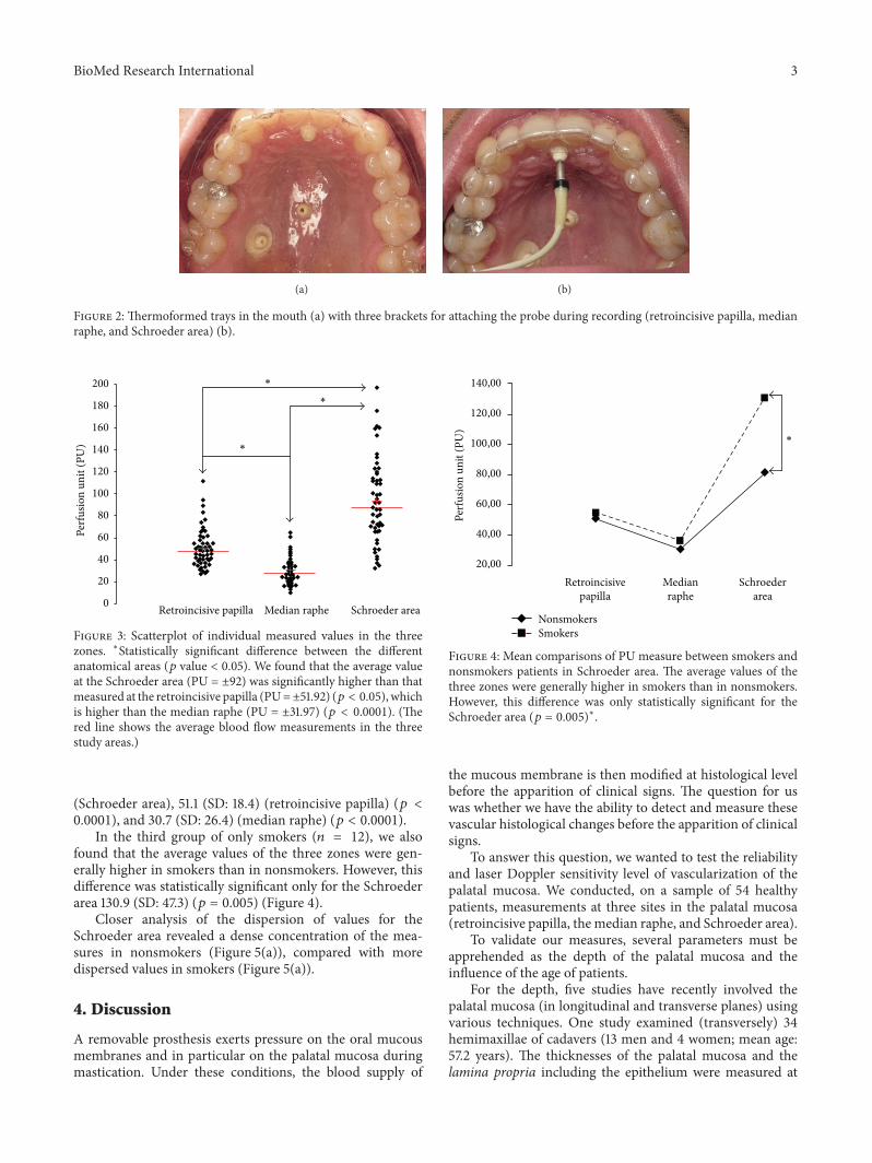
BioMed Research International 3
(a) (b)
Figure 2: Thermoformed trays in the mouth (a) with three brackets for attaching the probe during recording (retroincisive papilla, medianraphe, and Schroeder area) (b).
∗
∗
∗
Retroincisive papilla Median raphe Schroeder area0
20
40
60
80
100
120
140
160
180
200
Perf
usio
n un
it (P
U)
Figure 3: Scatterplot of individual measured values in the threezones. ∗Statistically significant difference between the differentanatomical areas (𝑝 value < 0.05). We found that the average valueat the Schroeder area (PU = ±92) was significantly higher than thatmeasured at the retroincisive papilla (PU=±51.92) (𝑝 < 0.05), whichis higher than the median raphe (PU = ±31.97) (𝑝 < 0.0001). (Thered line shows the average blood flow measurements in the threestudy areas.)
(Schroeder area), 51.1 (SD: 18.4) (retroincisive papilla) (𝑝 <0.0001), and 30.7 (SD: 26.4) (median raphe) (𝑝 < 0.0001).
In the third group of only smokers (𝑛 = 12), we alsofound that the average values of the three zones were gen-erally higher in smokers than in nonsmokers. However, thisdifference was statistically significant only for the Schroederarea 130.9 (SD: 47.3) (𝑝 = 0.005) (Figure 4).
Closer analysis of the dispersion of values for theSchroeder area revealed a dense concentration of the mea-sures in nonsmokers (Figure 5(a)), compared with moredispersed values in smokers (Figure 5(a)).
4. Discussion
A removable prosthesis exerts pressure on the oral mucousmembranes and in particular on the palatal mucosa duringmastication. Under these conditions, the blood supply of
140,00
120,00
100,00
80,00
60,00
40,00
20,00
Perf
usio
n un
it (P
U)
Retroincisivepapilla
Medianraphe
Schroederarea
∗
Nonsmokers Smokers
Figure 4: Mean comparisons of PU measure between smokers andnonsmokers patients in Schroeder area. The average values of thethree zones were generally higher in smokers than in nonsmokers.However, this difference was only statistically significant for theSchroeder area (𝑝 = 0.005)∗.
the mucous membrane is then modified at histological levelbefore the apparition of clinical signs. The question for uswas whether we have the ability to detect and measure thesevascular histological changes before the apparition of clinicalsigns.
To answer this question, we wanted to test the reliabilityand laser Doppler sensitivity level of vascularization of thepalatal mucosa. We conducted, on a sample of 54 healthypatients, measurements at three sites in the palatal mucosa(retroincisive papilla, themedian raphe, and Schroeder area).
To validate our measures, several parameters must beapprehended as the depth of the palatal mucosa and theinfluence of the age of patients.
For the depth, five studies have recently involved thepalatal mucosa (in longitudinal and transverse planes) usingvarious techniques. One study examined (transversely) 34hemimaxillae of cadavers (13 men and 4 women; mean age:57.2 years). The thicknesses of the palatal mucosa and thelamina propria including the epithelium were measured at

4 BioMed Research International
0 20 40 60
Perf
usio
n un
it (P
U)
0,00
20,00
40,00
60,00
80,00
100,00
120,00
140,00
160,00
180,00
200,00
(a) Nonsmokers, Schroeder area0 5 10 15
Perf
usio
n un
it (P
U)
0,00
20,00
40,00
60,00
80,00
100,00
120,00
140,00
160,00
180,00
200,00
(b) Smokers, Schroeder area
Figure 5: Scatterplot values in Schroeder area in nonsmokers (a) and smokers (b).
three points, starting from the alveolar crest, at intervals of4mm and with the aid of Adobe Photoshop®.The thicknessesof the palatal mucosa increase from the alveolar crest towardthe midpalatal suture. Conversely, the thicknesses of the lam-ina propria including the epithelium at these same positionsdecrease toward the midpalatal suture [18].
Kolliyavar et al. [19], Anuradha et al. [20], andYaman et al.[21] measured (longitudinally) the gingival margin andpalatal line area. A bone-sounding method using a peri-odontal probe was used to assess the thickness of thepalatal mucosa at 15 measurement sites, and the differencein mucosal thickness between the groups was determined.Themean thickness of the palatal masticatorymucosa rangedfrom 2.0 to 3.7mm.
Cho et al. [22] used light microscopy to investigate thelongitudinal depth from the surface of the palatal mucosa.The thickness of the epithelium and lamina propria of thepalatal mucosa was measured (from the canine distal area tothe first molar distal area) at three positions (starting from3mm below the alveolar crest and in 3mm intervals) alongthe path of the palatine artery. The mean depth from thesurface of the palatal mucosa to the greater palatine arterydecreased from the canine distal to the first premolar distalbut again increased towards the posterior molar. The meanlength from the alveolar crest to the greater palatine artery,however, increased toward the posterior molar.
Our measurements are effective to a depth of 1mm,which corresponds to a lamina propria volume of only 1mm3.Indeed, under this condition, the thickness of the palatalmucosa was not affected in our results.
Secondly, the effects of aging were not consistentlyreported in these three studies. Although Kolliyavar et al.[19] and Anuradha et al. [20] found that younger participantshad thinner mucosa than older participants, Yaman et al. [21]detected no significant difference between age groups. Anu-radha et al. [20] also found that, within the same age group,females had thinner mucosa than males whereas Yaman etal. [21] identified no differences according to gender or body
mass index.These conflicting findings are likely to result fromstudy differences in age, ethnicity, body mass index, varyingmeasurement methods, and the placement of measurementpoints.
If we compare these study groups to those of our study,our participants were relatively healthier and younger (mean23 years) and had a lower weight mean (65 kg).
Another interesting aspect is to compare the measuresobtained by laser Doppler between the skin and palatalmucosa; the buccal epithelium is relatively finer than the skinepidermis [23]. Despite this difference, the signal obtainedfrom contact with the oral mucous membrane is less pro-nounced than that of the skin of the cheek. The density ofcapillaries varies according to the anatomical zones of thehuman body, so at the level of the bowel the average densityof the capillaries is about 50 capillaries by square millimetreof mucous surface [17]. Another explanation is the fact thatthe vascular network of oral tissues is less rich than that ofthe skin [24]. Although it is possible that the presence orabsence of epithelial fingering can change the morphology ofthe epithelium, we propose that our measurements were notinfluenced by these histological characteristics because theoperating range of laser Doppler signal exceeds the thicknessof the epithelium.
Concerning the precision of the measures, in our study,for better recording probe support, an in-mouth stabilizingpatch was developed. The thickness of the gutters (<2mm)does not influencemeasures of blood flow in themucosa [25];indeed, amelioration of recording reliability over time couldbe verified by using this approach.
Another parameter is that the fluctuation in the lowerfrequencies at 0.1 Hz depends on the sympathetic nervoussystem at the level of the blood flow to the skin [26]. Underthe same conditions for the palatal mucosa, the precision ofour measures can be limited. This parameter is much moretransient at the level of the oral mucous membrane than atthe level of the skin. Histamine is quickly eliminated at thelevel of the vascular buccal network [24]. Furthermore, at the

BioMed Research International 5
level of the skin, the proportion of the nerve network that isinfluenced by histamine is large and with a prolonged effect[5, 8, 9]. It seems that, in the case of our experiment, thisparameter did not influence the result because every mea-surement was conducted on healthy participants after 30minof complete rest.
Our anatomical and histological findings enabled us todemonstrate a significant difference in the microcirculationin the three areas. The flow was much more important inthe Schroeder area compared to the retroincisive papilla andwas significantly higher than the median raphe. The averageflux values were higher in the Schroeder area, differences thatcould have been revealed only by the sensitivity of the laserDoppler device.
4.1. Smoking and Nonsmoking Patients. It is well establishedthat smoking modifies the vascular network, with a hyper-aemic response in the palatal mucosa of smokers comparedto nonsmokers. In the present investigation, the increased PUvalue of smokers was probably the result of a local vascularvasoconstriction effect, particularly at the most peripheralportion of the mucosa. The elevation of microcirculation inthe palatal mucosa serves as a trigger for angiogenesis of thepalatal vascular plexus, and our results confirm this otherresearch [27].
In our study, when comparing smokers to nonsmokers,we observed increased microcirculation in smokers, partic-ularly in the connective tissue of the Schroeder area. A localeffect of smoking on the palatal mucosa is well established[28, 29] and can potentiate interaction of the flow, particularlyin the Schroeder area. There we propose that the Schroederarea would likely be the most informative/interesting zone inwhich we measure the flow and compare findings betweenparticipant groups. Concerning our sample, we took intoaccount interindividual variability including general condi-tion, similar hemodynamic and ambient temperatures [30],age, gender, site, posture, and ethnicity.Other parameters thatcan influence intraindividual comparisons includemenstrualcycle, circadian rhythm, and physical activity whereas mentalactivity does not influence the results [31].
The relatively homogeneous age of the participants inthis study (22 to 26 years old) means that age had littleinfluence on our results [32]. The flexibility of blood vesselsis reduced with increased age. In the same way, a decreasein the thickness of the oral mucous membrane occurs withaging. Another aspect is fibrosis of connective tissue in thelamina propria, which appears with aging and can influencethe measures [33].
4.2. Influence of a Covered Removable Prosthetic Dentureon the Palatal Mucosa. In an interesting study [34], laserDoppler was used to investigate recovery after removal ofa partial denture (bilateral posterior maxilla edentulous) inthe palatal mucosa of the Schroeder area [34]. The resultsof this referenced study did not reveal significant changes inthe blood flow of the palatal mucosa in the Schroeder areaover a 12-minute period. It seems that wearing a resin partialdenture modifies the vascularization of the palatal mucosain the area of Schroeder, independently of disease. It would
also seem that bearing a partial removable prosthesis in resinincreases the stream of the vascularization of the palatalmucous membrane of the zone of Schroeder, independentlyof any clinically detectable sign of local pathologies.
It has been suggested that a denture on tissue surfacesmust irritate the mucosa, and the Schroeder area is oftenimplicated in the prevalence of denture-related stomatitis[35]. Changes to blood flow at the level of the palatinemucous membrane are not initially clinically detectable, andfor this reason laser Doppler can constitute an appropriatetool for diagnosis or early investigation of certain pathologies(e.g., burning mouth syndrome [9]) that present consider-able difficulties with visual detection. Recently, after clinicalobservations, a new laser Doppler perfusion tool, PeriScanPIM II, has been used to evaluate the distribution maps ofblood flow corresponding to specific areas [36].
5. Conclusion
Application of LDF to measure blood flow in the palatalmucosa can differentiate three anatomical areas, with mea-surement of the Schroeder area being the most sensitive. Theincidence of the mucosa thickness variability highlights thevalue of routine measurement of microcirculation by laserDoppler in patients presenting mucosal lesions. This studyrepresents a technical advance and opens new possibilitiesfor investigating diseases of the palatal mucosa, includingtissue changes related to dentures, such as denture-relatedstomatitis. The influence of certain technical impressionsor treatments (e.g., radiotherapy or bisphosphonates) canalter the subepithelial microcirculation, and these alterationscan be measured at the blood vessel level using LDF. Thispossibility also applies for more systemic diseases such asdiabetes. Therefore, further investigation will be required tobetter establish the clinical potential of LDF.We currently areextending this study of the palatal mucosa blood microcir-culation in the same areas of denture wearers with clinicallyhealthy mucosa and those with different denture-relatedstomatitis diseases.
Conflict of Interests
The authors declare that they have no conflict of interests.
Acknowledgments
The authors are grateful to ParoNantes association for finan-cial support, Jonathan Bodin for statistical advice, and SanFrancisco Edit for reviewing the paper.
References
[1] L. Orekhova and A. A. Barmasheva, “Doppler flowmetry as atool of predictive, preventive and personalised dentistry,” TheEPMA Journal, vol. 4, no. 1, article 21, 2013.
[2] D. Evans, J. Reid, R. Strang, and D. Strirrups, “A comparisonof laser Doppler flowmetry with other methods of assessingthe vitality of traumatised anterior teeth,” Endodontics & DentalTraumatology, vol. 15, no. 6, pp. 280–290, 1999.

6 BioMed Research International
[3] C. M. Choi and R. G. Bennett, “Laser Dopplers to determinecutaneous blood flow,” Dermatologic Surgery, vol. 29, no. 3, pp.272–280, 2003.
[4] G. E. Nilsson, U. Otto, and J. E. Wahlberg, “Assessment of skinirritancy in man by laser Doppler flowmetry,” Contact Dermati-tis, vol. 8, no. 6, pp. 401–406, 1982.
[5] S. Hellem, L. S. Jacobsson, G. E. Nilsson, and D. H. Lewis,“Measurement of microvascular blood flow in cancellous boneusing laser Doppler flowmetry and 133Xe-clearance,” Interna-tional Journal of Oral Surgery, vol. 12, no. 3, pp. 165–177, 1983.
[6] H. Svensson, P. Svedman, J. Holmberg, and S. Jacobsson, “LaserDoppler flowmetry and transcutaneously measured carbondioxide tension for observing changes of skin blood flow infingers,” Scandinavian Journal of Plastic and ReconstructiveSurgery, vol. 17, no. 3, pp. 183–186, 1983.
[7] D. B. Singh, G. Stansby, andD. K. Harrison, “Assessment of oxy-genation and perfusion in the tongue and oralmucosa by visiblespectrophotometry and laser doppler flowmetry in healthy sub-jects,” Advances in Experimental Medicine and Biology, vol. 614,pp. 227–233, 2008.
[8] E. Chen and P. V. Abbott, “Dental pulp testing: a review,”International Journal of Dentistry, vol. 2009, Article ID 365785,12 pages, 2009.
[9] M. L. Kaplan, M. K. Jeffcoat, and P. Goldhaber, “Blood flow ingingiva and alveolar bone in beagles with periodontal disease,”Journal of Periodontal Research, vol. 17, no. 4, pp. 384–389, 1982.
[10] H. Develioglu, B. Kesim, and A. Tuncel, “Evaluation of themarginal gingival health using laser Doppler flowmetry,”Brazil-ian Dental Journal, vol. 17, no. 3, pp. 219–222, 2006.
[11] M. L. Macedo Paizan and J. F. Vilela-Martin, “Is there anassociation between periodontitis and hypertension?” CurrentCardiology Reviews, vol. 10, no. 4, pp. 355–361, 2014.
[12] D. A. Curtis, S. A. Gansky, and O. Plesh, “Deep and superficialmasseter muscle blood flow in women,” Journal of Prosthodon-tics, vol. 21, no. 6, pp. 472–477, 2012.
[13] P. Broz, M. Aschwanden, S. Partovi et al., “Assessment ofcutaneous microcirculation in unaffected skin regions by tran-scutaneous oxygen saturation monitoring and Laser Dopplerflowmetry in systemic sclerosis,” Clinical Hemorheology andMicrocirculation, vol. 60, no. 3, pp. 263–271, 2015.
[14] Y.-K. Jan, B. Lee, F. Liao, and R. D. Foreman, “Local cool-ing reduces skin ischemia under surface pressure in rats: anassessment by wavelet analysis of laser Doppler blood flowoscillations,” Physiological Measurement, vol. 33, no. 10, pp.1733–1745, 2012.
[15] S. Juliusson andM. Bende, “Allergic reaction of the humannasalmucosa studied with laser Doppler flowmetry,” Clinical Allergy,vol. 17, no. 4, pp. 301–305, 1987.
[16] A. G.M. Hendriks,W. Steenbergen, E. Hondebrink, J. C. G. VanHespen, P. C. M. Van De Kerkhof, andM. M. B. Seyger, “Wholefield laser Doppler imaging of the microcirculation in psoriasisand clinically unaffected skin,” The Journal of DermatologicalTreatment, vol. 25, no. 1, pp. 18–21, 2014.
[17] D.A. L.Hoff,H.Gregersen, and J.G.Hatlebakk, “Mucosal bloodflowmeasurements using laser Doppler perfusion monitoring,”World Journal of Gastroenterology, vol. 15, no. 2, pp. 198–203,2009.
[18] S.-K. Yu, B.-H. Lee, M.-H. Lee, K.-H. Cho, D. K. Kim, and H.-J.Kim, “Histomorphometric analysis of the palatal mucosa asso-ciated with periodontal plastic surgery on cadavers,” Surgicaland Radiologic Anatomy, vol. 35, no. 6, pp. 463–469, 2013.
[19] B. Kolliyavar, S. Setty, and S. L.Thakur, “Determination of thick-ness of palatal mucosa,” Journal of Indian Society of Periodontol-ogy, vol. 16, no. 1, pp. 80–83, 2012.
[20] B. R. Anuradha, B. S. Shankar, B. John, K. A. R. V. Prasad, A.Gopinadh, and K. N. N. Devi, “Assessment of palatal mastica-tory mucosa: a cross-sectional study,”The Journal of Contempo-rary Dental Practice, vol. 14, no. 3, pp. 536–543, 2013.
[21] D. Yaman, S. Aksu, R. Disci, and K. Demirel, “Thickness ofpalatal masticatory mucosa and its relationship with differentparameters inTurkish subjects,” International Journal ofMedicalSciences, vol. 11, no. 10, pp. 1009–1014, 2014.
[22] K. H. Cho, S. K. Yu, M. H. Lee, D. S. Lee, and H. J. Kim,“Histological assessment of the palatal mucosa and greaterpalatine artery with reference to subepithelial connective tissuegrafting,” Anatomy & Cell Biology, vol. 46, no. 3, pp. 171–176,2013.
[23] C. A. Squier,M. J. Kremer, and P.W.Wertz, “Effect of ethanol onlipid metabolism and epithelial permeability barrier of skin andoral mucosa in the rat,” Journal of Oral Pathology & Medicine,vol. 32, no. 10, pp. 595–599, 2003.
[24] B. Bay, M. Hilliges, C. Weidner, and G. Sandborgh-Englund,“Response of human oral mucosa and skin to histamineprovocation: laser Doppler perfusion imaging discloses differ-ences in the nociceptive nervous system,” Acta OdontologicaScandinavica, vol. 67, no. 2, pp. 99–105, 2009.
[25] H. Akazawa and K. Sakurai, “Changes of blood flow in themucosa underlying a mandibular denture following pressureassumed as a result of light clenching,” Journal of Oral Reha-bilitation, vol. 29, no. 4, pp. 336–340, 2002.
[26] T. Soderstrom, A. Stefanovska, M. Veber, and H. Svensson,“Involvement of sympathetic nerve activity in skin blood flowoscillations in humans,” The American Journal of Physiology—Heart and Circulatory Physiology, vol. 284, no. 5, pp. H1638–H1646, 2003.
[27] M. M. Bornstein, K. Klingler, U. P. Saxer, C. Walter, and C.A. Ramseier, “Tobacco-associated lesions of the oral mucosa,”Schweizerische Monatsschrift fur Zahnmedizin, vol. 116, no. 12,pp. 1261–1274, 2006.
[28] D. A. Baab and P. A. Oberg, “The effect of cigarette smoking ongingival blood flow in humans,” Journal of Clinical Periodontol-ogy, vol. 14, no. 7, pp. 418–424, 1987.
[29] T.N.Meekin, R. F.Wilson,D.A. Scott,M. Ide, andR.M. Palmer,“Laser Doppler flowmeter measurement of relative gingival andforehead skin blood flow in light and heavy smokers during andafter smoking,” Journal of Clinical Periodontology, vol. 27, no. 4,pp. 236–242, 2000.
[30] J. Allen and K. Howell, “Microvascular imaging: techniques andopportunities for clinical physiological measurements,” Physio-logical Measurement, vol. 35, no. 7, pp. R91–R141, 2014.
[31] K. Zeghal, P. Geslin, A.Maurel, G. Lagrue, and F. Lhoste, “Laser-Doppler velocimetry: a new technic for evaluating of micro-circulation. A reproducibility study,” La Presse Medicale, vol.15, no. 40, pp. 1997–2000, 1986.
[32] G. Shklar, “The effects of aging upon oral mucosa,”The Journalof Investigative Dermatology, vol. 47, no. 2, pp. 115–120, 1966.
[33] M. W. Hill, “Influence of age on the morphology and transittime of murine stratified squamous epithelia,” Archives of OralBiology, vol. 33, no. 4, pp. 221–229, 1988.
[34] C. Okada, T. Ueda, and K. Sakurai, “Blood flow in denture-supportingmaxillarymucosa in response to simulatedmastica-tion by loading,” Journal of Prosthodontic Research, vol. 54, no.4, pp. 159–163, 2010.

BioMed Research International 7
[35] A. V. Newton, “Denture sore mouth. A possible aetology,”British Dental Journal, vol. 112, pp. 357–360, 1962.
[36] H. Hirai, “Application of a laser Doppler perfusion imagingPeriscan PIM II for measuring the blood flow of the oralmucosa,” Nihon Hotetsu Shika Gakkai zasshi, vol. 49, no. 1, pp.26–35, 2005.