optimizing a simple natural dye production method for dye
TRANSCRIPT
applied sciences
Article
Optimizing a Simple Natural Dye Production Methodfor Dye-Sensitized Solar Cells: Examples for Betalain(Bougainvillea and Beetroot Extracts) andAnthocyanin Dyes
María José García-Salinas * and María Jesús Ariza
Department of Chemistry and Physics, Applied Physics Area, University of Almería, 04120 Almería, Spain;[email protected]* Correspondence: [email protected]; Tel.: +34-950-01-5913
Received: 27 April 2019; Accepted: 18 June 2019; Published: 20 June 2019
Featured Application: After reviewing literature and showing our own results, this papersummarizes some steps to extract, characterize and use natural dyes, proposing a protocol toobtain and work with these dyes. As an example of application, we use dye-sensitized solarcells (DSSCs). Dyes are the ‘booster’ of this sustainable energy source, and our exhaustive studycontributes to the knowledge of dyes, and thus to improve cells’ environmentally friendly designand performance.
Abstract: We present a study about the sensitizers extracted from natural resources. This paperfocuses on how to select, extract and characterize natural dyes, giving some guides to establish aprotocol for the whole process of fabricating and using these dyes. The influence of the extractionsolvent and method, and of parameters such as pH are analyzed. Also, dye precursor and dye extractstability have been studied, as well as how the dye adsorbs onto substrates and the effect of mixing orconcentrating the extracts. Results concerning betalain pigments present in bougainvillea and beetrootextracts, and anthocyanins in eggplant extracts, analyzed by using UV-Vis spectrometry, are included.As an example of application, we report procedures intended to test and enhance the dye potential asa main component of dye-sensitized solar cells (DSSCs). DSSCs mimic nature’s photosynthesis andhave some advantages like an easy and low-cost fabrication procedure. Their efficiency depends onits design and fabrication process and also on the different components involved. Hence, optimizingeach component is essential to achieve the best performance, and thus the dye used as a sensitizer iscrucial. We fabricate cells by using a simple procedure: As the interest is focused on the sensitizer,the same consecutive steps are followed, varying only the dye extract. Among all the natural-dyestested, beetroot extract reaches up to 0.47% cell efficiency, which is near the highest values found inliterature for this pigment.
Keywords: natural dyes; UV-Vis spectrometry; bougainvillea; beetroot; betalains; anthocyanins;dye-sensitized solar cells (DSSCs)
1. Introduction
Dyes from bio-resources or natural dyes were the first ones used by human beings, and nowadays,ecological and health concerns have made natural dyes preferable to synthetic ones in different fieldssuch as cosmetic, pharmaceutical, textile, or food industries [1]. Most natural dyes are found in theroots, barks, leaves, bracts, flowers, skins, and shells of plants. These plant pigments are classifiedinto four major categories [1–3]: (i) Tetrapyrroles, such as green chlorophylls; (ii) Carotenoids, usually
Appl. Sci. 2019, 9, 2515; doi:10.3390/app9122515 www.mdpi.com/journal/applsci
Appl. Sci. 2019, 9, 2515 2 of 20
red, orange or yellow; (iii) Flavonoids, of which red, purple or blue anthocyanins are an importantsubgroup; and (iv) Betalains (yellow betaxanthins and red-purple betacyanins). The most abundantpigments are chlorophylls, followed by carotenoids. Carotenoids can coexist with other families,but betalains are incompatible with anthocyanins.
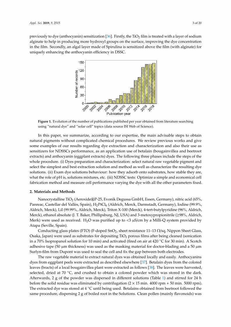
These natural colorants have been of interest in different fields and applications [1]. Particularlyinteresting is the use of natural dyes as sensitizers in dye-sensitized solar cells (DSSCs) [4–6], their mainadvantages being a simple extraction procedure, low cost, wide availability, and their environmentallyfriendly nature [7–9]. For almost three decades, DSSCs have been considered a focus line of renewableenergies, as a promising simple alternative power source. The increase in the number of publicationsreflects the interest on the subject [10]. The cell efficiency has been steadily improving due toextensive efforts in numerous experimental investigations and theoretical or computational studies,but there are many areas that deserve further research [11] before considering these cells as a feasiblecommercial product [12]. The DSSC device is an ensemble of various materials that undergo differentinteractions and processes, and researchers have focused on the study, modification or innovationof each component. For example, the dye or sensitizer, responsible for solar energy caption, shouldpossess certain features in order to effectively play its role for an efficient performance. The researchcommunity has attempted to find the best sensitizer and has developed different types of them:Ru-complex dyes, metal-free organic dyes, quantum dot sensitizers, perovskite-based sensitizers, andnatural dyes [6,13,14]. Dye molecules are adsorbed onto a nano-structured layer of a wide band gapsemiconductor (i.e., TiO2) to harvest photons. If these solar cells are to be considered a as a greenenergy generator, the more “natural” the dye, the more “green” the device. For NDSSCs (DSSCsusing natural dyes), the number of publications, scarce in the decade of 2000, has increased since 2009(Figure 1). As an example of the still alive controversy, the early work by Zhou [15] studied 20 naturaldyes and Narayan [3], only a few months later, published a review pointing out the requirements ofphotosensitizers, why the NDSSC efficiency is low and further recommendations. Something similaroccurs nowadays; reviews have been published stating recent progress in natural dyes [16,17] butthere are still challenges in the mass production of NDSSCs [12,16]. So this subject is still of interest toresearches, i.e., conferences and symposiums are being held, and since last year, almost 200 papersrelated to “natural dyes and DSSC” have been published, both from the experimental and theoreticalpoints of view, aimed to improve devices and understanding of the different materials and phenomenainvolved. In general, and some examples are cited next, most papers include firstly experimentaldetails about dyes. Pigments are obtained from a great variety of plants, some animals or minerals bydifferent methods, and then are characterized through UV–Vis and FTIR spectrophotometry [18–20].Further purification, combinations, analysis, and use constitute the body of the studies, together withnew ideas about cell implementation. Researches have focused on the influence of different factorson the final performance, such as: solvent for extraction and adsorption [21–23], extraction time [18],pH and temperature during dye adsorption onto the nanoporous film [24], dipping time [25], dyemixing or co-sensitization [26–28], together with “tandem” cells [28], film thickness [22,29] etc. Somepapers include also theoretical calculations or simulations [24,30,31], for example, adsorption isothermsof a dye onto TiO2 film are fitted by models established through statistical physics treatment [24]or chlorophyll is used as a reference dye to simulate absorption spectra and theoretically calculateground and excited state properties [30]. There have also been some interesting ideas related to theimprovement of cell efficiency. Due to its nature or different chemical structure, chlorophyll dyesperformed different in different liquid electrolytes [32] and this could be related to iodide mobilities [31].Exposing the photo anode 10 min to microwave frequency enhances electrode characteristics andefficiency and absorption of dye [33]. Studying the recombination processes for different natural dyes,the most efficient dyes can be discriminated [34]. To obtain dyes, a strategy to induce anthocyaninssynthesis in in-vitro cultures, has been presented: Stress induced by nutrient deficiencies leads to theproduction of secondary metabolites such as anthocyanins [35]. Also, to improve the efficiency andanchoring ability of natural dye, two algal buffer layers (sodium alginate and spirulina) are introduced
Appl. Sci. 2019, 9, 2515 3 of 20
previously to dye (anthocyanin) sensitization [36]. Firstly, the TiO2 film is treated with a layer of sodiumalginate to help in producing more hydroxyl groups on the surface, improving the dye concentrationin the film. Secondly, an algal layer made of Spirulina is sensitized above the film (with alginate) foruniquely enhancing the anthocyanin efficiency in DSSC.
Appl. Sci. 2019, 9, x 3 of 21
sensitization [36]. Firstly, the TiO2 film is treated with a layer of sodium alginate to help in producing more hydroxyl groups on the surface, improving the dye concentration in the film. Secondly, an algal layer made of Spirulina is sensitized above the film (with alginate) for uniquely enhancing the anthocyanin efficiency in DSSC.
Figure 1. Evolution of the number of publications published per year obtained from literature searching using “natural dye” and “solar cell” topics (data source ISI Web of Science).
In this paper, we summarize, according to our expertise, the main advisable steps to obtain natural pigments without complicated chemical procedures. We review previous works and give some examples of our results regarding dye extraction and characterization and also their use as sensitizers for NDSSCs performance, as an application use of betalain (bougainvillea and beetroot extracts) and anthocyanin (eggplant extracts) dyes. The following three phases include the steps of the whole procedure. i) Dyes preparation and characterization: select natural raw vegetable pigment and select the simplest and best extraction solution and method as well as characterize the resulting dye solutions. ii) Exam dye solutions behaviour: how they adsorb onto substrates, how stable they are, what the role of pH is, solutions mixtures, etc. iii) NDSSC tests: Optimize a simple and economical cell fabrication method and measure cell performance varying the dye with all the other parameters fixed.
2. Materials and Methods
Nanocrystalline TiO2 (Aeroxide® P-25, Evonik Degussa GmbH, Essen, Germany), nitric acid (65%, Panreac, Castellar del Vallès, Spain), H2PtCl6 (Aldrich, Merck, Darmstadt, Germany), Iodine (99.9%, Aldrich, Merck), LiI (99.99%, Aldrich, Merck), Triton X-100 (Merck), 4-tert-butylpyridine (96%, Aldrich, Merck), ethanol absolute (J. T. Baker, Phillipsburg, NJ, USA) and 3-metoxypropionitrile (≥98%, Aldrich, Merk) were used as received. H2O was purified up to <3 S/cm by a Milli-Q system provided by Atapa (Seville, Spain).
Conducting glass plates (FTO) (F-doped SnO2, sheet resistance 11–13 Ω/sq, Nippon Sheet Glass, Osaka, Japan) were used as substrates for depositing TiO2 porous films after being cleaned (sonication in a 70% Isopropanol solution for 10 min) and activated (fired on air at 420 °C for 30 min). A Scotch adhesive tape (50 µm thickness) was used as the masking material for doctor-blading and a 50 µm Surlyn-film from Dupont was used to seal the cell and fix the gap between both electrodes.
The raw vegetable material to extract natural dyes was obtained locally and easily. Anthocyanins dyes from eggplant peels were extracted as described elsewhere [37]. Betalain dyes from the colored leaves (bracts) of a local bougainvillea plant were extracted as follows [38]. The leaves were harvested, selected, dried at 70 °C, and crushed to obtain a colored powder which was stored in the dark. Afterwards, 2 g of the powder was dispersed in different solutions (Table 1) and stirred for 24 h before the solid residue was eliminated by centrifugation (2 × 15 min. 4000 rpm + 50 min. 5000 rpm). The extracted dye was stored at 4 °C until being used. Betalains obtained from beetroot followed the same procedure, dispersing 2 g of boiled root in the Solutions. Clean pollen
Figure 1. Evolution of the number of publications published per year obtained from literature searchingusing “natural dye” and “solar cell” topics (data source ISI Web of Science).
In this paper, we summarize, according to our expertise, the main advisable steps to obtainnatural pigments without complicated chemical procedures. We review previous works and givesome examples of our results regarding dye extraction and characterization and also their use assensitizers for NDSSCs performance, as an application use of betalain (bougainvillea and beetrootextracts) and anthocyanin (eggplant extracts) dyes. The following three phases include the steps of thewhole procedure. (i) Dyes preparation and characterization: select natural raw vegetable pigment andselect the simplest and best extraction solution and method as well as characterize the resulting dyesolutions. (ii) Exam dye solutions behaviour: how they adsorb onto substrates, how stable they are,what the role of pH is, solutions mixtures, etc. (iii) NDSSC tests: Optimize a simple and economical cellfabrication method and measure cell performance varying the dye with all the other parameters fixed.
2. Materials and Methods
Nanocrystalline TiO2 (Aeroxide®P-25, Evonik Degussa GmbH, Essen, Germany), nitric acid (65%,Panreac, Castellar del Vallès, Spain), H2PtCl6 (Aldrich, Merck, Darmstadt, Germany), Iodine (99.9%,Aldrich, Merck), LiI (99.99%, Aldrich, Merck), Triton X-100 (Merck), 4-tert-butylpyridine (96%, Aldrich,Merck), ethanol absolute (J. T. Baker, Phillipsburg, NJ, USA) and 3-metoxypropionitrile (≥98%, Aldrich,Merk) were used as received. H2O was purified up to <3 µS/cm by a Milli-Q system provided byAtapa (Seville, Spain).
Conducting glass plates (FTO) (F-doped SnO2, sheet resistance 11–13 Ω/sq, Nippon Sheet Glass,Osaka, Japan) were used as substrates for depositing TiO2 porous films after being cleaned (sonicationin a 70% Isopropanol solution for 10 min) and activated (fired on air at 420 C for 30 min). A Scotchadhesive tape (50 µm thickness) was used as the masking material for doctor-blading and a 50 µmSurlyn-film from Dupont was used to seal the cell and fix the gap between both electrodes.
The raw vegetable material to extract natural dyes was obtained locally and easily. Anthocyaninsdyes from eggplant peels were extracted as described elsewhere [37]. Betalain dyes from the coloredleaves (bracts) of a local bougainvillea plant were extracted as follows [38]. The leaves were harvested,selected, dried at 70 C, and crushed to obtain a colored powder which was stored in the dark.Afterwards, 2 g of the powder was dispersed in different solutions (Table 1) and stirred for 24 hbefore the solid residue was eliminated by centrifugation (2 × 15 min. 4000 rpm + 50 min. 5000 rpm).The extracted dye was stored at 4 C until being used. Betalains obtained from beetroot followed thesame procedure, dispersing 2 g of boiled root in the Solutions. Clean pollen (mainly flavonoids) was
Appl. Sci. 2019, 9, 2515 4 of 20
used as collected, dispersing also 2 g in the solutions. Finally, 100 g of ground coffee in 1L of water wasstirred at 80 C for 6 h.
Table 1. Solutions used for extracting the dye from vegetable raw materials.
ExtractionMethod
Acetone(% vol.)
Ethanol(% vol.)
HCl(M)
H2O(% vol.)
pHRange
Solution 1 32 — — 68 5.0–5.6Solution 2 — Solvent 0.1 — 1.6–1.7Solution 3 — — 0.1 Solvent 1.3–1.4Solution 4 — 80 — 20 6.5–7.0
Reflectance, transmittance and absorption spectra from solid materials (bougainvillea bracts)were obtained with a LICOR LI-1800 spectroradiometer. The dye extracts UV–Vis absorption spectrain dilution and onto TiO2 were recorded using a high-resolution spectrophotometer HR4000 (OceanOptics), following a systematic procedure for all measurements. Absorbed intensity data in all figuresare in arbitrary units.
DSSC fabrication followed the method proposed by Ito and collaborators [39,40]. In summary,commercial TiO2 nanoparticles (15% wt) were stirred in ethanol (61% wt) and diluted nitric acid (pH3–4, 24% wt) for 12 h at room temperature to obtain a paste of 150 g/l. Two drops of Triton X-100were added to 20 ml of paste, which was stirred for a further 12 h to obtain the final paste used inthis work. The electrolyte was a solution containing 0.05 M I2, 0.5 M LiI, 0.5 M 4-tert-butylpyridine in3-metoxypropionitrile [38]. Photovoltaic experiments were performed directly under solar radiationin Almeria (Spain), working with similar light irradiation in all tests: 660 W/m2 (admitting 6%error tolerance). This light source is natural, available in our geographical area, and quite in tunewith the possible final applications of devices based on DSSCs. However, some disadvantages likemeteorological changes, have to be taken into account. A calibrated high performance thermopile-basedpyranometer (Kipp&Zonnen, CMP 11) from CIESOL (Solar Energy Research Center, UAL-PSA, Almería,Spain) was used as a reference, measuring continuously the direct solar radiation. Digital sourcemeters were used to measure the I-V curves without any external bias. To determine the electricalparameters, at least three cells for each natural dye extract were built, and I-V curves were measuredto verify the reliability of data and the whole fabrication method. The detailed procedures for cellassembly and measurements are described elsewhere [37,38,41]
3. Results and Discussion
3.1. Dyes Preparation and Characterization
In a first approach to natural dyeing, at least three main steps should be followed. (i) Selectnatural raw vegetable pigment. As mentioned before, there are four main pigment families and severalimportant pigments in each of them. The pigments can be found in leaves, roots, bracts, flowers . . . andto select one or more pigments we have to consider the geographical area, availability, and extractionmethods that should be applied. (ii) Try several and select the simplest and best extraction solution andmethod. Once we have the raw vegetable material, we choose economical and environmentally friendlysolvents; consider different solvent combinations; test time spent in solutions, time and centrifugationvelocity, filtering processes, etc. (iii) Characterize the resulting dye solutions. The solutions can becharacterized initially by UV-Vis spectroscopy and the spectra could be compared to reference artificialdyes like N719 to check the dye’s potential. The spectra are also important to identify pigments orpigment families and to decide whether an extraction solution or method works.
To tackle this first phase, we have used different vegetable raw material: pollen, coffee grains,beetroot, bougainvillea bracts with different colors, and eggplant peels, all of them easily available.Solvent influence on natural dye extraction has been dealt with previously [21–23,42]. In our case,after a previous sequence of tests with different solvents and procedures, four simple and economical

Appl. Sci. 2019, 9, 2515 5 of 20
extraction methods have been selected using HCl, acetone, water and ethanol. As it is summarized inTable 1, the four Solutions prepared and numbered from 1 to 4 are: (1) acetone/water 32/68 (vol/vol); (2)0.1 M HCl in ethanol; (3) 0.1 M HCl in water; and (4) ethanol/water 80/20 (vol/vol). After previoussimple preparation (select, dry, crush), the vegetable material in the solution was stirred (800 rpm)for 24 h and the residue was eliminated by centrifugation. See Refs [37,38,41] for details. Differentfiltering processes and different concentrations have also been tested before determining dye extractspectra. Then, after a previous basic study, some of the materials can be ruled out, and the procedurecontinues only for the a priori good candidates for DSSC sensitizers, finding finally that Solution 1works better for betalains and Solutions 2 and 3 (much lower pH) go well for anthocyanins [37]. Theseresults are summarized next.
Coffee extract showed a spectrum with increasing absorbance towards lower wavelength, andpeaks for 450 nm and below. Similar results have already been shown [42], but in our case the spectrawere noisy in the UV values, with several peaks also affected by the presence of a caffeine-band around275 nm [43]. Recently, Setiawan and col. [44] extracted anthocyanins from coffee bean peels. The extractwas used as the natural dye, and the solid waste after the extraction process was carbonated and usedto prepare counter electrodes with different thicknesses. In spite of the fact that some previous resultssupport coffee as a dye with not bad efficiency (i.e., 0.33%, as mentioned by Zhou et al. [15]), in thiswork, it was decided not to continue with it, since it is not a local natural product in our region.
Something similar happened to pollen. Figure 2 shows spectra of pollen extracts with Solutions1 and 2. This figure also includes spectral photon density, Φλ, calculated from the standard ASTMG173-03 reference solar spectrum derived from SMARTS v. 2.9.2 and provided by Plataforma Solar deAlmería (PSA-CIEMAT, Almería, Spain). These data provide information to analyze dyes’ spectra.Since TiO2 absorbs at wavelengths below 387.5 nm (semiconductor bad gap Eg = 3.2 eV), there is anoptical window of interest for dyes in the visible region, from 400 to 750 nm, where the higher thewavelength the higher the number of photons despite them having less energy. Roughly speaking,a dye is presumed to work better when having wide absorption near red wavelengths.
The spectrum of N719, the most widely-used artificial dye for DSSCs, is also included in Figure 2as a reference. As can be seen, N719 has a peak around 400 nm and a wide peak around 550 nm, bothof which are in this spectral region. The wavelengths may change slightly, as they depend on dyeconcentration and pH [37].
Appl. Sci. 2019, 9, x 5 of 20
finding finally that Solution 1 works better for betalains and Solutions 2 and 3 (much lower pH) go well for anthocyanins [37]. These results are summarized next.
Table 1. Solutions used for extracting the dye from vegetable raw materials.
Extraction Method
Acetone (% vol.)
Ethanol (% vol.)
HCl (M)
H2O (% vol.)
pH Range
Solution 1 32 --- --- 68 5.0–5.6 Solution 2 --- Solvent 0.1 --- 1.6–1.7 Solution 3 --- --- 0.1 Solvent 1.3–1.4 Solution 4 --- 80 --- 20 6.5–7.0
Coffee extract showed a spectrum with increasing absorbance towards lower wavelength, and peaks for 450 nm and below. Similar results have already been shown [42], but in our case the spectra were noisy in the UV values, with several peaks also affected by the presence of a caffeine-band around 275 nm [43]. Recently, Setiawan and col. [44] extracted anthocyanins from coffee bean peels. The extract was used as the natural dye, and the solid waste after the extraction process was carbonated and used to prepare counter electrodes with different thicknesses. In spite of the fact that some previous results support coffee as a dye with not bad efficiency (i.e., 0.33%, as mentioned by Zhou et al. [15]), in this work, it was decided not to continue with it, since it is not a local natural product in our region.
Something similar happened to pollen. Figure 2 shows spectra of pollen extracts with Solutions 1 and 2. This figure also includes spectral photon density, Φλ, calculated from the standard ASTM G173-03 reference solar spectrum derived from SMARTS v. 2.9.2 and provided by Plataforma Solar de Almería (PSA-CIEMAT, Almería, Spain). These data provide information to analyze dyes’ spectra. Since TiO2 absorbs at wavelengths below 387.5 nm (semiconductor bad gap Eg = 3.2 eV), there is an optical window of interest for dyes in the visible region, from 400 to 750 nm, where the higher the wavelength the higher the number of photons despite them having less energy. Roughly speaking, a dye is presumed to work better when having wide absorption near red wavelengths.
The spectrum of N719, the most widely-used artificial dye for DSSCs, is also included in Figure 2 as a reference. As can be seen, N719 has a peak around 400 nm and a wide peak around 550 nm, both of which are in this spectral region. The wavelengths may change slightly, as they depend on dye concentration and pH [37].
Figure 2. Spectra of pollen extracts with Solutions 1 and 2 and artificial dye N719 spectrum. Right y axis: Spectral photon density, Φλ, calculated from the standard ASTM G173-03 reference solar spectrum.
Commented [M4]: 图 2 更换,移到这个位置 Figure 2. Spectra of pollen extracts with Solutions 1 and 2 and artificial dye N719 spectrum. Right y axis:Spectral photon density, Φλ, calculated from the standard ASTM G173-03 reference solar spectrum.
The spectrum obtained for pollen with Solution 1 (pollen 1) showed a wide and noisy peak around350 nm, as was also found by Masaya [45]. The spectrum obtained with Solution 2 gives two mainpeaks at 424 and 456 nm, then absorbance diminishes and slightly increases again for 718 nm up toa scarce 20% of the main peaks. Flavonoids can account for 2–5% of pollen dry weight [2], but the
Appl. Sci. 2019, 9, 2515 6 of 20
specific compounds depend on geographical area [46], and FT-IR spectroscopy or spectral fluorescenceanalysis should be required to identify them [47,48]. This would complicate our analysis. Besides,the main peak is found for wavelengths far below the optimum interval of interest. For these reasons,and also because no high efficiencies for pollen dyes have been reported in literature, pollen extractswere not finally used as NDSSC sensitizers.
The dye solutions extracted from other vegetable materials were also studied by UV-Visspectroscopy and results will be shown next. Clearly, the spectra contain pigments from two families:flavonoids (anthocyanins, in eggplant extracts) and belatains (in extracts from beetroot and differentcolored bougainvillea bracts).
3.1.1. Flavonoids
In general, when working with anthocyanins, alcoholic solvents are used [23,35,49–54].For example, anthocyanin extracts from five fruits were studied by Teoli and coworkers [54].To determine the best extraction protocol, they used five different solvent solutions and measured theanthocyanins content, finding that the best solvent was ethanol 70%.
For our eggplant extracts, only good results have been obtained with acidic solutions: Solutions 2and 3 (data and discussion already published [37]). The spectra showed the presence of anthocyaninswith absorbance peaks at 520–550 nm [37], related to the presence of nasunin [7,49]. After the wholestudy, it was found that solvent Solution 2 gave the highest cell efficiency.
3.1.2. Betalains from Bougainvillea Bracts
The effect of solvent on the extraction of betalains has also been tested in literature [55]. Acetone [56]and acid water [57] have been used as solvents for bougainvillea extracts, although other solvents arepossible, i.e., methanol [58] or ethanol [59] solutions, and of course, any of the extracts may undergofurther purification [60].
In our group, all the four different solvents listed in Table 1 have been tested in previous works.For instance, the spectra of dye Solutions 1 and 2 are compared with N719 dye spectra in the workby Maldonado and co-workers [38]. In our work, the best Solution is 1 (acetone/water). This bestsolvent and the corresponding method have been subsequently applied to four different-colored(yellow, orange, pink, and purple) bougainvillea (BG) bracts. The UV-Vis spectra of the four extractsobtained are compared in Figure 3a, which also includes N719 spectrum for reference. Quantitativedata, wavelength and intensity of the main peaks found are listed in Table 2.
Appl. Sci. 2019, 9, x 6 of 20
The spectrum obtained for pollen with Solution 1 (pollen 1) showed a wide and noisy peak around 350 nm, as was also found by Masaya [45]. The spectrum obtained with Solution 2 gives two main peaks at 424 and 456 nm, then absorbance diminishes and slightly increases again for 718 nm up to a scarce 20% of the main peaks. Flavonoids can account for 2–5% of pollen dry weight [2], but the specific compounds depend on geographical area [46], and FT-IR spectroscopy or spectral fluorescence analysis should be required to identify them [47,48]. This would complicate our analysis. Besides, the main peak is found for wavelengths far below the optimum interval of interest. For these reasons, and also because no high efficiencies for pollen dyes have been reported in literature, pollen extracts were not finally used as NDSSC sensitizers.
The dye solutions extracted from other vegetable materials were also studied by UV-Vis spectroscopy and results will be shown next. Clearly, the spectra contain pigments from two families: flavonoids (anthocyanins, in eggplant extracts) and belatains (in extracts from beetroot and different colored bougainvillea bracts).
3.1.1. Flavonoids
In general, when working with anthocyanins, alcoholic solvents are used [23,35,49–54]. For example, anthocyanin extracts from five fruits were studied by Teoli and coworkers [54]. To determine the best extraction protocol, they used five different solvent solutions and measured the anthocyanins content, finding that the best solvent was ethanol 70%.
For our eggplant extracts, only good results have been obtained with acidic solutions: Solutions 2 and 3 (data and discussion already published [37]). The spectra showed the presence of anthocyanins with absorbance peaks at 520–550 nm [37], related to the presence of nasunin [7,49]. After the whole study, it was found that solvent Solution 2 gave the highest cell efficiency.
3.1.2. Betalains from Bougainvillea Bracts
The effect of solvent on the extraction of betalains has also been tested in literature [55]. Acetone [56] and acid water [57] have been used as solvents for bougainvillea extracts, although other solvents are possible, i.e., methanol [58] or ethanol [59] solutions, and of course, any of the extracts may undergo further purification [60].
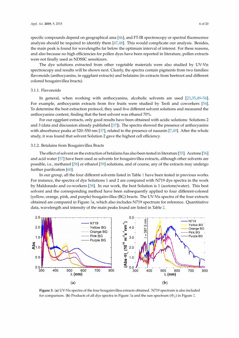
In our group, all the four different solvents listed in Table 1 have been tested in previous works. For instance, the spectra of dye Solutions 1 and 2 are compared with N719 dye spectra in the work by Maldonado and co-workers [38]. In our work, the best Solution is 1 (acetone/water). This best solvent and the corresponding method have been subsequently applied to four different-colored (yellow, orange, pink, and purple) bougainvillea (BG) bracts. The UV-Vis spectra of the four extracts obtained are compared in Figure 3a, which also includes N719 spectrum for reference. Quantitative data, wavelength and intensity of the main peaks found are listed in Table 2.
(a) (b)
Figure 3. (a) UV-Vis spectra of the four bougainvillea extracts obtained. N719 spectrum is also includedfor comparison. (b) Products of all dye spectra in Figure 3a and the sun spectrum (Φλ) in Figure 2.

Appl. Sci. 2019, 9, 2515 7 of 20
Table 2. Wavelength and intensity of the main absorbance peaks of the UV-Vis spectra shown inFigures 3–5.
Spectrum (Figure) Peak Type λ (nm) Abs
N719 (3) Peak 1 405 1.03Peak 2 553 0.96
Yellow BG (3)Shoulder 1 455 1.10
Peak 1 478 1.21Shoulder 2 543 0.12
Orange BG (3)Shoulder 1 455 1.01
Peak 1 478 1.08Shoulder 2 543 0.16
Pink BG (3)Shoulder 1 451 0.74
Peak 1 482 0.88Peak 2 535 0.74
Purple BG (3)Shoulder 1 454 0.40
Peak 1 484 0.47Peak 2 535 0.52
Pink BG bract (4)Shoulder 1 487 0.72
Peak 2 547 0.79Peak 3 674 0.13
Orange BG bract (4)
Shoulder 1 451 0.76Peak 1 484 0.81
Shoulder 2 553 0.54Peak 3 674 0.19
Beetroot 1 (5) Peak 1 526 0.62
Beetroot 2 (5) Peak 1 519 0.24
Beetroot 3 (5) Peak 1 516 0.56
Appl. Sci. 2019, 9, x 9 of 20
(responsible of absorption peaks) change with media, pH, or even with temperature, and consequently the amount of energy absorbed by those functional groups also changes, moving the position of the main peaks, in this case, to higher energies. iii) Finally, a remarkable fact is that the chlorophyll peak (around 670 nm, [62]) of the bract diminishes or even disappears in dye solution, probably because chlorophyll molecules present in bracts are eliminated when the dye is extracted following our process with acetone solvent solutions, as it is known that ethanol has been preferably used when the pigment of interest is chlorophyll [55,62].
(a) (b)
Figure 4. Dry bract and dye extract spectra for: (a) pink bougainvillea; (b) orange bougainvillea.
3.1.3. Betalains from Beetroots
Ethanol has been mainly used as an extraction solvent for beetroots [63–65]. However, we have obtained that Solutions 1, 2 and 3 all give good results. The three extracted dyes show an adsorption band around 520 nm (see Figure 5 and Table 2), comparable to the 533 nm peak of N719, so these natural extracts from beetroots could be eligible for sensitizing the NDSSCs.
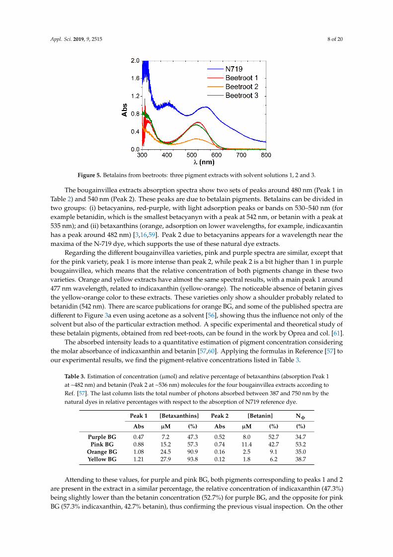
Figure 5. Betalains from beetroots: three pigment extracts with solvent solutions 1, 2 and 3.
It is noticeable that extract with Solution 2 (using ethanol) presents lower absorbance. In this case, the extremely low pH of the extract (pH = 1.7) may play an important role, as stated in previous works. Sengupta et al. [64] showed UV-Vis spectra of ethanol beetroot extracts with varying pH. In the pH range 6–9, the dyes showed similar pattern absorption, with the two peaks corresponding to betalain pigments, although their intensity and relative intensity within each pH varied. However, the absorbance fell drastically and the spectrum changed in shape at extreme pH levels of 3 and 12.
As we have prepared the three extracts with the same procedure, less concentration of pigment in Beetroot 2 is a priori presumed, which, apart from other considerations, would give a less efficient cell. This can be checked in Table 4: among NDSSCs using beetroot extracts, the number 2 is the less
Commented [M7]: 图 4 更换
Commented [M8]: 图 5 更换
Figure 4. Dry bract and dye extract spectra for: (a) pink bougainvillea; (b) orange bougainvillea.
Appl. Sci. 2019, 9, 2515 8 of 20
Appl. Sci. 2019, 9, x 9 of 20
(responsible of absorption peaks) change with media, pH, or even with temperature, and consequently the amount of energy absorbed by those functional groups also changes, moving the position of the main peaks, in this case, to higher energies. iii) Finally, a remarkable fact is that the chlorophyll peak (around 670 nm, [62]) of the bract diminishes or even disappears in dye solution, probably because chlorophyll molecules present in bracts are eliminated when the dye is extracted following our process with acetone solvent solutions, as it is known that ethanol has been preferably used when the pigment of interest is chlorophyll [55,62].
(a) (b)
Figure 4. Dry bract and dye extract spectra for: (a) pink bougainvillea; (b) orange bougainvillea.
3.1.3. Betalains from Beetroots
Ethanol has been mainly used as an extraction solvent for beetroots [63–65]. However, we have obtained that Solutions 1, 2 and 3 all give good results. The three extracted dyes show an adsorption band around 520 nm (see Figure 5 and Table 2), comparable to the 533 nm peak of N719, so these natural extracts from beetroots could be eligible for sensitizing the NDSSCs.
Figure 5. Betalains from beetroots: three pigment extracts with solvent solutions 1, 2 and 3.
It is noticeable that extract with Solution 2 (using ethanol) presents lower absorbance. In this case, the extremely low pH of the extract (pH = 1.7) may play an important role, as stated in previous works. Sengupta et al. [64] showed UV-Vis spectra of ethanol beetroot extracts with varying pH. In the pH range 6–9, the dyes showed similar pattern absorption, with the two peaks corresponding to betalain pigments, although their intensity and relative intensity within each pH varied. However, the absorbance fell drastically and the spectrum changed in shape at extreme pH levels of 3 and 12.
As we have prepared the three extracts with the same procedure, less concentration of pigment in Beetroot 2 is a priori presumed, which, apart from other considerations, would give a less efficient cell. This can be checked in Table 4: among NDSSCs using beetroot extracts, the number 2 is the less
Commented [M7]: 图 4 更换
Commented [M8]: 图 5 更换 Figure 5. Betalains from beetroots: three pigment extracts with solvent solutions 1, 2 and 3.
The bougainvillea extracts absorption spectra show two sets of peaks around 480 nm (Peak 1 inTable 2) and 540 nm (Peak 2). These peaks are due to betalain pigments. Betalains can be divided intwo groups: (i) betacyanins, red-purple, with light adsorption peaks or bands on 530–540 nm (forexample betanidin, which is the smallest betacyanyn with a peak at 542 nm, or betanin with a peak at535 nm); and (ii) betaxanthins (orange, adsorption on lower wavelengths, for example, indicaxantinhas a peak around 482 nm) [3,16,59]. Peak 2 due to betacyanins appears for a wavelength near themaxima of the N-719 dye, which supports the use of these natural dye extracts.
Regarding the different bougainvillea varieties, pink and purple spectra are similar, except thatfor the pink variety, peak 1 is more intense than peak 2, while peak 2 is a bit higher than 1 in purplebougainvillea, which means that the relative concentration of both pigments change in these twovarieties. Orange and yellow extracts have almost the same spectral results, with a main peak 1 around477 nm wavelength, related to indicaxanthin (yellow-orange). The noticeable absence of betanin givesthe yellow-orange color to these extracts. These varieties only show a shoulder probably related tobetanidin (542 nm). There are scarce publications for orange BG, and some of the published spectra aredifferent to Figure 3a even using acetone as a solvent [56], showing thus the influence not only of thesolvent but also of the particular extraction method. A specific experimental and theoretical study ofthese betalain pigments, obtained from red beet-roots, can be found in the work by Oprea and col. [61].
The absorbed intensity leads to a quantitative estimation of pigment concentration consideringthe molar absorbance of indicaxanthin and betanin [57,60]. Applying the formulas in Reference [57] toour experimental results, we find the pigment-relative concentrations listed in Table 3.
Table 3. Estimation of concentration (µmol) and relative percentage of betaxanthins (absorption Peak 1at ~482 nm) and betanin (Peak 2 at ~536 nm) molecules for the four bougainvillea extracts according toRef. [57]. The last column lists the total number of photons absorbed between 387 and 750 nm by thenatural dyes in relative percentages with respect to the absorption of N719 reference dye.
Peak 1 [Betaxanthins] Peak 2 [Betanin] Nφ
Abs µM (%) Abs µM (%) (%)
Purple BG 0.47 7.2 47.3 0.52 8.0 52.7 34.7Pink BG 0.88 15.2 57.3 0.74 11.4 42.7 53.2
Orange BG 1.08 24.5 90.9 0.16 2.5 9.1 35.0Yellow BG 1.21 27.9 93.8 0.12 1.8 6.2 38.7
Attending to these values, for purple and pink BG, both pigments corresponding to peaks 1 and 2are present in the extract in a similar percentage, the relative concentration of indicaxanthin (47.3%)being slightly lower than the betanin concentration (52.7%) for purple BG, and the opposite for pinkBG (57.3% indicaxanthin, 42.7% betanin), thus confirming the previous visual inspection. On the other

Appl. Sci. 2019, 9, 2515 9 of 20
hand, for yellow and orange BG dyes, betaxanthins (Peak 1) are present in more than 90%, and betaninis scarce. These relative concentrations may vary with aging or pH, as will be shown later. Isah et al.studied dyes from red bougainvillea as a function of pH [57], and they obtained for water extract(pH 5.7), concentration values of 15.17 µM betaxanthin (57.4%) and 11.26 µM betanin (42.6%), veryclose to our values for pink BG obtained with water-acetone solvent at similar pH.
Figure 3b shows the products of all BG dye spectra and the sun spectrum (the photon flux plottedin Figure 2), giving an idea of the total number of photons which can be converted in electrons bya NDSSC device using these dyes. Comparing with N719, the absorbance of BG dyes is poor atwavelengths above 600 nm, but it is quite good between 400 and 600 nm. The solar spectrum has amaximum of photons at 675 nm and enhances the dye absorbance peaks between 400 and roughly750 nm. The total number of visible photons (Nφ) potentially absorbed for every BG dye can beobtained by integrating Figure 3b curves in the range 387 (TiO2 limit) to 750 nm. Results for everyBG variety are listed in the last column of Table 3 in percentages relative to N φ obtained for N719.According to these values, the pink BG extract is the best bougainvillea dye and purple BG is the lessconvenient variety for NDSSCs applications, although factors such as the dye absorption onto theTiO2 semiconductor and dye stability in the device would obviously also determine the final solarcell performance.
Comparison of Dye Extracts and Dry Bracts Absorption Spectra
Before concluding the dye characterization phase for bougainvillea, we have interest inmeasuring the spectra of dry-colored bracts and comparing them with the corresponding dye solutionextract spectra.
Regarding the results for dry bracts, reflectance, transmittance and absorption spectra obtainedwith the spectroradiometer are quite similar for orange and yellow bracts, and on the other hand, forpink and purple as well. Then the absorption spectrum obtained for a bract with the spectroradiometer,is compared with the UV-Vis absorption spectrum of the corresponding dye extract.
Figure 4a,b show the comparison for pink and orange BG respectively. From both a and bgraphs, it can be concluded that: (i) The shape of both bract and extract spectra are similar, althoughthe peaks corresponding to the main betalains pigments are of course more evident in the extractthan in the solid sample. (ii) Besides, the spectra peaks in the dye solution appear in general atlower wavelengths (see Table 2). Chromophores containing electrons with low activation energies(responsible of absorption peaks) change with media, pH, or even with temperature, and consequentlythe amount of energy absorbed by those functional groups also changes, moving the position of themain peaks, in this case, to higher energies. iii) Finally, a remarkable fact is that the chlorophyll peak(around 670 nm, [62]) of the bract diminishes or even disappears in dye solution, probably becausechlorophyll molecules present in bracts are eliminated when the dye is extracted following our processwith acetone solvent solutions, as it is known that ethanol has been preferably used when the pigmentof interest is chlorophyll [55,62].
3.1.3. Betalains from Beetroots
Ethanol has been mainly used as an extraction solvent for beetroots [63–65]. However, we haveobtained that Solutions 1, 2 and 3 all give good results. The three extracted dyes show an adsorptionband around 520 nm (see Figure 5 and Table 2), comparable to the 533 nm peak of N719, so thesenatural extracts from beetroots could be eligible for sensitizing the NDSSCs.
It is noticeable that extract with Solution 2 (using ethanol) presents lower absorbance. In thiscase, the extremely low pH of the extract (pH = 1.7) may play an important role, as stated in previousworks. Sengupta et al. [64] showed UV-Vis spectra of ethanol beetroot extracts with varying pH. In thepH range 6–9, the dyes showed similar pattern absorption, with the two peaks corresponding tobetalain pigments, although their intensity and relative intensity within each pH varied. However,the absorbance fell drastically and the spectrum changed in shape at extreme pH levels of 3 and 12.
Appl. Sci. 2019, 9, 2515 10 of 20
As we have prepared the three extracts with the same procedure, less concentration of pigment inBeetroot 2 is a priori presumed, which, apart from other considerations, would give a less efficientcell. This can be checked in Table 4: among NDSSCs using beetroot extracts, the number 2 is theless efficient. However, the Beetroot 3 dye with a similar absorbance peak to Beetroot 1, gives alsolower efficiency than 1. Thus, the absorbance is not the only determinant factor: pH (pH Beetrrot 1 =
5.6; pH Beetroot 3 = 1.4) and the solvent [55] may influence the process of pigment anchoring to thesemiconductor surface, which should also be analyzed in each particular dye use.
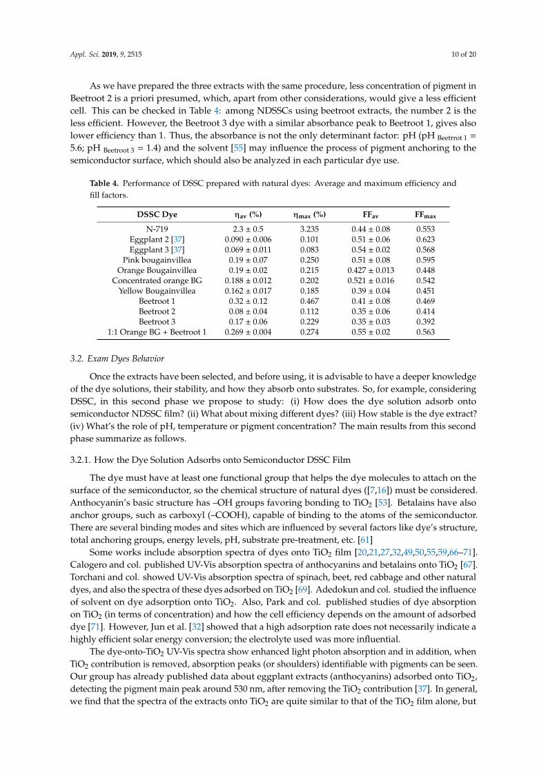
Table 4. Performance of DSSC prepared with natural dyes: Average and maximum efficiency andfill factors.
DSSC Dye ηav (%) ηmax (%) FFav FFmax
N-719 2.3 ± 0.5 3.235 0.44 ± 0.08 0.553Eggplant 2 [37] 0.090 ± 0.006 0.101 0.51 ± 0.06 0.623Eggplant 3 [37] 0.069 ± 0.011 0.083 0.54 ± 0.02 0.568
Pink bougainvillea 0.19 ± 0.07 0.250 0.51 ± 0.08 0.595Orange Bougainvillea 0.19 ± 0.02 0.215 0.427 ± 0.013 0.448
Concentrated orange BG 0.188 ± 0.012 0.202 0.521 ± 0.016 0.542Yellow Bougainvillea 0.162 ± 0.017 0.185 0.39 ± 0.04 0.451
Beetroot 1 0.32 ± 0.12 0.467 0.41 ± 0.08 0.469Beetroot 2 0.08 ± 0.04 0.112 0.35 ± 0.06 0.414Beetroot 3 0.17 ± 0.06 0.229 0.35 ± 0.03 0.392
1:1 Orange BG + Beetroot 1 0.269 ± 0.004 0.274 0.55 ± 0.02 0.563
3.2. Exam Dyes Behavior
Once the extracts have been selected, and before using, it is advisable to have a deeper knowledgeof the dye solutions, their stability, and how they absorb onto substrates. So, for example, consideringDSSC, in this second phase we propose to study: (i) How does the dye solution adsorb ontosemiconductor NDSSC film? (ii) What about mixing different dyes? (iii) How stable is the dye extract?(iv) What’s the role of pH, temperature or pigment concentration? The main results from this secondphase summarize as follows.
3.2.1. How the Dye Solution Adsorbs onto Semiconductor DSSC Film
The dye must have at least one functional group that helps the dye molecules to attach on thesurface of the semiconductor, so the chemical structure of natural dyes ([7,16]) must be considered.Anthocyanin’s basic structure has –OH groups favoring bonding to TiO2 [53]. Betalains have alsoanchor groups, such as carboxyl (–COOH), capable of binding to the atoms of the semiconductor.There are several binding modes and sites which are influenced by several factors like dye’s structure,total anchoring groups, energy levels, pH, substrate pre-treatment, etc. [61]
Some works include absorption spectra of dyes onto TiO2 film [20,21,27,32,49,50,55,59,66–71].Calogero and col. published UV-Vis absorption spectra of anthocyanins and betalains onto TiO2 [67].Torchani and col. showed UV-Vis absorption spectra of spinach, beet, red cabbage and other naturaldyes, and also the spectra of these dyes adsorbed on TiO2 [69]. Adedokun and col. studied the influenceof solvent on dye adsorption onto TiO2. Also, Park and col. published studies of dye absorptionon TiO2 (in terms of concentration) and how the cell efficiency depends on the amount of adsorbeddye [71]. However, Jun et al. [32] showed that a high adsorption rate does not necessarily indicate ahighly efficient solar energy conversion; the electrolyte used was more influential.
The dye-onto-TiO2 UV-Vis spectra show enhanced light photon absorption and in addition, whenTiO2 contribution is removed, absorption peaks (or shoulders) identifiable with pigments can be seen.Our group has already published data about eggplant extracts (anthocyanins) adsorbed onto TiO2,detecting the pigment main peak around 530 nm, after removing the TiO2 contribution [37]. In general,we find that the spectra of the extracts onto TiO2 are quite similar to that of the TiO2 film alone, but
Appl. Sci. 2019, 9, 2515 11 of 20
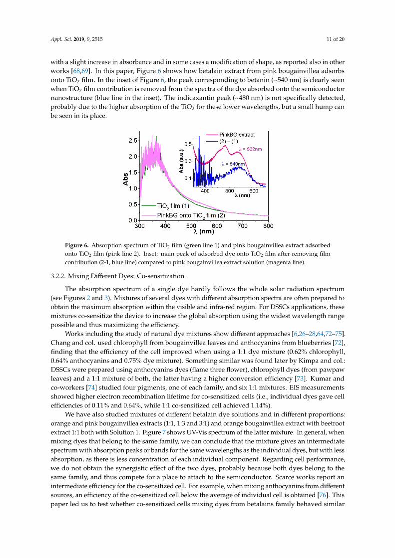
with a slight increase in absorbance and in some cases a modification of shape, as reported also in otherworks [68,69]. In this paper, Figure 6 shows how betalain extract from pink bougainvillea adsorbsonto TiO2 film. In the inset of Figure 6, the peak corresponding to betanin (~540 nm) is clearly seenwhen TiO2 film contribution is removed from the spectra of the dye absorbed onto the semiconductornanostructure (blue line in the inset). The indicaxantin peak (~480 nm) is not specifically detected,probably due to the higher absorption of the TiO2 for these lower wavelengths, but a small hump canbe seen in its place.
Appl. Sci. 2019, 9, x 11 of 21
The dye-onto-TiO2 UV-Vis spectra show enhanced light photon absorption and in addition, when TiO2 contribution is removed, absorption peaks (or shoulders) identifiable with pigments can be seen. Our group has already published data about eggplant extracts (anthocyanins) adsorbed onto TiO2, detecting the pigment main peak around 530 nm, after removing the TiO2 contribution [37] (p. 51, Figure 3). In general, we find that the spectra of the extracts onto TiO2 are quite similar to that of the TiO2 film alone, but with a slight increase in absorbance and in some cases a modification of shape, as reported also in other works [68,69]. In this paper, Figure 6 shows how betalain extract from pink bougainvillea adsorbs onto TiO2 film. In the inset of Figure 6, the peak corresponding to betanin (~540 nm) is clearly seen when TiO2 film contribution is removed from the spectra of the dye absorbed onto the semiconductor nanostructure (blue line in the inset). The indicaxantin peak (~480 nm) is not specifically detected, probably due to the higher absorption of the TiO2 for these lower wavelengths, but a small hump can be seen in its place.
Figure 6. Absorption spectrum of TiO2 film (green line 1) and pink bougainvillea extract adsorbed onto TiO2 film (pink line 2). Inset: main peak of adsorbed dye onto TiO2 film after removing film contribution (2-1, blue line) compared to pink bougainvillea extract solution (magenta line).
3.2.2. Mixing Different Dyes: Co-sensitization
The absorption spectrum of a single dye hardly follows the whole solar radiation spectrum (see Figures 2 and 3). Mixtures of several dyes with different absorption spectra are often prepared to obtain the maximum absorption within the visible and infra-red region. For DSSCs applications, these mixtures co-sensitize the device to increase the global absorption using the widest wavelength range possible and thus maximizing the efficiency
Works including the study of natural dye mixtures show different approaches [6,26–28,64,72–75]. Chang and col. used chlorophyll from bougainvillea leaves and anthocyanins from blueberries [72], finding that the efficiency of the cell improved when using a 1:1 dye mixture (0.62% chlorophyll, 0.64% anthocyanins and 0.75% dye mixture). Something similar was found later by Kimpa and col.: DSSC were prepared using anthocyanins dyes (flame three flower), chlorophyll dyes (from pawpaw leaves) and a 1:1 mixture of both, the latter having a higher conversion efficiency [73]. Kumar and co-workers [74] studied four pigments, one of each family, and six 1:1 mixtures. EIS measurements showed higher electron recombination lifetime for co-sensitized cells (i.e., individual dyes gave cell efficiencies of 0.11% and 0.64%, while 1:1 co-sensitized cell achieved 1.14%)
We have also studied mixtures of different betalain dye solutions and in different proportions: orange and pink bougainvillea extracts (1:1, 1:3 and 3:1) and orange bougainvillea extract with beetroot extract 1:1 both with Solution 1. Figure 7 shows UV-Vis spectrum of the latter mixture. In general, when mixing dyes that belong to the same family, we can conclude that the mixture gives an intermediate spectrum with absorption peaks or bands for the same wavelengths as the individual dyes, but with less absorption, as there is less concentration of each individual component. Regarding cell performance, we do not obtain the synergistic effect of the two dyes,
Figure 6. Absorption spectrum of TiO2 film (green line 1) and pink bougainvillea extract adsorbedonto TiO2 film (pink line 2). Inset: main peak of adsorbed dye onto TiO2 film after removing filmcontribution (2-1, blue line) compared to pink bougainvillea extract solution (magenta line).
3.2.2. Mixing Different Dyes: Co-sensitization
The absorption spectrum of a single dye hardly follows the whole solar radiation spectrum(see Figures 2 and 3). Mixtures of several dyes with different absorption spectra are often prepared toobtain the maximum absorption within the visible and infra-red region. For DSSCs applications, thesemixtures co-sensitize the device to increase the global absorption using the widest wavelength rangepossible and thus maximizing the efficiency.
Works including the study of natural dye mixtures show different approaches [6,26–28,64,72–75].Chang and col. used chlorophyll from bougainvillea leaves and anthocyanins from blueberries [72],finding that the efficiency of the cell improved when using a 1:1 dye mixture (0.62% chlorophyll,0.64% anthocyanins and 0.75% dye mixture). Something similar was found later by Kimpa and col.:DSSCs were prepared using anthocyanins dyes (flame three flower), chlorophyll dyes (from pawpawleaves) and a 1:1 mixture of both, the latter having a higher conversion efficiency [73]. Kumar andco-workers [74] studied four pigments, one of each family, and six 1:1 mixtures. EIS measurementsshowed higher electron recombination lifetime for co-sensitized cells (i.e., individual dyes gave cellefficiencies of 0.11% and 0.64%, while 1:1 co-sensitized cell achieved 1.14%).
We have also studied mixtures of different betalain dye solutions and in different proportions:orange and pink bougainvillea extracts (1:1, 1:3 and 3:1) and orange bougainvillea extract with beetrootextract 1:1 both with Solution 1. Figure 7 shows UV-Vis spectrum of the latter mixture. In general, whenmixing dyes that belong to the same family, we can conclude that the mixture gives an intermediatespectrum with absorption peaks or bands for the same wavelengths as the individual dyes, but with lessabsorption, as there is less concentration of each individual component. Regarding cell performance,we do not obtain the synergistic effect of the two dyes, probably because both dyes belong to thesame family, and thus compete for a place to attach to the semiconductor. Scarce works report anintermediate efficiency for the co-sensitized cell. For example, when mixing anthocyanins from differentsources, an efficiency of the co-sensitized cell below the average of individual cell is obtained [76]. Thispaper led us to test whether co-sensitized cells mixing dyes from betalains family behaved similar

Appl. Sci. 2019, 9, 2515 12 of 20
to co-sensitized cells with anthocyanins or achieved a higher efficiency. In our case, efficiency ofthe co-sensitized cells mixing betalain dyes is bit more than the average of the single sensitized cellsefficiency for the example in Figure 7. (see Table 4). The publications that report a notable increase inefficiency mix different dye families, i.e., betalain (beetroot) & chlorophyll (spinach) [64], chlorophyll &anthocyanins [72,73,75] or chlorophyll & betacyanins [74].Appl. Sci. 2019, 9, x 12 of 20
Figure 7. UV-Vis spectrum of dye mixture (green line) 1:1 orange bougainvillea extract with beetroot extract, both with Solution 1. Spectra of individual dyes are also included.
3.2.3. How Stable the Dye Extract is
Some researchers have published studies of DSSC stability or lifetime [8,12], showing how cell performance does not significantly change [60] (i.e., betacyanin dye cell was found stable for 12 weeks [63]) or diminishes with time [70,76–78].
Our group has also tested cell performance, keeping the cell working continuously for 350 h, with the aim to test not the dye, but the electrolyte. It was found that the cell with volatile electrolytes lasted for about 80 h, and when fresh electrolyte was injected, the cell could recover at 70% of the initial efficiency for almost 100 h. However, for cells with non-volatile electrolytes (i.e., ionic liquids), the efficiency kept constant at the beginning but diminished to 80% after 175 h and to 40% after 250 h [79].
In this work here, we have focused on dye extract stability, not the entire cell, just by recording UV-Vis spectra after a certain time, and the stability is not the same for the different extracts.
Anthocyanins can be affected by age, sugars, organic acids, co-pigments, etc. These molecules are susceptible to degradation under the influence of many factors, including temperature or basic pH in the presence of oxygen [2,80,81]. For example, Askar and col. [80] find degradation of the extract in 3 h of about 5% for the most stable extract or 20% for the less one. They measure degradation as the decrease of absorbance peaks in the dark. In contrast, our eggplant extract, with the extracting method and Solution 2, containing nasunin [37,82], seems to be stable for months even without any kind of purification, just keeping it preserved from air and light. Figure 8.a compares UV-Vis absorption spectra of a freshly prepared eggplant extract and of the same extract 3 months later. The spectra shape is the same and the absorption decreases by only 16% in the main peak at 550 nm. For this extract, pH is very acid, below 2 in all the samples. The influence of pH on anthocyanins absorption spectra has been previously reported [81,83]; the lower the pH, the lower the wavelength of the main absorbance peak and the more stable the dye.
Commented [M11]: 图 7 更换 Figure 7. UV-Vis spectrum of dye mixture (green line) 1:1 orange bougainvillea extract with beetrootextract, both with Solution 1. Spectra of individual dyes are also included.
3.2.3. How Stable the Dye Extract is
Some researchers have published studies of DSSC stability or lifetime [8,12], showing howcell performance does not significantly change [60] (i.e., betacyanin dye cell was found stable for12 weeks [63]) or diminishes with time [70,76–78].
Our group has also tested cell performance, keeping the cell working continuously for 350 h, withthe aim to test not the dye, but the electrolyte. It was found that the cell with volatile electrolytes lastedfor about 80 h, and when fresh electrolyte was injected, the cell could recover at 70% of the initialefficiency for almost 100 h. However, for cells with non-volatile electrolytes (i.e., ionic liquids), theefficiency kept constant at the beginning but diminished to 80% after 175 h and to 40% after 250 h [79].
In this work here, we have focused on dye extract stability, not the entire cell, just by recordingUV-Vis spectra after a certain time, and the stability is not the same for the different extracts.
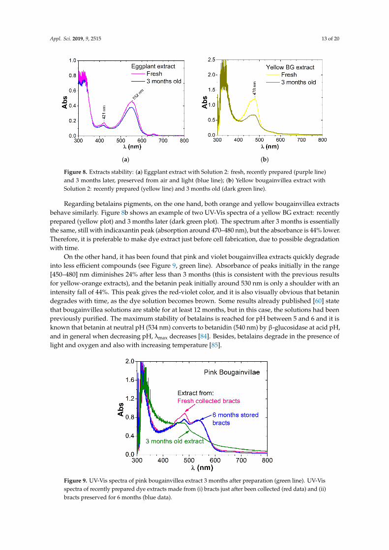
Anthocyanins can be affected by age, sugars, organic acids, co-pigments, etc. These molecules aresusceptible to degradation under the influence of many factors, including temperature or basic pH inthe presence of oxygen [2,80,81]. For example, Askar and col. [80] find degradation of the extract in3 h of about 5% for the most stable extract or 20% for the less one. They measure degradation as thedecrease of absorbance peaks in the dark. In contrast, our eggplant extract, with the extracting methodand Solution 2, containing nasunin [37,82], seems to be stable for months even without any kind ofpurification, just keeping it preserved from air and light. Figure 8a compares UV-Vis absorption spectraof a freshly prepared eggplant extract and of the same extract 3 months later. The spectra shape is thesame and the absorption decreases by only 16% in the main peak at 550 nm. For this extract, pH is veryacid, below 2 in all the samples. The influence of pH on anthocyanins absorption spectra has beenpreviously reported [81,83]; the lower the pH, the lower the wavelength of the main absorbance peakand the more stable the dye.
Appl. Sci. 2019, 9, 2515 13 of 20Appl. Sci. 2019, 9, x 13 of 21
(a) (b)
Figure 8. Extracts stability: (a) Eggplant extract with Solution 2: fresh, recently prepared (purple line) and 3 months later, preserved from air and light (blue line); (b) Yellow bougainvillea extract with Solution 2: recently prepared (yellow line) and 3 months old (dark green line).
Regarding betalains pigments, on the one hand, both orange and yellow bougainvillea extracts behave similarly. Figure 8.b shows an example of two UV-Vis spectra of a yellow BG extract: recently prepared (yellow plot) and 3 months later (dark green plot). The spectrum after 3 months is essentially the same, still with indicaxantin peak (absorption around 470–480 nm), but the absorbance is 44% lower. Therefore, it is preferable to make dye extract just before cell fabrication, due to possible degradation with time.
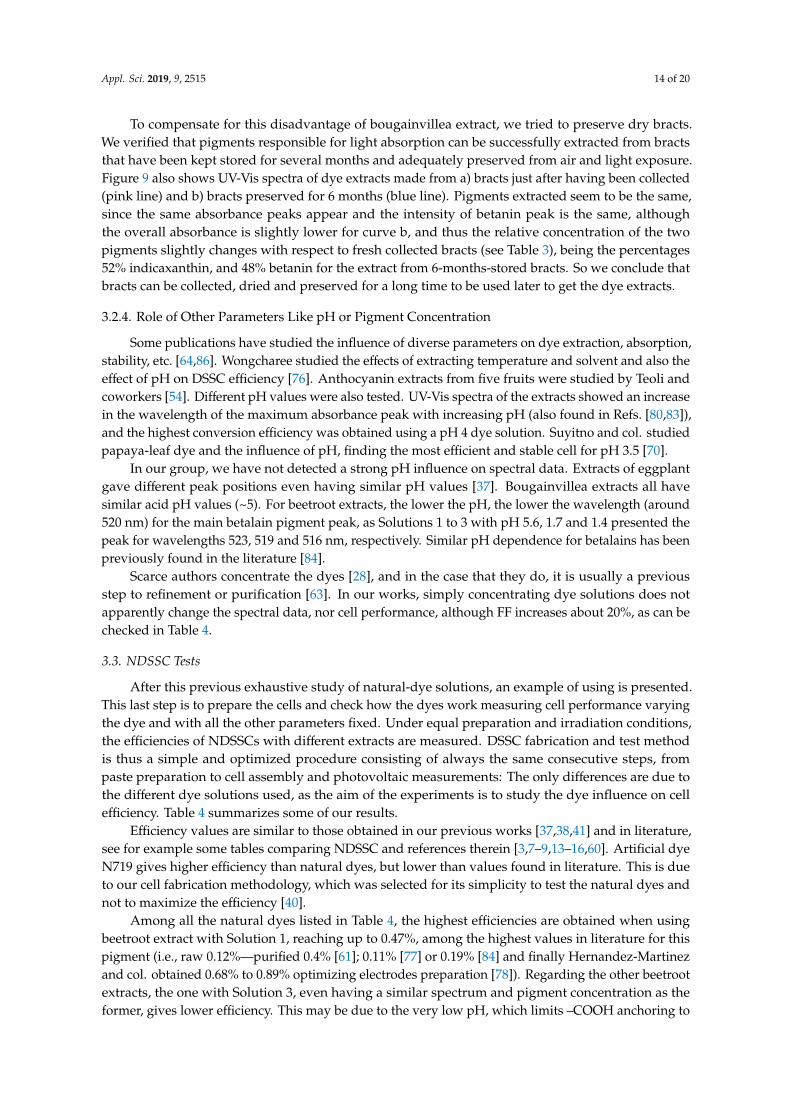
On the other hand, it has been found that pink and violet bougainvillea extracts quickly degrade into less efficient compounds (see Figure 9, green line). Absorbance of peaks initially in the range [450–480] nm diminishes 24% after less than 3 months (this is consistent with the previous results for yellow-orange extracts), and the betanin peak initially around 530 nm is only a shoulder with an intensity fall of 44%. This peak gives the red-violet color, and it is also visually obvious that betanin degrades with time, as the dye solution becomes brown. Some results already published [60] state that bougainvillea solutions are stable for at least 12 months, but in this case, the solutions had been previously purified. The maximum stability of betalains is reached for pH between 5 and 6 and it is known that betanin at neutral pH (534 nm) converts to betanidin (540 nm) by β-glucosidase at acid pH, and in general when decreasing pH, λmax decreases [84]. Besides, betalains degrade in the presence of light and oxygen and also with increasing temperature [85].
Figure 9. UV-Vis spectra of pink bougainvillea extract 3 months after preparation (green line). UV-Vis spectra of recently prepared dye extracts made from i) bracts just after been collected (red data) and ii) bracts preserved for 6 months (blue data).
Commented [M12]: 图 8 更换
Commented [M13]: 图 9 更换
Figure 8. Extracts stability: (a) Eggplant extract with Solution 2: fresh, recently prepared (purple line)and 3 months later, preserved from air and light (blue line); (b) Yellow bougainvillea extract withSolution 2: recently prepared (yellow line) and 3 months old (dark green line).
Regarding betalains pigments, on the one hand, both orange and yellow bougainvillea extractsbehave similarly. Figure 8b shows an example of two UV-Vis spectra of a yellow BG extract: recentlyprepared (yellow plot) and 3 months later (dark green plot). The spectrum after 3 months is essentiallythe same, still with indicaxantin peak (absorption around 470–480 nm), but the absorbance is 44% lower.Therefore, it is preferable to make dye extract just before cell fabrication, due to possible degradationwith time.
On the other hand, it has been found that pink and violet bougainvillea extracts quickly degradeinto less efficient compounds (see Figure 9, green line). Absorbance of peaks initially in the range[450–480] nm diminishes 24% after less than 3 months (this is consistent with the previous resultsfor yellow-orange extracts), and the betanin peak initially around 530 nm is only a shoulder with anintensity fall of 44%. This peak gives the red-violet color, and it is also visually obvious that betanindegrades with time, as the dye solution becomes brown. Some results already published [60] statethat bougainvillea solutions are stable for at least 12 months, but in this case, the solutions had beenpreviously purified. The maximum stability of betalains is reached for pH between 5 and 6 and it isknown that betanin at neutral pH (534 nm) converts to betanidin (540 nm) by β-glucosidase at acid pH,and in general when decreasing pH, λmax decreases [84]. Besides, betalains degrade in the presence oflight and oxygen and also with increasing temperature [85].
Appl. Sci. 2019, 9, x 13 of 20
(a) (b)
Figure 8. Extracts stability: (a) Eggplant extract with Solution 2: fresh, recently prepared (purple line) and 3 months later, preserved from air and light (blue line); (b) Yellow bougainvillea extract with Solution 2: recently prepared (yellow line) and 3 months old (dark green line).
Regarding betalains pigments, on the one hand, both orange and yellow bougainvillea extracts behave similarly. Figure 8.b shows an example of two UV-Vis spectra of a yellow BG extract: recently prepared (yellow plot) and 3 months later (dark green plot). The spectrum after 3 months is essentially the same, still with indicaxantin peak (absorption around 470–480 nm), but the absorbance is 44% lower. Therefore, it is preferable to make dye extract just before cell fabrication, due to possible degradation with time.
On the other hand, it has been found that pink and violet bougainvillea extracts quickly degrade into less efficient compounds (see Figure 9, green line). Absorbance of peaks initially in the range [450–480] nm diminishes 24% after less than 3 months (this is consistent with the previous results for yellow-orange extracts), and the betanin peak initially around 530 nm is only a shoulder with an intensity fall of 44%. This peak gives the red-violet color, and it is also visually obvious that betanin degrades with time, as the dye solution becomes brown. Some results already published [60] state that bougainvillea solutions are stable for at least 12 months, but in this case, the solutions had been previously purified. The maximum stability of betalains is reached for pH between 5 and 6 and it is known that betanin at neutral pH (534 nm) converts to betanidin (540 nm) by β-glucosidase at acid pH, and in general when decreasing pH, λmax decreases [84]. Besides, betalains degrade in the presence of light and oxygen and also with increasing temperature [85].
Figure 9. UV-Vis spectra of pink bougainvillea extract 3 months after preparation (green line). UV-Vis spectra of recently prepared dye extracts made from i) bracts just after been collected (red data) and ii) bracts preserved for 6 months (blue data).
To compensate for this disadvantage of bougainvillea extract, we tried to preserve dry bracts. We verified that pigments responsible for light absorption can be successfully extracted from bracts that have been kept stored for several months and adequately preserved from air and light exposure. Figure 9 also shows UV-Vis spectra of dye extracts made from a) bracts just after having been collected (pink line) and b) bracts preserved for 6 months (blue line). Pigments extracted seem to be the same, since the same absorbance peaks appear and the intensity of betanin peak is the same, although the overall absorbance is slightly lower for curve b, and thus the relative concentration of the two pigments slightly changes with respect to fresh collected bracts (see Table 3), being the percentages 52% indicaxanthin, and 48% betanin for the extract from 6-months-stored bracts. So we conclude that bracts can be collected, dried and preserved for a long time to be used later to get the dye extracts.
Commented [M12]: 图 8 更换
Commented [M13]: 图 9 更换 Figure 9. UV-Vis spectra of pink bougainvillea extract 3 months after preparation (green line). UV-Visspectra of recently prepared dye extracts made from (i) bracts just after been collected (red data) and (ii)bracts preserved for 6 months (blue data).
Appl. Sci. 2019, 9, 2515 14 of 20
To compensate for this disadvantage of bougainvillea extract, we tried to preserve dry bracts.We verified that pigments responsible for light absorption can be successfully extracted from bractsthat have been kept stored for several months and adequately preserved from air and light exposure.Figure 9 also shows UV-Vis spectra of dye extracts made from a) bracts just after having been collected(pink line) and b) bracts preserved for 6 months (blue line). Pigments extracted seem to be the same,since the same absorbance peaks appear and the intensity of betanin peak is the same, althoughthe overall absorbance is slightly lower for curve b, and thus the relative concentration of the twopigments slightly changes with respect to fresh collected bracts (see Table 3), being the percentages52% indicaxanthin, and 48% betanin for the extract from 6-months-stored bracts. So we conclude thatbracts can be collected, dried and preserved for a long time to be used later to get the dye extracts.
3.2.4. Role of Other Parameters Like pH or Pigment Concentration
Some publications have studied the influence of diverse parameters on dye extraction, absorption,stability, etc. [64,86]. Wongcharee studied the effects of extracting temperature and solvent and also theeffect of pH on DSSC efficiency [76]. Anthocyanin extracts from five fruits were studied by Teoli andcoworkers [54]. Different pH values were also tested. UV-Vis spectra of the extracts showed an increasein the wavelength of the maximum absorbance peak with increasing pH (also found in Refs. [80,83]),and the highest conversion efficiency was obtained using a pH 4 dye solution. Suyitno and col. studiedpapaya-leaf dye and the influence of pH, finding the most efficient and stable cell for pH 3.5 [70].
In our group, we have not detected a strong pH influence on spectral data. Extracts of eggplantgave different peak positions even having similar pH values [37]. Bougainvillea extracts all havesimilar acid pH values (~5). For beetroot extracts, the lower the pH, the lower the wavelength (around520 nm) for the main betalain pigment peak, as Solutions 1 to 3 with pH 5.6, 1.7 and 1.4 presented thepeak for wavelengths 523, 519 and 516 nm, respectively. Similar pH dependence for betalains has beenpreviously found in the literature [84].
Scarce authors concentrate the dyes [28], and in the case that they do, it is usually a previousstep to refinement or purification [63]. In our works, simply concentrating dye solutions does notapparently change the spectral data, nor cell performance, although FF increases about 20%, as can bechecked in Table 4.
3.3. NDSSC Tests
After this previous exhaustive study of natural-dye solutions, an example of using is presented.This last step is to prepare the cells and check how the dyes work measuring cell performance varyingthe dye and with all the other parameters fixed. Under equal preparation and irradiation conditions,the efficiencies of NDSSCs with different extracts are measured. DSSC fabrication and test methodis thus a simple and optimized procedure consisting of always the same consecutive steps, frompaste preparation to cell assembly and photovoltaic measurements: The only differences are due tothe different dye solutions used, as the aim of the experiments is to study the dye influence on cellefficiency. Table 4 summarizes some of our results.
Efficiency values are similar to those obtained in our previous works [37,38,41] and in literature,see for example some tables comparing NDSSC and references therein [3,7–9,13–16,60]. Artificial dyeN719 gives higher efficiency than natural dyes, but lower than values found in literature. This is dueto our cell fabrication methodology, which was selected for its simplicity to test the natural dyes andnot to maximize the efficiency [40].
Among all the natural dyes listed in Table 4, the highest efficiencies are obtained when usingbeetroot extract with Solution 1, reaching up to 0.47%, among the highest values in literature for thispigment (i.e., raw 0.12%—purified 0.4% [61]; 0.11% [77] or 0.19% [84] and finally Hernandez-Martinezand col. obtained 0.68% to 0.89% optimizing electrodes preparation [78]). Regarding the other beetrootextracts, the one with Solution 3, even having a similar spectrum and pigment concentration as theformer, gives lower efficiency. This may be due to the very low pH, which limits –COOH anchoring to

Appl. Sci. 2019, 9, 2515 15 of 20
the semiconductor surface. Solution 2 behaves similarly, and, in addition, the absorbance spectrumshows the lowest pigment concentration, so in this case the efficiency is even lower.
Eggplant extracts gave low efficiencies compared with already published data (i.e., 0.48% [82]).Other works obtain lower efficiency for eggplant (0.15%) compared to other natural dyes, and attributeit to more pronounced recombination reactions for eggplant dye than for the other dyes [34].
The different bougainvillea extracts achieved similar efficiencies (0.16% to 0.19%) no matter whatbract color, which is due to the presence of different betalain groups and in different proportions.Red-purple bracts extracts gave also efficiencies of the same order or up to (0.29 ± 0.02) % in ourprevious works, and the small differences found were due to other factors like film thickness, surfactantused in paste preparation, and porosity of the semiconductor film [38,41].
The fill factor does not change noticeably when varying the dye, although in our experiments thehighest FF (0.62) has been found for eggplant extracts, while the lowest FF (0.35) is obtained for cellsmade with beetroot extract.
We have also concentrated some of the dyes, but no clear increase or change in efficiencies hasbeen seen. In Table 4, we give as an example the cell efficiency when using orange bougainvilleaextract concentrated 10% wt. Efficiency does not change, although the fill factor does increase from0.43 (initial extract) to 0.52 (concentrated).
4. Conclusions
Based on our experience and after doing a wide literature review, we propound a general protocolto optimize a simple natural dye production method, without complicating the chemical procedures,with the aim of enhancing the dye potential to whatever subsequent use. In particular, as a possibleapplication, NDSSCs performance is considered.
In conclusion, the protocol consists of these main and minimum advisable steps to obtain and testnatural pigments: (i) Dye solutions preparation and characterization by UV-Vis spectrometry: Selectnatural raw vegetable pigment, preferably to be obtained locally and easily, and select the simplestand best extraction solution and method. (ii) Exam dye solutions behavior i.e., how they adsorbonto semiconductor film, how stable they are, or what is the influence of different parameters onextraction and adsorption. And finally (iii) NDSSC tests: As an example of application of the previousexhaustive study, we have designed a simple and economical cell fabrication method and measuredcell performance in environmental conditions varying the dye and with all the other parameters fixed.The novelty of this approach is that it gives a previous deeper knowledge of the dye, crucial to the finalNDSSC performance.
We have successfully employed this protocol with betalain pigments (a series of four bougainvilleadyes and three beetroot dyes) and anthocyanins (three eggplant dyes). The previous UV-Vis spectraanalysis gives useful information about:
• The best extraction solution and method: for betalains, Solution 1 (acetone/water, moderatelyacid), for anthocyanins, Solutions 2 and 3 (very acidic solutions).
• The relative concentration of different pigment molecules for the different extracts.• Some candidate vegetable material can be a priori selected or ruled out by inspection of the extract
spectra. Besides, comparison with the corresponding spectra of dry raw material performed witha spectroradiometer, gives reliable information about the dye molecules which can be potentiallyextracted. We show the example of bougainvillea-colored bracts spectra.
• The dye stability or ageing: Anthocyanins are more stable and betalain molecules degrade, butconservation of the dry precursor (collected bracts) for at least 6 months is possible.
• How the dye adsorbs onto photoelectrode film, and the effect of mixing dye extracts. In this paper,we focus on mixing dyes from the betalain family to complement results in the bibliography.
For the selected cell fabrication method, NDSSC tests show that for anthocyanins the bestperformances were obtained with Solutions 2 and 3, but with very low values compared to literature.
Appl. Sci. 2019, 9, 2515 16 of 20
In this work, betalains give higher efficiencies than anthocyanins. For betalains, the best extract isobtained with Solution 1, as initially found from UV-Vis spectrometry analysis. Within the betalainfamily, for bougainvillea dyes, similar performances were obtained for the different varieties, andfor beetroots the best result reached up to 0.47% cell efficiency, which is among the highest valuesin literature.
Author Contributions: Conceptualization, M.J.G.-S.; Methodology, M.J.G.-S.; Investigation, M.J.G.-S.; Resources,M.J.A.; Writing—Original Draft Preparation, M.J.G.-S.; Writing—Review & Editing, M.J.G.-S. and M.J.A.;Visualization, M.J.A.; Supervision, M.J.G.-S. and M.J.A.; Project Administration, M.J.A.; Funding Acquisition,M.J.A. Both authors have read and approved the manuscript.
Funding: The research work leading to this article received ERDF funds from the Spanish government withinthe framework of the SOLTERMIN project (Ref. ENE2017-83973-R) of the Ministerio de Economía, Industria yCompetitividad (Spanish Ministry of Economy, Industry and Competitiveness).
Acknowledgments: The authors sincerely thank S. Rosiek and J. Alonso from CIESOL (Solar Energy ResearchCenter, UAL-PSA, Almería, Spain); and J. Fernández-Reche from Plataforma Solar de Almería (PSA-CIEMAT,Almería, Spain); S. Galera and J. Barbero are acknowledged for the technical support.
Conflicts of Interest: “The authors declare no conflict of interest.”.
References
1. Mohd, Y.; Mohd, S.; Faqeer, M. Natural Colorants: Historical, Processing and Sustainable Prospects. Nat. Prod.Bioprospect. 2017, 7, 123–145. [CrossRef]
2. Miller, R.; Owens, S.J.; Rørslett, B. Plants and colour: Flowers and pollination. Opt. Laser Technol. 2011, 43,282–294. [CrossRef]
3. Narayan, M.R. Review: Dye sensitized solar cells based on natural photosensitizers. Renew. Sust. Energ. Rev.2012, 16, 208–215. [CrossRef]
4. Sugathan, V.; John, E.; Sudhakar, K. Recent improvements in dye sensitized solar cells: A review. Renew. Sust.Energ. Rev. 2015, 52, 54–64. [CrossRef]
5. Sharma, S.; Siwach, B.; Ghoshal, S.K.; Mohan, D. Dye sensitized solar cells: From genesis to recent drifts.Renew. Sust. Energ. Rev. 2017, 70, 529–537. [CrossRef]
6. Mohiuddin, O.; Obaidullah, M.; Sabah, C. Improvement in dye sensitized solar cells from past to present.Opt. Quant. Electron. 2018, 50, 377. [CrossRef]
7. Hug, H.; Bader, M.; Mair, P.; Glatzel, T. Biophotovoltaics: Natural pigments in dye-sensitized solar cells.Appl. Energ. 2014, 115, 216–225. [CrossRef]
8. Ludin, N.A.; Mahmoud, A.M.A.A.; Mohamad, A.B.; Amir, H.K.A.; Sopian, K.; Karim, N.S.A. Review on thedevelopment of natural dye photosensitizer for dye-sensitized solar cells. Renew. Sust. Energ. Rev. 2014, 31,386–396. [CrossRef]
9. Shalini, S.; Balasundara Prabhu, R.; Prasanna, S.; Mallick, T.K.; Senthilarasu, S. Review on natural dyesensitized solar cells: Operation, materials and methods. Renew. Sust. Energ. Rev. 2015, 51, 1306–1325.[CrossRef]
10. Gong, J.; Sumathy, K.; Qiao, Q.; Zhou, Z. Review on dye-sensitized solar cells (DSSCs): Advanced techniquesand research trends. Renew. Sust. Energ. Rev. 2017, 68, 234–246. [CrossRef]
11. Ye, M.; Wen, X.; Wang, M.; Iocozzia, J.; Zhang, N.; Lin, C.; Lin, Z. Recent advances in dye-sensitized solarcells: From photoanodes, sensitizers and electrolytes to counter electrodes. Mater. Today 2015, 18, 155–162.[CrossRef]
12. Mozaffari, S.; Nateghi, M.R.; Zarandi, M.B. An overview of the challenges in the commercialization of dyesensitized solar cells. Renew. Sust. Energ. Rev. 2017, 71, 675–686. [CrossRef]
13. Chawla, P.; Tripathi, M. Novel improvements in the sensitizers of dye-sensitized solar cells for enhancementin efficiency—A review. Int. J. Energy Res. 2015, 39, 1579–1596. [CrossRef]
14. Shalini, S.; Balasundaraprabhu, R.; Satish-Kumar, T.; Prabavathy, N.; Senthilarasu, S.; Prasanna, S. Status andoutlook of sensitizers/dyes used in dye sensitized solar cells (DSSC): A review. Int. J. Energy Res. 2016, 40,1303–1320. [CrossRef]
15. Zhou, H.; Wu, L.; Gao, Y.; Ma, T. Dye-sensitized solar cells using 20 natural dyes as sensitizers. J. Photoch.Photobio. A 2011, 219, 188–194. [CrossRef]
Appl. Sci. 2019, 9, 2515 17 of 20
16. Kumara, N.T.R.N.; Lim, A.; Lim, S.M.; Petra, M.I.; Ekanayake, P. Recent progress and utilization of naturalpigments in dye sensitized solar cells: A review. Renew. Sust. Energ. Rev. 2017, 78, 301–317. [CrossRef]
17. Richhariya, G.; Kumar, A.; Tekasakul, P.; Gupta, B. Natural dyes for dye sensitized solar cell: A review.Renew. Sust. Energ. Rev. 2017, 69, 705–718. [CrossRef]
18. Gu, P.; Yang, D.; Zhu, X.; Sun, H.; Li, J. Fabrication and characterization of dye-sensitized solar cells based onnatural plants. Chem. Phys. Lett. 2018, 693, 16–22. [CrossRef]
19. Mahmoud, A.M.A.A.; Ludin, N.A.; Mohamad, A.B.; Kadhum, A.A.H.; Mukhlus, A. Application ofdyes extracted from Alternanthera dentata leaves and Musa acuminata bracts as natural sensitizers fordye-sensitized solar cells. Spectrochim. Acta A Mol. Biomol. Spectrosc. 2018, 192, 487–498. [CrossRef]
20. Maurya, I.C.; Singh, S.; Neetu; NeetuGupta, A.K.; Srivastava, P.; Bahadur, L. Dye-sensitized solar cellsemploying extracts from four cassia flowers as natural sensitizers: Studies on dye ingredient effect onphotovoltaic performance. J. Electron. Mater. 2018, 47, 225–232. [CrossRef]
21. Adedokun, O.; Sanusi, Y.K.; Awodugba, A.O. Solvent dependent natural dye extraction and its sensitizationeffect for dye sensitized solar cells. Optik 2018, 174, 497–507. [CrossRef]
22. Calogero, G.; Barichello, J.; Citro, I.; Mariani, P.; Vesce, L.; Bartolotta, A.; Di Carlo, A.; Di Marco, G.Photoelectrochemical and spectrophotometric studies on dye-sensitized solar cells (DSCs) and stablemodules (DSCMs) based on natural apocarotenoids pigments. Dyes Pigments 2018, 155, 75–83. [CrossRef]
23. Sanjay, P.; Deepa, K.; Madhavan, J.; Senthil, S. Optical, spectral and photovoltaic characterization of naturaldyes extracted from leaves of Peltophorum pterocarpum and Acalypha amentacea used as sensitizers forZnO based dye sensitized solar cells. Opt. Mater. 2018, 83, 192–199. [CrossRef]
24. Manaa, M.B.; Bouaziz, N.; Schmaltz, B.; Van, F.T.; Lamine, A.B. Study of the effect of variation in temperatureand pH on the adsorption process of natural Gardenia yellow dye into TiO2 mesoporous for dye sensitizedsolar cells using the statistical physics formalism: Physicochemical and thermodynamic investigation.Micropor. Mesopor. Mat. 2018, 270, 82–92. [CrossRef]
25. Gu, P.; Yang, D.; Zhu, X.; Sun, H.; Li, J. Performance of dye-sensitized solar cells based on natural dyes.Opt. Quant. Electron. 2018, 50, 223. [CrossRef]
26. Tahir, D.; Satriani, W.; Gareso, P.L.; Abdullah, B. Dye sensitized solar cell (DSSC) with natural dyes extractedfrom Jatropha leaves and purple Chrysanthemum flowers as sensitizer. IOP Conf. Ser. J. Phys. Conf. Ser.2018, 979, 012056. [CrossRef]
27. Rajkumar, S.; Nirmal Kumar, M.; Suguna, K.; Muthulakshmi, S.; Ashok Kumar, R. Enhanced performance ofdye-sensitized solar cells using natural cocktail dye as sensitizer. Optik 2019, 178, 224–230. [CrossRef]
28. Hosseinnezhad, M.; Rouhani, S.; Gharanjig, K. Extraction and application of natural pigments for fabricationof green dye-sensitized solar cells. Opto. Electron. Rev. 2018, 26, 165–171. [CrossRef]
29. Yildiz, Z.K.; Atilgan, A.; Atli, A.; Özel, K.; Altinkaya, C.; Yildiz, A. Enhancement of efficiency of natural andorganic dye sensitized solar cells using thin film TiO2 photoanodes fabricated by spin-coating. J. Photochem.Photobiol. A 2019, 368, 23–29. [CrossRef]
30. Liu, Q.; Gao, N.; Liu, D.; Liu, J.; Li, Y. Structure and photoelectrical properties of natural photoactive dyes forsolar cells. Appl. Sci. 2018, 8, 1697. [CrossRef]
31. Cassone, G.; Calogero, G.; Sponer, J.; Saija, F. Mobilities of iodide anions in aqueous solutions for applicationsin natural dye-sensitized solar cells. Phys. Chem. Chem. Phys. 2018, 20, 13038–13046. [CrossRef] [PubMed]
32. Jun, H.K.; Arof, A.K. Performance of natural dyes as sensitizer in dye-sensitized solar cells employingLiBOB-based liquid electrolyte. Ionics 2018, 24, 915–922. [CrossRef]
33. Swathi, K.E.; Jinchu, I.; Sreelatha, K.S.; Sreekala, C.O.; Menon, S.K. Effect of microwave exposure on the photoanode of DSSC sensitized with natural dye. IOP Conf. Ser. Mater. Sci. Eng. 2018, 310, 012141. [CrossRef]
34. Tractz, G.T.; Viomar, A.; Dias, B.V.; de Lima, C.A.; Banczek, E.P.; da Cunha, M.T.; Antunes, S.R.M.;Rodrigues, P.R.P. Recombination Study of Dye Sensitized Solar Cells with Natural Extracts. J. Braz. Chem. Soc.2019, 30, 371–378. [CrossRef]
35. Lucioli, S.; Di Bari, C.; Forni, C.; Di Carlo, A.; Barrajón-Catalán, E.; Micol, V.; Nota, P.; Teoli, F.; Matteocci, F.;Frattarelli, A.; et al. Anthocyanic pigments from elicited in vitro grown shoot cultures of Vacciniumcorymbosum L., cv. Brigitta Blue, as photosensitizer in natural dye-sensitized solar cells (NDSSC). J. Photochem.Photobiol. B 2018, 188, 69–76. [CrossRef]
Appl. Sci. 2019, 9, 2515 18 of 20
36. Prabavathy, N.; Shalini, S.; Balasundaraprabhu, R.; Velauthapillai, D.; Prasanna, S.; Balaji, G.;Muthukumarasamy, N. Algal buffer layers for enhancing the efficiency of anthocyanins extracted from rosepetals for natural dye-sensitized solar cell (DSSC). Int. J. Energy Res. 2018, 42, 790–801. [CrossRef]
37. Galera-Moreno, S.; González-Galindo, E.; Ariza, M.J.; García-Salinas, M.J. Influence of natural dyes in low-costdye-sensitized solar cell’s efficiency. In Materials and Technologies for Energy Efficiency; Méndez-Vilas, A., Ed.;BrownWalker Press: Boca Ratón, FL, USA, 2015; pp. 48–52.
38. Maldonado-Valdivia, A.I.; Galindo, E.G.; Ariza, M.J.; Garcia-Salinas, M.J. Surfactant influence in theperformance of titanium dioxide photoelectrodes for dye-sensitized solar cells. Sol. Energy 2013, 91, 263–272.[CrossRef]
39. Ito, S.; Kitamura, T.; Wada, Y.; Yanagida, S. Facile fabrication of mesoporous TiO2 electrodes for dye solarcells: Chemical modification and repetitive coating. Sol. Energy Mater. Sol. Cells 2003, 76, 3–13. [CrossRef]
40. Ito, S.; Murakami, T.N.; Comte, P.; Liska, P.; Grätzel, C.; Nazeeruddin, M.K.; Grätzel, M. Fabrication of thinfilm dye sensitized solar cells with solar to electric power conversion efficiency over 10%. Thin Solid Films2008, 516, 4613–4619. [CrossRef]
41. Galindo, E.G.; Ariza, M.J.; De las Nieves, F.J.; García-Salinas, M.J. Effects of multilayer coating and calcinationprocedures on the morphology of dye-sensitized solar cell semiconductor photoelectrodes. Thin Solid Films2015, 590, 230–240. [CrossRef]
42. Syafinar, R.; Gomesh, N.; Irwanto, M.; Fareq, M.; Irwan, Y.M. Potential of Purple Cabbage, Coffee, Blueberryand Turmeric as Nature Based Dyes for Dye Sensitized Solar Cell (DSSC). Energy Procedia 2015, 79, 799–807.[CrossRef]
43. Belay, A.; Ture, K.; Redi, M.; Asfaw, A. Measurement of caffeine in coffee beans with UV/vis spectrometer.Food Chem. 2008, 108, 310–315. [CrossRef]
44. Setiawan, T.; Subekti, W.Y.; Nur’Adya, S.S.; Ilmiah, K.; Ulfa, S.M. Performance of natural dye and counterelectrode from robusta coffee beans peel waste for fabrication of dye-sensitized solar cell (DSSC). IOP Conf.Ser.: Mat. Sci. Eng. 2018, 299, 012073. [CrossRef]
45. Kawase, M.; Takahashi, M. Chemical composition of sporopollenin in magnolia grandiflora (Magnoliaceae)and hibiscus syriacus (Malvaceae). Grana 1995, 34, 242–245. [CrossRef]
46. Di Paola-Naranjo, R.D.; Sánchez-Sánchez, J.; González-Paramás, A.M.; Rivas-Gonzalo, J.C. Liquidchromatographic–mass spectrometric analysis of anthocyanin composition of dark blue bee pollen fromEchium plantagineum. J. Chromatogr. A 2004, 1054, 205–210. [CrossRef] [PubMed]
47. Urbanczyk, J.; Fernandez-Casado, M.A.; Díaz, T.E.; Heras, P.; Infante, M.; Borrego, A.G. Spectral fluorescencevariation of pollen and spores from recent peat-forming plants. Int. J. Coal Geol. 2014, 131, 263–273. [CrossRef]
48. Mularczyk-Oliwa, M.; Bombalska, A.; Kaliszewski, M.; Włodarski, M.; Trafny, A.; Kopczynski, K.; Kwasny, M.;Szpakowska, M.; Zbieta, E. Comparison of fluorescence spectroscopy and FTIR in differentiation of plantpollens. Spectrochim. Acta A Mol. Biomol. Spectrosc. 2012, 97, 246–254. [CrossRef] [PubMed]
49. Calogero, G.; Di Marco, G.; Caramori, S.; Cazzanti, S.; Argazzi, R.; Bignozzi, C.A. Natural dye sensitizers forphotoelectrochemical cells. Energy Environ. Sci. 2009, 2, 1162–1172. [CrossRef]
50. Shanmugam, V.; Manoharan, S.; Anandan, S.; Murugan, R. Performance of dye-sensitized solar cellsfabricated with extracts from fruits of ivy gourd and flowers of red frangipani as sensitizers. Spectrochim.Acta A Mol. Biomol. Spectrosc. 2013, 104, 35–40. [CrossRef] [PubMed]
51. Gholamrezaei, S.; Salavati-Niasari, M. Natural sensitizer for low cost dye sensitized solar cell based onStrontium Titanate nanoparticles. J. Mater. Sci. Mater. Electron. 2016, 27, 2467–2472. [CrossRef]
52. Sashank, T.V.S.S.P.; Manikanta, B.; Pasula, A. Fabrication and experimental investigation on dye sensitizedsolar cells using titanium dioxide nano particles. Mater. Today Proc. 2017, 4, 3918–3925. [CrossRef]
53. Liao, S.K.; Chang, Y.H.; Wu, C.T.; Lai, Y.R.; Chen, W.Y. Fabrication of anthocyanin-sensitized nanocrystallinetitanium dioxide solar cells using supercritical carbon dioxide. J. CO2 Util. 2017, 21, 513–520. [CrossRef]
54. Teoli, F.; Lucioli, S.; Nota, P.; Frattarelli, A.; Matteocci, F.; Di Carlo, A.; Caboni, E.; Forni, C. Role of pH andpigment concentration for natural dye-sensitized solar cells treated with anthocyanin extracts of commonfruits. J. Photoch. Photobio. A 2016, 316, 24–30. [CrossRef]
55. Mahmoud, A.M.A.A.; Mohamad, A.B.; Kadhum, A.A.H.; Ludin, N.A. Effect of solvents on the extraction ofnatural pigments and adsorption onto TiO2 for dye-sensitized solar cell applications. Spectrochim. Acta AMol. Biomol. Spectrosc. 2015, 138, 130–137. [CrossRef]
Appl. Sci. 2019, 9, 2515 19 of 20
56. Maabong, K.; Muiva, C.M.; Monowe, P.; Sathiaraj, T.S.; Hopkins, M.; Nguyen, L.; Malungwa, K.; Thobega, M.Natural pigments as photosensitizers for dye-sensitized solar cells with TiO2 thin films. Int. J. Renew. Sustain.Energy Res. 2015, 5, 54–60.
57. Isah, K.U.; Ahmadu, U.; Idris, A.; Kimpa, M.I.; Uno, U.E.; Ndamitso, M.M.; Alu, N. Betalain pigments asnatural photosensitizers for dye-sensitized solar cells: The effect of dye pH on the photoelectric parameters.Mater. Renew. Sustain. Energy 2015, 4, 39. [CrossRef]
58. Kumar, S.N.A.; Ritesh, S.K.; Sharmila, G.; Muthukumaran, C. Extraction optimization and characterizationof water soluble red purple pigment from floral bracts of Bougainvillea glabra. Arab. J. Chem. 2017, 10,2145–2150. [CrossRef]
59. Damit, D.N.F.P.; Galappaththi, K.; Lim, A.; Petra, M.I.; Ekanayake, P. Formulation of water to ethanol ratio asextraction solvents of Ixora coccinea and Bougainvillea glabra and their effect on dye aggregation in relationto DSSC performance. Ionics 2017, 23, 485–495. [CrossRef]
60. Hernandez-Martinez, A.R.; Estevez, M.; Vargas, S.; Quintanilla, F.; Rodriguez, R. New dye-sensitized solarcells obtained from extracted bracts of bougainvillea Glabra and Spectabilis Betalain Pigments by DifferentPurification Processes. Int. J. Mol. Sci. 2011, 12, 5565–5576. [CrossRef]
61. Oprea, C.I.; Dumbravă, A.; Enache, I.; Georgescu, A.; Gîrtu, M.A. A combined experimental and theoreticalstudy of natural betalain pigments used in dye-sensitized solar cells. J. Photochem. Photobio. A 2012, 240,5–13. [CrossRef]
62. Shanmugam, V.; Manoharan, S.; Sharafali, A.; Anandan, S.; Murugan, R. Green grasses as light harvestersin dye sensitized solar cells. Spectrochim. Acta A Mol. Biomol. Spectrosc. 2015, 135, 947–952. [CrossRef][PubMed]
63. Tripathi, M.; Upadhyay, R.; Pandey, A. Novel dye based photoelectrode for improvementof solar cellconversion efficiency. Appl. Sol. Energy 2013, 49, 54–57. [CrossRef]
64. Sengupta, D.; Mondal, B.; Mukherjee, K. Visible light absorption and photo-sensitizing properties of spinachleaves and beetroot extracted natural dyes. Spectrochim. Acta A 2015, 148, 85–92. [CrossRef]
65. Hemmatzadeh, R.; Mohammadi, A. Improving optical absorptivity of natural dyes for fabrication of efficientdye-sentitized solar cells. J. Theor. Appl. Phys. 2013, 7, 57. [CrossRef]
66. Senthil, T.S.; Muthukumarasamy, N.; Velauthapillai, D.; Agilan, S.; Thambidurai, M.; Balasundaraprabhu, R.Natural dye (cyaniding 3-O-glucoside) sensitized nanocrystalline TiO2 solar cell fabricated using liquidelectrolyte/quasi-solid-state polymer electrolyte. Renew. Energy 2011, 36, 2484–2488. [CrossRef]
67. Calogero, G.; Yum, J.H.; Sinopoli, A.; Di Marco, G.; Grätzel, M.; Nazeeruddin, M.K. Anthocyanins andbetalains as light-harvesting pigments for dye-sensitized solar cells. Sol. Energy 2012, 86, 1563–1575.[CrossRef]
68. Hemalatha, K.V.; Karthick, S.N.; Justin-Raj, C.; Hong, N.Y.; Kim, S.K.; Kim, H.-J. Performance of Kerriajaponica and Rosa chinensis flower dyes as sensitizers for dye-sensitized solar cells. Spectrochim. Acta A Mol.Biomol. Spectrosc. 2012, 96, 305–309. [CrossRef]
69. Torchani, A.; Gharbi, R.; Fathallah, M. Study of natural dyes for sensitized solar cells applications.Sens. Transducers 2014, 27, 185–189.
70. Suyitno, S.; Saputra, T.J.; Supriyanto, A.; Arifin, Z. Stability and efficiency of dye-sensitized solar cells basedon papaya-leaf dye. Spectrochim. Acta A Mol. Biomol. Spectrosc. 2015, 148, 99–104. [CrossRef]
71. Park, K.H.; Kim, T.Y.; Park, J.Y.; Jin, E.M.; Yim, S.H.; Choi, D.Y.; Lee, J.W. Adsorption characteristics ofgardenia yellow as natural photosensitizer for dye-sensitized solar cells. Dyes Pigments 2013, 96, 595–601.[CrossRef]
72. Chang, H.; Kao, M.J.; Chen, T.L.; Fuo, H.G.; Choand, K.C.; Lin, X.P. Natural sensitizer for dye-sensitizedsolar cells using three layers of photoelectrode thin films with a Schottky barrier. Am. J. Eng. Appl. Sci. 2011,4, 214–222. [CrossRef]
73. Kimpa, M.I.; Momoh, M.; Isah, K.U.; Yahya, H.N.; Ndamitso, M.M. Photoelectric characterization of dyesensitized solar cells using natural dye from pawpaw leaf and flame tree flower as sensitizers. Mater. Sci. Appl.2012, 3, 281–286. [CrossRef]
74. Kumar, K.A.; Subalakshmi, K.; Senthilselvan, J. Effect of mixed valence state of titanium on reducedrecombination for natural dye-sensitized solar cell applications. J. Solid State Electrochem. 2016, 20, 1921–1932.[CrossRef]
Appl. Sci. 2019, 9, 2515 20 of 20
75. Chang, H.; Lo, Y.J. Pomegranate leaves and mulberry fruit as natural sensitizers for dye-sensitized solar cells.Sol. Energy 2010, 84, 1833–1837. [CrossRef]
76. Wongcharee, K.; Meeyoo, V.; Chavadej, S. Dye-sensitized solar cell using natural dyes extracted from rosellaand blue pea flowers. Sol. Energy Mater. Sol. Cell 2007, 91, 566–571. [CrossRef]
77. Torchani, A.; Saadaoui, S.; Gharbi, R.; Fathallah, M. Sensitized solar cells based on natural dyes.Curr. Appl. Phys. 2015, 15, 307–312. [CrossRef]
78. Hernandez-Martinez, A.R.; Estevez, M.; Vargas, S.; Rodriguez, R. Stabilized conversion efficiency anddye-sensitized solar cells from beta vulgaris pigment. Int. J. Mol. Sci. 2013, 14, 4081–4093. [CrossRef]
79. Galindo, E.G. Estudio de la Dinámica de Liquidos Ionicos Confinados en Medios Nanoporosos MedianteExperimentos y Simulaciones. Aplicación a las Celulas Solares de Colorante. Ph.D. Thesis, University ofAlmeria, Almeria, Spain, 2015.
80. Askar, K.A.; Alsawad, Z.H.; Khalaf, M.N. Evaluation of the pH and thermal stabilities of rosella anthocyaninextracts under solar light. Beni Suef Univ. J. Basic Appl. Sci. 2015, 4, 262–268. [CrossRef]
81. Norman, M.; Bartczak, P.; Zdarta, J.; Ehrlich, H.; Jesionowski, T. Anthocyanin dye conjugated withHippospongia communis marine demosponge skeleton and its antiradical activity. Dyes Pigments 2016, 134,541–552. [CrossRef]
82. Calogero, G.; Di Marco, G. Red Sicilian orange and purple eggplant fruits as natural sensitizers fordye-sensitized solar cells. Sol. Energy Mater. Sol. Cell 2008, 92, 1341–1346. [CrossRef]
83. Vankar, P.S.; Shukla, D. Natural Dyeing with Anthocyanins from Hibiscus rosa sinensis Flowers. J. Appl.Polym. Sci. 2011, 122, 3361–3368. [CrossRef]
84. Zhang, D.; Lanier, S.M.; Downing, J.A.; Avent, J.L.; Lum, J.; McHale, J.L. Betalain pigments for dye-sensitizedsolar cells. J. Photochem. Photobiol. A 2008, 195, 72–80. [CrossRef]
85. Azeredo, H.M.C. Betalains: Properties, sources, applications and stability—A review. Int. J. Food Sci. Technol.2009, 44, 2365–2376. [CrossRef]
86. Chien, C.Y.; Hsu, B.D. Optimization of the dye-sensitized solar cell with anthocyanin as photosensitizer.Sol. Energy 2013, 98, 203–211. [CrossRef]
© 2019 by the authors. Licensee MDPI, Basel, Switzerland. This article is an open accessarticle distributed under the terms and conditions of the Creative Commons Attribution(CC BY) license (http://creativecommons.org/licenses/by/4.0/).