optimization of a dried residue specimen preparation - icdd
TRANSCRIPT
OPTIMIZATION OF A DRIED RESIDUE SPECIMEN PREPARATION METHOD FOR QUANTIFYING ANALYTES IN PLUTONIUM METAL
USING WDXRF
Christopher G. Worley and Lisa P. Colletti
Los Alamos National Laboratory, MS G740, Los Alamos, NM 87545
ABSTRACT
A novel method for preparing thin films was investigated for quantifying gallium and iron in plutonium solutions using WDXRF. This technique was developed to eliminate the potential for radioactive liquid to leak into the spectrometer, decrease specimen preparation time, and minimize waste. Samples were cast in µL quantities onto Kapton, and a surfactant was added to disperse the solution uniformly across the Kapton. After drying the specimens, they were sealed in a cell for analysis. Results to date indicate the method can provide a relative precision of ~0.5% for gallium and ~2% for iron, which is more than sufficient for routine sample analyses.
INTRODUCTION
Analytical methods for quantifying elements in plutonium metal are critical to numerous nuclear materials applications. A straightforward method for quantifying gallium in plutonium metal by WDXRF has been previously reported [1,2]. In this process the metal was dissolved with HCl, and the plutonium was removed with ion exchange chromatography to minimize the amount of radioactive material analyzed outside of a glove box and eliminate matrix effects. Although plutonium metal can be analyzed directly by WDXRF, it oxidizes rapidly, an internal standard cannot be added, and the analyte signal is affected by severe matrix effects. Thus, dissolving the metal is desirable.
While the plutonium dissolution method typically provides relative precision and accuracy values <0.5%, [2] the specimens are radioactive due to residual plutonium as well as trace americium and uranium that are not removed by chromatography. Thus, the potential exists for radioactive solution to leak inside the instrument. The casting of solutions into dry residues for XRF analysis has been investigated by others for preconcentrating trace elements to improve the detection limits [3–6]. A similar dried residue technique was developed here to eliminate the need to analyze radioactive solutions [2,7–9]. Also, since no acidic solutions were analyzed, the instrument steel cups were not exposed to corrosive vapors. In the most recent studies, the plutonium was dissolved, removed with ion exchange chromatography, and µL sized spots were cast from the eluate and dried [9]. Acceptable error was obtained with the plutonium matrix removed, but the sample and specimen preparation times were undesirably long.
In the present work a new thin film specimen preparation approach was examined. Instead of casting multiple solution spots, alconox detergent was added to break the casting solution surface tension, and a thin, uniform film resulted. Such specimens should be more reproducible than dried spots because the spot thicknesses can vary as they dry, but the films dry uniformly. Whereas multiple spots must be cast per specimen to maintain tolerable error, a thin film can be
29Copyright ©JCPDS-International Centre for Diffraction Data 2007 ISSN 1097-0002Advances in X-ray Analysis, Volume 50

This document was presented at the Denver X-ray Conference (DXC) on Applications of X-ray Analysis. Sponsored by the International Centre for Diffraction Data (ICDD). This document is provided by ICDD in cooperation with the authors and presenters of the DXC for the express purpose of educating the scientific community. All copyrights for the document are retained by ICDD. Usage is restricted for the purposes of education and scientific research. DXC Website – www.dxcicdd.com
ICDD Website - www.icdd.com
Advances in X-ray Analysis, Volume 50
prepared with one drop, which is less tedious and time consuming. The films also dry more rapidly than cast spots which further speeds up the specimen preparation process. The uniformity and thin film nature of this specimen preparation approach also minimizes matrix effects. Hence, the plutonium was left in the casting solution, and the time consuming chromatography process was eliminated. In addition to gallium, iron is among a suite of elements that are quantified in the plutonium. Currently, iron is quantified by spectrophotometry, but quantifying both elements by WDXRF would reduce waste and increase process efficiency. This new thin film method was therefore examined for quantifying both elements by WDXRF. EXPERIMENTAL Instrument. A PANalytical PW2404 wavelength-dispersive XRF spectrometer with a 4000 W rhodium anode was used. The data were collected while flushing the analysis chamber with helium. A LiF(200) crystal was used for wavelength separation. A 150 µm collimator was used in front of a scintillation detector, and a 750 µm aluminum tube filter was employed. The gallium and zinc Kα peak intensities were collected at 60 kV and 66 mA until a 0.25% relative counting statistical error was reached. A background channel was collected for each element. The iron and cobalt Kα peak intensities were collected at 40 kV and 100 mA until a 0.75% relative counting statistical error was reached. A background channel was collected for each element. The total analysis time for each specimen was ~16 min. The interior of the steel instrument cups was lined with 0.001” thick molybdenum foil to remove the iron signal. Test sample. An ~0.5g sample from a plutonium test metal previously quantified for gallium and iron by other elemental methods was dissolved in ~3 mL of 6 M HCl, and ~1 mL of 10 M HNO3 was added to convert the plutonium to the +4 oxidation state. The solution was spiked with 1 mL of a NIST traceable standard (Inorganic Ventures) containing 8018 µg Zn and 600.4 µg Co to serve as an internal standard for gallium and iron respectively. Reproducibility test #1 specimen preparation. A 7.5 µm Kapton film (Chemplex) was mounted on one end of a 40 mm diameter double open ended specimen cup (Chemplex), and silicone paste (Wacker-Chemie) was applied in a ring around the outer edge of the Kapton on the inside surface of the cup using a cotton tipped applicator. A 0.01 g/mL alconox solution was prepared with powdered alconox (Alconox, Inc) in 18 MΩ water, and a 15 µL spot of the solution was cast on the Kapton inside the cup. Working inside a radioactive material open front box, a 50 µL drop of the plutonium test solution was cast on top of the alconox drop, and the solution was rolled around to cover the Kapton uniformly inside the silicone paste ring. A minimal casting volume was chosen to provide a very thin film, but wetting the entire surface was sometimes difficult when rolling the film around, and a pipette tip was used to help guide the solution to wet the entire surface. The specimens were dried with a heat lamp on a flat surface (measured with a leveler) for a few minutes. After the films dried, the open end of the cups were sealed with three pieces of microporous Teflon (Spex Certiprep). Four specimens were prepared in this manner. The exteriors of the cups were confirmed to be free of contamination using an α radiation meter, and they were transferred to the instrument. A secondary 4 µm Prolene film (Chemplex) was placed over the Kapton film end of the specimen cups as a safety precaution for analysis due to
30Copyright ©JCPDS-International Centre for Diffraction Data 2007 ISSN 1097-0002Advances in X-ray Analysis, Volume 50
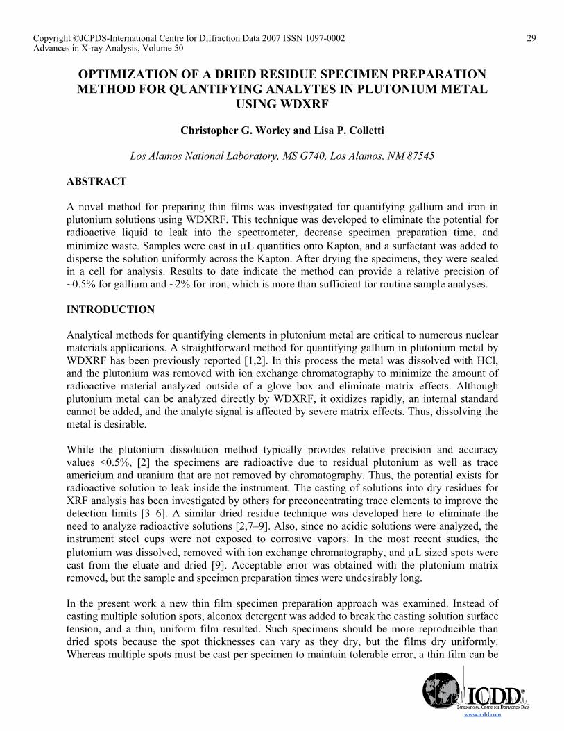
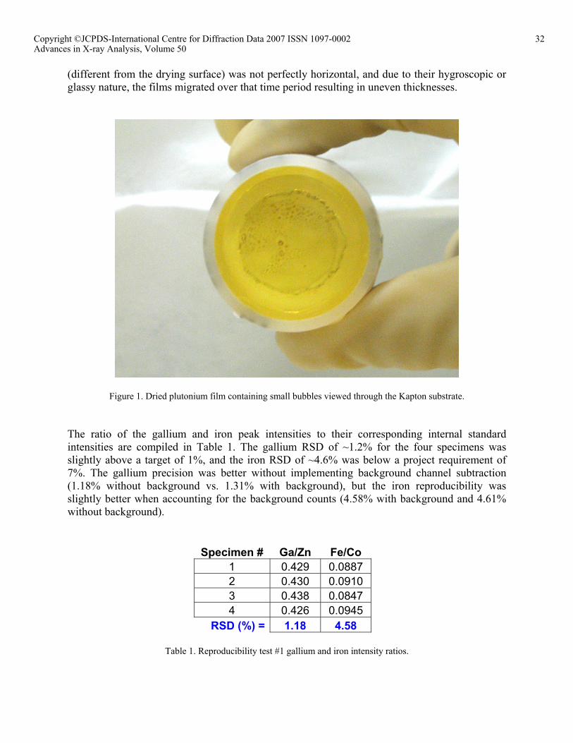
the radioactive specimens inside the cups. The specimens were analyzed four days after preparation. Reproducibility test #2 specimen preparation. Most of the preparation steps were the same as in test #1 for the second set of test specimens. The drying temperature was reduced and maintained at ~50°C. The specimens were dried for ~20 minutes. Five specimens were prepared, and they were analyzed within a few hours after preparation. RESULTS AND DISCUSSION The detergent alconox was added to the casting solutions to break the surface tension and wet the hydrophobic Kapton surface. A film of alconox was cast on Kapton containing 5 times the amount used for spreading the plutonium spots. The count rates for Ga, Zn, Fe, and Co detected from this film were no greater than those detected from a blank cup. Thus, the detergent purity was deemed to be adequate for purposes here. The concentration of alconox used was also important; a high concentration was found to react with the acidic plutonium solution and induce precipitation, and the drops would not spread into a thin film if too low of a concentration was used. A 0.01 g/mL alconox solution was found to work well. Several options were investigated for applying the alconox to the Kapton surface. An excess of solution was applied, and the excess was removed with a pipette. Other options attempted included swabbing the solution on with a cotton-tipped applicator and adding a drop of the plutonium casting solution to a drop of alconox solution. The latter was chosen as the method of choice because it was reproducible and quick. A hydrophobic barrier was applied to the outer edge of the Kapton on the inside of the specimen cup to prevent the casting solution from drying unevenly at the edges of the cup. Preventing the plutonium solution from reaching the cup/Kapton seal also eliminated the possibility of any radioactive material leaking out of the cup. Several substances were tested to provide this barrier including Apiezon L vacuum grease, Wacker silicone paste P4, Vaseline, high vacuum grease (Dow Corning), and Rain-X (SOPUS Products). To check for impurities, a film of each was smeared on 4 µm Prolene film, and the Ga, Zn, Fe, and Co channels intensities were collected for 60 sec each. The intensities were comparable to those from a blank cup except that the iron counts were ~2.5 greater from the high vacuum grease than from the blank. Alconox solutions were found to penetrate the Rain-X barrier, and Vaseline and Apiezon melted when using a heat lamp for drying. Thus, the silicone paste was chosen to produce the barrier ring. Reproducibility test #1. In this first test of the method, the height of the heat lamps used for drying could not be adjusted to control the temperature, and the films formed numerous tiny bubbles as they dried (Figure 1) (in a subsequent reproducibility test, an adjustable height heat lamp assembly was used to control the drying temperature in an attempt to prevent these bubbles from forming as discussed later). After drying the films displayed a glassy appearance. The specimens could not be analyzed for ~4 days after preparation due to the radioactive laboratory area being closed over a holiday weekend, and unfortunately the films flowed to one side of the cups forming a thicker region. Apparently the surface on which the specimens were left
31Copyright ©JCPDS-International Centre for Diffraction Data 2007 ISSN 1097-0002Advances in X-ray Analysis, Volume 50
(different from the drying surface) was not perfectly horizontal, and due to their hygroscopic or glassy nature, the films migrated over that time period resulting in uneven thicknesses.
Figure 1. Dried plutonium film containing small bubbles viewed through the Kapton substrate.
The ratio of the gallium and iron peak intensities to their corresponding internal standard intensities are compiled in Table 1. The gallium RSD of ~1.2% for the four specimens was slightly above a target of 1%, and the iron RSD of ~4.6% was below a project requirement of 7%. The gallium precision was better without implementing background channel subtraction (1.18% without background vs. 1.31% with background), but the iron reproducibility was slightly better when accounting for the background counts (4.58% with background and 4.61% without background).
Specimen # Ga/Zn Fe/Co 1 0.429 0.0887 2 0.430 0.0910 3 0.438 0.0847 4 0.426 0.0945
RSD (%) = 1.18 4.58
Table 1. Reproducibility test #1 gallium and iron intensity ratios.
32Copyright ©JCPDS-International Centre for Diffraction Data 2007 ISSN 1097-0002Advances in X-ray Analysis, Volume 50
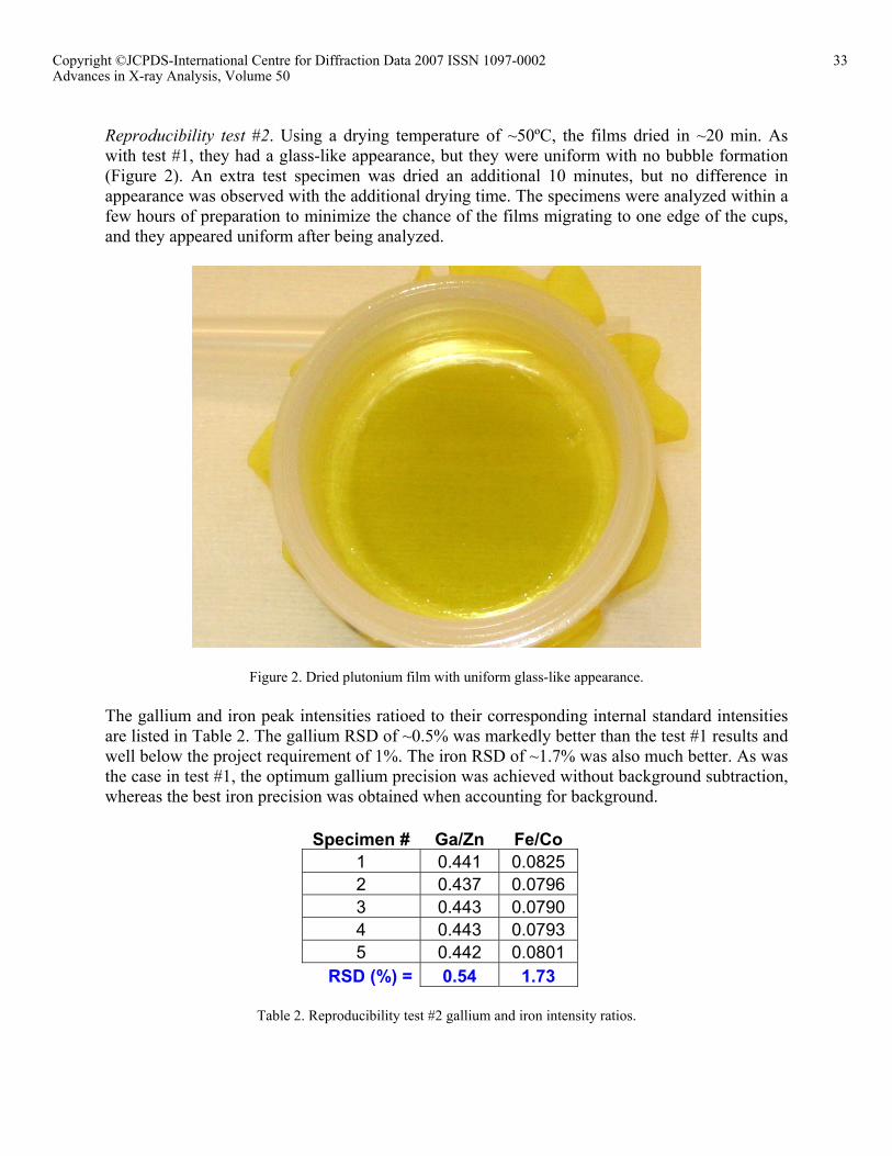
Reproducibility test #2. Using a drying temperature of ~50ºC, the films dried in ~20 min. As with test #1, they had a glass-like appearance, but they were uniform with no bubble formation (Figure 2). An extra test specimen was dried an additional 10 minutes, but no difference in appearance was observed with the additional drying time. The specimens were analyzed within a few hours of preparation to minimize the chance of the films migrating to one edge of the cups, and they appeared uniform after being analyzed.
Figure 2. Dried plutonium film with uniform glass-like appearance.
The gallium and iron peak intensities ratioed to their corresponding internal standard intensities are listed in Table 2. The gallium RSD of ~0.5% was markedly better than the test #1 results and well below the project requirement of 1%. The iron RSD of ~1.7% was also much better. As was the case in test #1, the optimum gallium precision was achieved without background subtraction, whereas the best iron precision was obtained when accounting for background.
Specimen # Ga/Zn Fe/Co 1 0.441 0.0825 2 0.437 0.0796 3 0.443 0.0790 4 0.443 0.0793 5 0.442 0.0801
RSD (%) = 0.54 1.73
Table 2. Reproducibility test #2 gallium and iron intensity ratios.
33Copyright ©JCPDS-International Centre for Diffraction Data 2007 ISSN 1097-0002Advances in X-ray Analysis, Volume 50
Future plans. Now that a reproducible specimen preparation method has been developed, standards will be prepared using electrorefined (ER) high purity plutonium spiked with known amounts of the analytes, cast as thin films, and used to generate a calibration curve. Several test samples will be prepared using ER metal to determine the relative precision and accuracy. CONCLUSION A method was developed for casting thin, uniform dry films from dissolved plutonium samples to quantify the gallium and iron content while avoiding the need to analyze radioactive liquids. In this new technique a surfactant was used to break the casting solution surface tension so that it spread evenly into a thin film. While a previous dried residue spotting process was found to be effective, the plutonium matrix had to be removed with chromatography to achieve acceptable error. The specimen film thicknesses are more reproducible using this new method, and acceptable precision can be realized without removing the plutonium. Hence, sample preparation is considerably faster, and less waste is generated. ACKNOWLEDGMENTS The authors would like to thank the Los Alamos Pollution Prevention Generator Set Aside Fee Program and the Department of Energy for funding this work. REFERENCES [1] Martell, C. J.; Hansel, J. M., Los Alamos Unclassified Report, Report# LA-11435, 1988. [2] Worley, C. G., Adv. X-ray Anal., 2002, 46, 369–374. [3] Murata, M.; Omatsu, M.; Mushimoto, S., X-Ray Spectrom., 1984, 13, 83–86. [4] Meltzer, C.; King, B.-S., Adv. X-ray Anal., 1990, 34, 41–55. [5] Colletti, L. P.; Havrilla, G. J., Adv. X-ray Anal., 1997, 41, 291–300. [6] Nielson, A. J.; Turner, D. C.; Wilson, A.; Wherry, D. C.; Wong, R., Adv. X-ray Anal.,
1995, 39, 799–804. [7] Worley, C. G.; Havrilla, G. J., Adv. X-ray Anal., 2000, 44, 392–397. [8] Worley, C. G., Adv. X-ray Anal., 2001, 45, 421–426. [9] Worley, C. G.; Colletti, L. P., Adv. X-ray Anal., 2003, 47, 98–103.
34Copyright ©JCPDS-International Centre for Diffraction Data 2007 ISSN 1097-0002Advances in X-ray Analysis, Volume 50