ophthalmology nothing but toronto...
TRANSCRIPT

OphthalmologyNothing but Toronto notes
with pictures
Omar Zainal

Globe Displacement
• Exophthalmos: most common cause in adults is Graves
• Most common cause in children is orbital cellulitis
• Enophthalmos:
• Causes: blow-out fracture, orbital fat atrophy, congenital abnormality, metastatic disease.
Page: 10

Cellulitis
• Preseptal cellulitis:
• Rx: systemic Augmentin (amox+clav)
• Complication: Orbital cellulitis.
• Orbital cellulitis: ocular and medical emergency!
• Causes: sinus, facial or tooth infection, trauma or even preseptal cellulitis
• Complications: loss of vision, death, meningitis, brain abscess, cavernous sinus thrombosis.
• Rx: IV ceftriaxone and vancomycin
Page: 10

Keratoconjunctivitis sicca (actually it’s a symptom rather than a picture!)
• Causes:• Decreased production of aqueous layer: SLE,
RA, CN7 palsy, idiopathic, anticholinergics, antihistamines, diuretics and beta blockers.
• Increased evaporation of aqueous layer: meibomina gland dysfunction, vitA def., ectropion, contact lens use.
Clinical: dry, red, foreign body sensation, blurred and tearing.
Investigation: fluorescein stain, Schirmer’s test.
Complication: scarring, erosion and ulceration.
Rx: Artificial tears, cyclosporine emulsion. Treat underlying cause.
Page: 11

Dacryocystitis – lacrimal sac infla.
• Inflammation of lacrimal sac because of nasolacrimal duct blockage.
• By: S. aureus, S. pneumoniae.
• Picture: swelling and redness at medial canthus. Pain with or without fever.
• If chronic: patient only has epiphora.
• Rx: warm compress, nasal decongestant, systemic and topical Ab. Consider dacryocystorhinostomy.
Page: 12

Dacryoadenitis – lacrimal gland infla.
• By: S. aureus, mumps, EBV, HZV, N. gono…
• Chronic type will present as painless enlargement. Chronic cases associated with sarcoidosis, TB, lymphoma and leukemia.
• Acute will have pain redness swelling tearing and maybe discharge.
• Rx: warm compress, NSAIDs, systemic Ab.
Page: 12

Lids and Lashes

Ptosis
• Causes:• Aponeurotic: disinsertion of levator
aponeurosis• Mechanical: mass preventing lid
opening• Neuromascular: myasthenia gravis,
CN3 palsy, Horner’s syndrome.• Congenital• Pseudoptosis: contralateral
expothlamia, dermatochalasis• Opioids, heroin, pregabalin.
Rx: blepharoplasty, levator resection, muller’s muscle resection, frontalis sling.
Page: 13

Trichiasis
• Eyelashes turned inwards
• Causes: blepharitis, stevens-Johnson syn. Trauma and burns
• Red, foreign body sensation and tearing
• Complication: ulceration of cornea
• Rx: lubrication, eyelash plucking, electrolysis, cryotherapy.
Page: 13

Entropion
• Lid margin turned inwards, mostly lower lid.
• Symptoms: tearing and red eye.
• Complications: abrasion and scarring of cornea.
• Causes: aging, HZV, surgery, trauma, burn, Orbicularis oculi muscle spasm, congenital.
• Rx: lubrication, lid taping, surgery
Page: 13

Ectropion
• Lid margin turned outwards
• LEADS TO EXPOSURE KERATITIS.
• Causes: aging, burns, trauma, surgery, CN7 palsy.
• Rx: lubrication and surgery.
Page: 13

Hordeolum
• ACUTE infla. of Meibomian gland, gland of Zeis, or Moll gland
• By: S. aureus
• Pain red swelling
• Rx: warm compress, topical Ab
Page: 13
Chalazion
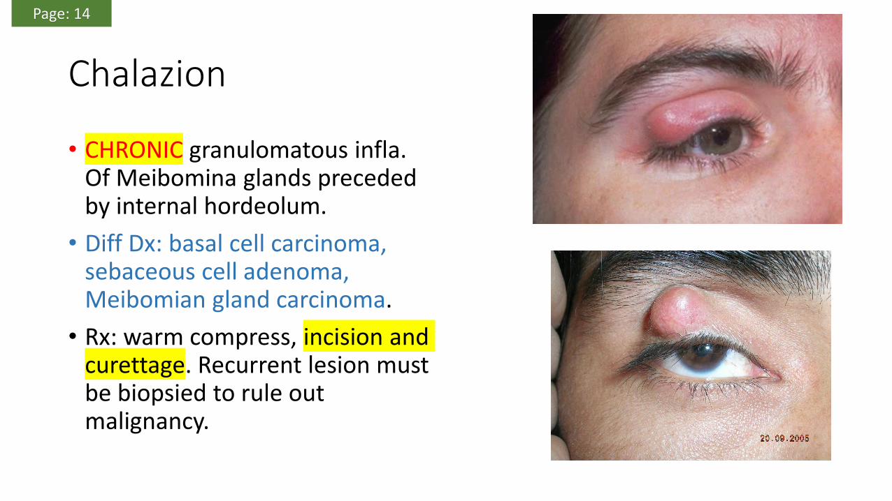
• CHRONIC granulomatous infla. Of Meibomina glands preceded by internal hordeolum.
• Diff Dx: basal cell carcinoma, sebaceous cell adenoma, Meibomian gland carcinoma.
• Rx: warm compress, incision and curettage. Recurrent lesion must be biopsied to rule out malignancy.
Page: 14

Blepharitis
• Infla. of lid margin• Two types:
• Staph: dry scales• Seborrheic: greasy scales
Clinical features: crusting, thickened red lid margins, discharge, itching , tearing, foreign body sensationComplication: recurrent chalazion, conjunctivitis, keratitis, corneal ulcer.Rx: warm compress, lid wipes with diluted baby shampoo, topical or systemic Ab, topical corticosteroids.
Page: 14

Xanthelasma
• Lipid deposits
• Site: medial upper lids.
• Hyperlipidemia
• Common in elderly
• Rx: excision
Page: 14

ConjunctivaThin vascular mucous epithelium

Pinguecula
• Associated with SUN and WINDexposure.
• Rx: for cosmetic purposes go for surgery, otherwise symptomatically treated with lubricating drops.
• Subepithelial deposition of hyaline and elastic tissue. Spares the cornea
Page: 14

Pterygium
• Rx: excision (surgery) and lubricating drops.
• Fibrovascular epithelial growth onto the cornea. Usually nasally.
• Decreased vision
Page: 15

Subconjunctival hemorrhage
• Rx: resolves in 2-3 wks
• Can be idiopathic or caused by trauma, bleeding disorder, HTN, Valsalva maneuver, anticoagulants.
Page: 15

Giant papillary conjunctivitis
• Immune reaction in contact lenses wearers.
• Rx: clean or change lens.
Page: 15

Atopic conjunctivitis
• Associated with asthma, rhinitis, dermatitis, hay fever.
• Clinical: chemosis, corneal neovascularization, small papillae.
• Seasonal
• Rx: antihistamine, mast cell stabilizer, or topical CS.
Page: 15

Vernal Conjunctivitis
• Cobblestones-like papillae on superior palpebral conjunctiva with corneal ulcer.
• In WARM weather
• In children
• Rx: topical CS, topical cyclosporin
Page: 15

Viral conjunctivitis
• Watery discharge.
• By Adenovirus
• Preauricular node palpable and tender.
• Rx: cool compress, lubrication and good hygiene.
Page: 15

Bacterial conjunctivitis
• By: S. aureus, S. pneumoniae, H. influenzae and M. catarrhalis.
• C. trachomatis the most common cause in neonates.
• Rx: topical or systemic Ab.
Page: 16

ScleraWhite fibrous outer protective layer composed of irregularly distributed
collagen bundles.
Episclera is a thin vascularized layer between sclera and conjunctiva

Episcleritis
• 1/3 bilateral.
• Rx: Topical steroids or oral NSAIDs if PAINFUL. Otherwise its self-limited.
• Usually asymptomatic, heat sensation and red eyes.
• Diffuse injection of radially-directed vessels, chemosis.
• BLANCHES WITH PHENYLEPHRINE
Page: 16

Scleritis• Rx: systemic NSAIDs or systemic
steroids, treat underlying etiology.
• Posterior scleritis causes blindness through exudative retinal detachment.
• Symptoms: pain, photophobia, decreased vision
• Causes: SLE, RA, Ankylosing spondylitis, TB, Sarcoidosis, Syphilis, infection, chemical burn.
Page: 16

CorneaMade of 6 layers: out to in Epithelium, bowman’s membrane, stroma, dua’s layer,
Descemet’s layer and Endothelium.
Function: transmission of light, protection, refraction of light (accounts for 2/3 of total refractive power)
Why is it transparent? Due to avascularity, uniform collagen, and deturgescence

Corneal foreign body
• Rx: remove it under magnification using local anesthetic.
• Complication: iritis, corneal abrasion and ulceration and scarring.
Page: 17

Corneal abrasion
• Nothing but epithelial defect.
• Pain red tearing photophobia
• De-epithelialized area will stain with fluorescein dye
• Complication: corneal erosion and ulceration and infection. Iritis.
• Rx: topical antibiotics, Topical NSAIDs, Patch
Page: 17

Corneal ulcer – positive seidel’s test
• Necrosis of corneal tissue due to infection.
• This infection is usually caused by: lens, corneal abrasion, foreign body
• Pain redness photophobia tearing and decreased visual acuity. And corneal hypoesthesia
• Upon examination: anterior chamber cells, corneal edema, hypopyon.
• Associated with conjunctivitis, blepharitis, keratitis, vitA def.
• Complications: corneal perforation, iritis, ENDOPHTHALMITIS
• Rx: urgent referral, culture, topical Ab every hour.
Page: 18

Herpes Simplex Keratitis
• Caused by HSV-1
• Triggered by stress fever immunosuppression
• Pain red tearing and decreased vision, eyelid edema and corneal hypoesthesia.
• DENDRITIC LESION in epithelium (just like abrasion)
• Complications: scarring, iritis, keratitis, secondary glaucoma
• Rx: trifluridine or acyclovir
Page: 18

Herpes Zoster
• Nothing but a dermatitis of the CN V1 territory that MAY involve the eye.
• Picture: (Hutchinson’s sign **the nose**), if present means the eye will be involved in 75% of cases.
• If not present it means, only 33% of cases will have the eye involved.
• Pain, tearing photophobia and red eye.
• Complications: corneal ulceration, iritis with secondary glaucoma, cataract.
• Rx: acyclovir, valcyclovir, famiciclovir. Topical steroids.
Page: 19

keratoconus
• Bulging of cornea
• Associated with down syndrome, contact lens use.
• There is a break in the Descemet’s and Bowman’s layers of the cornea.
• Rx: cross-linking treatment, intrastromal corneal ring segment, penetrating keratoplasty.
Page: 19

Arcus senilis
• Corneal degeneration due to lipid deposition.
• AGING
Page: 19

Kayser-Fleischer ring
• Deposition of copper in Descemet’s layer of cornea.
• In WILSONS disease.
Page: 19

The Uveal TractIris – ciliary body – choroid
Vascularized pigmented layer of the eye, between the sclera and retina

Anterior uveitis – iritis (iris), or iridocyclitis (iris & ciliary body)• Unilateral
• Causes:• Idiopathic• Reactive arthritis, ankylosing spondylitis, psoriatic arthritis, IBD• Juvenile idiopathic arthritis• Syphilis, lyme disease, toxoplasmosis, TB, HSV, HZV• Sarcoidosis, Trauma, large abrasion, post ocular surgery
• Symptoms: photophobia, pain, tenderness, decreased VA.
• Signs: ciliary flush, hypopyon, anterior chamber cells, keratic precipitate.
• Low intraocular pressure, but in severe iritis and HSV and HZV iritis the pressure will increase because of secondary glaucoma.
• Complication: angle closure glaucoma, posterior synechiae or peripheral anterior synechiae, cataracts, band keratopathy, macular edema.
• Rx: cycloplegics to dilate pupil, steroids, analgesia.
Page: 20

You can see ciliary flush and conjunctival hyperemia, posterior synechiae as indicated by the irregular pupil shape, hypopyon.
Band keratopathyPage: 20

Intermediate uveitis – vitreous inflammation
• Causes: idiopathic, sarcoidosis, Lyme disease, MULTIPLE SCLEROSIS
• Symptoms: blurred vision, floaters, unilateral then bilateral.
• Examination: vitreous cells, posterior segment snowbank.
• Complications: cystoid macular edema, cataract, glaucoma
• Rx: steroids, cryotherapy, laser, vitrectomy.
Page: 20

Page: 20

Posterior uveitis – infla. of choroid and/or retina• Cause: bacterial, viral, fungal,
toxoplasmosis (MOST COMMON), Behcet’s disease, melanoma.
• PAINLESS, NO CONJUNCTIVAL INJECTION.
• Decreased VA, floaters, vitreous cells and hypopyon.
• Complications: macular edema, vitritis, neovascularization, visual field loss and EXUDATIVE RETINAL DETACHMENT.
• Rx: Steroids.
Page: 20

LensOuter capsule surrounding a soft cortex and a firm inner nucleus
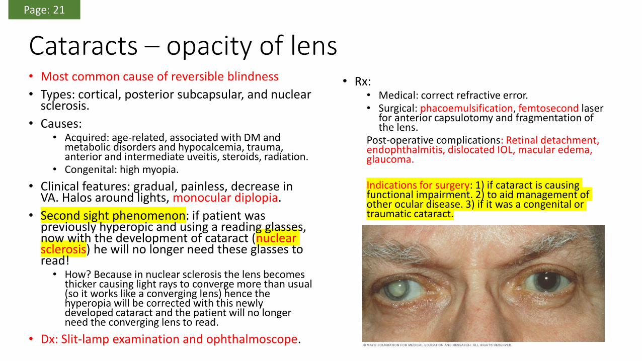
Cataracts – opacity of lens• Most common cause of reversible blindness
• Types: cortical, posterior subcapsular, and nuclear sclerosis.
• Causes:• Acquired: age-related, associated with DM and
metabolic disorders and hypocalcemia, trauma, anterior and intermediate uveitis, steroids, radiation.
• Congenital: high myopia.
• Clinical features: gradual, painless, decrease in VA. Halos around lights, monocular diplopia.
• Second sight phenomenon: if patient was previously hyperopic and using a reading glasses, now with the development of cataract (nuclear sclerosis) he will no longer need these glasses to read!• How? Because in nuclear sclerosis the lens becomes
thicker causing light rays to converge more than usual (so it works like a converging lens) hence the hyperopia will be corrected with this newly developed cataract and the patient will no longer need the converging lens to read.
• Dx: Slit-lamp examination and ophthalmoscope.
• Rx:• Medical: correct refractive error.• Surgical: phacoemulsification, femtosecond laser
for anterior capsulotomy and fragmentation of the lens.
Post-operative complications: Retinal detachment, endophthalmitis, dislocated IOL, macular edema, glaucoma.
Indications for surgery: 1) if cataract is causing functional impairment. 2) to aid management of other ocular disease. 3) if it was a congenital or traumatic cataract.
Page: 21

Page: 21

Dislocated lens
• Occurs in Marfan syndrome, Ehlers-Danlos syndrome type VI, homocystinuria, syphilis, lens coloboma. Or traumatic.
• Clinical: decreased VA, monocular diplopia (means double vision with one eye opened), iridodenesis
• Abnormal red reflex.• Complications: cataract, glaucoma,
uveitis.• Rx: surgical correction, lens
replacement.
Page: 22

VitreousClear gel 99% water plus collagen fibrils, glycosaminoglycans and
hyaluronic acid.

Posterior vitreous detachment
• Causes: aging (central vitreous shrinks and liquid vitreous moves between posterior vitreous and retina and the vitreous is peeled away.)
• Clinical: FLOATERS and FLASHES OF LIGHT.
• Picture: Weiss ring seen in PVD
• Complications: retinal detachment, vitreous hemorrhage.
• Rx: no treatment, just rule out RD.
Page: 22

Vitreous hemorrhage:
• Causes: PVD, retinal detachment, trauma, proliferative diabetic retinopathy, retinal vein occlusion.
• Clinical: SUDDEN LOSS OF VISION. Floaters and flashes of light
• NO RED REFLEX.
• Rx: surgical vitrectomy, retinal endolaser.
Page: 22

Endophthalmitis and vitritis
• Usually post-operative• Also caused by penetrating injury to eye,
intravitreal injection.• Clinical: painful, red eye, photophobia,
discharge, decreased VA, lid edema, proptosis, corneal edema, anterior chamber cells, hypopyon, reduced red reflex.
• Maybe signs of ruptured globe: reduced IOP, hyphema and subconjunctival hemorrhage.
• Rx: vitrectomy, intravitreal antibiotic.
Page: 23

RetinaMade of two parts: neurosensory retina and retinal pigmented epithelium.
Macula: rich in cons
Fovea: center of macula
Optic disk: through which retinal vessels pass
The choroidal layer is located between the sclera and the retina, it is vascular and nourishes the outer layer with oxygen and nutrients.

Central retinal artery occlusion• Causes: emboli, thrombosis, arteritis
• Clinical: sudden painless, Relative afferent pupillary defect, amaurosis fugax.
• Fundoscopy: cherry-red spots, retinal pallor, cotton wool spots, narrowed arterioles, cholesterol emboli, Hollenhorst plaques.
• WHY CHERRY-RED SPOTS: this spot is basically located at the center of the macula. Because when the retinal artery gets occluded, the whole retina will become pale except the macula, because the macula receives blood supply from different artery called posterior ciliary artery.
• Rx: decrease IOP (b-blocker, IV acetazolamide, IV mannitol, anterior chamber paracentesis, inhaled oxygen-carbon dioxide mixture)• Intraarterial or intravenous thrombolysis• ND:YAG laser embolectomy
Page: 23

Page: 23

Central/Branch retinal vein occlusion• Second most frequent vascular retinal disorder after diabetic
retinopathy.
• Usually occurs with HTN and diabetes, where there is thrombus formation.
• Predisposing factors: HTN, DM, glaucoma, hyperviscosity, drugs.
• Clinical: painless, sudden or gradual loss of vision, RAPD.
• Fundoscopy: blood & thunder appearance, cotton wool spots, SWOLLEN OPTIC DISC, MACULAR EDEMA.
• Two groups:• Venous stasis (non-ischemic): no RAPD, mild hemorrhage, few cotton
wool spots, resolves in few weeks, chance of regaining normal vision if macula is still intact.
• Hemorrhagic retinopathy: old patients, RAPD, low VA, reduced peripheral vision, massive hemorrhage, cotton wool spots, poor prognosis.
• Complications: degeneration of retinal pigmented epithelium, secondary rubeosis leading to glaucoma, vitreous hemorrhage.
• Rx: retinal laser photocoagulation. Intravitreal anti-VEGF.
Page: 24

Retinal detachment
• Cleavage between neurosensory layer and RPE layer.
• Three types:1. Rhegmatogenous: most common, a hole in the neurosensory retina allowing vitreous fluid to go
into subretinal space. Due to PVD, trauma.2. Tractional: occurs in DR, CRVO, SCD, retinopathy of prematurity, and trauma.3. Exudative: a damage that happen to the RPE not the neurosensory layer (as opposed to the
Rhegamentegenous type), occurs in posterior uveitis, intraocular tumors.
• Clinical: sudden, flash of lights, and floaters, curtain of blackness, decreased IOP, loss of red reflex, RAPD.
• Complications: loss of vision, vitreous hemorrhage
• Rx:1. Rhegmatogenous: scleral buckle procedure, pneumatic retinopexy and cryotherapy/laser to
create adhesions. Vitrectomy.2. Tractional: vitrectomy, scleral buckling.3. Exudative: treat underlying cause.
Page: 24

OCT of retinal detachment
Page: 24

Retinitis pigmentosa
• Most commonly autosomal recessive.
• It is a retinal degeneration disease.• Clinical: NIGHT BLINDNESS,
TUNNEL VISION, glare.• Fundoscopy: areas of bone-spicule,
pale optic disk.• Electroretinography and
electrooculography assist in diagnosis
• Rx: Vitamin A and E may reduce disease progression.
Page: 25

Leber’s congenital amaurosis
• Autosomal recessive
• Degenerative disease of retina.
• Clinical: resting nystagmus, no pupillary response, SEVERE vision loss.
• Dx: genetic testing
Page: 25
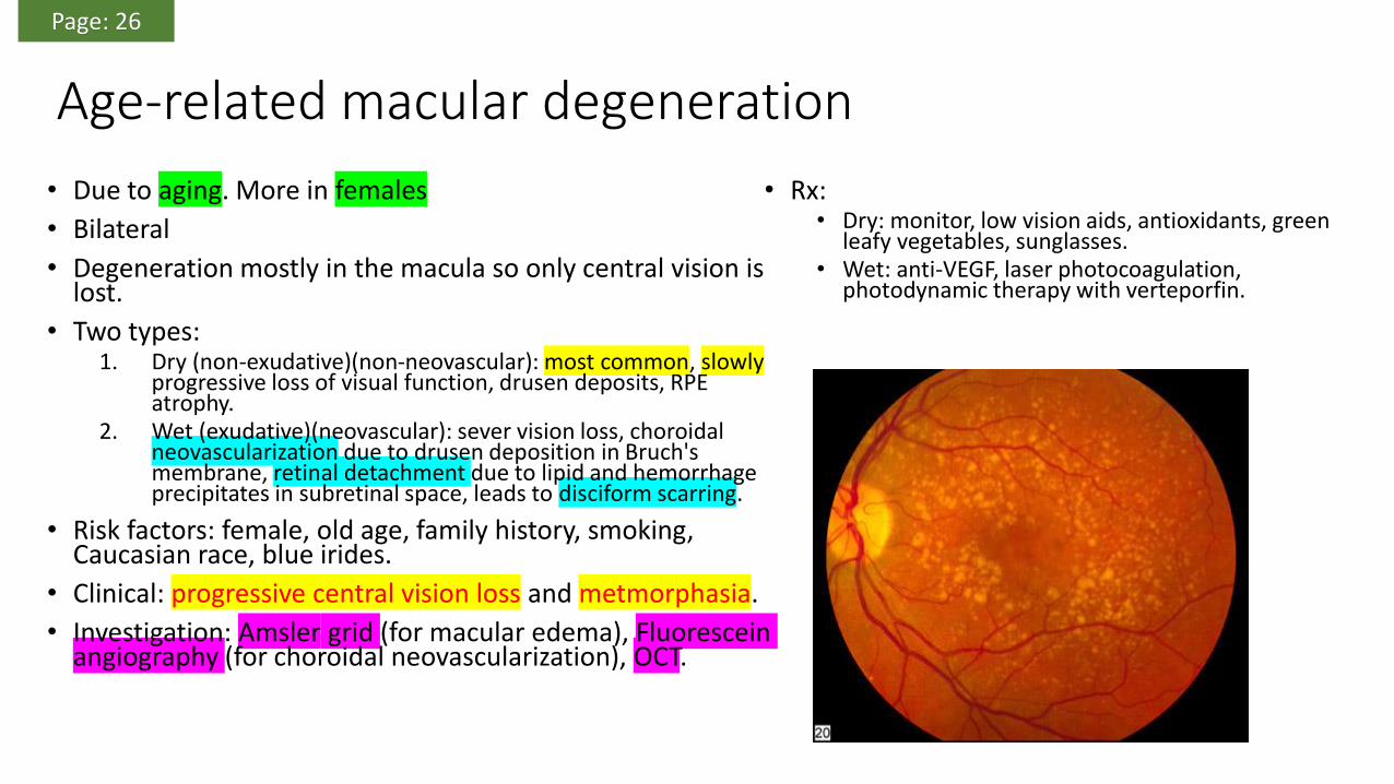
Age-related macular degeneration
• Due to aging. More in females
• Bilateral
• Degeneration mostly in the macula so only central vision is lost.
• Two types:1. Dry (non-exudative)(non-neovascular): most common, slowly
progressive loss of visual function, drusen deposits, RPE atrophy.
2. Wet (exudative)(neovascular): sever vision loss, choroidal neovascularization due to drusen deposition in Bruch's membrane, retinal detachment due to lipid and hemorrhage precipitates in subretinal space, leads to disciform scarring.
• Risk factors: female, old age, family history, smoking, Caucasian race, blue irides.
• Clinical: progressive central vision loss and metmorphasia.
• Investigation: Amsler grid (for macular edema), Fluorescein angiography (for choroidal neovascularization), OCT.
• Rx:• Dry: monitor, low vision aids, antioxidants, green
leafy vegetables, sunglasses.• Wet: anti-VEGF, laser photocoagulation,
photodynamic therapy with verteporfin.
Page: 26
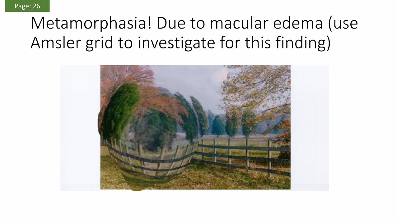
Metamorphasia! Due to macular edema (use Amsler grid to investigate for this finding)
Page: 26

GlaucomaIts optic neuropathy resulting in peripheral vision loss then central vision loss.
How to investigate? Visual acuity test, slit-lamp, ophthalmoscopy, tonometry, visual field test, pachymetry (to asses for corneal thickness because a thin
central cornea is a major risk factor for glaucoma).

Primary open-angle glaucoma (most common)
• Obstruction to aqueous drainage within trabecular meshwork and Canal of Schlemm.
• ASYMPTOMATIC (gradual).
• MAJOR RISK FACTORS: high IOP(>21), old age, African, familial, thin central cornea.
• MINOR RISK FACTORS: myopia, HTN, DM, hyperthyroid, steroid use, ocular trauma, anemia/hemodynamic crisis.
• Clinical: often found incidentally, the first sign is increased C:D ratio more than 60%, C:D asymmetry between eyes, thinning and notching of neuroretinal rim, flame-shaped disc hemorrhage, 360 degree peripapillary atrophy, nasal displacement of blood vessels.
• PARACENTRAL AND ARCUATE SCOTOMA DEFECTS ARE CHARACTERISTICS.
• Rx: decrease IOP, laser trabeculectomy.
Page: 28

Increased C:D ratio
Page: 28

Flame-shaped disc hemorrhage
Page: 28

Secondary open-angle glaucoma
• Steroid induced
• Traumatic
• Pigmentary dispersion syndrome
• Pseudoexfoliation syndrome
Page: 29

Primary closed-angle glaucoma (5%)
• Iris bows forward and attaches to the cornea so preventing aqueous flow to the trabecular meshwork.
• Sudden increase in IOP.
• Risk factors: (75 years old female wearing glasses because she is Hyperopic, she fights with her husband then decides to go to the cinema for a horror movie), so the risk factors are: old age, female, Hyperopia, pupil dilation (stress due to fight) (and due to lack of light being in cinema) (horror movie will stimulate sympathetic system causing pupil dilation).
• Clinical: very red and painful, unilateral, decreased VA, halos around lights, fixed mid-dilated pupil, nausea and vomiting, abdominal pain, increased IOP by tonometry. Shallow anterior chamber and anterior cells as seen by Slit-lamp exam, corneal edema.
• Complication: loss of vision, permanent peripheral anterior synechiae.
• Rx: Pilocarpine, decrease IOP, laser iridotomy
Page: 29

Fixed mid dilated right pupil.
Page: 29

Laser iridotomy
Page: 29

Secondary causes of angle closure glaucoma
• Uveitis (iris adheres to lens causing pupillary block)
• Proliferative diabetic retinopathy (Rubeosis iridis)
• Central retinal vein occlusion (Rubeosis iridis)
Page: 29

Best pic to elaborate difference in mechanism

PupilsControlled by the balance of two muscles: sphincter and dilator muscle, both of these muscles are located in the iris. NEVER mix with the ciliary muscle that is
intended to control the curvature of the LENS.
*Sphincter receives parasympathetic innervation by CN3 (ciliary ganglion).
*Dilator receives sympathetic innervation by nasociliary nerve (a branch of CN V1) (through superior cervical ganglion).

Anisocoria (unequal pupil size) (conditions in which pupils cannot dilate)• Argyll-Robertson Pupil
• both pupils irregular and <3 mm in diameter• does not respond to light stimulation• responds to accommodation (light-near dissociation)• neurosyphilis
• Horner’s Syndrome• lesion in sympathetic pathway• difference in pupil size greater in dim light• associated with ptosis, anhydrosis of ipsilateral face/neck• application of cocaine 4-10% (blocks reuptake of norepinephrine) to eye does not result in pupildilation (vs. physiologic anisocoria), therefore confirms diagnosis• causes: carotid or subclavian aneurysm, brainstem infarct, demyelinating disease, cervical or mediastinal tumour, Pancoast tumour, goiter, cervical lymphadenopathy, surgical sympathectomy, Lyme disease, cervical ribs, tabes dorsalis, cervical vertebral fractures
Page: 30

Argyll-Robertson Pupil – bilateral pupil constriction
Page: 33

Horner’s syndrome affecting right eye, ptosis of right eyelid and constricted pupil as opposed to the left eye.
Page: 32

• CN III palsy
• Adie’s Tonic Pupil
Anisocoria (unequal pupil size) (conditions in which pupils cannot constrict)
Page: 32

Malignancies

• Lid Carcinoma• basal cell carcinoma (rodent ulcer) (90%)• squamous cell carcinoma (<5%)• sebaceous cell carcinoma (1-5%)• Kaposi’s sarcoma, malignant melanoma, Merkel cell tumour, metastatic tumour• Treatment
• incisional or excisional biopsies• may require cryotherapy, radiotherapy, chemotherapy, immunotherapy• surgical reconstruction
• Malignant Melanoma• most common 1° intraocular malignancy in adults• arise from uveal tract, 90% choroidal melanoma• hepatic metastases predominate• Treatment
• imaging to investigate spread• depending on the size of the tumour, either radiotherapy, enucleation, limited surgery
• Metastases• most common intraocular malignancy in adults• most commonly from breast and lung in adults, neuroblastoma in children• usually infiltrate the choroid, but may also affect the optic nerve or extraocular muscles• may present with decreased or distorted vision, irregularly shaped pupil, iritis, hyphema• Treatment
• local radiation, chemotherapy• enucleation if blind, painful eye
Page: 34

Lid Carcinoma
Page: 34

Malignant uveal melanoma
Page: 34

Ocular Manifestations of Systemic Disease
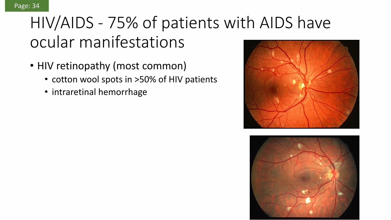
HIV/AIDS - 75% of patients with AIDS have ocular manifestations• HIV retinopathy (most common)
• cotton wool spots in >50% of HIV patients
• intraretinal hemorrhage
Page: 34

CMV retinitis• develops in late stages of HIV when severely
immunocompromised (CD4 count ≤50)• a necrotizing retinitis, with retinal hemorrhage and
vasculitis, “brushfire” or “pizza pie” appearance• presents with scotomas (macular involvement and RD),
blurred vision, and floaters• untreated infection will progress to other eye in 4-6 wk• treatment: virostatic agents (e.g. gancyclovir or
foscarnet) via IV or intravitreal injection
Page: 35

DIABETIC RETINOPATHY
• loss of vision due to (IMPORTANT)• progressive microangiopathy leading to macular edema
• progressive DR > neovascularization > traction > Retinal detachment and vitreous hemorrhage
• rubeosis iridis leading to neovascular glaucoma (poor prognosis)
• macular ischemia
Page: 35

non-proliferative: increased vascular permeability and retinal ischemia
• microaneurysms
• dot and blot hemorrhages
• hard exudates (lipid deposits), non-specific for DR
• macular edema
Page: 35

Proliferative DR - 5% of patients with DM will reach this stage• neovascularization of iris, disc, retina
to vitreous• rubeosis iridis can lead to neovascular
glaucoma
• vitreous hemorrhage from bleeding, fragile new vessels, fibrous tissue can contract causing tractional retinal detachment
• high risk of severe vision loss secondary to vitreous hemorrhage, retinal detachment
Page: 35

Treatment
• focal laser for clinically significant macular edema
• intravitreal injection of corticosteroid or anti-VEGF for foveal involved diabetic macular edema
• panretinal laser photocoagulation for PDR: reduces neovascularization, hence reducing the angiogenic stimulus from ischemic retina by decreasing retinal metabolic demand > reduces risk of blindness
• vitrectomy for non-clearing vitreous hemorrhage and tractional RD in PDR
Page: 36
Screening Guidelines for Diabetic Retinopathy
• type 1 DM• screen for retinopathy beginning annually 5 yr after disease onset
• annual screening indicated for all patients over 12 yr and/or entering puberty
• type 2 DM• initial examination at time of diagnosis, then annually
• pregnancy• ocular exam in 1st trimester, close follow-up throughout as pregnancy can
exacerbate DR
• gestational diabetics are not at risk for DR
Page: 36

Strabismus

Heterotropia
• It is a manifest deviation (means deviation is apparent when both eyes are in use –both eyes uncovered-)
• Hirschberg test (corneal light reflex): positive if the light reflex on both corneas is asymmetrical
• Cover test. (cover the normal eye, you will see the other eye correcting)
Page: 38

Heterophoria
• Latent deviation: no deviation when both eyes are uncovered.
• Hirschberg test will be normal (light reflexes symmetrical)
• very common – majority are asymptomatic
• Test• Cover-uncover test.
Page: 39

Leukocoria DDx:
1. cataract
2. retinoblastoma
3. retinal coloboma
4. ROP
5. persistent hyperplastic primary vitreous
6. Coat’s disease (exudative retinal telangiectasis)
7. toxocariasis
8. RD
Page: 41

Leukocoria (white reflex) – Think retinoblastoma
Retinoblastoma:• most common primary intraocular malignancy
in children• unilateral (2/3) or bilateral (1/3)• sporadic or genetic transmission• Diagnosis: often presents with leukocoria or
strabismus.• U/S or CT scan may demonstrate calcified
mass (present in most cases)Treatment
• radiotherapy, chemotherapy combined with laser, cryopexy, and/or enucleation
Page: 41