ophthal. (1961) 45, 117.bjo.bmj.com/content/bjophthalmol/45/2/117.full.pdf · brit. j. ophthal....
TRANSCRIPT
Brit. J. Ophthal. (1961) 45, 117.
END-TO-SIDE ANASTOMOSIS FOR OBSTRUCTIONOF THE NASO-LACRIMAL DUCT*
BY
R. A. BURNThe Royal Eye Hospital, London
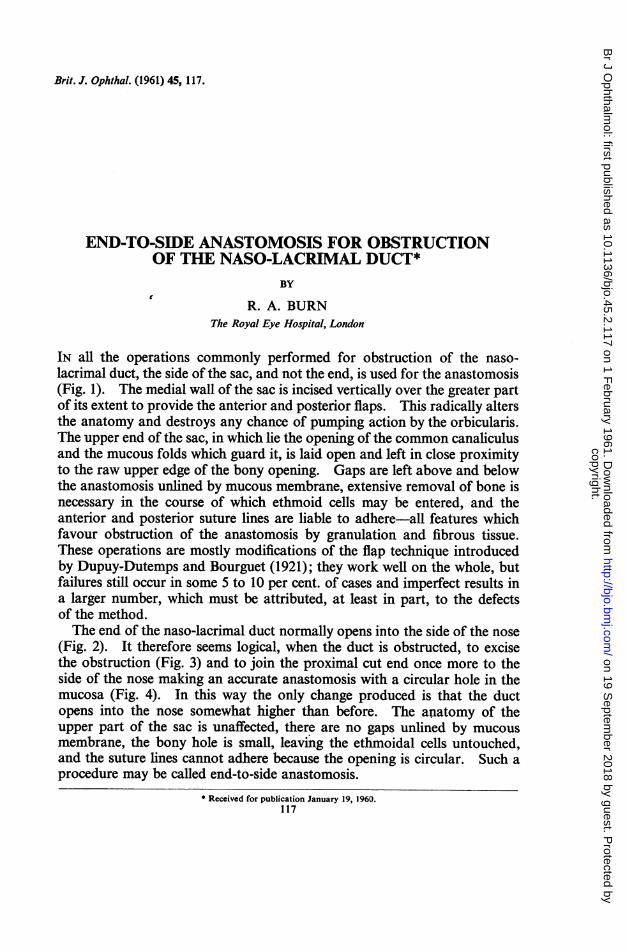
IN all the operations commonly performed for obstruction of the naso-lacrimal duct, the side of the sac, and not the end, is used for the anastomosis(Fig. 1). The medial wall of the sac is incised vertically over the greater partof its extent to provide the anterior and posterior flaps. This radically altersthe anatomy and destroys any chance of pumping action by the orbicularis.The upper end of the sac, in which lie the opening of the common canaliculusand the mucous folds which guard it, is laid open and left in close proximityto the raw upper edge of the bony opening. Gaps are left above and belowthe anastomosis unlined by mucous membrane, extensive removal of bone isnecessary in the course of which ethmoid cells may be entered, and theanterior and posterior suture lines are liable to adhere-all features whichfavour obstruction of the anastomosis by granulation and fibrous tissue.These operations are mostly modifications of the flap technique introducedby Dupuy-Dutemps and Bourguet (1921); they work well on the whole, butfailures still occur in some 5 to 10 per cent. of cases and imperfect results ina larger number, which must be attributed, at least in part, to the defectsof the method.The end of the naso-lacrimal duct normally opens into the side of the nose
(Fig. 2). It therefore seems logical, when the duct is obstructed, to excisethe obstruction (Fig. 3) and to join the proximal cut end once more to theside of the nose making an accurate anastomosis with a circular hole in themucosa (Fig. 4). In this way the only change produced is that the ductopens into the nose somewhat higher than before. The anatomy of theupper part of the sac is unaffected, there are no gaps unlined by mucousmembrane, the bony hole is small, leaving the ethmoidal cells untouched,and the suture lines cannot adhere because the opening is circular. Such aprocedure may be called end-to-side anastomosis.
* Received for publication January 19, 1960.117
copyright. on 19 S
eptember 2018 by guest. P
rotected byhttp://bjo.bm
j.com/
Br J O
phthalmol: first published as 10.1136/bjo.45.2.117 on 1 F
ebruary 1961. Dow
nloaded from
R. A. BURN
FIG. 1.-Side-to-side anastomosis. FIG. 2.-Left lacrimal passages and nose.Coronal section.
FIG. 3.-Common site of obstruction. FIG. 4.-End-to-side anastomosis.
Technique
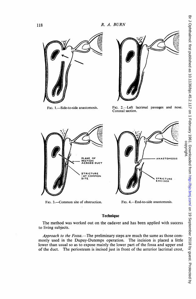
The method was worked out on the cadaver and has been applied with successto living subjects.Approach to the Fossa.-The preliminary steps are much the same as those com-
monly used in the Dupuy-Dutemps operation. The incision is placed a littlelower than usual so as to expose mainly the lower part of the fossa and upper endof the duct. The periosteum is incised just in front of the anterior lacrimal crest,
118
copyright. on 19 S
eptember 2018 by guest. P
rotected byhttp://bjo.bm
j.com/
Br J O
phthalmol: first published as 10.1136/bjo.45.2.117 on 1 F
ebruary 1961. Dow
nloaded from
NASO-LACRIMAL DUCT OBSTRUCTION
and the periosteal sheet is swept laterally taking the sac with it and exposing thefossa up to the posterior crest (Fig. 5).
FIG. 5.-Approach tolacrimal fossa.
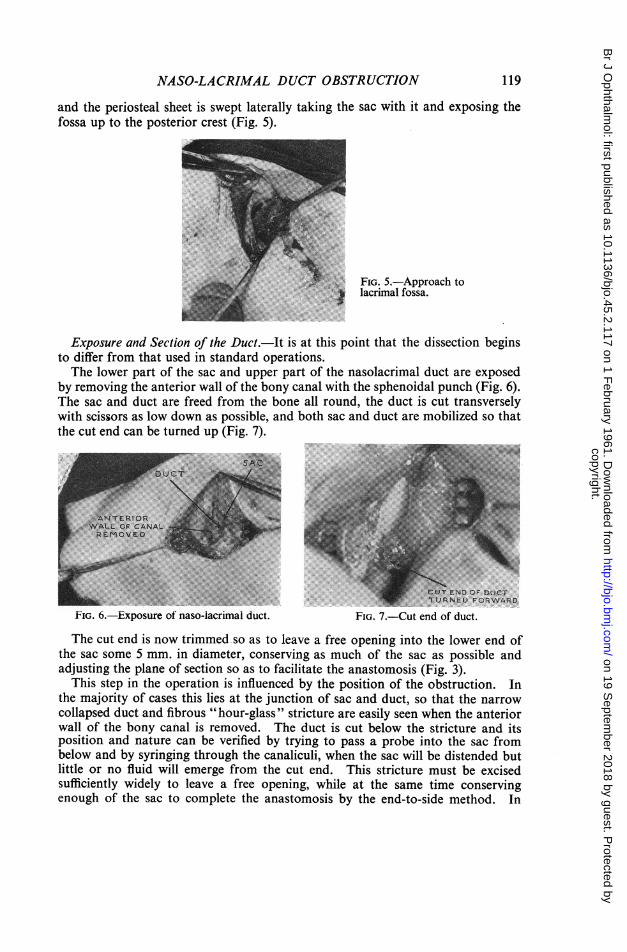
Exposure and Section of the Duct. It is at this point that the dissection beginsto differ from that used in standard operations.The lower part of the sac and upper part of the nasolacrimal duct are exposed
by removing the anterior wall of the bony canal with the sphenoidal punch (Fig. 6).The sac and duct are freed from the bone all round, the duct is cut transverselywith scissors as low down as possible, and both sac and duct are mobilized so thatthe cut end can be turned up (Fig. 7).
FIG. 6.-Exposure of naso-lacrimal duct.
:.. ........''.CUT END OF DUCTTURNED FORWARD
FIG. 7.-Cut end of duct.
The cut end is now trimmed so as to leave a free opening into the lower end ofthe sac some 5 mm. in diameter, conserving as much of the sac as possible andadjusting the plane of section so as to facilitate the anastomosis (Fig. 3).
This step in the operation is influenced by the position of the obstruction. Inthe majority of cases this lies at the junction of sac and duct, so that the narrowcollapsed duct and fibrous "hour-glass" stricture are easily seen when the anteriorwall of the bony canal is removed. The duct is cut below the stricture and itsposition and nature can be verified by trying to pass a probe into the sac frombelow and by syringing through the canaliculi, when the sac will be distended butlittle or no fluid will emerge from the cut end. This stricture must be excisedsufficiently widely to leave a free opening, while at the same time conservingenough of the sac to complete the anastomosis by the end-to-side method. In
119
copyright. on 19 S
eptember 2018 by guest. P
rotected byhttp://bjo.bm
j.com/
Br J O
phthalmol: first published as 10.1136/bjo.45.2.117 on 1 F
ebruary 1961. Dow
nloaded from
some cases, however, the obstruction is situated lower down and then the duct isseen to be dilated, filling the bony canal, and little different in width from the sac.A probe passes easily through the cut end into the sac and fluid emerges freelyfrom it on syringing. In these cases no excision is needed and a greater lengthof sac and duct is available for the anastomosis.
Patency of the Upper Lacrimal Passages.-Syringing with the cannula just withinthe canaliculus should produce a free flow of fluid from the cut end of the sac. Ifit does not do so, an obstruction must be present in the upper end of the sac orin the sinus of Maier and conversion to the side-to-side operation is necessary.
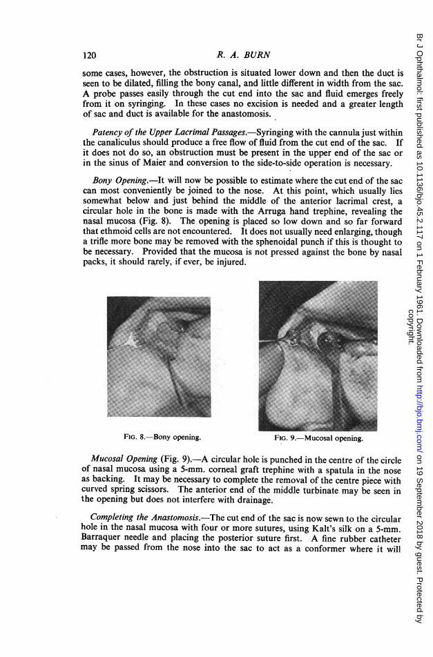
Bony Opening.-It will now be possible to estimate where the cut end of the saccan most conveniently be joined to the nose. At this point, which usually liessomewhat below and just behind the middle of the anterior lacrimal crest, acircular hole in the bone is made with the Arruga hand trephine, revealing thenasal mucosa (Fig. 8). The opening is placed so low down and so far forwardthat ethmoid cells are not encountered. It does not usually need enlarging, thougha trifle more bone may be removed with the sphenoidal punch if this is thought tobe necessary. Provided that the mucosa is not pressed against the bone by nasalpacks, it should ra,rely, if ever, be injured.
FIG. 8. Bony opening. FIG. 9. Mucosal opening.
Mucosal Opening (Fig. 9). A circular hole is punched in the centre of the circleof nasal mucosa using a 5-mm. corneal graft trephine with a spatula in the noseas backing. It may be necessary to complete the removal of the centre piece withcurved spring scissors. The anterior end of the middle turbinate may be seen inthe opening but does not interfere with drainage.
Completing the Anastomosis. The cut end of the sac is now sewn to the circularhole in the nasal mucosa with four or more sutures, using Kalt's silk on a 5-mm.Barraquer needle and placing the posterior suture first. A fine rubber cathetermay be passed from the nose into the sac to act as a conformer where it will
R. A. BURN120
copyright. on 19 S
eptember 2018 by guest. P
rotected byhttp://bjo.bm
j.com/
Br J O
phthalmol: first published as 10.1136/bjo.45.2.117 on 1 F
ebruary 1961. Dow
nloaded from
NASO-LACRIMAL DUCT OBSTRUCTION
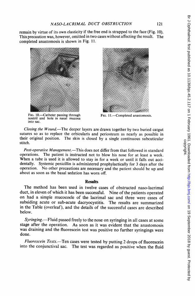
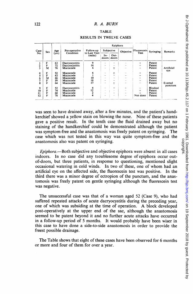
remain by virtue of its own elasticity if the free end is strapped to the face- (Fig. 10).This precaution was, however, omitted in two cases without affecting the result. Thecompleted anastomosis is shown in Fig. 11.
FIG. 10.-Catheter passing throughnostril and hole in nasal mucosainto sac.
Closing thesutures so astheir originalstitch.
FIG. 11.-Completed anastomosis.
Wound.-The deeper layers are drawn together by two buried catgutto replace the orbicularis and periosteum as nearly as possible inposition. The skin is closed by a single continuous subcuticular
Post-operative Management.-This does not differ from that followed in standardoperations. The patient is instructed not to blow his nose for at least a week.When a tube is used it is allowed to stay in for a week or until it falls out acci-dentally. Systemic penicillin is administered prophylactically for 3 days after theoperation. No other precautions are necessary and the patient should be up andabout as soon as the basal sedation has worn off.
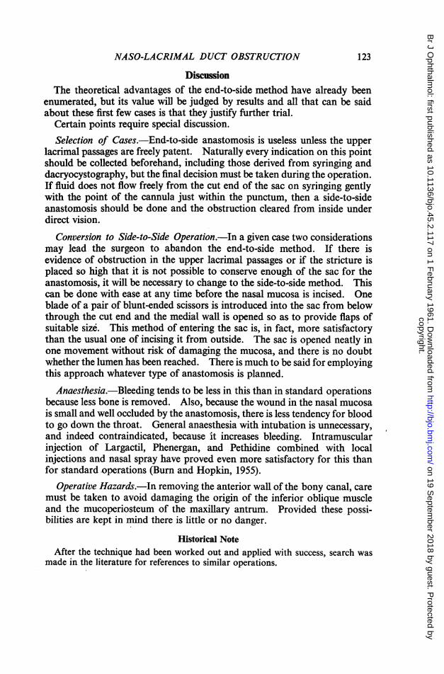
ResultsThe method has been used in twelve cases of obstructed naso-lacrimal
duct, in eleven of which it has been successful. Nine of the patients operatedon had a simple mucocoele of the lacrimal sac and three were cases ofsubsiding acute or sub-acute dacryocystitis. The results are summarizedin the Table (overleaf), and the details of the successful cases are describedbelow.
Syringing.-Fluid passed freely to the nose on syringing in all cases at somestage after the operation. As soon as it was evident that the anastomosiswas draining and the fluorescein test was positive no further syringings weredone.
Fluorescein Tests.-Ten cases were tested by putting 2 drops of fluoresceininto the conjunctival sac. The test was regarded as positive when the fluid
121
copyright. on 19 S
eptember 2018 by guest. P
rotected byhttp://bjo.bm
j.com/
Br J O
phthalmol: first published as 10.1136/bjo.45.2.117 on 1 F
ebruary 1961. Dow
nloaded from
TABLE
RESULTS IN TWELVE CASES
Epiphora
Case Sex Age Pre-onpeative Follow-up Subjective Objective Fluorescein Syringing RemarksNo. (yrs) Condition to Last Visit Teat Srgg e
(mths) In- Out-
1 F 63 Dacryocystitis 9 - _ - + Patent2 F 72 Dacryocystitis 16 - -- - Patent3 M 76 Mucocoele 6 - ± _ + Patent Artificial
eye4 F 52 Mucocoele 6 - + _ + Patent5 F 43 Mucocoele I - -_ - Patent6 M 46 Mucocoele 10 - -_ Patent7 F 59 Mucocoele 18 - _ - - Patent8 F 64 Mucocoele 17 - + - - Patent Everted
punctum9 F 52 Dacryocystitis 4 _ + _ Blocked10 F 74 Mucocoele 12 - - + Patent11 F 68 Mucocoele 2 - -_ + Patent12 F 78 Mucocoele 4 - - - Not done Patent12
_. _ _
was seen to have drained away, after a few minutes, and the patient's hand-kerchief showed a yellow stain on blowing the nose. Nine of these patientsgave a positive result. In the tenth case the fluid drained away but nostaining of the handkerchief could be demonstrated although the patientwas symptom-free and the anastomosis was freely patent on syringing. Thecase which was not tested in this way was quite symptom-free and theanastomosis also was patent on syringing.
Epiphora.-Both subjective and objective epiphora were absent in all casesindoors. In no case did any troublesome degree of epiphora occur out-of-doors, but three patients, in response to questioning, mentioned slightoccasional watering in cold winds. In two of these, one of whom had anartificial eye on the affected side, the fluorescein test was positive. In thethird there was a minor degree of ectropion of the punctum, and the anas-tomosis was freely patent on gentle syringing although the fluorescein testwas negative.
The unsuccessful case was that of a woman aged 52 (Case 9), who hadsuffered repeated attacks of acute dacryocystitis during the preceding year,one of which was subsiding at the time of operation. A block developedpost-operatively at the upper end of the sac, although the anastomosisseemed to be patent beyond it and no further acute attacks have occurredin a follow-up period of 5 months. It would probably have been wiser inthis case to have done a side-to-side anastomosis in order to provide thefreest possible drainage.
The Table shows that eight of these cases have been observed for 6 monthsor more and four of them for over a year.
122 R. A. BURN
copyright. on 19 S
eptember 2018 by guest. P
rotected byhttp://bjo.bm
j.com/
Br J O
phthalmol: first published as 10.1136/bjo.45.2.117 on 1 F
ebruary 1961. Dow
nloaded from
NASO-LACRIMAL DUCT OBSTRUCTION
DiscussionThe theoretical advantages of the end-to-side method have already been
enumerated, but its value will be judged by results and all that can be saidabout these first few cases is that they justify further trial.
Certain points require special discussion.Selection of Cases.-End-to-side anastomosis is useless unless the upper
lacrimal passages are freely patent. Naturally every indication on this pointshould be collected beforehand, including those derived from syringing anddacryocystography, but the final decision must be taken during the operation.If fluid does not flow freely from the cut end of the sac on syringing gentlywith the point of the cannula just within the punctum, then a side-to-sideanastomosis should be done and the obstruction cleared from inside underdirect vision.
Conversion to Side-to-Side Operation.-In a given case two considerationsmay lead the surgeon to abandon the end-to-side method. If there isevidence of obstruction in the upper lacrimal passages or if the stricture isplaced so high that it is not possible to conserve enough of the sac for theanastomosis, it will be necessary to change to the side-to-side method. Thiscan be done with ease at any time before the nasal mucosa is incised. Oneblade of a pair of blunt-ended scissors is introduced into the sac from belowthrough the cut end and the medial wall is opened so as to provide flaps ofsuitable size. This method of entering the sac is, in fact, more satisfactorythan the usual one of incising it from outside. The sac is opened neatly inone movement without risk of damaging the mucosa, and there is no doubtwhether the lumen has been reached. There is much to be said for employingthis approach whatever type of anastomosis is planned.
Anaesthesia.-Bleeding tends to be less in this than in standard operationsbecause less bone is removed. Also, because the wound in the nasal mucosais small and well occluded by the anastomosis, there is less tendency for bloodto go down the throat. General anaesthesia with intubation is unnecessary,and indeed contraindicated, because it increases bleeding. Intramuscularinjection of Largactil, Phenergan, and Pethidine combined with localinjections and nasal spray have proved even more satisfactory for this thanfor standard operations (Burn and Hopkin, 1955).
Operative Hazards.-In removing the anterior wall of the bony canal, caremust be taken to avoid damaging the origin of the inferior oblique muscleand the mucoperiosteum of the maxillary antrum. Provided these possi-bilities are kept in mind there is little or no danger.
Historical NoteAfter the technique had been worked out and applied with success, search was
made in the literature for references to similar operations.
123
copyright. on 19 S
eptember 2018 by guest. P
rotected byhttp://bjo.bm
j.com/
Br J O
phthalmol: first published as 10.1136/bjo.45.2.117 on 1 F
ebruary 1961. Dow
nloaded from
One account was found of an end-to-side anastomosis of this type (Juge, 1955),but no mention is made of numbers or results.The principle of using the end instead of the side of the sac to form a fresh
passage into the nose has been incorporated in a number of transplantationoperations in which the cut end is drawn into the nose through a hole in the boneand mucosa, and held there by a suture emerging from the nostril. Such anoperation was first performed by Speciale-Cirincione (1913) in two cases, in Italy,and more successfully and in larger numbers by Forsmark (1911) in Sweden. Stock(1934) in Germany, Burch (1920), Stokes (1938), and Gifford (1944) in America,and MacMillan (1932) in Canada, have advocated similar methods. The resultswere not, on the whole, so good as those achieved at the same period by side-to-sidemethods, and this must be attributed mainly to the absence of an accurate anas-tomosis so that the implanted end was left to collapse and seal off.The logical consequence of Forsmark's work would have been the development
of such an anastomosis, but at that time anaesthetic methods, needles, and suturematerials were not such as to make fine plastic procedures easy. The more roughand ready side-to-side techniques introduced by rhinologists like Toti and Moscherwere achieving good results and the attention of ophthalmic surgeons becameconcentrated upon them until, with the introduction of anterior and posteriormuco-mucous flaps by Dupuy-Dutemps and Bourguet (1921), the results becameso good that they have held the field ever since. Nevertheless, now that moderninstruments and anaesthetic methods have made accurate end-to-side anastomosisrelatively easy, it is perhaps worth considering whether the standard methodscannot be improved upon in this way.
SummaryA method of treating obstruction of the nasolacrimal duct by anastomosing
the cut end of the sac to a circular hole in the nasal mucosa is described.Twelve cases have been treated, of which eleven have proved successful.
I wish to express my gratitude to Miss P. Turnbull, of the Charing Cross Hospital PhotographicDepartment, for the photographs, and Miss Thorogood, S.R.N., of the Royal Eye Unit, LambethHospital, for her help with the theatre technique.
REFERENCESBURCH, F. E. (1920). Trans. Amer. Acad. Ophthal. Otolaryng., 25, 137.BURN, R. A., HOPKIN, D. A. B., EDWARDS, G., and JONES, C. M. (1955). Brit. J. Ophthal., 39,
333.DUPUY-DUTEMPS, P., and BOURGUET, J. (1921). Ann. Oculist. (Paris), 158, 241.FORSMARK, E. (1911). Hygiea (Stockh.), 73, 1432.GIFFORD, H. (1944). Arch. Ophthal. (Chicago), 32, 485.JUGE, P. (1955). Arch. Ophtal., 15, 732.MACMILLAN, J. H. (1932). Arch. Ophthal. (Chicago), 8, 831.
(1921). Amer. J. Ophthal., 4, 448.SPECIALE-CIRINCIONE, F. (1913). Clin. oculist. (Palermo), 3 ser., p. 1369.STOCK, W. (1934). Klin. Mbl. Augenheilk., 92, 433.STOKES, W. H. (1938). Trans. Amer. Acad. Ophthal. Otolarvng., 43, 342.
124 R. A. BURN
copyright. on 19 S
eptember 2018 by guest. P
rotected byhttp://bjo.bm
j.com/
Br J O
phthalmol: first published as 10.1136/bjo.45.2.117 on 1 F
ebruary 1961. Dow
nloaded from