open reduction and internal fixation compared with
TRANSCRIPT

The PDF of the article you requested follows this cover page.
This is an enhanced PDF from The Journal of Bone and Joint Surgery
2009;91:74-88. doi:10.2106/JBJS.G.01165 J Bone Joint Surg Am.Jeremy A. Hall, Murray J. Beuerlein, Michael D. McKee and the Canadian Orthopaedic Trauma Society
TechniqueApplication for Bicondylar Tibial Plateau Fractures. Surgical Open Reduction and Internal Fixation Compared with Circular Fixator
This information is current as of March 27, 2011
Reprints and Permissions
Permissions] link. and click on the [Reprints andjbjs.orgarticle, or locate the article citation on
to use material from thisorder reprints or request permissionClick here to
Publisher Information
www.jbjs.org20 Pickering Street, Needham, MA 02492-3157The Journal of Bone and Joint Surgery

COPYRIGHT © 2 009 BY THE JOURNAL OF BONE AND JOINT SURGERY, I NCORPORATED
74
Open Reduction and Internal Fixation Compared with Circular Fixator Application for Bicondylar Tibial Plateau FracturesSurgical Technique
By Jeremy A. Hall, MD, FRCS(C), Murray J. Beuerlein, MD, FRCS(C), Michael D. McKee, MD, FRCS(C), and the Canadian Orthopaedic Trauma Society
Investigation performed at St. Michael’s Hospital and the University of Toronto, Toronto, Ontario, Canada
The original scientific article in which the surgical technique was presented was published in JBJS Vol. 88-A, pp. 2613-23, December 2006
DISCLOSURE: In support of their research for or preparation of this work, one or more of the authors received, in any one year, outside funding or grants in excess of $10,000 from Smith and Nephew Ltd, Simon Fraser Orthopaedic Fund. Neither they nor a member of their immediate families received payments or other bene-fits or a commitment or agreement to provide such benefits from a commercial entity. No commercial entity paid or directed, or agreed to pay or direct, any benefits to any research fund, foundation, division, center, clinical practice, or other charitable or nonprofit organization with which the authors, or a member of their imme-diate families, are affiliated or associated.
ABSTRACT FROM THE ORIGINAL ARTICLE
BACKGROUND: Standard open reduction and internal fixation techniques have been successful in restoring osseous align-ment for bicondylar tibial plateau fractures; however, surgical morbidity, especially soft-tissue infection and wound necro-sis, has been reported frequently. For this reason, several investigators have proposed minimally invasive methods of fracture reduction followed by circular external fixation as an alternative approach. To our knowledge, there has been no direct comparison of the two operative approaches.
METHODS: We performed a multicenter, prospective, randomized clinical trial in which standard open reduction and inter-nal fixation with medial and lateral plates was compared with percutaneous and/or limited open fixation and application of a circular fixator for displaced bicondylar tibial plateau fractures (Schatzker types V and VI and Orthopaedic Trauma As-sociation types C1, C2, and C3). Eighty-three fractures in eighty-two patients were randomized to operative treatment (forty-three fractures were randomized to circular external fixation and forty to open reduction and internal fixation). Fol-low-up consisted of obtaining a history, physical examination, and radiographs; completion of the Hospital for Special Sur-gery (HSS) knee score, the Western Ontario and McMaster Universities Osteoarthritis Index (WOMAC), and the Short Form-36 (SF-36) General Health Survey; and recording of complication and reoperation rates.
RESULTS: There were no significant differences between the groups in terms of demographic variables, mechanism of injury, or fracture severity and/or displacement. However, patients in the circular fixator group had less intraoperative blood loss than those in the open reduction and internal fixation group (213 mL and 544 mL, respectively; p = 0.006) and spent less time in the hospital (9.9 days and 23.4 days, respectively; p = 0.024). The quality of osseous reduction was similar in the groups. There was a trend for patients in the circular fixator group to have superior early outcome in terms of HSS scores at six months (p = 0.064) and the ability to return to preinjury activities at six months (p = 0.031) and twelve months (p = 0.024). These outcomes were not significantly different at two years. There was no difference in total arc of knee motion, and the WOMAC scores at two years after the injury were not significantly different between the groups with regard to the pain (p = 0.923), stiffness (p = 0.604), or function (p = 0.827) categories. The SF-36 scores at two years after the injury were significantly decreased compared with the controls for both groups (p = 0.001 for the cir-cular fixator group and p = 0.014 for the open reduction and internal fixation group), although there was less impairment
continued
J Bone Joint Surg Am. 2009;91 Suppl 2 (Part 1):74-88 • doi:10.2106/JBJS.G.01165
Hall.fm Page 74 Friday, February 6, 2009 3:39 PM
75
TH E JO UR N AL O F BO N E & JO IN T SU RG E R Y · SU R G IC A L TE CH N I Q U E S MARCH 2009 · VOLUME 91-A · SUPPLEMENT 2, PART 1 · JBJS.ORG
INTRODUCTIONDisplaced bicondylar fractures of the proximal part of the tibia in-
volving the articular surface re-main difficult to treat. Previously, the standard ac-
cepted treatment for such frac-tures was open reduction and internal fixation with plates and
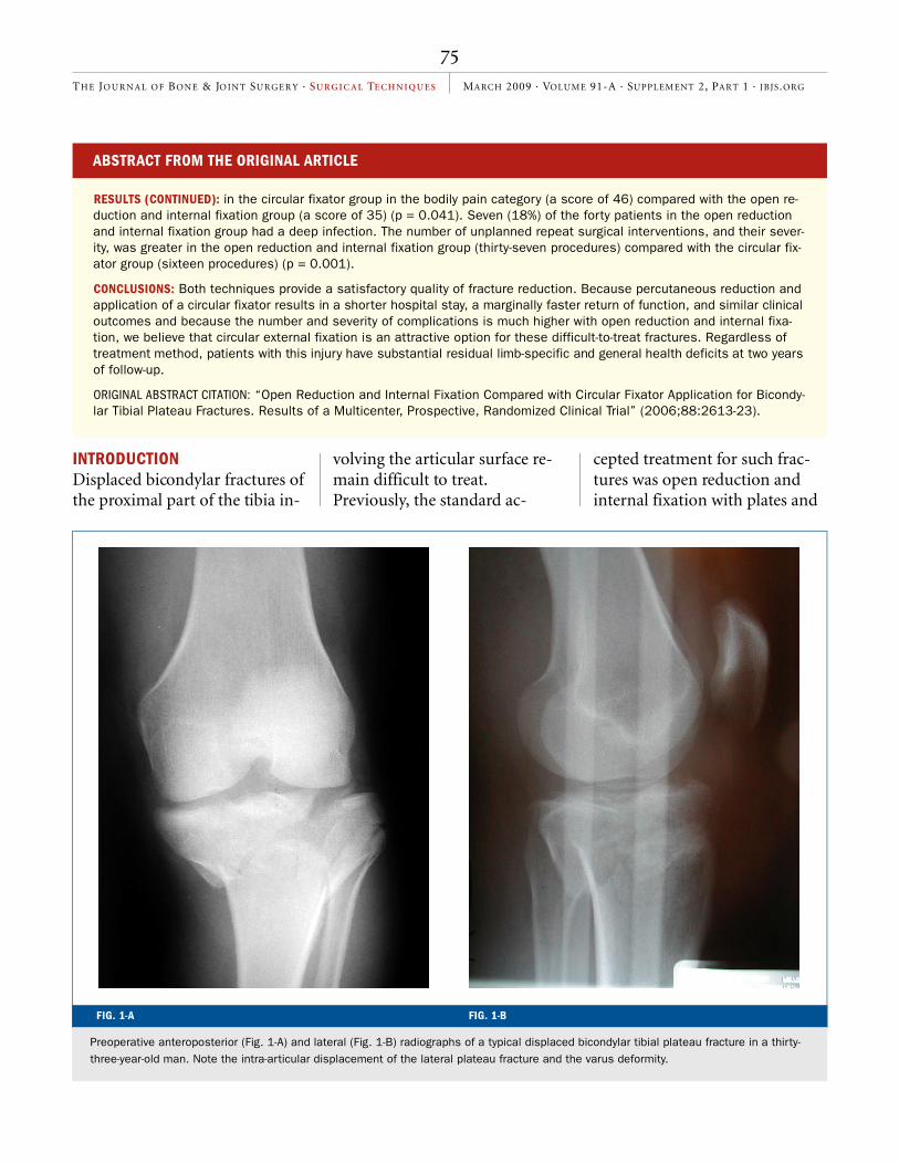
FIG. 1-A FIG. 1-B
Preoperative anteroposterior (Fig. 1-A) and lateral (Fig. 1-B) radiographs of a typical displaced bicondylar tibial plateau fracture in a thirty-three-year-old man. Note the intra-articular displacement of the lateral plateau fracture and the varus deformity.
ABSTRACT FROM THE ORIGINAL ARTICLE
RESULTS (CONTINUED): in the circular fixator group in the bodily pain category (a score of 46) compared with the open re-duction and internal fixation group (a score of 35) (p = 0.041). Seven (18%) of the forty patients in the open reduction and internal fixation group had a deep infection. The number of unplanned repeat surgical interventions, and their sever-ity, was greater in the open reduction and internal fixation group (thirty-seven procedures) compared with the circular fix-ator group (sixteen procedures) (p = 0.001).
CONCLUSIONS: Both techniques provide a satisfactory quality of fracture reduction. Because percutaneous reduction and application of a circular fixator results in a shorter hospital stay, a marginally faster return of function, and similar clinical outcomes and because the number and severity of complications is much higher with open reduction and internal fixa-tion, we believe that circular external fixation is an attractive option for these difficult-to-treat fractures. Regardless of treatment method, patients with this injury have substantial residual limb-specific and general health deficits at two years of follow-up.
ORIGINAL ABSTRACT CITATION: “Open Reduction and Internal Fixation Compared with Circular Fixator Application for Bicondy-lar Tibial Plateau Fractures. Results of a Multicenter, Prospective, Randomized Clinical Trial” (2006;88:2613-23).
Hall.fm Page 75 Friday, February 6, 2009 3:39 PM

76
TH E JO UR N AL O F BO N E & JO IN T SU RG E R Y · SU R G IC A L TE CH N I Q U E S MARCH 2009 · VOLUME 91-A · SUPPLEMENT 2, PART 1 · JBJS.ORG
FIG. 2
A preoperative computed tomography scan clarifies the main fracture lines. This step is essential if a percutaneous or limited open reduc-tion and fixation technique is chosen. It allows planning for closed reduction and fine-wire placement.
FIG. 3
Patient positioning. The patient is placed on a radiolucent operating room table to allow unhindered access for the image intensifier, and a bolster is placed under the affected thigh.
Hall.fm Page 76 Friday, February 6, 2009 3:39 PM
77
TH E JO UR N AL O F BO N E & JO IN T SU RG E R Y · SU R G IC A L TE CH N I Q U E S MARCH 2009 · VOLUME 91-A · SUPPLEMENT 2, PART 1 · JBJS.ORG
screws through an extensile ante-rior incision1-6. However, while this technique was optimal for fracture visualization, reduction, and fixation, it required substan-tial soft-tissue dissection over the predominantly subcutaneous proximal part of the tibia. The combination of damage to the soft tissues resulting from the en-ergy of the original injury and the extensive surgical dissection led to a high rate of complica-tions, including skin slough, ne-crosis, and infection7-9.
As the detrimental effects of excessive dissection of the soft-tissue envelope and devascular-ization of the osseous fragments have become apparent, a number of alternative methods of treat-ment, including percutaneous
reduction and circular frame sta-bilization, minimally invasive techniques and implants, and temporary external fixation fol-lowed by delayed definitive sur-gery, have been popularized10-19. The advantages of circular frame fixation (with or without percu-taneous lag-screw fixation) in-clude minimal soft-tissue dissection, the ability to correct deformity in multiple planes, early knee joint motion, the op-tion of spanning the knee joint in cases of concomitant ligament injury, and the possibility of sta-bilizing more distal fractures with the same device. However, the quality of articular reduction with circular fixation remains unproven, and, to our knowl-edge, a direct comparison with
standard reduction techniques has not been performed.
We performed a prospec-tive, randomized, multicenter clinical trial of patients with dis-placed bicondylar tibial plateau fractures in which the outcome of standard open reduction and internal fixation with use of me-dial and lateral plates was com-pared with the outcome of percutaneous reduction and cir-cular fixation. In the present re-port, we describe the techniques used for each surgical approach.
SURGICAL TECHNIQUECircular External FixatorWhile proper anteroposterior and lateral radiographs centered on the knee are important (Figs. 1-A and 1-B), a preoperative
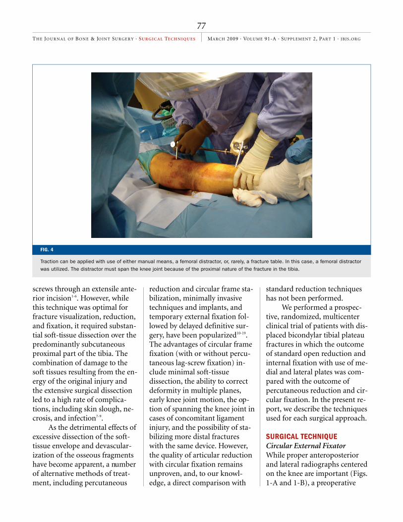
FIG. 4
Traction can be applied with use of either manual means, a femoral distractor, or, rarely, a fracture table. In this case, a femoral distractor was utilized. The distractor must span the knee joint because of the proximal nature of the fracture in the tibia.
Hall.fm Page 77 Friday, February 6, 2009 3:39 PM

78
TH E JO UR N AL O F BO N E & JO IN T SU RG E R Y · SU R G IC A L TE CH N I Q U E S MARCH 2009 · VOLUME 91-A · SUPPLEMENT 2, PART 1 · JBJS.ORG
FIG. 5-B
Figs. 5-A and 5-B A small stab incision (Fig. 5-A) is made over the fracture site. This allows percutaneous placement of a periosteal elevator to reduce the depressed fragments (Fig. 5-B). When reduction is achieved, a reduction clamp is used to hold the reduction prior to fixation.
FIG. 5-A
Hall.fm Page 78 Friday, February 6, 2009 3:39 PM

79
TH E JO UR N AL O F BO N E & JO IN T SU RG E R Y · SU R G IC A L TE CH N I Q U E S MARCH 2009 · VOLUME 91-A · SUPPLEMENT 2, PART 1 · JBJS.ORG
computed tomography scan is essential to evaluate the details of the fracture configuration, to identify the main fragments, and to help to plan wire placement (Fig. 2). The patient is positioned supine on either a fracture table or a long radiolucent table under general anesthesia (Fig. 3). The limb is prepared and is draped free appropriately with a tourni-quet applied. While the tourni-quet may not be used, it can be a useful adjunct in certain situa-tions. Bolsters are placed about the limb to allow 360° visualiza-tion. Longitudinal traction is ap-plied to the fracture with use of either manual means, a fracture table, or a femoral distractor (Fig. 4). The choice of traction depends on the complexity and chronicity of the fracture and the
FIG. 6
Following reduction of the articular surface, a fracture clamp is applied percutaneously. The fracture is then stabilized with the insertion of large-fragment cannulated screws in a lag fashion across the main fracture lines.
FIG. 7
Initial application of the frame. The length, circumference, and ring position are checked both visually and with the image intensifier. It is important to pad the rings posteriorly to prevent the leg from “sagging” posteriorly and coming into contact with the rings. There should be approximately two fingerbreadths between the skin and the rings at the narrowest point.
Hall.fm Page 79 Friday, February 6, 2009 3:39 PM

80
TH E JO UR N AL O F BO N E & JO IN T SU RG E R Y · SU R G IC A L TE CH N I Q U E S MARCH 2009 · VOLUME 91-A · SUPPLEMENT 2, PART 1 · JBJS.ORG
preference of the surgeon. Image intensification is used to assess fracture alignment. Fracture fragments are reduced into posi-tion with use of gentle, closed pressure or percutaneously in-serted periosteal elevators or re-duction forceps (Figs. 5-A and 5-B). Percutaneous lag screws are inserted to stabilize the articular fragments (Fig. 6). An anterome-dial or anterolateral mini-arthro-tomy is made to repair tibial
spine fractures with use of gentle manipulation with periosteal ele-vators or reduction clamps; the fractures are fixed with use of percutaneous screws or sutures tied over a metaphyseal screw distally. With the articular sur-face reduced, the circular exter-nal fixator frame is applied next to secure the metaphysis to the diaphysis of the tibia.
The frame is assembled with use of three or four rings,
depending on the proximity of the fracture to the joint. Rings are sized to the limb to provide roughly two fingerbreadths of clearance circumferentially (Fig. 7). The leg tends to “sag” poste-riorly against the frame, and therefore sterile towels or sponges are used to maintain this clearance posteriorly throughout the procedure. Fine wires are then placed across the proximal part of the tibia, paral-
FIG. 8
Wire insertion. After the positioning of the frame, the first transfixing olive wires are placed in the proximal fragments with use of the orien-tation of the main fracture lines as seen on the preoperative computed tomography scan for guidance. Typically, one wire is passed through the fibular head from lateral to medial and a second olive wire is then passed from posteromedial to anterolateral to “capture” the proxi-mal fracture assembly. If possible, the proximal wires are kept 1 cm from the joint line to avoid transgressing the reflections of the joint capsule. This is done in an attempt to avoid having a pin-track infection spread intra-articularly. In some cases, fracture geometry may force such proximal placement and the patient should be watched carefully for signs and symptoms of intra-articular infection.
Hall.fm Page 80 Friday, February 6, 2009 3:39 PM

81
TH E JO UR N AL O F BO N E & JO IN T SU RG E R Y · SU R G IC A L TE CH N I Q U E S MARCH 2009 · VOLUME 91-A · SUPPLEMENT 2, PART 1 · JBJS.ORG
lel to the joint surface. The first is placed from lateral to medial through the fibular head (Fig. 8). As the proximal tibiofibular joint communicates with the knee joint in approximately 10% of individuals, the inser-tion of this wire theoretically can lead to septic arthritis of the knee if the area around the wire becomes infected. However, es-pecially in cases of proximal fractures, the added stability of the proximal fragment attrib-uted to this wire usually justi-fies its use. If the area around the wire becomes infected, the wire must be removed and/or replaced promptly. Care is taken to place the wires at least 1 cm from the joint surface to avoid intra-articular penetration un-
less this is absolutely necessary, so that the wires are not intra-articular during their course. The wires are first tightened on the insertion end with use of a cannulated bolt and nut and then are tensioned to 90 lb with use of the tensioner on the op-posite end. The opposite cannu-lated nut and bolt are then tightened with the tensioner in place. Once tightened, the ten-sioner is removed. The olive end of the wire is used to compress and maneuver displaced frag-ments of the metaphysis. In this setting, the insertion end of the wire remains untightened while the wire is gently drawn to the opposite side of the frame with use of the tensioner. Then the olive end of the wire is tightened
with a cannulated bolt and nut, and the tensioner is removed, reset, and used to tension the in-sertion end of the wire (Fig. 9). The opposite cannulated bolt and nut are then tightened, and the tensioner is removed. This procedure is repeated with sev-eral wires (usually four), alter-nating from lateral to medial and medial to lateral, to provide stability to the proximal seg-ment. Image intensification is used to assess the articular re-duction and the alignment of the tibia. Fixation of the shaft to the distal rings can then be per-formed with transfixion wires or percutaneous threaded half-pins, depending on local factors, the associated injuries, and the preference of the surgeon (Fig.
FIG. 9
Fixation of the wires to the frame. A tensioner is used on the non-olive end of the wire to pull the fragments and to correct any deformity (typically varus). Once the wires are tensioned, they are secured to the frame with a cannulated bolt and nut.
Hall.fm Page 81 Friday, February 6, 2009 3:39 PM

82
TH E JO UR N AL O F BO N E & JO IN T SU RG E R Y · SU R G IC A L TE CH N I Q U E S MARCH 2009 · VOLUME 91-A · SUPPLEMENT 2, PART 1 · JBJS.ORG
10). In general, half-pins are used with the distal rings once alignment of the fracture has been achieved. Half-pin cubes are fixed to the rings with nuts. The drill-guide is positioned, and the skin is cut and spread with a hemostat, after which the guide is placed against the dia-physis and a 4.0-mm drill is passed across both cortices. A 5.0-mm threaded half-pin is then inserted and fixed to the cube with use of two set screws. This is repeated over the two distal rings, for a total of four half-pins. Image intensification is used to assess the position of the half-pins to ensure place-ment across both cortices and to evaluate the overall alignment of the tibia. With this complete,
the fine wires are cut and are bent over the rings. The pin sites and surgical wounds are then closed and dressed. Radiographs are made to confirm reduction of the intra-articular fracture, alignment with the remainder of the tibia, and proper placement of the screws and wires (Figs. 11-A, 11-B, and 11-C).
We prefer this method for the treatment of fractures that are associated with compart-ment syndrome as it eliminates the problem of exposed fixation devices in open fasciotomy wounds. On the other hand, al-though transfixion pin place-ment can be planned to facilitate eventual wound closure, the use of local rotational flaps such as the gastrocnemius can be com-
promised by the use of the circu-lar frame.
Open Reduction and Internal FixationFormal open reduction and in-ternal fixation of bicondylar tib-ial plateau fractures (Fig. 12) may need to be delayed until the anterior soft-tissue structures are healthy and viable. Positioning is similar to that for the circular fixator technique. The patient is placed supine under general an-esthesia. A fracture table or long radiolucent operating table is used to facilitate visualization. The limb is prepared and draped free with a tourniquet applied proximally and bolstered appro-priately; we have found that an adjustable triangular frame is
FIG. 10
Distal fixation of the fixator. Fixation of the distal ring can involve the use of either transfixion wires or percutaneous threaded half-pins. In this case, a half-pin construct was used.
Hall.fm Page 82 Friday, February 6, 2009 3:39 PM

83
TH E JO UR N AL O F BO N E & JO IN T SU RG E R Y · SU R G IC A L TE CH N I Q U E S MARCH 2009 · VOLUME 91-A · SUPPLEMENT 2, PART 1 · JBJS.ORG
useful in this situation. In gen-eral, widely separate posterome-dial and anterolateral incisions
are made to minimize the soft-tissue dissection (Fig. 13) and yet allow access to the major fracture
lines and fragments that are of-ten difficult to address from an anterior approach. Anterior frac-
FIG. 11-C
FIG. 11-A FIG. 11-B
Fig. 11-A Anteroposterior intraoperative radio-graph made after limited open reduction, percu-taneous screw fixation, and application of a circular fixator for the treatment of the fracture depicted in Figures 1-A, 1-B, and 2. The intra-articular fractures have been reduced, and the varus deformity has been corrected. Although this fracture pattern required olive wire place-ment close to the joint line, which may have re-sulted in penetration of the capsule, no infection developed. Fig. 11-B Anteroposterior radiograph made two years postoperatively. While there is some residual deformity of the medial plateau, the knee is stable, the leg is straight, and the pa-tient had returned to all preinjury activities with-out pain. Fig. 11-C Lateral radiograph made two years postoperatively.
Hall.fm Page 83 Friday, February 6, 2009 3:39 PM

84
TH E JO UR N AL O F BO N E & JO IN T SU RG E R Y · SU R G IC A L TE CH N I Q U E S MARCH 2009 · VOLUME 91-A · SUPPLEMENT 2, PART 1 · JBJS.ORG
tures (as from a hyperextension type of injury) with mainly ante-rior comminution can be treated through a single anterior inci-sion. The main goal is to avoid extensive soft-tissue dissection that devascularizes bone frag-ments or creates precarious soft-tissue flaps leading to wound breakdown. The incisions should be only as long as necessary to visualize and reduce the frac-ture. While concerns regarding possible later conversion to total knee arthroplasty are valid, the conversion rate is surprisingly low and the surgical approach should not compromise expo-sure of the fracture site. An ar-throtomy is created to visualize
FIG. 13
Two operative ap-proaches can be used: (1) a single anterior in-cision (left), which usu-ally involves substantial soft-tissue stripping, or (2) the preferred two-incision approach (right). One anterolateral inci-sion and one postero-medial incision are made. This allows for a sufficient skin bridge to decrease the chance of wound complications.
FIG. 12
Preoperative radiograph of a displaced bicondylar tibial pla-teau fracture in a thirty-two-year-old man.
Hall.fm Page 84 Friday, February 6, 2009 3:39 PM

85
TH E JO UR N AL O F BO N E & JO IN T SU RG E R Y · SU R G IC A L TE CH N I Q U E S MARCH 2009 · VOLUME 91-A · SUPPLEMENT 2, PART 1 · JBJS.ORG
the joint. This is usually per-formed just proximal to the ar-ticular surface of the plateau and below the meniscus by section-ing the coronary ligament. The articular surface is visualized and reduced by elevating any de-
pressed fracture fragments (Figs. 14-A and 14-B). Kirschner wires are used to hold the reduction while lag screws are placed across the fragments. The articular re-duction is assessed directly through the arthrotomy as well
as with image intensification. The cruciate ligaments can be visualized by extending the ar-throtomy anteriorly or by per-forming a formal anterolateral arthrotomy in a vertical plane. The overall alignment of the
FIG. 14-A
Fracture reduction is performed by work-ing through the anterior fracture lines to elevate depressed osseous fragments and to secure their reduction with tem-porary Kirschner wire fixation and a re-duction clamp (Fig. 14-A). After reduction, lag screws are inserted. Later, buttress plate application pro-vides definitive fixation (Fig. 14-B).
FIG. 14-B
Hall.fm Page 85 Friday, February 6, 2009 3:39 PM

86
TH E JO UR N AL O F BO N E & JO IN T SU RG E R Y · SU R G IC A L TE CH N I Q U E S MARCH 2009 · VOLUME 91-A · SUPPLEMENT 2, PART 1 · JBJS.ORG
tibia should be assessed in the anteroposterior and lateral planes. When the alignment is satisfactory, the metaphysis is fixed to the diaphysis with me-dial and lateral plates and screws. This fixation follows conven-tional plating techniques with an emphasis on minimizing soft-tissue dissection. The lateral pla-teau is fixed to the diaphysis with use of a precontoured 3.5 or 4.5-mm proximal tibial periarticular plate, depending on the size of the patient and the size of the fracture fragments. Image inten-sification is used to ensure ap-propriate placement in the anteroposterior and lateral planes. Bicortical screws are
placed from lateral to medial in the metaphysis and diaphysis. The medial plateau is fixed to the diaphysis with use of a hand-contoured 3.5 or 4.5-mm low-contact dynamic compression buttress plate. Bicortical screws are placed across the metaphysis and diaphysis. At least four corti-ces are required in both the metaphysis and the diaphysis (Figs. 15-A and 15-B). Large os-seous defects created by the ele-vation of depressed articular fragments can be filled with a number of compounds, includ-ing autogenous bone graft or any one of a number of newer bone substitutes. Tibial spine or cruci-ate ligament injuries are re-
paired through the arthrotomy with use of screw fixation or su-tures woven through the liga-ment and tied over a well-fixed screw in the tibial metaphysis. Meniscal injuries are repaired to the capsule with use of inter-rupted nonabsorbable sutures. Image intensification is used for a final check in the anteroposte-rior and lateral planes. The wounds are irrigated with sterile saline solution and are closed in layers after the tourniquet has been deflated and hemostasis has been obtained. Postoperatively, the limb is placed in a long-leg plaster splint. Early range of mo-tion is instituted on the basis of the stability of the fracture.
FIG. 15-A FIG. 15-B
Fig. 15-A Postoperative radiograph following open reduction and internal fixation through two incisions utilizing medial and lateral plates. Fig. 15-B Radiograph made two years postoperatively. There is mild residual deformity of the lateral plateau, but the leg is clinically straight, the knee stable and pain-free, and the patient had returned to preinjury activities.
Hall.fm Page 86 Friday, February 6, 2009 3:39 PM
87
TH E JO UR N AL O F BO N E & JO IN T SU RG E R Y · SU R G IC A L TE CH N I Q U E S MARCH 2009 · VOLUME 91-A · SUPPLEMENT 2, PART 1 · JBJS.ORG
CRITICAL CONCEPTS
INDICATIONS:
While indications vary depending on the age, health status, and functional demands of the patient, operative treatment for displaced intra-articular bicondylar tibial plateau fractures is generally appropriate for patients with the following findings.
• Intra-articular displacement of ≥2 mm
• Metaphyseal-diaphyseal translation of >1 cm
• Angular deformity of >10° in the coronal (varus-valgus) or sagittal plane
• Open fracture
• Associated compartment syndrome
• Associated ligament injury requiring repair
• Associated fractures of the ipsilateral tibia or fibula
CONTRAINDICATIONS:
Contraindications to the operative fixation of bicondylar tibial plateau fractures include the following.
• Undisplaced fracture
• Lack of familiarity with either surgical technique
• A patient who is medically unfit for an anesthetic
• A patient who is unable to comply with the rehabilitation protocol or benefit from the procedure (for example, because of dementia)
• Active infection in or around the surgical site
• Severe soft-tissue swelling that would jeopardize the healing of incisions
• Open growth plates
PITFALLS:
Circular External Fixation
• Care must be taken to ensure adequate reduction of intra-articular fragments prior to the application of the frame. Once the frame is applied, access to the proximal fragments is limited.
• The tibia has a tendency to sag posteriorly during frame application and must be bolstered posteriorly with towels to pre-vent frame-leg contact.
• Fine wires should be placed 1 cm distal to the joint line to avoid intra-articular placement as pin-track infections are fre-quent and may result in septic arthritis if the pins are intracapsular.
• The overall alignment of the tibia must be assessed radiographically in both the anteroposterior and lateral planes to en-sure the adequacy of the reduction.
• If proximal purchase is inadequate despite multiple transfixion wires and/or if there is substantial ligamentous injury in the knee, spanning the knee with an extension of the circular frame to the distal part of the femur should be considered with a hinge placed at the center axis of the knee and connected to the distal part of the femur with half-pins or transfixion wires.
Open Reduction and Internal Fixation
• The extensive soft-tissue disruption of the fracture combined with the secondary soft-tissue injury associated with surgery can result in complications, including wound dehiscence and infection. Care must be taken when handling the proximal soft tissues; gentle handling of tissue, minimizing incision length and flap formation, and meticulous, layered closure are required. This may mean an extended period of time with the limb in a splint or in a spanning external fixator before the soft tissues are robust enough for surgical intervention.
continued
Hall.fm Page 87 Friday, February 6, 2009 3:39 PM

88
TH E JO UR N AL O F BO N E & JO IN T SU RG E R Y · SU R G IC A L TE CH N I Q U E S MARCH 2009 · VOLUME 91-A · SUPPLEMENT 2, PART 1 · JBJS.ORG
Jeremy A. Hall, MD, FRCS(C)Murray J. Beuerlein, MD, FRCS(C)Michael D. McKee, MD, FRCS(C)Division of Orthopaedics, Department of Surgery, St. Michael’s Hospital and the University of Tor-onto, 55 Queen Street East, Suite 800, Toronto, ON M5C 1R6, Canada. E-mail address for M.D. McKee: [email protected]
The line drawings in this article are the work of Jennifer Fairman ([email protected]).
REFERENCES1. Covall DJ, Fowble CD, Foster TE, Whitelaw GP. Bicondylar tibial plateau fractures: princi-ples of treatment. Contemp Orthop. 1994;28:115-22.
2. Agnew SG, Benirschke SK, Mayo KA, Henley MB, Santoro VM. Open reduction and internal fixation of complex tibial plateau fractures. J Orthop Trauma. 1991;5:236.
3. Schatzker J. Fractures of the tibial plateau. In: Schatzker J, Tile M, editors. The rationale of operative fracture care. New York: Springer; 1987. p 279-95.
4. Benirschke SK, Agnew SG, Mayo KA, San-toro VM, Henley MB. Immediate internal fixa-tion of open, complex tibial plateau fractures: treatment by a standard protocol. J Orthop Trauma. 1992;6:78-86.
5. Waddell JP, Johnston DW, Neidre A. Frac-tures of the tibial plateau: a review of ninety-five patients and comparison of treatment methods. J Trauma. 1981;21:376-81.
6. Tscherne H, Lobenhoffer P. Tibial plateau fractures. Management and expected results. Clin Orthop Relat Res. 1993;292:87-100.
7. Young MJ, Barrack RL. Complications of in-ternal fixation of tibial plateau fractures. Or-thop Rev. 1994;23:149-54.
8. Moore TM, Patzakis MJ, Harvey JP. Tibial plateau fractures: definition, demographics, treatment rationale, and long-term results of closed traction management or operative re-duction. J Orthop Trauma. 1987;1:97-119.
9. Mallik AR, Covall DJ, Whitelaw GP. Internal versus external fixation of bicondylar tibial plateau fractures. Orthop Rev. 1992;21:1433-6.
10. Stamer DT, Schenk R, Staggers B, Aurori K, Aurori B, Behrens FF. Bicondylar tibial pla-teau fractures treated with a hybrid ring exter-nal fixator: a preliminary study. J Orthop Trauma. 1994;8:455-61.
11. Watson JT. High-energy fractures of the tibial plateau. Orthop Clin North Am. 1994;25:723-52.
12. Buckle R, Blake R, Watson JT, Morandi M, Browner BD. Treatment of the complex tibial plateau fractures with the Ilizarov external fix-
ator. J Orthop Trauma. 1993;7:167-8.
13. Mikulak SA, Gold SM, Zinar DM. Small wire external fixation of high energy tibial pla-teau fractures. Clin Orthop Relat Res. 1998;356:230-8.
14. Morandi MM, Pearse MF. Management of complex plateau tibial fractures with the Ilizarov external fixator. Tech Orthop. 1996;11:125-31.
15. Watson JT, Ripple S, Hoshaw SJ, Fhyrie D. Hybrid external fixation for tibial plateau frac-tures: clinical and biomechanical correlation. Orthop Clin North Am. 2002;33:199-209, ix.
16. Marsh JL, Smith ST, Do TT. External fixa-tion and limited internal fixation for complex fractures of the tibial plateau. J Bone Joint Surg Am. 1995;77:661-73.
17. Dendrinos GK, Kontos S, Katsenis D, Dalas A. Treatment of high-energy tibial pla-teau fractures by the Ilizarov circular fixator. J Bone Joint Surg Br. 1996;78:710-7.
18. Watson JT, Coufal C. Treatment of com-plex lateral plateau fractures using Ilizarov techniques. Clin Orthop Relat Res. 1998;353:97-106.
19. Kumar A, Whittle AP. Treatment of com-plex (Schatzker Type VI) fractures of the tibial plateau with circular wire external fixation: retrospective case review. J Orthop Trauma. 2000;14:339-44.
CRITICAL CONCEPTS
PITFALLS (CONTINUED):
• Care must be taken to directly assess the articular reduction through a submeniscal or parapatellar tendon arthrotomy as image intensification alone can be misleading.
• Overall alignment of the tibia must be assessed in both the anteroposterior and lateral planes to ensure the adequacy of reduction.
AUTHOR UPDATE:
We continue to use both surgical techniques for the fixation of displaced bicondylar tibial plateau fractures, and we have found the two techniques to be complementary rather than mutually exclusive. Fractures associated with severe soft-tis-sue injury or swelling, predominantly metaphyseal-diaphyseal comminution and displacement, fracture extension into the tibial shaft, and less severe intra-articular fracture displacement are ideal for closed or limited open reduction and circu-lar frame fixation. Fractures associated with extensive intra-articular displacement and comminution are ideally suited for formal open reduction and internal fixation with use of a two-incision technique. The utility of this method has been aug-mented by the advent of precontoured plates for the proximal part of the tibia and the use of minimally invasive tech-niques for the application of these plates distally along the shaft.
Locking plates are gaining in popularity for the treatment of complex fractures, particularly those of the proximal part of the tibia. While it is tempting to minimize soft-tissue dissection in cases of bicondylar tibial plateau fractures by using a single axially stable locking plate to secure both condyles, we have not been uniformly satisfied with that technique. We have observed collapse or settling of the far (usually medial) condyle when a single (usually lateral) plate is used alone for fixation. At the present time, we do not routinely recommend solitary lateral plate fixation; this is especially true for posteromedial fractures with a predominantly coronal fracture line.
Hall.fm Page 88 Friday, February 6, 2009 3:39 PM