open access research screening for seemingly healthy
TRANSCRIPT

Screening for seemingly healthynewborns with congenitalcytomegalovirus infection byquantitative real-time polymerasechain reaction using newborn urine:an observational study
Akira Yamaguchi,1,2 Tsutomu Oh-ishi,3 Takashi Arai,1,2 Hideaki Sakata,4,5
Nodoka Adachi,6 Satoshi Asanuma,6 Eiji Oguma,7 Hirofumi Kimoto,8
Jiro Matsumoto,9 Hidetoshi Fujita,10 Tadashi Uesato,10 Jutaro Fujita,10
Ken Shirato,11 Hideki Ohno,11 Takako Kizaki11
To cite: Yamaguchi A, Oh-ishi T, Arai T, et al. Screeningfor seemingly healthynewborns with congenitalcytomegalovirus infection byquantitative real-timepolymerase chain reactionusing newborn urine:an observational study. BMJOpen 2017;7:e013810.doi:10.1136/bmjopen-2016-013810
▸ Prepublication history forthis paper is available online.To view these files pleasevisit the journal online(http://dx.doi.org/10.1136/bmjopen-2016-013810).
Received 23 August 2016Revised 29 December 2016Accepted 30 December 2016
For numbered affiliations seeend of article.
Correspondence toDr Takako Kizaki;[email protected]
ABSTRACTObjective: Approximately 8–10% of newborns withasymptomatic congenital cytomegalovirus (cCMV)infection develop sensorineural hearing loss (SNHL).However, the relationship between CMV load, SNHLand central nervous system (CNS) damage in cCMVinfection remains unclear. This study aimed to examinethe relationship between urinary CMV load, SNHL andCNS damage in newborns with cCMV infection.Study design: The study included 23 368 newbornsfrom two maternity hospitals in Saitama Prefecture,Japan. Urine screening for cCMV infection (quantitativereal-time PCR) and newborn hearing screening(automated auditory brainstem response (AABR)testing) were conducted within 5 days of birth toexamine the incidence of cCMV infection and SNHL,respectively. CNS damage was assessed by MRI ofcCMV-infected newborns.Results: The incidence of cCMV infection was 60/23 368 (0.257%; 95% CI 0.192% to 0.322%). Thegeometric mean urinary CMV DNA copy number innewborns with cCMV was 1.79×10⁶ copies/mL (95%CI 7.97×10⁵ to 4.02×10⁶). AABR testing revealedabnormalities in 171 of the 22 229 (0.769%) newbornswhose parents approved hearing screening. Of these171 newborns, 22 had SNHL (12.9%), and 5 of these22 were infected with cCMV (22.7%). Newborns withboth cCMV and SNHL had a higher urinary CMV DNAcopy number than newborns with cCMV without SNHL(p=0.036). MRI revealed CNS damage, including whitematter abnormalities, in 83.0% of newborns withcCMV. Moreover, newborns with CNS damage had asignificantly greater urinary CMV load than newbornswithout CNS damage (p=0.013).Conclusions: We determined the incidence of cCMVinfection and urinary CMV DNA copy number inseemingly healthy newborns from two hospitals in
Saitama Prefecture. SNHL and CNS damage wereassociated with urinary CMV DNA copy number.Quantification of urinary CMV load may effectivelypredict the incidence of late-onset SNHL andneurodevelopmental disorders.
Strengths and limitations of this study
▪ Verification of the risk factors for sensorineuralhearing loss (SNHL) in newborns and late-onsetSNHL caused by congenital cytomegalovirus(cCMV) infection as well as for developmental orintellectual disability is important for predicting apatient’s prognosis. In this study we examinedthe association of urinary CMV load in cCMVinfection cases with SNHL and central nervoussystem (CNS) damage.
▪ Urinary CMV load was evaluated by calculatingthe CMV DNA copy number in a urine sample byusing quantitative real-time PCR (qPCR) whileensuring sensitivity and quantitative capability.
▪ The CMV DNA copy number of newborns withSNHL and CNS damage was significantly morethan that of newborns without any findings. Thissuggests that measuring CMV load in urinesamples could be useful for predicting prognosisof cCMV infection.
▪ Based on the current research, it is unclearwhether CNS damage related to cCMV infectionis associated with future developmental or intel-lectual disability, which indicates that follow-upexaminations that include intelligence and psy-chological evaluation are required.
▪ The use of qPCR for a urine sample as a screen-ing test requires improvements in usability andeconomic efficiency.
Yamaguchi A, et al. BMJ Open 2017;7:e013810. doi:10.1136/bmjopen-2016-013810 1
Open Access Research
on Decem
ber 26, 2021 by guest. Protected by copyright.
http://bmjopen.bm
j.com/
BM
J Open: first published as 10.1136/bm
jopen-2016-013810 on 20 January 2017. Dow
nloaded from

INTRODUCTIONHuman cytomegalovirus (CMV) is known to cause con-genital infection and is the most commonly implicatedpathogen in the group of congenital infections knownas TORCH syndrome (toxoplasmosis, rubella, CMVinfection, herpes simplex and other infections). Theincidence of CMV is 0.2–2.4% in all newborns, andapproximately 5–10% of CMV-infected newborns havesymptomatic congenital CMV (cCMV) infection. Incases of severe infection, newborns can present withsymptoms of cytomegalic inclusion disease (intrauterinegrowth retardation, hepatosplenomegaly, jaundice, blue-berry muffin rash, thrombocytopenia, purpura, micro-cephaly, intracranial calcification and developmentaldelay). In contrast, cCMV infection is asymptomatic in90–95% of infected newborns.1
In the USA the incidence of perinatal sensorineuralhearing loss (SNHL) is estimated to be 0.186%, and theprevalence of hearing loss (HL) among 4-year-olds isestimated to be 0.27%. Twenty-one per cent of cases ofperinatal SNHL and 25% of cases of HL among4-year-olds are estimated to be caused by cCMVinfection.2 Fowler et al3 reported that 7.2% of childrenwith asymptomatic cCMV infection developed congenitalSNHL and that 18.2% of asymptomatic children withHL developed late-onset SNHL. Thus, the high inci-dence of SNHL in 4-year-olds could be attributed tolate-onset SNHL, occurring as a result of cCMV infec-tion. However, newborn hearing screening cannotpredict or identify late-onset SNHL due to asymptomaticcCMV infection.4
CMV infection of the central nervous system (CNS)presents as encephalitis and periependymitis, and cancause ventriculomegaly, gliosis and calcification onceencephalitis resolves. CMV infection is also known tocause developmental delay and seizures. MRI has beenused to show the CNS damage associated with cCMVinfection,5 6 but the relationship between CMV load andSNHL and CNS damage has not yet been fullyascertained.Several techniques have been used to screen for CMV
infection in newborns. Numazaki et al7 used traditionalculturing of CMV from urine to determine the inci-dence of cCMV infection in Sapporo, Japan. In anothermulticentre study conducted in Japan, Koyano et al8
used FTA-Elute filter cards (GE Healthcare LifeSciences, Piscataway, New Jersey, USA) placed in thediapers of newborns to collect urine and then dried thecards and subjected them to quantitative real-time PCR(qPCR) to determine the incidence of cCMV infection.Isolating CMV from urine represents a reliable screeningtechnique for CMV infection, but involves a complicatedand time-consuming procedure and the result may notbe ascertained for up to 2 weeks. In contrast, the use ofFTA-Elute filter cards is easier, economical and forceful,but the correlation between urinary CMV loads, seque-lae and MRI findings of neurological damage is equivo-cal in the newborn screening for cCMV infection. The
accurate determination of CMV loads in urine usingqPCR may therefore be a good tool to assess newbornswith a risk of sequelae, and could be especially helpfulin determining the need for antiviral treatment.In this study, DNA was extracted from liquid urine col-
lected within 5 days of birth and was subjected to qPCRto screen for cCMV infection. Subsequently, the associ-ation between the screening results and incidence ofSNHL was examined. This study also used MRI to inves-tigate CNS damage in newborns with cCMV.
METHODSSubjects and initial testingThe subjects were newborn infants from two maternityhospitals in Saitama Prefecture (Aiwa Hospital, Kawagoeand Sannoh Clinic, Shiraoka, Saitama) and wererecruited between 2 December 2008 and 31 May 2015.The mothers of 23 368 newborns agreed to participatein this study. Of these 23 368 newborns, 22 229 (95.1%)underwent newborn hearing screening using automatedauditory brainstem response (AABR) testing.Head circumference (HC) at birth was measured in
addition to weight and height and was compared withthat addressed under the 2010 Japan Child GrowthStandards (Ministry of Health, Labour and Welfare). HCz score modified by Noyola et al9 was calculated, and HCz score <−2 was considered microcephaly.
Hearing screeningIf the hearing screening revealed abnormalities, thenewborn underwent more precise testing of the auditorybrainstem response (ABR), auditory steady-stateresponses and otoacoustic emissions at the MejiroUniversity Audiology Clinic. When a newborn was diag-nosed with SNHL based on this precise testing, the gapjunction protein β 2 (GJB2) or connexin 26 gene wassequenced to identify mutations. Mutations in thesegenes are responsible for roughly 20% of SNHL cases.10
Urine collection and testingUrine was collected in a urine collection bag 0–5 daysafter birth and then transferred to a sterile plastic urinecollection tube. Urine was then frozen at −20°C andtransported to Saitama Children’s Medical Center,where DNA was extracted within 10 days. Newborns inwhose urine CMV was detected underwent MRI atSaitama Children’s Medical Center to identify CNSdamage. The CMV load in their urine was then retested.
DNA extraction and qPCRA QIAamp DNA Blood Mini Kit (Qiagen, Hilden,Germany) was used to extract DNA from urine samplesin accordance with the manufacturer’s instructions.11
CMV DNA was subjected to qPCR to detect the pp65gene (GenBank: accession number NC_006273), as pre-viously described by Griscelli et al.12 Primers and afluorescence-labelled probe were added to the reaction
2 Yamaguchi A, et al. BMJ Open 2017;7:e013810. doi:10.1136/bmjopen-2016-013810
Open Access
on Decem
ber 26, 2021 by guest. Protected by copyright.
http://bmjopen.bm
j.com/
BM
J Open: first published as 10.1136/bm
jopen-2016-013810 on 20 January 2017. Dow
nloaded from

mixture (TaqMan Real-Time qPCR Master Mix, LifeTechnologies, Carlsbad, California, USA) and CMVDNA was amplified. The PCR products were quantifiedusing an ABI 7900 HT FAST Real-time PCR System(ABI, Foster City, California, USA). The sensitivity ofqPCR detection was ≥100 copies/mL.
Identification of mutations in GJB2DNA samples were extracted from peripheral bloodmononuclear cells using a QIAamp DNA Blood Mini Kit(Qiagen) and the GJB2 gene was directly sequenced aspreviously described.13
Identification of CNS damage using MRIHead MRI was performed using a 1.5 T MRI scanner(Philips, MRI Systems Achieva 1.5T, Amsterdam, TheNetherlands) and the images were analysed by twopaediatric radiologists with over 15 years of experience.CNS damage was identified in images acquired usingT1-weighted spin-echo (SE) sequences in the sagittaland axial planes and a T2-weighted fast SE sequence inthe axial or coronal plane. CNS damage was assessed onthe basis of a combination of criteria for white matter(signal abnormality, loss of volume, presence of a cyst,ventriculomegaly, corpus callosal thinning or myelin-ation) and grey matter (grey matter signal abnormality,gyration or presence of subarachnoid spaces). In someinstances, a T2*-weighted or T1-weighted inversionrecovery (IR) sequence was added to accurately assesscalcification of soft tissue or myelin abnormalities.Myelination is thought to be mostly completed by18 months when assessed using a fluid-attenuated IRsequence. White matter abnormalities (WMAs) wereassessed on T2-weighted images,5 14 15 essentially usingthe criteria defined by van der Voorn et al.16 The criteriafor patients with cerebral WMAs only were as follows:(1) multifocal white matter lesions were present; (2) thelesions were predominantly located in the deep whitematter; and (3) the largest lesions were located in theparietal region. The criteria for patients with both cere-bral white matter and gyral abnormalities were asfollows: (1) dysgyria and MRI findings suggestive of poly-microgyria were present; and (2) either multifocalWMAs (like those described for the patients with cere-bral WMAs only) or diffuse WMAs were present.CT was performed on newborns with suspected calcifi-
cation revealed by MRI.
Statistical analysisStatistical analyses were performed using SPSS V.19.0(IBM, Tokyo, Japan). The number of copies of CMV wasexpressed as a geometric mean (GM) (95% CI). Datawere compared using a two-sided non-parametricMann-Whitney U test, and p<0.05 was considered to rep-resent statistical significance. Mother’s age, gestationalage, newborn’s birth weight and HC at birth wereexpressed as an arithmetic mean (AM)±2 SE. Pearson’sχ2 test was used for testing the proportion.
Ethical considerationsThis study was fully explained to the parents of the new-borns before obtaining parental consent, and wasapproved by the ethics committee of Saitama Children’sMedical Center.
RESULTSGeneral characteristics of the screened newbornsSixty of the 23 368 newborns tested positive for cCMV(0.257%; 95% CI 0.192% to 0.322%). At birth, newbornswith cCMV infection weighed 2895±96 g (AM±2SE), hada HC of 32.8±0.5 cm and a gestational age of 39.0±0.4 weeks. At the birth of newborns with cCMV infec-tion the mothers were aged 30.2±1.5 years. The urine ofnewborns with cCMV infection contained 1.79×10⁶
copies/mL CMV DNA (GM) (95% CI 7.97×10⁵ to4.02×106 copies/mL). No CMV DNA was detected in theurine of the other 23 308.
Clinical manifestations of cCMV infectionThe clinical manifestations of cCMV in 60 newborns areshown in table 1. The first or second MRI examinationand CT revealed CNS damage in 44 newborns(figures 1–3). Calcification was noted in one newborn,polymicrogyria in one, ventriculomegaly in four, a peri-ventricular cyst in two, hippocampal dysplasia in oneand WMAs in all 44, and some newborns had multipleabnormal manifestations. In addition, microcephaly wasnoted in one newborn with a modified HC z score of−2.95. SNHL was noted in five newborns and hemiple-gia in one. At birth, all newborns appeared normalwithout any signs of cCMV infection aside from micro-cephaly. One newborn with microcephaly, however, didnot present with any other features of symptomaticcCMV infection and thus was not suspected of havingcCMV infection before her urine screen tested positivefor CMV.
Newborn hearing screeningA total of 22 229 newborns underwent AABR testing,which revealed abnormalities in 171 (0.769%). Moreprecise testing revealed abnormalities in 22 of these 171(12.9%), and these 22 newborns were thus diagnosedwith SNHL. Of the 22 newborns diagnosed with SNHL,five (22.7%) were infected with cCMV. All 22 newbornswith SNHL underwent genetic testing to detect GJB2mutations, but no newborns had both GJB2 geneticmutations and cCMV infection. Of the 60 newbornsinfected with cCMV, however, seven did not undergoAABR testing.
Relationship between cCMV and SNHLThe urinary CMV DNA copy number in five newbornswith SNHL (3.23×10⁷ copies/mL, 95% CI 3.02×10⁶ to3.45×108 copies/mL) was significantly higher than thatin the 48 newborns with cCMV without SNHL (1.65×10⁶
copies/mL, 95% CI 7.02×10⁵ to 3.86×10⁶ copies/mL;
Yamaguchi A, et al. BMJ Open 2017;7:e013810. doi:10.1136/bmjopen-2016-013810 3
Open Access
on Decem
ber 26, 2021 by guest. Protected by copyright.
http://bmjopen.bm
j.com/
BM
J Open: first published as 10.1136/bm
jopen-2016-013810 on 20 January 2017. Dow
nloaded from
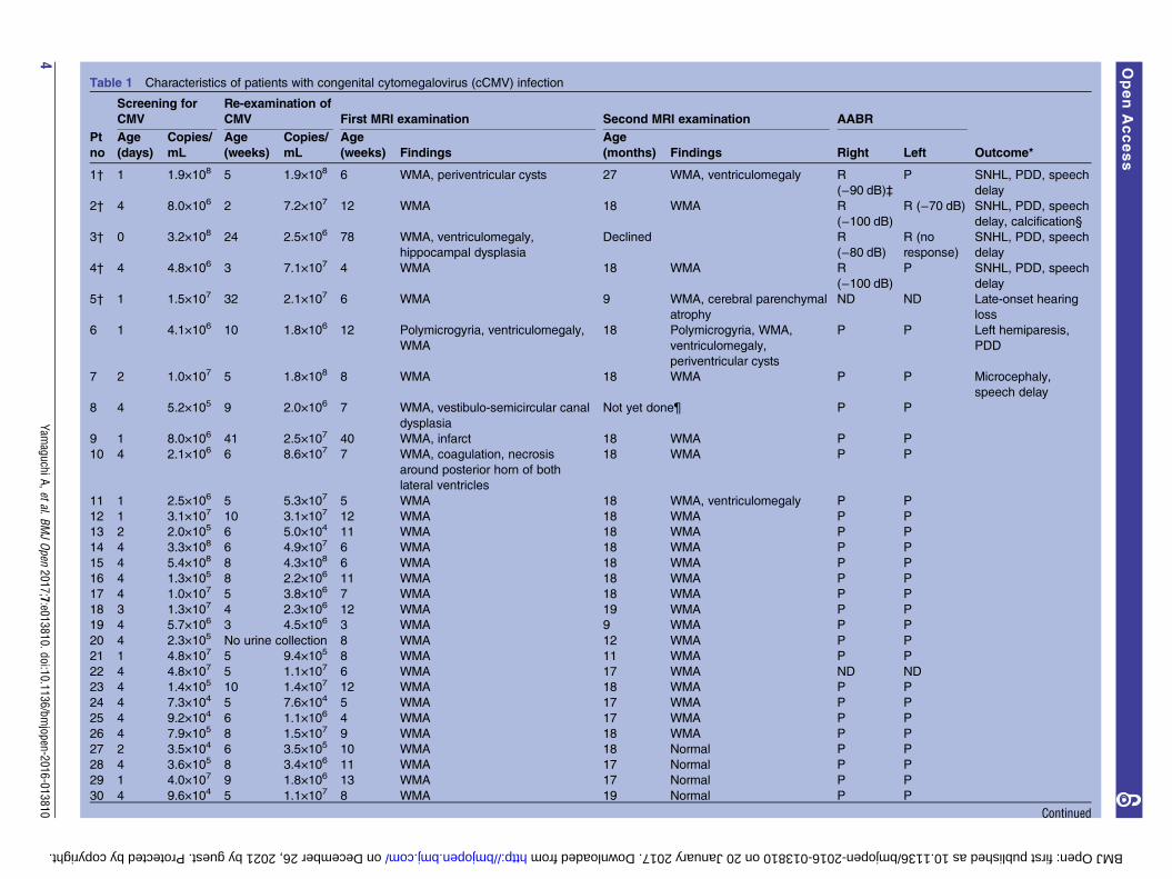
Table 1 Characteristics of patients with congenital cytomegalovirus (cCMV) infection
Screening for
CMV
Re-examination of
CMV First MRI examination Second MRI examination AABR
Pt
no
Age
(days)
Copies/
mL
Age
(weeks)
Copies/
mL
Age
(weeks) Findings
Age
(months) Findings Right Left Outcome*
1† 1 1.9×10⁸ 5 1.9×10⁸ 6 WMA, periventricular cysts 27 WMA, ventriculomegaly R
(−90 dB)‡
P SNHL, PDD, speech
delay
2† 4 8.0×10⁶ 2 7.2×10⁷ 12 WMA 18 WMA R
(−100 dB)
R (−70 dB) SNHL, PDD, speech
delay, calcification§
3† 0 3.2×10⁸ 24 2.5×10⁶ 78 WMA, ventriculomegaly,
hippocampal dysplasia
Declined R
(−80 dB)
R (no
response)
SNHL, PDD, speech
delay
4† 4 4.8×10⁶ 3 7.1×10⁷ 4 WMA 18 WMA R
(−100 dB)
P SNHL, PDD, speech
delay
5† 1 1.5×10⁷ 32 2.1×10⁷ 6 WMA 9 WMA, cerebral parenchymal
atrophy
ND ND Late-onset hearing
loss
6 1 4.1×10⁶ 10 1.8×10⁶ 12 Polymicrogyria, ventriculomegaly,
WMA
18 Polymicrogyria, WMA,
ventriculomegaly,
periventricular cysts
P P Left hemiparesis,
PDD
7 2 1.0×10⁷ 5 1.8×10⁸ 8 WMA 18 WMA P P Microcephaly,
speech delay
8 4 5.2×10⁵ 9 2.0×10⁶ 7 WMA, vestibulo-semicircular canal
dysplasia
Not yet done¶ P P
9 1 8.0×10⁶ 41 2.5×10⁷ 40 WMA, infarct 18 WMA P P
10 4 2.1×10⁶ 6 8.6×10⁷ 7 WMA, coagulation, necrosis
around posterior horn of both
lateral ventricles
18 WMA P P
11 1 2.5×10⁶ 5 5.3×10⁷ 5 WMA 18 WMA, ventriculomegaly P P
12 1 3.1×10⁷ 10 3.1×10⁷ 12 WMA 18 WMA P P
13 2 2.0×10⁵ 6 5.0×10⁴ 11 WMA 18 WMA P P
14 4 3.3×10⁸ 6 4.9×10⁷ 6 WMA 18 WMA P P
15 4 5.4×10⁸ 8 4.3×10⁸ 6 WMA 18 WMA P P
16 4 1.3×10⁵ 8 2.2×10⁶ 11 WMA 18 WMA P P
17 4 1.0×10⁷ 5 3.8×10⁶ 7 WMA 18 WMA P P
18 3 1.3×10⁷ 4 2.3×10⁶ 12 WMA 19 WMA P P
19 4 5.7×10⁶ 3 4.5×10⁶ 3 WMA 9 WMA P P
20 4 2.3×10⁵ No urine collection 8 WMA 12 WMA P P
21 1 4.8×10⁷ 5 9.4×10⁵ 8 WMA 11 WMA P P
22 4 4.8×10⁷ 5 1.1×10⁷ 6 WMA 17 WMA ND ND
23 4 1.4×10⁵ 10 1.4×10⁷ 12 WMA 18 WMA P P
24 4 7.3×10⁴ 5 7.6×10⁴ 5 WMA 17 WMA P P
25 4 9.2×10⁴ 6 1.1×10⁶ 4 WMA 17 WMA P P
26 4 7.9×10⁵ 8 1.5×10⁷ 9 WMA 18 WMA P P
27 2 3.5×10⁴ 6 3.5×10⁵ 10 WMA 18 Normal P P
28 4 3.6×10⁵ 8 3.4×10⁶ 11 WMA 17 Normal P P
29 1 4.0×10⁷ 9 1.8×10⁶ 13 WMA 17 Normal P P
30 4 9.6×10⁴ 5 1.1×10⁷ 8 WMA 19 Normal P P
Continued
4Yam
aguchiA,etal.BMJOpen
2017;7:e013810.doi:10.1136/bmjopen-2016-013810
OpenAccess
on December 26, 2021 by guest. Protected by copyright. http://bmjopen.bmj.com/ BMJ Open: first published as 10.1136/bmjopen-2016-013810 on 20 January 2017. Downloaded from

Table 1 Continued
Screening for
CMV
Re-examination of
CMV First MRI examination Second MRI examination AABR
Pt
no
Age
(days)
Copies/
mL
Age
(weeks)
Copies/
mL
Age
(weeks) Findings
Age
(months) Findings Right Left Outcome*
31 1 1.1×10⁶ 9 2.1×10⁵ 11 WMA 18 Normal ND ND
32 1 1.6×10⁸ 4 6.9×10⁷ 5 WMA Declined P P
33 4 1.9×10⁵ 9 6.6×10⁵ 14 WMA Declined P P
34 4 5.1×10⁶ 15 2.9×10⁵ 7 WMA Not yet done P P
35 4 3.7×10⁵ 8 3.4×10⁵ 8 WMA Declined P P
36 4 1.1×10⁴ 6 4.8×10⁵ 8 WMA Not yet done P P
37 1 3.9×10⁴ 6 1.3×10⁵ 5 WMA Not yet done ND ND
38 1 1.5×10⁷ No urine collection 7 WMA Not yet done P P
39 4 6.9×10⁷ 15 9.8×10⁶ 14 Normal 20 WMA P P
40 1 1.1×10⁹ 16 2.0×10⁷ 18 Normal 18 WMA P P
41 1 3.5×10⁷ 10 6.2×10⁶ 13 Normal 17 WMA P P
42 4 6.7×10⁵ 8 6.2×10⁵ 10 Normal 24 WMA P P
43 4 3.6×10⁵ 9 4.7×10⁶ 11 Normal 22 WMA P P Cavum vergae
44 2 1.4×10⁷ 8 4.4×10⁵ 9 Normal 18 WMA ND ND
45 4 6.1×10⁴ 8 6.1×10⁴ 7 Normal 16 Normal P P
46 4 3.8×10⁴ 11 2.2×10⁶ 16 Normal 18 Normal P P
47 4 1.2×10⁶ 12 1.6×10⁷ 12 Normal 17 Normal P P
48 1 4.5×10³ 4 3.1×10⁵ 4 Normal Declined P P
49 1 6.4×10⁶ 14 4.1×10⁵ 15 Normal Declined P P
50 4 7.4×10⁵ 13 2.2×10⁶ 28 Normal Declined P P
51 4 6.1×10⁴ 3 1.2×10⁵ 5 Normal Declined P P
52 4 1.1×10⁶ 13 6.0×10⁶ 12 Normal Not yet done P P
53 4 9.3×10⁵ 7 3.9×10⁶ 8 Normal Not yet done P P
54 1 1.1×10⁴ Not examined** Not examined Not examined P P
55 1 3.1×10⁶ Not examined Not examined Not examined P P
56 3 2.4×10⁶ Not examined Not examined Not examined P P
57 4 7.7×10⁶ Not examined Not examined Not examined P P
58 4 2.4×10³ Not examined Not examined Not examined P P
59 1 1.0×10⁴ Not examined Not examined Not examined ND ND
60 2 1.1×10⁸ Not examined Not examined Not examined ND ND
Microcephaly was noted in one newborn with modified HC z score of −2.95.*No data means the patient had shown no apparent clinical manifestations until documentation of this paper.†The oral antiviral drug valganciclovir was administered to patients 1–5 with SNHL for 5–6 months. Patient 5 presented with late-onset hearing loss and was then treated with valganciclovir.‡Figures in parentheses indicate hearing loss measured in decibel (dB).§Calcification was confirmed with CT scan.¶Not yet done means that the second MRI examination is scheduled but had not yet been done until documentation of this paper.**Not examined means no visit or examinations.AABR, automatic auditory brainstem response; HC, nead circumference; nt, not tested; P, pass; PDD, psychomotor developmental delays; Pt no, patient number; R, refer; SNHL, sensorineuralhearing loss; WMA, white matter abnormality.
YamaguchiA,etal.BM
JOpen
2017;7:e013810.doi:10.1136/bmjopen-2016-013810
5
OpenAccess
on December 26, 2021 by guest. Protected by copyright. http://bmjopen.bmj.com/ BMJ Open: first published as 10.1136/bmjopen-2016-013810 on 20 January 2017. Downloaded from

p=0.036, Mann-Whitney U test). When newborns wereretested at a mean age of 7.7 weeks (range 2–16 weeks),the urinary CMV DNA copy number of newborns withSNHL (9.90×10⁷ copies/mL, 95% CI 2.44×10⁷ to4.02×108 copies/mL) was still significantly higher thanin newborns who had cCMV infection but no SNHL(2.61×10⁶ copies/mL, 95% CI 2.88×10⁴ to 2.37×10⁸
copies/mL; p=0.010) (figure 4).
Relationship between cCMV infection and MRI findingsOf the 60 newborns with cCMV, 53 underwent MRIscans. Of these 53 newborns, 44 (83.0%) were found tohave some form of CNS damage (figure 5). Fifty of the53 newborns underwent an initial MRI scan before theage of 5 months; two of the remaining three newbornsunderwent the scan at 7 or 10 months while the otherwas scanned at 18 months. Of the 50 newborns whounderwent MRI scans by the age of 6 months, 36 werefound to have some form of CNS damage (72.0%). Themean urinary CMV DNA copy number of newborns whowere found to have CNS damage during screening(2.30×10⁶ copies/mL, 95% CI 8.78×10⁵ to 6.01×10⁶
copies/mL) did not differ significantly from that in 14newborns who had no such abnormalities (1.31×10⁶
copies/mL, 95% CI 1.90×10⁵ to 9.07×10⁶ copies/mL)(figure 6A).Thirty-one newborns underwent a second scan at the
age of 18 months (range 16–20 months). During thesecond MRI scan, 23 newborns were found to have CNSdamage (74.2%) and these newborns had higherurinary CMV DNA copy numbers (6.19×10⁶ copies/mL,95% CI 1.90×10⁶ to 2.02×10⁷ copies/mL) than the eightnewborns in which no such abnormalities were detected(3.32×10⁵ copies/mL, 95% CI 4.48×10⁴ to 2.46×10⁶
copies/mL; p=0.013) (figure 6B).The MRI scans performed before 6 months of age
and around the age of 18 months were compared for 30infants. The mean CMV DNA copy number of the 18newborns who were found to have abnormal CNS find-ings in the first and second scans (3.48×10⁶ copies/mL,
95% CI 9.14×10⁵ to 1.32×10⁷ copies/mL) was signifi-cantly higher than in three newborns with normal CNSfindings in both scans (1.41×10⁵ copies/mL, 95% CI1.35×103 to 1.47×10⁷ copies/mL; p=0.035). Five new-borns were found to have WMAs on an initial scan butno abnormalities were detected on the second scan. Theurinary CMV DNA copy number of these five newborns(5.56×10⁵ copies/mL, 95% CI 1.89×10⁴ to 1.64×10⁷
copies/mL) did not differ significantly from that of new-borns without CNS abnormalities in both scans(p=0.67). In contrast, four newborns had no abnormal-ities initially but were found to have WMAs on thesecond scan. In these four newborns, the urinary CMVDNA copy number was significantly higher (7.81×10⁷
copies/mL, 95% CI 3.92×10⁶ to 1.56×10⁹ copies/mL)than the three infants who had no abnormalities(p=0.034) (figure 7). All five newborns with SNHL(100%) and 39 of 48 children without SNHL (81%) had
Figure 1 MRI findings in patient number 4 with congenital
cytomegalovirus infection at the age of 1 month (A) and at the
age of 18 months (B). (A) Axial T2-weighted images show
diffuse abnormal high intensity. (B) Follow-up axial
T2-weighted images at the age of 18 months show residual
abnormal T2 high intensity areas (arrowheads).
Figure 2 MRI findings in patient number 6 with congenital
cytomegalovirus infection at the age of 3 months (A and B)
and at the age of 18 months (C and D). (A) Axial T2-weighted
images at the level of the basal ganglia show polymicrogyria
involving bilateral frontal lobes and perisylvian and insular
cortices (arrows) and bilateral occipital ventricular septations
(arrowheads). Bilateral enlargement of the lateral ventricles
shows volume loss of white matter. (B) Axial T2-weighted
images at the level of the central semiovale show right
predominant frontal lobe polymicrogyria (arrows) and diffuse
abnormal T2 high intensity areas (arrowheads). (C, D)
Follow-up MRI study at the age of 18 months also shows
bilateral polymicrogyria (arrows), but abnormal T2 high
intensity has been obscured and there are only some
scattered abnormal T2 high intensity areas (arrowheads).
Abnormally thin centrum semiovale suggests white matter
volume loss.
6 Yamaguchi A, et al. BMJ Open 2017;7:e013810. doi:10.1136/bmjopen-2016-013810
Open Access
on Decem
ber 26, 2021 by guest. Protected by copyright.
http://bmjopen.bm
j.com/
BM
J Open: first published as 10.1136/bm
jopen-2016-013810 on 20 January 2017. Dow
nloaded from

abnormal CNS findings including WMAs, thus the inci-dences of abnormal CNS findings were not significantlydifferent between the two groups (p=0.288). Childrenwith SNHL, however, showed significantly far moreabnormal CNS findings other than WMAs (3/5 withSNHL) than children without SNHL (5/48 withoutSNHL) (p=0.003) (table 1).
DISCUSSIONThis study examined the incidence of cCMV infectionand SNHL in seemingly healthy newborns with noobvious clinical manifestations of cCMV infection. Oneof 60 newborns (1.7%) who tested positive for CMVexhibited microcephaly but no other clinical manifesta-tions. As microcephaly has been reported to be acommon single clinical finding in newborns with cCMVinfection,17 nearly asymptomatic newborns with milderor non-specific findings can be missed in the absence offurther screening. Therefore, this study used MRI toidentify CNS damage in newborns with cCMV infectionand examined the relationship between CNS damageand urinary CMV DNA copy number.Various methods have been used to screen for cCMV
infection.18–20 However, no previous study for cCMVscreening has described the direct extraction ofCMV DNA from the liquid urine of newborns. We sub-jected CMV DNA to qPCR and screened for cCMV infec-tion in over 23 000 newborns. Unlike blood, urine canbe collected non-invasively and the viral load in urine islikely to be elevated even when a virus is inactive.Real-time PCR was used to detect CMV DNA in urinesamples in order to ensure specificity, quantitative reli-ability and rapidity. The method used in this study alsoallows accurate quantitative determination of urinaryvolume, unlike the technique employing FTA-Elute filtercards described in previous studies.8 The real-time PCRassay of saliva has excellent sensitivity and specificity andcan easily be adapted for large-scale screening of new-borns.19 However, the real-time PCR assay of saliva is lessable to provide the quantitative capability than that ofliquid urine. Thus, the novel method described hereallows more accurate examination of the relationshipbetween viral load and SNHL/CNS damage.In this study the incidence of cCMV infection was
0.257%, about 17% lower than that reported in otherstudies conducted in Japan. Numazaki et al7 used a con-ventional cell culture method to screen 11 938 indivi-duals and reported an incidence of 0.31%, whereasKoyano et al8 used FTA-Elute filter cards embedded in adiaper to screen 21 272 individuals and reported an inci-dence of 0.31%. The higher level of cCMV infectiondetected in these studies could be explained by the factthat they recruited newborns from university hospitals, apopulation likely to include newborns at a greater risk ofcCMV infection. In fact, Koyano et al8 reported that theincidence of cCMV infection at primary obstetric clinicswas 0.24%. Our sample of seemingly healthy newborns,born at typical maternity hospitals, might provide a CMVincidence that is closer to the typical incidence of cCMVinfection.Newborn hearing screening was performed using
AABR testing. Of the screened newborns, 171 werereferred for further testing and 22 (12.9%) were finallydiagnosed with SNHL. Four newborns underwent bothnewborn hearing screening and screening for cCMVinfection, and all four of these newborns and one other
Figure 3 Basal ganglia calcification. Unenhanced CT image
of patient number 2 shows punctate calcification in the head
of the left caudate nucleus (arrows).
Figure 4 Comparison of cytomegalovirus (CMV) load in
urine samples of newborns with congenital CMV (cCMV)
infection and hearing loss and in those of newborns with
cCMV infection but no hearing loss. DNA was extracted from
urine samples collected from newborns within 0–5 days of
birth and CMV DNA was quantified by quantitative real-time
PCR. (A, B) Number of copies of CMV in 48 newborns with
CMV infection but no hearing loss (without hearing loss) and
five newborns with CMV infection and hearing loss (with
hearing loss) in the screening (p=0.036) and in the follow-up
examination (p=0.010). Upper and lower borders of box plots
represent the 25th and 75th percentiles and bars in the boxes
represent median viral loads. Upper and lower bars indicate
maximum and minimum viral load, respectively.
Yamaguchi A, et al. BMJ Open 2017;7:e013810. doi:10.1136/bmjopen-2016-013810 7
Open Access
on Decem
ber 26, 2021 by guest. Protected by copyright.
http://bmjopen.bm
j.com/
BM
J Open: first published as 10.1136/bm
jopen-2016-013810 on 20 January 2017. Dow
nloaded from

newborn with late-onset HL had SNHL. In thisnewborn, because the AABR test was not conductedwithin 5 days of birth, ABR was performed but HL wasnot confirmed. HL detected during follow-up was diag-nosed as late-onset HL. Thus, performing hearingscreening and CMV screening at the same time shouldallow early prediction of SNHL prior to more detailedhearing testing. In other words, simultaneous screeningmay allow early detection and treatment of SNHL due tocCMV infection.In 60 newborns with cCMV infection, those with the
highest urinary CMV DNA copy number had4.6×105-fold more copies than those with the lowest CMVDNA copy number (range 2.4×10³–1.1×10⁹ copies/mL).Newborns with cCMV infection and SNHL had a signifi-cantly higher urinary CMV DNA copy number thanthose with cCMV infection but no SNHL (figure 4).Of the 60 newborns with cCMV infection, one hadlate-onset SNHL. It is well known that SNHL due tocCMV infection appears as a form of late auditorydysfunction.21 22 Thus, infants with cCMV infection, par-ticularly with high urinary CMV DNA copy number,need to be carefully monitored for the development oflate-onset SNHL.Of the 60 newborns with cCMV infection, 53 under-
went MRI. Abnormal findings were noted in 44 of theseinfants (83.0%), and most of the findings were WMAsthat have been attributed to myelination failure inducedby CMV infection of the CNS.5 14 23
The first MRI examination performed by the age of6 months identified no significant difference betweenthe urinary CMV DNA copy number of newborns withnormal MRI findings and those with abnormal MRI
Figure 5 Enrolment in the head MRI scan and outcomes. ‘Around the age of 18 months’ means that the second MRI scans
were performed between the ages of 16 months and 20 months.
Figure 6 Comparison of urinary cytomegalovirus (CMV)
screening based on the difference in MRI manifestations in
congenital CMV infection. Urine samples were collected from
newborns within 0–5 days of birth. (A) Of the 60 newborns, 53
underwent MRI examination. Three newborns had the first
MRI scans carried out much later than the other 50 at 7, 10
and 18 months, respectively 50. Thus, three were excluded
from the comparison. Of the 50 newborns on whom MRI was
performed at less than 5 months of age, 14 (normal) did not
have central nervous system (CNS) damage and 36
(abnormal) had CNS damage. No significant difference in the
viral load is shown between normal and abnormal (p=0.560).
(B) Thirty-one newborns underwent a second MRI scan
between 16 months and 20 months of age. Twenty-three
newborns (abnormal) were found to have CNS damage
during the second scan and eight newborns (normal) had no
such abnormalities. A significant difference in the viral load is
shown between normal and abnormal (p=0.013). Upper and
lower borders of box plots represent the 25th and 75th
percentiles and bars represent median viral loads. Upper and
lower bars indicate maximum and minimum viral load,
respectively.
8 Yamaguchi A, et al. BMJ Open 2017;7:e013810. doi:10.1136/bmjopen-2016-013810
Open Access
on Decem
ber 26, 2021 by guest. Protected by copyright.
http://bmjopen.bm
j.com/
BM
J Open: first published as 10.1136/bm
jopen-2016-013810 on 20 January 2017. Dow
nloaded from

findings. The second examination carried out at aboutthe age of 18 months showed that newborns with abnor-mal MRI findings had a significantly higher urinary CMVDNA copy number than those with normal MRI findings(figure 6). High CMV DNAemia has been reported topredict CMV infection sequelae in newborns with asymp-tomatic cCMV infection,24 so high urinary CMV DNAcopy numbers could predict late CNS damage.The urinary CMV viral load of newborns with initial
abnormal results who subsequently had normal resultsdid not differ significantly from that of newbornswithout abnormal results. Conversely, the urinary CMVviral load of newborns who initially had normal resultsbut subsequently had abnormal results was significantlyhigher than that of newborns who had normal results(figure 7). Newborns with a high urinary CMV DNAcopy number during screening should be followed upwith MRI.In this study, MRI examination revealed that WMAs
occurred with surprising frequency of 83% in newbornsand infants with cCMV infection. Although nearly halfof WMAs found in the children with cCMV infectionwere considered to be mild, WMAs could be the criticalsign of asymptomatic cCMV infection detectable withneuroimaging examination. WMAs are very often seenin children with symptomatic cCMV infection.6 23 25 26
Uematsu et al27 retrospectively investigated asymptomaticcCMV infection with neurological sequelae using qPCRof CMV DNA from dried umbilical cord and reportedthat all the 54 patients (100%) had cerebral WMAs inMRI study. In contrast, there are very few referencesabout the incidence of WMAs in newborns and infantswith cCMV infection found through neonatal screening.Recently, Krakar et al28 described the apparent regres-
sion of WMAs in a case report of serial postnatal MRIsin a child and proposed that leukoencephalopathy incCMV infection is not only non-progressive or static aspreviously reported, but also might even evolve in partof WMAs, suggesting both disturbed and delayed myelin-ation. Consequently, it could be considered that mostWMAs due to cCMV infection, especially mild WMAs,would regress or disappear in late childhood, althoughmany WMAs of varying severity and extent exist in theneonatal period and infancy. However, the qualitativeand quantitative evaluations of WMAs caused by cCMVinfection remain to be well-defined.Milewska-Bobula et al29 indicated increased emotional
sensitivity and problems with school maturity in6-year-old children with asymptomatic or mildly symp-tomatic cCMV infection, and Karltorp et al30 showed that88% of 26 children with congenital CMV infection hadbalance disturbances including walking at a later age.Whether WMAs in cCMV infection cause a subsequentdevelopmental disorder is still unclear,14 25 26 and new-borns and infants with SNHL in our study showed farmore abnormal CNS findings other than WMAs thanthose without SNHL; thus SNHL could occur in chil-dren with more severely damaged CNS. WMAs in
newborns and infants could be related to the subse-quent developmental disorders such as balance distur-bances and emotional problems, aside from thewell-known clinical manifestations such as intellectualdisability and cerebral palsy that could be referred to thesevere CNS damage.Further follow-up of the patients in this study is there-
fore needed, and the relationship between the sitewhere a WMA was noted, the severity of WMA, the CMVDNA copy number in the urine and the occurrence ofdevelopmental disorders needs to be examined ingreater detail.This study reports the incidence of cCMV infection
and the typical range of urinary CMV viral load of
Figure 7 Comparison of cytomegalovirus (CMV) load in
urine samples of newborns with congenital CMV infection with
respect to the changes in MRI findings. Urine samples were
collected from newborns within 0–5 days of birth. The first
MRI scans were performed at less than 5 months of age. The
second MRI scans were performed between the age of
16 months and 20 months. One of 31 newborns who
underwent the first and second MRI scans was excluded from
the comparison because the first MRI scan was performed at
10 months of age. In the first and second MRI scans, three
newborns (normal) showed no abnormalities while 18
newborns (abnormal) showed central nervous system
damage. The initial scan revealed abnormalities but the
second scan showed no abnormalities in five newborns
(changed to normal). In contrast, the initial scan revealed no
abnormalities but the second scan showed abnormalities in
four newborns (change to abnormal). The 18 (abnormal MRI)
and four newborns (change to abnormal MRI from normal)
who had abnormalities in the second MRI scans showed a
significant difference (p=0.035 and p=0.034, respectively)
compared with three newborns (normal) who had no
abnormalities in both the first and second MRI scans while
the five newborns who had no abnormalities in the second
MRI scans showed no significant difference (p=0.67)
compared with newborns with no abnormalities in both the
first and second MRI scans. Upper and lower borders of box
plots represent the 25th and 75th percentiles and bars
represent median viral loads. Upper and lower bars indicate
maximum and minimum viral load, respectively.
Yamaguchi A, et al. BMJ Open 2017;7:e013810. doi:10.1136/bmjopen-2016-013810 9
Open Access
on Decem
ber 26, 2021 by guest. Protected by copyright.
http://bmjopen.bm
j.com/
BM
J Open: first published as 10.1136/bm
jopen-2016-013810 on 20 January 2017. Dow
nloaded from

seemingly healthy newborns in Japan. Newborns withcCMV infection and SNHL had a higher urinary CMVviral load. Newborns who had CNS abnormalities includ-ing WMAs detected by MRI also had a significantlyhigher urinary CMV viral load. Because the urinaryCMV viral load could be directly related to illness, theuse of qPCR to determine the CMV viral load in urinespecimens may identify cCMV infection and predict itssequelae. However, this study has an important limita-tion in the implementation. Urine collection from new-borns is laborious and time-consuming compared witha saliva swab or a FTA-Elute filter card embedded in adiaper, and thus the use of qPCR for a urine sample as ascreening test requires improvements in usability andeconomic efficiency. The saliva swab or FTA-Elute filtercard would be better suited for the screening of cCMVinfection. The CMV viral load in liquid urine from anewborn found to be positive by screening should thenbe determined to assess CNS abnormalities, predictsequelae or follow late-onset HL.
Author affiliations1Laboratory of Clinical Research, Saitama Children’s Medical Center, Saitama,Japan2Department of Radiological Technology, Saitama Children’s Medical Center,Saitama, Japan3Division of Infectious Disease, Saitama Children’s Medical Center, Saitama,Japan4Division of Otorhinolaryngology, Kawagoe Otology Institute, Saitama, Japan5Mejiro University Audiology Clinic, Saitama, Japan6Division of Otolaryngology, Saitama Children’s Medical Center, Saitama,Japan7Division of Radiology, Saitama Children’s Medical Center, Saitama, Japan8Division of Pediatrics, Sannoh Doom Clinic, Saitama, Japan9Division of Obstetrics and Gynecology, Sannoh Clinic, Saitama, Japan10Division of Obstetrics and Gynecology, Aiwa Hospital, Saitama, Japan11Department of Molecular Predictive Medicine and Sport Science, School ofMedicine, Kyorin University, Tokyo, Japan
Acknowledgements The authors would like to thank the newborns and theirparents who agreed to take part in the study and medical colleagues whocollaborated in this study.
Contributors TO and HS designed the study. AY and TO participated in dataanalysis and writing the manuscript. AY and TA conducted all initial urinescreening. HS, NA and SA diagnosed hearing loss. EO performed MRI and CTimage analysis. HK, JM, HF, TU, JF and KS enrolled newborns andcontributed to hearing screening. KS, HO and TK contributed to criticalrevision of the manuscript.
Funding This study was supported by grants for the Research on ChildDevelopment and Diseases (H20-Kodomo-007; H23-Jisedai-Ippan-001) fromthe Ministry of Health, Labour and Welfare, Japan.
Competing interests None declared.
Patient consent Patient consents were obtained from the parents.
Ethics approval Ethics approval was provided by the ethical committees ofthe participating hospitals.
Provenance and peer review Not commissioned; externally peer reviewed.
Data sharing statement No additional data are available.
Open Access This is an Open Access article distributed in accordance withthe Creative Commons Attribution Non Commercial (CC BY-NC 4.0) license,which permits others to distribute, remix, adapt, build upon this work non-commercially, and license their derivative works on different terms, provided
the original work is properly cited and the use is non-commercial. See: http://creativecommons.org/licenses/by-nc/4.0/
REFERENCES1. Stagno S, Whitley RJ. Herpesvirus infection of pregnancy. Part I:
Cytomegalovirus and Epstein-Barr virus infections. N Engl J Med1985;313:1270–4.
2. Morton CC, Nance WE. Newborn hearing screening--a silentrevolution. N Engl J Med 2006;354:2151–64.
3. Fowler K, McCollister F, Dahle A, et al. Progressive and fluctuatingsensorineural hearing loss in children with asymptomatic congenitalcytomegalovirus infection. J Pediatr 1997;130:624–30.
4. Hicks T, Fowler K, Richardson M, et al. Congenital cytomegalovirusinfection and neonatal auditory screening. J Pediatr1993;123:779–82.
5. van der Knaap MS, Vermeulen G, Barkhof F, et al. Pattern of whitematter abnormalities at MR imaging: use of polymerase chainreaction testing of Guthrie cards to link pattern with congenitalcytomegalovirus infection. Radiology 2004;230:529–36.
6. de Vries LS, Gunardi H, Barth PG, et al. The spectrum of cranialultrasound and magnetic resonance imaging abnormalities incongenital cytomegalovirus infection. Neuropediatrics2004;35:113–19.
7. Numazaki K, Fujikawa T. Chronological changes of incidence andprognosis of children with asymptomatic congenital cytomegalovirusinfection in Sapporo, Japan. BMC Infect Dis 2004;4:22.
8. Koyano S, Inoue N, Oka A, et al., Japanese Congenital CytomegalovirusStudy Group. Screening for congenital cytomegalovirus infection usingnewborn urine samples collected on filter paper: feasibility and outcomesfrom a multicentre study. BMJ Open 2011;1:e000118.
9. Noyola DE, Demmler GJ, Nelson CT, et al. Early predictors ofneurodevelopmental outcome in symptomatic congenitalcytomegalovirus infection. J Pediatr 2001;138:325–31.
10. Nance WE, Lim BG, Dodson KM. Importance of congenitalcytomegalovirus infections as a cause for pre-lingual hearing loss.J Clin Virol 2006;35:221–5.
11. Wirgart BZ, Andersson P, Grillner L. Evaluation of the ReSSQ assayin relation to the COBAS AMPLICOR CMV MONITOR test and anin-house nested PCR method for detection of cytomegalovirus DNA.J Clin Microbiol 2005;43:4057–63.
12. Griscelli F, Barrois M, Chauvin S, et al. Quantification of humancytomegalovirus DNA in bone marrow transplant recipients byreal-time PCR. J Clin Microbiol 2001;39:4362–9.
13. Kelley PM, Harris DJ, Comer BC, et al. Novel mutations in theconnexin 26 gene (GJB2) that cause autosomal recessive (DFNB1)hearing loss. Am J Hum Genet 1998;62:792–9.
14. Fink KR, Thapa MM, Ishak GE, et al. Neuroimaging of pediatriccentral nervous system cytomegalovirus infection. Radiographics2010;30:1779–96.
15. Pascual-Castroviejo I, Pascual-Pascual SI, Velasquez-Fragua R,et al. Congenital cytomegalovirus infection and cortical/subcorticalmalformations. Neurologia 2012;27:336–42.
16. van der Voorn JP, Pouwels PJ, Vermeulen RJ, et al. QuantitativeMR imaging and spectroscopy in congenital cytomegalovirusinfection and periventricular leukomalacia suggests a comparableneuropathological substrate of the cerebral white matter lesions.Neuropediatrics 2009;40:168–73.
17. Dreher AM, Arora N, Fowler KB, et al. Spectrum of disease andoutcome in children with symptomatic congenital cytomegalovirusinfection. J Pediatr 2014;164:855–9.
18. Kenneson A, Cannon MJ. Review and meta-analysis of theepidemiology of congenital cytomegalovirus (CMV) infection. RevMed Virol 2007;17:253–76.
19. Boppana SB, Ross SA, Shimamura M, et al., Saliva polymerase-chain-reaction assay for cytomegalovirus screening in newborns.N Engl J Med 2011;364:2111–18.
20. Paixão P, Almeida S, Videira PA, et al. Screening of congenitalcytomegalovirus infection by real-time PCR in urine pools. EurJ Pediatr 2012;171:125–9.
21. Foulon I, Naessens A, Foulon W, et al. A 10-year prospective studyof sensorineural hearing loss in children with congenitalcytomegalovirus infection. J Pediatr 2008;153:84–8.
22. Amir J, Attias J, Pardo J. Treatment of late-onset hearing loss ininfants with congenital cytomegalovirus infection. Clin Pediatr (Phila)2014;53:444–8.
23. Barkovich AJ, Lindan CE. Congenital cytomegalovirus infection ofthe brain: imaging analysis and embryological considerations. AJNRAm J Neuroradiol 1994;15:703–15.
10 Yamaguchi A, et al. BMJ Open 2017;7:e013810. doi:10.1136/bmjopen-2016-013810
Open Access
on Decem
ber 26, 2021 by guest. Protected by copyright.
http://bmjopen.bm
j.com/
BM
J Open: first published as 10.1136/bm
jopen-2016-013810 on 20 January 2017. Dow
nloaded from

24. Forner G, Abate D, Mengoli C, et al. High cytomegalovirus (CMV)DNAemia predicts CMV sequelae in asymptomatic congenitallyinfected newborns born to women with primary infection duringpregnancy. J Infect Dis 2015;212:67–71.
25. Manara R, Balao L, Baracchini C, et al. Brain magnetic resonancefindings in symptomatic congenital cytomegalovirus infection. PediatrRadiol 2011;41:962–70.
26. Inder TE, Wells SJ, Mogridge NB, et al. Defining the nature of thecerebral abnormalities in the premature infant: a qualitative magneticresonance imaging study. J Pediatr 2003;143:171–9.
27. Uematsu M, Haginoya K, Kikuchi A, et al. Asymptomaticcongenital cytomegalovirus infection with neurological
sequelae: a retrospective study using umbilical cord. Brain Dev2016;38:819–26.
28. Krakar G, Dakovic I, Delin S, et al. Evolutive leukoencephalopathyin congenital cytomegalovirus infection. J Child Neurol2015;30:93–5.
29. Milewska-Bobula B, Zebrowska J, Olszaniecka M, et al. Evaluationof intellectual development of children following congenital, mildlysymptomatic cytomegalovirus (CMV) infection. A prospective study.Med Wieku Rozwoj 2010;14:370–3.
30. Karltorp E, Löfkvist U, Lewensohn-Fuchs I, et al. Impaired balanceand neurodevelopmental disabilities among children with congenitalcytomegalovirus infection. Acta Paediatr 2014;103:1165–73.
Yamaguchi A, et al. BMJ Open 2017;7:e013810. doi:10.1136/bmjopen-2016-013810 11
Open Access
on Decem
ber 26, 2021 by guest. Protected by copyright.
http://bmjopen.bm
j.com/
BM
J Open: first published as 10.1136/bm
jopen-2016-013810 on 20 January 2017. Dow
nloaded from