open access original article effect of ischaemic brain
TRANSCRIPT
Effect of ischaemic brain injuryon sexual function in adult mice
Yaohui Tang,1 Falei Yuan,1 Beibei Cai,1 Weiliang Xia,1 Yongting Wang,1
Guo-Yuan Yang1,2
To cite: Tang Y, Yuan F,Cai B, et al. Effect ofischaemic brain injuryon sexual function in adultmice. Stroke and VascularNeurology 2016;1:e000013.doi:10.1136/svn-2016-000013
Received 7 March 2016Revised 30 June 2016Accepted 4 July 2016Published Online First29 August 2016
1Neuroscience andNeuroengineering Center,Med-X Research InstituteShanghai Jiao TongUniversity, Shanghai, China2Shanghai Ruijin Hospital,School of Medicine,Shanghai Jiao TongUniversity, Shanghai, China
Correspondence toDr Guo-Yuan Yang;[email protected]
ABSTRACTObjective: Priapism refers to a condition withpersistent abnormal erection of the penis, which isusually caused by disease or injury in the brain orspinal cord, or obstruction to the outflow of bloodthrough the dorsal vein at the root of the penis,without sexual desires. The effect of cerebral ischaemiaon sexual function is unknown. The aim of this studyis to explore whether priapism occurs in adult mice.Furthermore, we examined the relationship betweenpriapism and the region of infarct in the brain.Design: Adult male CD-1 mice who underwentpermanent middle cerebral artery occlusion (pMCAO)were closely examined from 2 hours to 14 dayspostoperation.Results: We found that priapism occurs in ∼80% ofthe mice with pMCAO, which could persist up to14 days. Further study has demonstrated that theoccurrence of priapism is related to the infarctregion: priapism is found only in mice with ischaemicinjury extending to the hypothalamus and thehippocampus regions.Conclusion: Our result suggested priapism may beused as a deep brain injury marker for evaluatingbrain injury in mice after pMCAO.
INTRODUCTIONStroke is one of the leading causes of disabil-ity and mortality in the ageing populationworldwide.1 Ischaemia or hypoxic braininjury often results in irreversible braindamage, mediated by multiple mechanismsincluding oxidative stress, inflammatoryresponse, apoptosis, neuronal death, etc.2–4
Priapism is a persistent, painful penile erec-tion, which can last for hours in the absenceof sexual stimulation. It is found in 40% ofpatients with sickle cell disease, because ofimpaired drainage of the corpora cavern-osa.5 6 Priapism often leads to erectile tissuefibrosis and ultimately results in erectile dys-function.7 Several research groups studiedthe molecular mechanisms of priapism toobtain a better understanding of this dis-order both experimentally and clinically.They found that opiorphin-induced priapisminvolves the activation of the polyamine
synthetic pathway.8 Increased adenosine alsocontributes to penile fibrosis.9 Brainischaemia-induced priapism is not wellunderstood. Till now there are only a fewcase reports on brain injury induced priap-ism in clinic. Takaku et al10 reported a35-year-old man had three attacks of sub-arachnoid haemorrhage in three consecutiveyears and suffered priapism with intolerablepain after the last attack of subarachnoidhaemorrhage. Monga et al11 reported a53-year-old man with an old temporal lobeinfarction involving the anteromedial basalportion increase in libido and coital fre-quency with a tendency towards priapism.However, there is no animal model to mimicclinical priapism at present. In our group, wehave consistently established middle cerebralartery occlusion (MCAO) models in mice asa stroke model.12–16 In this study, we reportour observation that permanent MCAO(pMCAO) can induce priapism in adultmice. We explored the causative relationshipbetween focal cerebral ischaemia andpriapism occurrence. Priapism may serve asan indication for severe stroke damage inanimal models.
MATERIALS AND METHODSAnimal stroke modelAnimal procedures for the use of laboratoryanimals were approved by the InstitutionalAnimal Care and Use Committee ofShanghai Jiao Tong University. Twenty-fouradult male CD-1 mice (Shanghai SLACLaboratory Animal CO) weighing 25–32 gwere used. Focal cerebral ischaemia was con-ducted by pMCAO using the suture model asdescribed previously.17 Briefly, mice wereanaesthetised with 4% isoflurane and main-tained with 1.5% isoflurane in an oxygen/airmixture using a gas anaesthesia mask in astereotaxic frame (RWD Life Science,Shenzhen, China) and the body temperaturewas maintained at 37°C by using a heatingpad (RWD Life Science, Shenzhen, China).
Tang Y, et al. Stroke and Vascular Neurology 2016;1:e000013. doi:10.1136/svn-2016-000013 127
Open Access Original article
on January 12, 2022 by guest. Protected by copyright.
http://svn.bmj.com
/S
troke Vasc N
eurol: first published as 10.1136/svn-2016-000013 on 29 August 2016. D
ownloaded from
A midline incision was made on the neck under anoperating microscope (Leica, Germany). Externalcarotid artery (ECA), common carotid artery (CCA) andinternal carotid artery (ICA) were isolated. After tempor-arily clamping the CCA, a 6–0 silicon-coated round-tipmonofilament nylon suture was introduced into the ICAthrough the ECA until slight resistance was felt, indicat-ing a complete middle cerebral artery (MCA) occlusion.Then cerebral blood flow was measured by laserDoppler flowmetry (Moor Instruments, UK) to confirma successful MCA occlusion that has regional blood flowof <15% of baseline blood flow.
TTC staining and H&E stainingAfter sacrificing the animals, the brains were sectionedwith brain matrix, and stained with a 2% solution of2,3,5-triphenyltetrazolium (TTC) dye (Sigma, T8877–100G) at room temperature for 20 min as described pre-viously.18 Then stained sections were immersed in 4%phosphate-buffered paraformaldehyde and imaged witha digital camera. The infarct area was measured as previ-ously described.19 Then the brains were embedded inparaffin. Coronal sections were processed and stainedwith H&E (Beyotime Institute of Biotechnology). UsingNIH Image 1.63 software, the ischaemic lesion area wascalculated as the difference between the area of the non-ischaemic hemisphere and the normal area in the ischae-mic hemisphere. Infarct volume was calculated by multi-plying the infarct area by the thickness of the section.
Priapism recordingThe mice were observed every day after the pMCAO pro-cedure. The time of priapism occurrence and the recov-ery of flaccidity (disappearance of priapism) were bothrecorded. Photographs were taken using a Canon digitalcamera (Canon EOS 450D).
Statistical analysisThe values were given as mean±SE. The significanceof difference was calculated by one-way analysis ofvariance, followed by Student-Newman-Keuls posthoc comparisons. p Values <0.05 were consideredsignificant.
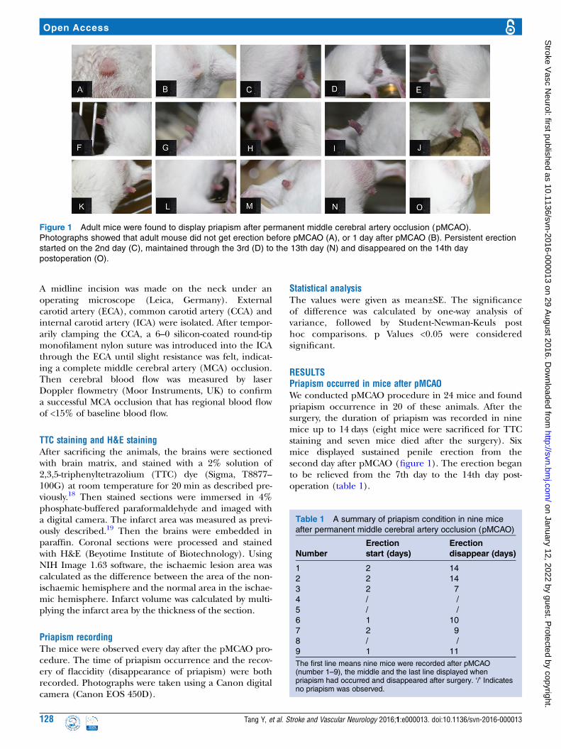
RESULTSPriapism occurred in mice after pMCAOWe conducted pMCAO procedure in 24 mice and foundpriapism occurrence in 20 of these animals. After thesurgery, the duration of priapism was recorded in ninemice up to 14 days (eight mice were sacrificed for TTCstaining and seven mice died after the surgery). Sixmice displayed sustained penile erection from thesecond day after pMCAO (figure 1). The erection beganto be relieved from the 7th day to the 14th day post-operation (table 1).
Figure 1 Adult mice were found to display priapism after permanent middle cerebral artery occlusion (pMCAO).
Photographs showed that adult mouse did not get erection before pMCAO (A), or 1 day after pMCAO (B). Persistent erection
started on the 2nd day (C), maintained through the 3rd (D) to the 13th day (N) and disappeared on the 14th day
postoperation (O).
Table 1 A summary of priapism condition in nine mice
after permanent middle cerebral artery occlusion (pMCAO)
Number
Erection
start (days)
Erection
disappear (days)
1 2 14
2 2 14
3 2 7
4 / /
5 / /
6 1 10
7 2 9
8 / /
9 1 11
The first line means nine mice were recorded after pMCAO(number 1–9), the middle and the last line displayed whenpriapism had occurred and disappeared after surgery. ‘/’ Indicatesno priapism was observed.
128 Tang Y, et al. Stroke and Vascular Neurology 2016;1:e000013. doi:10.1136/svn-2016-000013
Open Access
on January 12, 2022 by guest. Protected by copyright.
http://svn.bmj.com
/S
troke Vasc N
eurol: first published as 10.1136/svn-2016-000013 on 29 August 2016. D
ownloaded from

The difference of brain infarct volume between priapismand non-priapism ischaemic miceTo compare brain infarct volume in the mice with andwithout priapism, the brain tissues were processed byTTC staining and H&E staining, and the infarct area wasrecorded, in parallel to animals with sham operation(no ischaemia and priapism). Mice after pMCAO hadbrain ischaemia as demonstrated by TTC staining (leftand right columns of figure 2). However, the infractvolume are significantly different in mice with priapismfrom those without priapism, consistent with H&Eresults (figure 3). We found that in the stroke mice withpriapism, the infarct area was significantly larger thanthose displaying no priapism (figure 4). Furthermore,the specific infarct area was also different between them.In the ischaemic mice with priapism, the hippocampusand hypothalamus were damaged by ischaemia; however,in the non-priapism mice with ischaemia, the hippocam-pus area and the hypothalamus area were normal(figures 2 and 3).
DISCUSSIONIn this study, we demonstrated that: (1) pMCAO couldinduce a high occurrence of priapism in mice (80%);(2) ischaemia-induced erection often occurs 2 days afterpMCAO (67%) and disappears between the 7th and14th day after pMCAO; and (3) hypothalamus andhippocampus damage may contribute to the occurrence
of priapism. Our data suggested that cerebral ischaemiais closely related with priapism occurrence.Priapism is defined as prolonged penile erection
without sexual stimulation, and it often induces irrevers-ible damage to erectile tissues and results in erectiondysfunction. To the best of our knowledge, we firstreported that pMCAO can induce priapism in rodents inthis study. Our data showed that most ischaemic micedisplayed priapism 2 days after pMCAO, and the detu-mescence time varied from the 7th day to the 14th day.As our TTC staining result revealed, there were some dif-ferences between the two groups (with and without pri-apism in stroke mice). The infarct volume in the micewith priapism was much larger, and the infarct areaincluded the hypothalamus and hippocampus. However,these areas in the non-priapism mice were normal, sug-gesting that priapism is closely related to the lesion siteof stroke, especially related to hippocampus and hypo-thalamus. We propose these differences are caused byposterior communicating artery (PComA) variation,which supplies blood to the hippocampus and hypothal-amus regions.20 Our results might also indicate thathypothalamus and hippocampus injury are responsiblefor priapism. Indeed, these results are consistent withsome previous studies. Some researchers have demon-strated that stimulation of brain can induce penileerection, and hippocampus and hypothalamus areimportant erection mediators.21 Melis et al22 reportedthat microinjection of oxytocin into the paraventricular
Figure 2 Priapism closely
correlated with infarct area.
Photographs showed that the
sham group did not get priapism
and infarct in the brain (middle
column, B–K); ischaemic mice
that also displayed priapism were
found to have extensive infarct
affecting both hypothalamus
(arrowhead) and hippocampus
(arrow) areas (right column, C–L);
in contrast, ischaemic mice that
did not display priapism were
found to have non-infarct
hypothalamus and hippocampus
areas (left column, A–J). All mice
were sacrificed 3 days after
permanent middle cerebral artery
occlusion, n=4 for each group.
Tang Y, et al. Stroke and Vascular Neurology 2016;1:e000013. doi:10.1136/svn-2016-000013 129
Open Access
on January 12, 2022 by guest. Protected by copyright.
http://svn.bmj.com
/S
troke Vasc N
eurol: first published as 10.1136/svn-2016-000013 on 29 August 2016. D
ownloaded from

nucleus of the hypothalamus or into the CA1 field ofthe hippocampus induced penile erection in male rats.Arrow suggested that penile erection was associated withbrain region activation, including the claustrum, leftcaudate and putamen, right middle occipital/middletemporal gyri, bilateral cingulated gyrus, right sensori-motor, premotor regions and right hypothalamus.23
Other studies showed that anaesthesia could also inducepriapism. For instance, Senthilkumaran et al24 reportedthat propofol induced priapism in a 25-year-old man,Ravindran et al25 reported two cases of priapism inducedby 0.5% mg/kg ketamine and 1.5 mg of physostigmine.
However, in these clinical cases, the reasons why priap-ism responds to anaesthesia are not explored.To further investigate the association between the
brain region and priapism, functional MRI may berequired in follow-up studies. Besides the damage to thespecific brain region, we suggest that priapism can berelated to some chemicals induced by stroke, such asnitric oxide (NO), adenosine and so on. These chemi-cals are closely related with stroke, and they also playimportant roles in erection. NO is reported to be animportant factor in stroke26 and penile erection.27 It isclosely associated with stroke and shows beneficialactions,28 including enhancing angiogenesis via the syn-thesis of vascular endothelial growth factor and cyclicguanosine monophosphate after stroke.29 NO is also animportant mediator in penile erection. Azadzoi et al30
stated that neuronal nitric oxide synthase (nNOS),endothelial nitric oxide synthase (eNOS) and induciblenitric oxide synthase (iNOS) levels in cavernosum wereupregulated after atherosclerosis-induced ischaemia, andNO-mediated smooth muscle relaxation in a rabbit.However, we peritoneally injected the NOS inhibitorLNMMA (Beyotime, S0011) into the stroke mice, priap-ism did not disappear (data not shown). We cannot fullyexclude the possible involvement of NO in causing pri-apism by this simple inhibitor study though. Furtherexperiments are needed to clarify its role in the phe-nomenon reported here. Adenosine is an inhibitorymodulator of brain activity with neuroprotective proper-ties,31 and recent studies have reported that adenosine
Figure 3 Priapism occurrence
was related with different infarct
area. H&E staining showed that
the sham group did not show
priapism, and the nuclei of cells
in these brains were normal (B
and E); in the group of ischaemic
mice with priapism (C and F), the
nuclei of the cells in the
hippocampus (arrow head) and
hypothalamus (white arrow)
displayed shrinkage; however, in
the group of ischaemic mice
without priapism (A and D), the
hippocampus (arrowhead) and
hypothalamus (white arrow)
showed normal nuclei size;
photographs A, C, D, F were
taken from a, c, d and f,
respectively. Bar=20 µm, n=4 for
each group.
Figure 4 The infarct volume was significantly larger in stroke
mice with priapism than those without priapism. Bar graph
showed significant difference of infarct volume between the
stroke priapism mice and stroke non-priapism mice. **p<0.01,
n=4 for each group.
130 Tang Y, et al. Stroke and Vascular Neurology 2016;1:e000013. doi:10.1136/svn-2016-000013
Open Access
on January 12, 2022 by guest. Protected by copyright.
http://svn.bmj.com
/S
troke Vasc N
eurol: first published as 10.1136/svn-2016-000013 on 29 August 2016. D
ownloaded from
contributed to animal priapism.9 Intracavernous injec-tion of adenosine resulted in tumescence and penileerection.32 Adenosine accumulated in the penis coupledwith its receptor A2BR, contributed to priapism,33 andophylline, an adenosine receptor antagonist, inhibitedadenosine-induced penile tumescence.34 However,whether adenosine is a potential agent which mediatesboth stroke and priapism needs to be further discussed.Recent studies showed that in corpus cavernosumischaemia-reperfusion mouse, many oxidative injuryparameters like superoxide dismutase (SOD), catalase,malondialdehyde (MDA) and NO in the tissues were allupregulated, and melatonin, which also played neuro-protective actions in stroke,35 could attenuate the activityof SOD and the levels of MDA and NO.36 These conclu-sions strongly suggested that melatonin may be involvedin the stroke-induced priapism pathway. If these chemi-cals are indeed associated with stroke and priapism,drugs used to cure priapism may also interfere in signal-ling pathways in stroke and some of them could possiblyshow beneficial effects to the ischaemic brain. This lineof study might also be worth exploring.In conclusion, this investigation has demonstrated the
correlation between stroke and priapism. Most mice gotsustained erection after pMCAO though their hypothal-amus and hippocampus area were damaged. It is con-ceivable that priapism may serve as a physiologicalmarker for adult stroke mouse. Since the patency ofPComA plays an important role in the infarct results ofthe murine pMCAO models,37 an effective evaluationmethod is urgently needed to reduce the fluctuation ofthe infarct area among different individual animals.38 Itis likely that we can use priapism as a physiologicalmarker to identify stable pMCAO models more conveni-ently, which may facilitate the study of stroke.
CONCLUSIONOur study demonstrated that the occurrence of priapismis related to the infarct region: priapism is found only inmice with ischaemic injury extending to the hypothal-amus and the hippocampus regions. Our results sug-gested priapism may be used as a deep brain injurymarker for evaluating brain injury in mice afterpMCAO.
Acknowledgements The authors thank Caibin Sheng for his help taking thephotographs, Minjie Shen for editorial assistance and the staff ofNeuroscience and Neuroengineering Center for their collaborative support.
Contributors YT, FY and BC participated in the experimental design and dataanalysis. They prepared animal ischaemia model, analysed the data anddrafted the manuscript. WX and YW participated in the experimental designand manuscript discussion. G-YY took care of all aspects, includingparticipating in the study design, supervising tissue assays, analysing theresults, and organising and finalising the manuscript.
Funding The study is supported by the National Natural Science Foundationof China, 81471178(GYY), U1232205 (GYY) and 81371305 (YW).
Competing interests None declared.
Provenance and peer review Commissioned; externally peer reviewed.
Data sharing statement No additional data are available.
Open Access This is an Open Access article distributed in accordance withthe Creative Commons Attribution Non Commercial (CC BY-NC 4.0) license,which permits others to distribute, remix, adapt, build upon this work non-commercially, and license their derivative works on different terms, providedthe original work is properly cited and the use is non-commercial. See: http://creativecommons.org/licenses/by-nc/4.0/
REFERENCES1. Rosamond W, Flegal K, Furie K, et al. Heart disease and stroke
statistics—2008 update: a report from the American HeartAssociation Statistics Committee and Stroke StatisticsSubcommittee. Circulation 2008;117:e25–146.
2. Chan PH. Role of oxidants in ischemic brain damage.Stroke 1996;27:1124–9.
3. Ankarcrona M, Dypbukt JM, Bonfoco E, et al. Glutamate-inducedneuronal death: a succession of necrosis or apoptosis depending onmitochondrial function. Neuron 1995;15:961–73.
4. Huang J, Li Y, Tang Y, et al. CXCR4 antagonist AMD3100 protectsblood-brain barrier integrity and reduces inflammatory response afterfocal ischemia in mice. Stroke 2013;44:190–7.
5. Montague DK, Jarow J, Broderick GA, et al. American UrologicalAssociation guideline on the management of priapism. J Urol2003;170:1318–24.
6. Serjeant GR, de Ceulaer K, Maude GH. Stilboestrol and stutteringpriapism in homozygous sickle-cell disease. Lancet 1985;2:1274–6.
7. El-Bahnasawy MS, Dawood A, Farouk A. Low-flow priapism: riskfactors for erectile dysfunction. BJU Int 2002;89:285–90.
8. Kanika ND, Tar M, Tong Y, et al. The mechanism ofopiorphin-induced experimental priapism in rats involves activation ofthe polyamine synthetic pathway. Am J Physiol Cell Physiol2009;297:C916–27.
9. Wen J, Jiang X, Dai Y, et al. Increased adenosine contributes topenile fibrosis, a dangerous feature of priapism, via A2B adenosinereceptor signaling. FASEB J 2010;24:740–9.
10. Takaku A, Fukawa O, Suzuki J. A case of priapism with rupturedintracranial aneurysm. J Neurol 1979;221:279–83.
11. Monga TN, Monga M, Raina MS, et al. Hypersexuality in stroke.Arch Phys Med Rehabil 1986;67:415–17.
12. Fan Y, Shen F, Frenzel T, et al. Endothelial progenitor celltransplantation improves long-term stroke outcome in mice.Ann Neurol 2010;67:488–97.
13. Yang G, Chan PH, Chen J, et al. Human copper-zinc superoxidedismutase transgenic mice are highly resistant to reperfusion injuryafter focal cerebral ischemia. Stroke 1994;25:165–70.
14. Yang GY, Gong C, Qin Z, et al. Inhibition of TNFalpha attenuatesinfarct volume and ICAM-1 expression in ischemic mouse brain.Neuroreport 1998;9:2131–4.
15. Yuan F, Tang Y, Lin X, et al. Optimizing suture middle cerebralartery occlusion model in C57BL/6 mice circumvents posteriorcommunicating artery dysplasia. J Neurotrauma 2012;29:1499–505.
16. Lin X, Miao P, Wang J, et al. Surgery-related thrombosis criticallyaffects the brain infarct volume in mice following transient middlecerebral artery occlusion. PLoS ONE 2013;8:e75561.
17. Zhang L, Wang Y, Tang Y, et al. High MRI performance fluorescentmesoporous silica-coated magnetic nanoparticles for tracking neuralprogenitor cells in an ischemic mouse model. Nanoscale2013;5:4506–16.
18. Bederson JB, Pitts LH, Germano SM, et al. Evaluation of2,3,5-triphenyltetrazolium chloride as a stain for detection andquantification of experimental cerebral infarction in rats. Stroke1986;17:1304–8.
19. Lin TN, He YY, Wu G, et al. Effect of brain edema on infarct volumein a focal cerebral ischemia model in rats. Stroke 1993;24:117–21.
20. Dawson BH. The blood vessels of the human optic chiasma andtheir relation to those of the hypophysis and hypothalamus. Brain1958;81:207–17.
21. Argiolas A, Melis MR. Central control of penile erection: role of theparaventricular nucleus of the hypothalamus. Prog Neurobiol2005;76:1–21.
22. Melis MR, Argiolas A, Gessa GL. Oxytocin-induced penile erectionand yawning: site of action in the brain. Brain Res 1986;398:259–65.
23. Arnow BA, Desmond JE, Banner LL, et al. Brain activation andsexual arousal in healthy, heterosexual males. Brain2002;125:1014–23.
24. Senthilkumaran S, Shah S, Ganapathysubramanian, et al. Propofoland priapism. Indian J Pharmacol 2010;42:238–9.
Tang Y, et al. Stroke and Vascular Neurology 2016;1:e000013. doi:10.1136/svn-2016-000013 131
Open Access
on January 12, 2022 by guest. Protected by copyright.
http://svn.bmj.com
/S
troke Vasc N
eurol: first published as 10.1136/svn-2016-000013 on 29 August 2016. D
ownloaded from
25. Ravindran RS, Dryden GE, Somerville GM. Treatment of priapismwith ketamine and physostigmine. Anesth Analg 1982;61:705–7.
26. Samdani AF, Dawson TM, Dawson VL. Nitric oxide synthase inmodels of focal ischemia. Stroke 1997;28:1283–8.
27. Burnett AL, Lowenstein CJ, Bredt DS, et al. Nitric oxide: aphysiologic mediator of penile erection. Science 1992;257:401–3.
28. Ignarro LJ. Nitric oxide as a unique signaling molecule in thevascular system: a historical overview. J Physiol Pharmacol2002;53:503–14.
29. Zhang R, Wang L, Zhang L, et al. Nitric oxide enhancesangiogenesis via the synthesis of vascular endothelial growth factorand cGMP after stroke in the rat. Circ Res 2003;92:308–13.
30. Azadzoi KM, Master TA, Siroky MB. Effect of chronic ischemia onconstitutive and inducible nitric oxide synthase expression in erectiletissue. J Androl 2004;25:382–8.
31. Boison D. Adenosine kinase, epilepsy and stroke: mechanisms andtherapies. Trends Pharmacol Sci 2006;27:652–8.
32. Chiang PH, Wu SN, Tsai EM, et al. Adenosine modulation ofneurotransmission in penile erection. Br J Clin Pharmacol1994;38:357–62.
33. Dai Y, Zhang Y, Phatarpekar P, et al. Adenosine signaling, priapismand novel therapies. J Sex Med 2009;6(Suppl 3):292–301.
34. Noto T, Inoue H, Mochida H, et al. Role of adenosine and P2receptors in the penile tumescence in anesthetized dogs.Eur J Pharmacol 2001;425:51–5.
35. Reiter RJ, Sainz RM, Lopez-Burillo S, et al. Melatonin amelioratesneurologic damage and neurophysiologic deficits in experimentalmodels of stroke. Ann N Y Acad Sci 2003;993:35–47; discussion48–53.
36. Uluocak N, Atilgan D, Erdemir F, et al. An animal model of ischemicpriapism and the effects of melatonin on antioxidant enzymes andoxidative injury parameters in rat penis. Int Urol Nephrol2010;42:889–95.
37. Kitagawa K, Matsumoto M, Yang G, et al. Cerebral ischemia afterbilateral carotid artery occlusion and intraluminal suture occlusion inmice: evaluation of the patency of the posterior communicatingartery. J Cereb Blood Flow Metab 1998;18:570–9.
38. Belayev L, Busto R, Zhao W, et al. Middle cerebral artery occlusionin the mouse by intraluminal suture coated with poly-L-lysine:neurological and histological validation. Brain Res 1999;833:181–90.
132 Tang Y, et al. Stroke and Vascular Neurology 2016;1:e000013. doi:10.1136/svn-2016-000013
Open Access
on January 12, 2022 by guest. Protected by copyright.
http://svn.bmj.com
/S
troke Vasc N
eurol: first published as 10.1136/svn-2016-000013 on 29 August 2016. D
ownloaded from