open access expression and function of junctional adhesion
TRANSCRIPT

Available online http://arthritis-research.com/content/9/4/R65
Open AccessVol 9 No 4Research articleExpression and function of junctional adhesion molecule-C in human and experimental arthritisGaby Palmer1*, Nathalie Busso2*, Michel Aurrand-Lions3, Dominique Talabot-Ayer1, Véronique Chobaz-Péclat2, Claudia Zimmerli3, Philippe Hammel3, Beat A Imhof3 and Cem Gabay1
1Division of Rheumatology, Department of Internal Medicine, University Hospital, 26 avenue Beau-Séjour, 1211 Geneva 14, Switzerland and Department of Pathology and Immunology, University of Geneva School of Medicine, 1 rue Michel-Servet, 1211 Geneva 4, Switzerland2Division of Rheumatology, Department of Medicine, University Hospital, Nestlé 05-5029, 1011 Lausanne, Switzerland3Department of Pathology and Immunology, University of Geneva School of Medicine, 1 rue Michel-Servet, 1211 Geneva 4, Switzerland* Contributed equally
Corresponding author: Cem Gabay, [email protected]
Received: 9 Mar 2007 Revisions requested: 11 Apr 2007 Revisions received: 11 Jun 2007 Accepted: 5 Jul 2007 Published: 5 Jul 2007
Arthritis Research & Therapy 2007, 9:R65 (doi:10.1186/ar2223)This article is online at: http://arthritis-research.com/content/9/4/R65© 2007 Palmer et al.; licensee BioMed Central Ltd. This is an open access article distributed under the terms of the Creative Commons Attribution License (http://creativecommons.org/licenses/by/2.0), which permits unrestricted use, distribution, and reproduction in any medium, provided the original work is properly cited.
Abstract
Junctional adhesion molecule-C (JAM-C) is an adhesionmolecule involved in transendothelial migration of leukocytes. Inthis study, we examined JAM-C expression in the synovium andinvestigated the role of this molecule in two experimental mousemodels of arthritis. JAM-C expression was investigated byreverse transcriptase-polymerase chain reaction andimmunohistochemistry. The effects of a monoclonal anti-JAM-Cantibody were assessed in antigen-induced arthritis (AIA) and K/BxN serum transfer-induced arthritis. JAM-C was expressed bysynovial fibroblasts in the lining layer and associated withvessels in the sublining layer in human and mouse arthriticsynovial tissue. In human tissue, JAM-C expression wasincreased in rheumatoid arthritis (RA) as compared toosteoarthritis synovial samples (12.7 ± 1.3 arbitrary units in RAversus 3.3 ± 1.1 in OA; p < 0.05). Treatment of mice with a
monoclonal anti-JAM-C antibody decreased the severity of AIA.Neutrophil infiltration into inflamed joints was selectivelyreduced as compared to T-lymphocyte and macrophageinfiltration (0.8 ± 0.3 arbitrary units in anti-JAM-C-treated versus2.3 ± 0.6 in isotype-matched control antibody-treated mice; p <0.05). Circulating levels of the acute-phase protein serumamyloid A as well as antigen-specific and concanavalin A-induced spleen T-cell responses were significantly decreased inanti-JAM-C antibody-treated mice. In the serum transfer-inducedarthritis model, treatment with the anti-JAM-C antibody delayedthe onset of arthritis. JAM-C is highly expressed by synovialfibroblasts in RA. Treatment of mice with an anti-JAM-C antibodysignificantly reduced the severity of AIA and delayed the onsetof serum transfer-induced arthritis, suggesting a role for JAM-Cin the pathogenesis of arthritis.
IntroductionThe recruitment of leukocytes to inflamed tissues is a highlyregulated multistep process, which includes leukocyte rollingon the vascular endothelium, activation of leukocytes and sub-sequent firm adhesion to endothelial ligands, transendothelialmigration from the vascular lumen into the surrounding tissue,and migration of inflammatory cells through the tissue inresponse to chemokine gradients [1,2]. The successive
events in this cascade are mediated by coordinated interac-tion of adhesion molecules expressed by leukocytes, endothe-lial cells, and the surrounding tissues. In particular, endothelialtransmigration involves the interaction of leukocytes withadhesion molecules expressed on the endothelial cell surface,whereas their retention likely involves interaction with adhe-sion molecules present on different cell types residing withinthe target tissue.
Page 1 of 12(page number not for citation purposes)
AIA = antigen-induced arthritis; ConA = concanavalin A; DMEM = Dulbecco's modified Eagle's medium; ELISA = enzyme-linked immunosorbent assay; ICAM-1 = intracellular adhesion molecule-1; IFN-γ = interferon-gamma; Ig = immunoglobulin; IHC = immunohistochemistry; IL-10 = interleukin-10; i.p. = intraperitoneal; JAM = junctional adhesion molecule; LFA-1 = lymphocyte function-associated antigen-1; mBSA = methylated bovine serum albumin; OA = osteoarthritis; PBS = phosphate-buffered saline; PCR = polymerase chain reaction; PECAM-1 = platelet endothelial cell adhesion molecule-1; RA = rheumatoid arthritis; RT = reverse transcriptase; SAA = serum amyloid A; Tc = technetium; VEGF = vascular endothelial growth factor.
Arthritis Research & Therapy Vol 9 No 4 Palmer et al.
Transendothelial migration of leukocytes involves severalendothelial adhesion molecules regulating the paracellulartrafficking, such as CD99, platelet endothelial cell adhesionmolecule-1 (PECAM-1), or the junctional adhesion molecules(JAMs) [3-6]. The JAM protein family consists of three mem-bers called JAM-A, JAM-B, and JAM-C, which are immu-noglobulin (Ig) superfamily molecules with two extracellular Igdomains and a short cytoplasmic tail, ending with a PDZ-bind-ing motif, involved in cytoskeletal and signal transduction inter-actions [7]. JAM-C was initially described as an adhesionmolecule localized at interendothelial contacts and as anintegrin ligand mediating interactions between vascular cellsand leukocytes [5,8]. JAM-C is also expressed in mesenchy-mal and epithelial cells, suggesting that in addition to its rolein inflammatory cell recruitment, it might contribute to theretention of leukocytes within inflamed tissues [9,10].
Soluble JAM-C has been demonstrated to inhibit neutrophiltransmigration both in vitro and in vivo [6]. Similarly, mono-clonal antibodies directed against JAM-C reduced the accu-mulation of leukocytes in alveoli during acute pulmonaryinflammation in mice [11], prevented leukocyte influx in amurine model of allergic contact dermatitis [12], anddecreased inflammatory cell recruitment and tissue injury incerulein-induced acute pancreatitis [13].
Uncontrolled activation of leukocytes and endothelial cells is afeature of pathologic chronic inflammation, such as observedin rheumatoid arthritis (RA). The mechanisms regulatingrecruitment and retention of leukocytes in the joint in experi-mental models of inflammatory arthritis and the role of variousadhesion molecules in human RA are still poorly understood.The aim of the present study was to investigate the role ofJAM-C in arthritis. We describe the expression of JAM-C inhuman and mouse synovium and synovial fibroblasts. Further-more, we observed that a monoclonal anti-JAM-C antibodydecreased the severity of mouse antigen-induced arthritis(AIA) and delayed the onset of K/BxN serum transfer-inducedarthritis.
Materials and methodsMiceMale C57BL/6 mice were obtained from Janvier (Le Genest-St-Isle, France) and used between 9 and 11 weeks of age.KRN T-cell receptor transgenic mice, developed in the labora-tory of Diane Mathis and Christophe Benoist, were kindly pro-vided by the Institut de Génétique et de Biologie Moléculaireet Cellulaire (Strasbourg, France) [14] and were maintainedon a C57BL/6 background (K/B). Progeny bearing the Vβ6transgenic T-cell receptor were identified by cytofluorometryof peripheral blood lymphocytes using antibodies labeled withanti-CD4 phycoerythrin (clone L3T4; BD Pharmingen, SanDiego, CA, USA) and anti-Vβ6 fluorescein isothiocyanate(clone RR4-7; BD Pharmingen). NOD/Lt mice were pur-chased from The Jackson Laboratory (Bar Harbor, ME, USA).
All mice were housed under conventional conditions, andwater and standard laboratory chow were provided ad libitum.All animal experiments were approved by the Animal EthicsCommittee of the Geneva University School of Medicine andby the Geneva Veterinarian Office.
Antigen-induced arthritisMice were injected intradermally at the base of the tail with100 μg of methylated bovine serum albumin (mBSA) (Fluka,part of Sigma-Aldrich, St. Louis, MO, USA), emulsified in com-plete Freund's adjuvant (Difco Laboratories Inc., now part ofBecton Dickinson and Company, Franklin Lakes, NJ, USA)containing 5 mg/ml Mycobacterium tuberculosis. Heat-killedBordetella pertussis organisms (0.2 × 10⁹) (Berna, Bern,Switzerland) were injected intraperitoneally as an additionaladjuvant. On day 7, a booster injection of 100 μg of mBSA inincomplete Freund's adjuvant (Becton Dickinson and Com-pany) was given at the base of the tail. On day 21, arthritis wasinduced by intra-articular injection of 100 μg of mBSA in 10 μlof phosphate-buffered saline (PBS) into the left knee joint ofmBSA-immunized mice, the right knee being injected withsterile PBS alone. The monoclonal anti-JAM-C antibody H36[15] or an isotype-matched control antibody (9B5, rat IgG2aanti-human CD44) was injected (150 μg/mouse, intraperito-neal [i.p.]) 1 hour before intra-articular injection of mBSA intothe left knee and of PBS into the right knee. Mice were sacri-ficed 4 or 8 days after induction of arthritis, the latter groupreceiving a second injection of antibodies on day 4. The devel-opment of arthritis was followed by measuring technetium-99m (Tc) uptake in the knees on days 1, 3, and 7 after intra-articular mBSA injection as previously described [16].
K/BxN serum transfer-induced arthritisArthritic K/BxN mice were obtained by crossing K/B mice withNOD/Lt (N) animals. Arthritic adult K/BxN mice were bled andthe sera were pooled. Recipient C57BL/6 mice were injectedwith pooled serum (100 μl of serum i.p. on days 0 and 6). Themonoclonal anti-JAM-C antibody H36 or an isotype-matchedcontrol antibody (9B5) was injected (150 μg/mouse, i.p.) 1hour before the first injection of serum on day 0 and then againon days 4 and 8. Mice were sacrificed on day 13. The devel-opment of arthritis was assessed daily, and the severity ofarthritis was scored in a blinded fashion for each paw on a 3-point scale, in which 0 = normal appearance, 1 = localizededema/erythema on one digit or over one surface of the paw,2 = edema/erythema involving more than one surface of thepaw, and 3 = marked edema/erythema involving the wholepaw. The scores of all four paws were added for a compositescore.
Histological grading of arthritisAt sacrifice, the knees (AIA) or the paws (K/BxN serum trans-fer-induced arthritis) were dissected and fixed in 10% bufferedformalin for 7 days. Fixed tissues were decalcified for 3 weeksin 15% EDTA (ethylenediaminetetraacetic acid), dehydrated,
Page 2 of 12(page number not for citation purposes)

Available online http://arthritis-research.com/content/9/4/R65
and embedded in paraffin. Sagittal sections (5 μm) of thewhole joint were stained with safranin O and counterstainedwith fast green/iron hematoxilin. Histological sections weregraded independently by two observers unaware of the treat-ment group by using the following parameters. For AIA, syno-vial membrane thickness, which reflects the degree of synovialinflammation and hyperplasia, was scored on a scale of 0 to 6(0 = normal thickness to 6 = maximal thickness). Cartilage pro-teoglycan depletion, reflected by loss of safranin O stainingintensity, was scored on a scale of 0 (fully stained cartilage) to6 (totally unstained cartilage). For the K/BxN serum transfer-induced arthritis, synovial membrane thickness around theankle, exudates in the ankle region, and ankle edema werescored on a scale of 0 to 3 (0 = normal to 3 = maximal).
Human synovial tissue samplesSpecimens of synovial tissue from osteoarthritis (OA) and RApatients undergoing joint surgery of the knee or the hip wereobtained from the Department of Orthopedics of the CentreHospitalier Universitaire Vaudois (Lausanne, Switzerland).Patients with RA fulfilled at least four of the seven AmericanCollege of Rheumatology revised criteria for RA. All tissueswere cut into small pieces and immediately frozen in precooledhexane and stored at -70°C until use. All subsequent analyseswere performed on consecutive cryostat sections (one repre-sentative piece analyzed per patient). Samples were obtainedafter appropriate informed consent, and their use for researchwas approved by the Ethics Committee.
ImmunohistochemistryJAM-C expression was studied by immunohistochemistry(IHC) using rabbit polyclonal antibodies against human ormurine JAM-C on cryostat sections of human or murine syno-vial tissues, respectively. Antibodies against mouse andhuman JAM-C have been previously described [17,18]. Lym-phocyte, macrophage, and neutrophil infiltrations into mousesynovium were detected by IHC using anti-CD3, anti-MAC-2,or anti-MPO antibodies, respectively, on paraffin-embeddedsections. Briefly, frozen or deparaffinized and rehydrated sec-tions were incubated for 30 minutes at room temperature with5% BSA and 20% normal serum. Endogenous peroxidaseactivity was blocked with 3% H2O2 for 10 minutes. Slideswere then overlaid with the primary antibody for 1 hour at roomtemperature. Bound antibody was visualized using the avidin-biotin-peroxidase complex (Vectastain Elite ABC kit; VectorLaboratories, Burlingame, CA, USA). The color was devel-oped by 3,3'-diaminobenzidine (Sigma-Aldrich) containing0.01% H2O2. After extensive washing in water, slides werecounterstained with Papanicolaou (Merck AG, Dietikon, Swit-zerland) and mounted in Merckoglass (Merck AG, Dietikon,Switzerland). Staining specificity was confirmed using preim-mune serum (for anti-JAM-C IHC), isotype-matched antibodies(for anti-CD3 and anti-MAC-2 IHC), or matched serum (foranti-MPO IHC) as primary antibodies. An incubation withoutthe first antibody served as a negative control. Infiltration of
lymphocytes, macrophages, and neutrophils was assessed bysemi-quantitative visual scoring of CD3, MAC-2, and MPOimmunostaining in the synovial membrane. For each marker,staining was graded independently by two observers (una-ware of animal treatment) on a scale of 0 (no staining at all) to3 (maximal staining). To estimate JAM-C levels in human syno-vial tissues, sections were magnified 400 times through amicroscope (Olympus, Mont-sur-Lausanne, Switzerland),scanned using a JVC TK-C1381 color video camera (Olym-pus) and analyzed using Semper 6P image analysis software(Synoptics Ltd, Cambridge, UK). The results were expressedas the ratio between the number of pixels associated to immu-noreactive regions and between the number of pixels of thetotal area examined.
Culture of human and mouse synovial fibroblastsHuman synovial fibroblasts were isolated by collagenasedigestion as reported previously [19]. Murine synovial fibrob-lasts were prepared from synovial tissue dissected from AIAknee joints. The tissue was finely minced and then digested in0.1% collagenase (Gibco-BRL, now part of Invitrogen Corpo-ration, Carlsbad, CA, USA) in Dulbecco's modified Eagle'smedium (DMEM) for 2 hours at 37°C. Undigested materialwas removed, and cells were collected by centrifugation. Bothhuman and murine synovial fibroblasts were kept in primaryculture, at 37°C, in a humidified atmosphere containing 5%CO2, in DMEM supplemented with 10% fetal calf serum and100 U/ml penicillin and 100 μg/ml streptomycin. Non-adher-ent cells were removed by repeated washings during cell cul-ture, and cells were used at the third passage.
Reverse transcriptase-polymerase chain reactionTotal RNA was isolated from human synovial tissue samples,knees of mice with AIA or collagen-induced arthritis [20], pawsof mice with K/BxN serum transfer-induced arthritis, andhuman and mouse synovial fibroblasts using the TRIzol rea-gent (Invitrogen Corporation). Total RNA (1 to 3 μg) wasdigested with DNAse I (Promega AG, Wallisellen, Switzer-land) and reverse-transcribed using avian myeloblastosis virusreverse transcriptase (RT) (Promega AG) and random hex-amer primers. Polymerase chain reaction (PCR) amplification(40 cycles for JAM-C and 30 cycles for β-actin) was per-formed using Taq DNA polymerase (Qiagen AG, Hombrech-tikon, Switzerland) and the following primers: murine Jam-Cforward primer 5'-TGC TGC TGC TCT TCA GGG GC-3' andmurine Jam-C reverse primer 5'-GAC AGG GGT CAC TGGCTT C-3' (GenBank accession number: NM023844), humanJAM-C forward primer 5'-CTG GGG AAG ACA TCC CTGAAG-3' and human JAM-C reverse primer 5'-AGT GCG GATGTA GTT AAC TCC-3' (GenBank accession number:NM032801), and β-actin forward primer 5'-CCAAG-GCCAACCGCGAGAAGATGAC-3' and β-actin reverseprimer 5'-AGGGTACATGGTGGTGCCGCCAGAC-3' (Gen-Bank accession number: M10277). Annealing temperatureswere 55°C for JAM-C and 60°C for β-actin. The absence of
Page 3 of 12(page number not for citation purposes)
Arthritis Research & Therapy Vol 9 No 4 Palmer et al.
DNA contamination in RNA preparations was tested by includ-ing RNA samples, which had not been reverse-transcribed,and distilled water was used as a negative control for PCRamplification. The identity of the amplified products was con-firmed by DNA sequencing.
Measurement of serum amyloid A levelsBlood was taken at the end of experiment by cardiac puncture.Serum levels of serum amyloid A (SAA) were determinedusing a direct enzyme-linked immunosorbent assay (ELISA) aspreviously described [21]. The detection limit for this test is 13μg/ml.
T-cell proliferation assaySpleen cells were harvested on day 4 after intra-articularmBSA injection and seeded at 4 × 10⁵ cells per well in 96-wellplates in 200 μl of RPMI 1640 medium containing 100 IU/mlpenicillin, 100 μg/ml streptomycin, 5 × 10⁻⁵ M β-mercaptoeth-anol, and 1% mouse serum. Cells were incubated at 37°Cwith 5% CO2 for 72 hours without or with 10 μg/ml mBSA or5 μg/ml concanavalin A (ConA) (Amersham Pharmacia Bio-tech, now part of GE Healthcare, Little Chalfont, Buckingham-shire, UK). During the final 18 hours of incubation, 3H-thymidine was added at 1 μCi/well. Cells were harvested andradioactivity was counted to determine 3H-thymidine incorpo-ration into DNA as a measure of cell proliferation.
Determination of cytokine and antibody productionSpleen cells were harvested on day 4 after intra-articularmBSA injection and cultured for 72 hours without or with 10μg/ml mBSA or 5 μg/ml ConA. Culture supernatants were har-vested, and interferon-gamma (IFN-γ) and interleukin-10 (IL-10) levels were quantified by ELISA by means of a commercialDuoSet ELISA Development System (R&D Systems, Abing-don, UK) for IFN-γ and a BD OptEIA Set (BD Biosciences,Heidelberg, Germany) for IL-10. The detection limits for bothtests are 31 pg/ml. Serum levels of total anti-mBSA antibodieswere measured as described previously [16].
Statistical analysisSignificance of differences was calculated by analysis of vari-ance, chi-square test, or Wilcoxon rank sum test as indicated.A difference between experimental groups was consideredsignificant when the p value was less than 0.05.
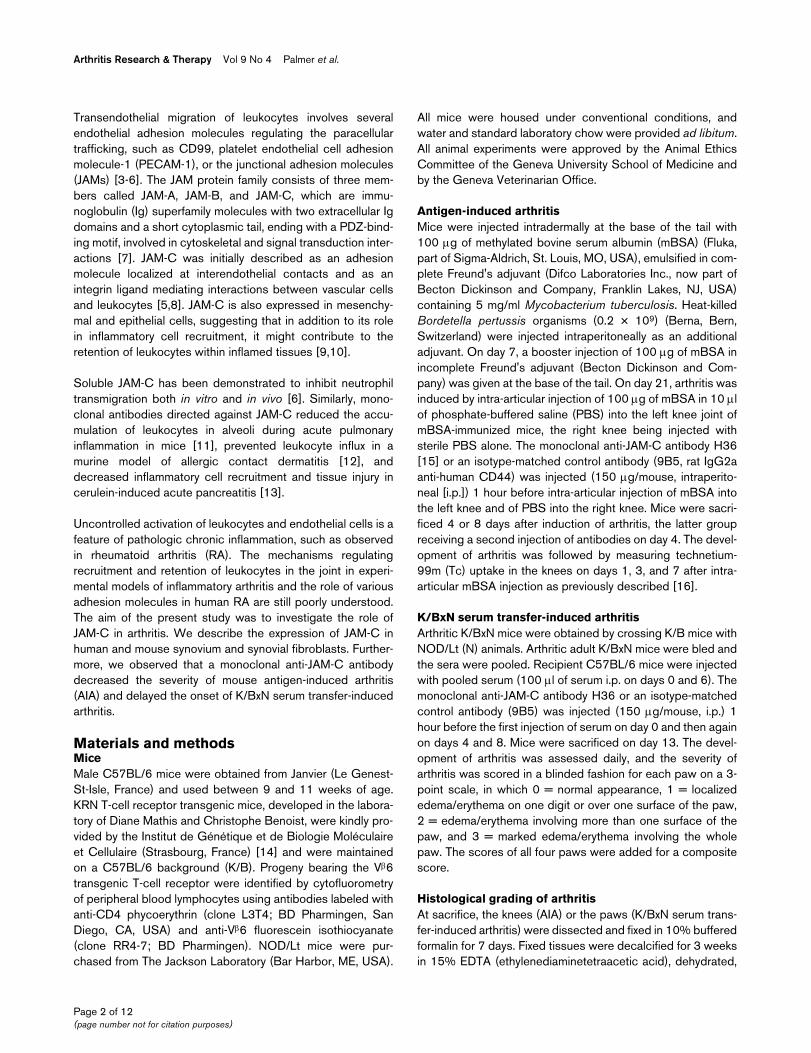
ResultsExpression of junctional adhesion molecule-C in human and mouse synovial tissuesWe investigated JAM-C expression in human OA and RA syn-ovial samples by IHC using polyclonal antibodies againsthuman JAM-C. Interestingly, expression of JAM-C was mainlyfound in the lining layer and was associated with vessels in thesublining layer (Figure 1a,b) in OA and in RA synovial biopsies.JAM-C expression, as quantified by histomorphometry, wassignificantly higher in RA than in OA samples (Figure 1c). RT-
PCR analysis showed JAM-C mRNA expression in OA andRA synovial biopsies as well as in purified human synovialfibroblasts (Figure 1d), suggesting that these cells account forat least part of the JAM-C expression observed in the lininglayer by IHC.
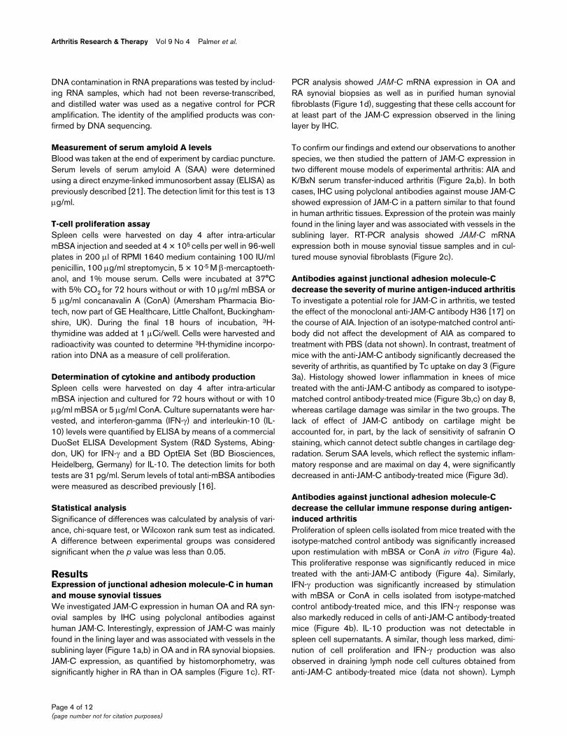
To confirm our findings and extend our observations to anotherspecies, we then studied the pattern of JAM-C expression intwo different mouse models of experimental arthritis: AIA andK/BxN serum transfer-induced arthritis (Figure 2a,b). In bothcases, IHC using polyclonal antibodies against mouse JAM-Cshowed expression of JAM-C in a pattern similar to that foundin human arthritic tissues. Expression of the protein was mainlyfound in the lining layer and was associated with vessels in thesublining layer. RT-PCR analysis showed JAM-C mRNAexpression both in mouse synovial tissue samples and in cul-tured mouse synovial fibroblasts (Figure 2c).
Antibodies against junctional adhesion molecule-C decrease the severity of murine antigen-induced arthritisTo investigate a potential role for JAM-C in arthritis, we testedthe effect of the monoclonal anti-JAM-C antibody H36 [17] onthe course of AIA. Injection of an isotype-matched control anti-body did not affect the development of AIA as compared totreatment with PBS (data not shown). In contrast, treatment ofmice with the anti-JAM-C antibody significantly decreased theseverity of arthritis, as quantified by Tc uptake on day 3 (Figure3a). Histology showed lower inflammation in knees of micetreated with the anti-JAM-C antibody as compared to isotype-matched control antibody-treated mice (Figure 3b,c) on day 8,whereas cartilage damage was similar in the two groups. Thelack of effect of JAM-C antibody on cartilage might beaccounted for, in part, by the lack of sensitivity of safranin Ostaining, which cannot detect subtle changes in cartilage deg-radation. Serum SAA levels, which reflect the systemic inflam-matory response and are maximal on day 4, were significantlydecreased in anti-JAM-C antibody-treated mice (Figure 3d).
Antibodies against junctional adhesion molecule-C decrease the cellular immune response during antigen-induced arthritisProliferation of spleen cells isolated from mice treated with theisotype-matched control antibody was significantly increasedupon restimulation with mBSA or ConA in vitro (Figure 4a).This proliferative response was significantly reduced in micetreated with the anti-JAM-C antibody (Figure 4a). Similarly,IFN-γ production was significantly increased by stimulationwith mBSA or ConA in cells isolated from isotype-matchedcontrol antibody-treated mice, and this IFN-γ response wasalso markedly reduced in cells of anti-JAM-C antibody-treatedmice (Figure 4b). IL-10 production was not detectable inspleen cell supernatants. A similar, though less marked, dimi-nution of cell proliferation and IFN-γ production was alsoobserved in draining lymph node cell cultures obtained fromanti-JAM-C antibody-treated mice (data not shown). Lymph
Page 4 of 12(page number not for citation purposes)

Available online http://arthritis-research.com/content/9/4/R65
node cells obtained from isotype-matched control antibody-treated mice produced detectable levels of IL-10 upon stimu-lation with mBSA, and this IL-10 production was also reducedin cells isolated from anti-JAM-C antibody-treated mice. Totalanti-BSA IgG and anti-BSA IgG2a levels were comparable inanti-JAM-C antibody-treated and isotype-matched control anti-body-treated mice on days 4 and 8 (Figure 4c,d). Anti-BSAIgG1 levels were transiently elevated at day 4, but not at day8, in the anti JAM-C antibody-treated mice.
Neutrophil infiltration in the inflamed synovium was selectivelyreduced in anti-JAM-C antibody-treated mice, whereas lym-
phocyte and macrophage infiltrations per field were similar inanti-JAM-C antibody and isotype-matched control antibody-treated mice (Figure 5).
Treatment with anti-junctional adhesion molecule-C antibody delays the onset of K/BxN serum transfer-induced arthritisTo specifically investigate a potential role of JAM-C in theeffector phase of arthritis, we tested the impact of anti-JAM-Cantibody treatment in a model of K/BxN serum transfer-induced arthritis. Treatment with the anti-JAM-C antibodydelayed the onset of K/BxN serum transfer-induced arthritis,
Figure 1
Expression of junctional adhesion molecule-C (JAM-C) in human arthritic synoviumExpression of junctional adhesion molecule-C (JAM-C) in human arthritic synovium. Expression of JAM-C was examined by immunohistochemistry in human osteoarthritis (OA) (a) and rheumatoid arthritis (RA) (b) synovial tissue. The panels labeled 1 show the original magnification (× 100). The panels labeled 2 show expression of JAM-C in the synovial lining layer (magnification × 400), and panels labeled 3 show JAM-C associated with blood vessels in the sublining (magnification × 400). The panels labeled 4 show negative control sections incubated with preimmune serum. Scale bars = 60 μm (panels 1 and 4) and 15 μm (panels 2 and 3). (c) Quantification of JAM-C expression in OA and RA synovial tissues. JAM-C immuno-histochemical synovial tissue sections from four different OA and RA patients were scanned, and the surface of immunoreactive areas was deter-mined and expressed as the percentage of the surface of the image examined. Results are expressed as the mean ± standard error of the mean. *p < 0.001 OA versus RA as assessed using the Wilcoxon rank sum test. (d) Reverse transcriptase-polymerase chain reaction (PCR) analysis of JAM-C mRNA expression in human synovial tissue samples and in cultured human synovial fibroblasts. A representative agarose gel electrophoresis of PCR products is shown. bp, base pairs; H2O, polymerase chain reaction negative control; hSF1 and hSF2, human rheumatoid arthritis synovial fibroblast cultures from two different patients used after the third passage; OA, osteoarthritis synovial tissue; RA, rheumatoid arthritis synovial tissue; RT neg, non-reverse-transcribed sample.
Page 5 of 12(page number not for citation purposes)

Arthritis Research & Therapy Vol 9 No 4 Palmer et al.
as indicated by a significantly lower incidence of arthritis inanti-JAM-C antibody-treated mice as compared to isotype-matched control antibody-treated mice on day 3 (Figure 6a). Incontrast, the severity of arthritis was not affected by the treat-ment in this model (Figure 6b). We also verified that injectionof the isotype-matched control antibody 9B5 per se had noeffect on the development of serum transfer-induced arthritis(data not shown).
Histological analysis of the paws showed no differencebetween anti-JAM-C antibody-treated and control mice at day13 (Figure 6c), although we cannot exclude the possibility thathistological differences, and in particular differences in neu-trophil infiltration, could have been detectable at an earlier timepoint. The serum SAA levels, which are not significantly
increased in this model, were not affected by treatment withthe anti-JAM-C antibody. In contrast, ConA-induced spleencell proliferation in vitro was decreased in anti-JAM-C anti-body-treated arthritic mice as compared to isotype-matchedantibody-treated controls (fold increase in proliferation overunstimulated control cultures: 11.3 ± 0.67 for anti-JAM-C anti-body-treated mice, n = 10; 37.1 ± 4.49 for control mice, n =10; p < 0.05).
DiscussionThe protein JAM-C is an adhesion molecule involved in junc-tional interaction between adjoining endothelial, epithelial, orfibroblastic cells [7]. It has also been reported to regulatemonocyte and neutrophil adhesion to and transmigrationthrough various types of cells, including fibroblast, epithelial,
Figure 2
Expression of junctional adhesion molecule-C (JAM-C) in mouse arthritic synovium and synovial fibroblastsExpression of junctional adhesion molecule-C (JAM-C) in mouse arthritic synovium and synovial fibroblasts. Expression of JAM-C was examined by immunohistochemistry in knee joints of mice with antigen-induced arthritis (AIA) (a) and in paws of mice with K/BxN serum transfer-induced arthritis (b). The left panels show expression of JAM-C in the synovial lining layer (upper panel) and associated with blood vessels in the sublining (lower panel). The right panels show negative control sections incubated with preimmune serum. Original magnification × 400 (scale bar = 20 μm). (c) Reverse transcriptase-polymerase chain reaction (PCR) analysis of JAM-C mRNA expression in mouse synovial tissue samples and in cultured mouse synovial fibroblasts. A representative agarose gel electrophoresis of PCR products is shown. AIA1 and AIA2, antigen-induced arthritis syno-vial tissue; bp, base pairs; H2O, polymerase chain reaction negative control; K/BxN1 and K/BxN2, K/BxN serum transfer-induced arthritis synovial tissue; mSF1 and mSF2, two different mouse synovial fibroblast cultures isolated from antigen-induced arthritis synovial tissue; RTneg, non reverse-transcribed sample.
Page 6 of 12(page number not for citation purposes)

Available online http://arthritis-research.com/content/9/4/R65
and endothelial cells [6,11,22,23]. Although these results sug-gest that JAM-C is involved in leukocyte recruitment duringinflammation, the role of JAM-C in human inflammatory dis-eases has not been well studied. Here, we found that JAM-Cis present on vessels and synovial fibroblasts of human andmouse arthritic lesions. An antibody against JAM-C inhibits therecruitment of neutrophils to the joints during AIA and delaysthe onset of serum transfer-induced arthritis. Finally, treatmentwith anti-JAM-C antibody decreases the T-cell response inAIA.
Recruitment of inflammatory cells, maintenance of the inflam-matory state, and the sustained retention of leukocytes withinarthritic lesions are mandatory for the development of arthritis.The invasion of leukocytes into the synovial tissue is controlled
at the molecular level by interactions between adhesion mole-cules expressed on endothelial cells and leukocytes [24]. Theadhesion molecule JAM-C expressed by endothelial cells hasbeen shown to bind the leukocyte integrin Mac-1 expressed byneutrophils and monocytes [6,8]. Although Mac-1 is not theonly leukocyte adhesion molecule involved in leukocyte migra-tion to arthritic lesions, this integrin, together with lymphocytefunction-associated antigen-1 (LFA-1) and α4β1 integrins,has been shown to contribute to the development of the dis-ease [25-27]. We know from previous studies that our anti-JAM-C monoclonal antibody does not prevent direct interac-tion between Mac-1 and JAM-C [15]. Nevertheless, it stillblocks neutrophil recruitment to inflamed tissues [13]. Onepossibility is that treatment with the anti-JAM-C antibody mod-ulates the contribution of Mac-1 or α4β1 integrins to leukocyte
Figure 3
Treatment with anti-JAM-C antibody decreases the severity of antigen-induced arthritisTreatment with anti-JAM-C antibody decreases the severity of antigen-induced arthritis. (a) Joint inflammation on days 1, 3, and 7 after intra-articular methylated bovine serum albumin (mBSA) injection. Results are expressed as the ratio of technetium-99m (99mTc) uptake in the arthritic over the non-inflamed knee. The mean ± standard error of the mean (SEM) of the ratios is shown for anti-JAM-C antibody-treated (n = 10 for days 1 and 3, n = 6 for day 7; black symbols) and isotype-matched control antibody-treated (n = 10 for days 1 and 3, n = 5 for day 7; open symbols) mice. Joint inflammation was significantly reduced in anti-JAM-C antibody-treated mice on day 3. (b) Representative histological sections for anti-JAM-C anti-body-treated (upper panel) and isotype-matched control antibody-treated (lower panel) mice 8 days after intra-articular mBSA injection (original magnification × 40, scale bar = 125 μm). Arrowheads: cartilage erosions; asterisks: pannus; broken lines: synovial thickness. (c) Histological scores shown as the mean ± SEM for anti-JAM-C antibody-treated (n = 6, black columns) and isotype-matched control antibody-treated (n = 5, open col-umns) mice 8 days after intra-articular mBSA injection. Synovial inflammation was significantly reduced in anti-JAM-C antibody-treated mice. (d) Cir-culating levels of serum amyloid A (SAA) on days 4 and 8 after intra-articular mBSA injection. Results shown represent the mean ± SEM for anti-JAM-C antibody-treated (n = 5 on day 4, n = 6 on day 8, black columns) and isotype-matched control antibody-treated (n = 5 at both time points, open columns) mice. Serum SAA levels were significantly decreased in anti-JAM-C antibody-treated mice on day 4. *p < 0.05 versus control mice, as assessed by analysis of variance. Ab, antibody; JAM-C, junctional adhesion molecule-C.
Page 7 of 12(page number not for citation purposes)

Arthritis Research & Therapy Vol 9 No 4 Palmer et al.
Figure 4
Treatment with anti-JAM-C antibody decreases the T-cell responseTreatment with anti-JAM-C antibody decreases the T-cell response. (a) Proliferation of spleen cells isolated from anti-JAM-C antibody-treated (n = 5) and isotype-matched control antibody-treated (n = 5) mice. Cells were restimulated in vitro with methylated bovine serum albumin (mBSA) (10 μg/ml, hatched columns) or concanavalin A (ConA) (5 μg/ml) or were left unstimulated (white columns). Results, expressed as fold increase in prolifera-tion of stimulated over unstimulated cultures, represent the mean ± standard error of the mean (SEM) for each group of mice. Proliferation was sig-nificantly increased by stimulation with mBSA or ConA in cells isolated from isotype-matched control antibody-treated mice; *p < 0.05 versus unstimulated cultures. This proliferative response to mBSA and ConA was significantly reduced in cells of anti-JAM-C antibody-treated mice as com-pared to cells isolated from isotype-matched control antibody-treated mice; &p < 0.05 versus cells isolated from isotype-matched control antibody-treated mice with similar stimulation, as assessed by analysis of variance (ANOVA). (b) Interferon-gamma (IFN-γ) production by spleen cells isolated from anti-JAM-C antibody-treated (n = 5) and isotype-matched control antibody-treated (n = 5) mice. Cells were restimulated in vitro with mBSA (10 μg/ml, hatched columns) or ConA (5 μg/ml) or were left unstimulated (white columns). Results shown represent the mean ± SEM for each group of mice. IFN-γ production was significantly increased by stimulation with mBSA or ConA in cells isolated from isotype-matched control antibody-treated mice; *p < 0.05 versus unstimulated cultures. This IFN-γ response to mBSA and ConA was markedly reduced in cells of anti-JAM-C antibody-treated mice as compared to cells isolated from isotype-matched control antibody-treated mice; &p < 0.05 versus cells isolated from isotype-matched con-trol antibody-treated mice with similar stimulation, as assessed by ANOVA. Serum levels of anti-mBSA total IgG (c), IgG1 ([d], left panel), and IgG2a ([d], right panel) on days 4 and 8 after intra-articular mBSA injection. Results shown represent the mean ± SEM for anti-JAM-C antibody-treated (n = 6, black columns) and isotype-matched control antibody-treated (n = 5, open columns) mice. *p < 0.05 versus control mice, as assessed by ANOVA. Ab, antibody; A.U., arbitrary units; Ig = immunoglobulin; JAM-C, junctional adhesion molecule-C; O.D., optical density.
Page 8 of 12(page number not for citation purposes)

Available online http://arthritis-research.com/content/9/4/R65
trafficking indirectly by interfering with the function of JAM-C[6,28]. Alternatively, the anti-JAM-C antibody may act directlyon resident synovial fibroblasts to disrupt retention of granulo-cytes within lesions. Synovial fibroblasts are indeed potentproducers of cytokines, adhesion molecules, and chemokinesthat attract and retain large numbers of leukocytes in theinflamed synovium, thus sustaining chronic inflammation andpreventing the resumption of normal tissue homeostasis [29].
The mouse AIA model can be divided into two phases. Intra-articular antigen injection first results in an acute inflammatoryreaction, characterized by joint swelling and leukocyte infiltra-tion, which later proceeds to a chronic destructive arthritis with
synovial hyperplasia, and cartilage and bone erosion. In addi-tion to an inhibitory effect of anti-JAM-C antibody treatment onthe acute inflammatory response, which is in line with previousfindings [11-13], we observed an anti-inflammatory effect inthe chronic phase of the disease, as shown by reduced inflam-mation in the knees of mice treated with anti-JAM-C antibodyas compared to isotype-matched control antibody-treatedmice on day 8 (Figure 3b,c).
The systemic suppressive effect of anti-JAM-C on T-cellresponse is an unexpected result. JAM-C is not expressed onmouse T cells and in vitro addition of anti-JAM-C antibodiesonto spleen cell cultures did not alter cell proliferation (data
Figure 5
Treatment with anti-JAM-C antibody decreases neutrophil infiltration into the joints during antigen-induced arthritisTreatment with anti-JAM-C antibody decreases neutrophil infiltration into the joints during antigen-induced arthritis. Infiltration of neutrophils (a), lym-phocytes (b), and macrophages (c) into the synovium was detected by immunohistochemistry using anti-MPO, anti-CD3, and anti-MAC-2 antibod-ies, respectively. The left panels show representative knee joint sections of control and anti-JAM-C antibody-treated mice. Original magnification × 400 (scale bar = 25 μm). In the right panels, leukocyte infiltration per field was evaluated by semi-quantitative scoring for anti-JAM-C antibody-treated (n = 6, black columns) and isotype-matched control antibody-treated (n = 5, open columns) mice. There was a significant decrease in syno-vial neutrophil infiltration in anti-JAM-C antibody-treated mice as compared to isotype-matched antibody-treated controls. *p < 0.05 versus control mice, as assessed by analysis of variance. Ab, antibody; JAM-C, junctional adhesion molecule-C.
Page 9 of 12(page number not for citation purposes)

Arthritis Research & Therapy Vol 9 No 4 Palmer et al.
not shown), suggesting that the immunomodulatory effect ofthe anti-JAM-C antibody may be mediated by stromal cells invivo. Consistent with this observation, other investigatorsfound no difference regarding the number of circulating lym-phocytes between anti-JAM-C-treated and isotype control-treated groups in a model of allergic contact dermatitisresponsive to anti-JAM-C administration [12]. We alsoobserved that circulating levels of lymphocytes were not sig-
nificantly decreased in naive and arthritic mice (K/BxN experi-ments) following the administration of the anti-JAM-C or theisotype-matched control antibody. The absolute lymphocytecounts (mean ± standard error of the mean) were 4.5 ± 0.57g/L (naive anti-JAM-C antibody-treated), 5.5 ± 0.41 g/L (naiveisotype-matched control antibody-treated), 6.89 ± 0.55 g/L(arthritic anti-JAM-C antibody-treated), and 5.5 ± 0.57 g/L(arthritic isotype-matched control antibody-treated). In addi-
Figure 6
Treatment with anti-JAM-C antibody delays onset of K/BxN serum transfer-induced arthritisTreatment with anti-JAM-C antibody delays onset of K/BxN serum transfer-induced arthritis. (a) Incidence of K/BxN serum transfer-induced arthritis is shown for anti-JAM-C antibody-treated (n = 10, black symbols) and isotype-matched control antibody-treated (n = 10, open symbols) mice. Results are expressed as the percentage of arthritic mice per group. Incidence of arthritis was significantly lower in anti-JAM-C antibody-treated mice as compared to isotype-matched antibody-treated controls on day 3. *p < 0.05 versus control mice, as assessed by chi-square test. (b) Severity of K/BxN serum transfer-induced arthritis. Arthritis was evaluated by clinical assessment of arthritis severity scores for anti-JAM-C antibody-treated (n = 10, black symbols) and isotype-matched control antibody-treated (n = 10, open symbols) mice. Results shown represent the mean ± standard error of the mean (SEM) for each group of mice. There were no significant differences between the groups. (c) Histological assessment of arthritis. Rep-resentative sections are shown for anti-JAM-C antibody-treated (upper panels) and isotype-matched control antibody-treated (lower panels) mice 13 days after serum transfer. Original magnifications × 40 (left panel) (scale bar = 125 μm) and × 100 (right panel) (scale bar = 50 μm). (d) Histologi-cal scores for synovial thickness, exudates, and edema 13 days after serum transfer. Results shown represent the mean ± SEM for anti-JAM-C anti-body-treated (n = 10, black columns) and isotype-matched control antibody-treated (n = 10, open columns) mice. No significant differences were observed between the groups. Ab, antibody; JAM-C, junctional adhesion molecule-C.
Page 10 of 12(page number not for citation purposes)

Available online http://arthritis-research.com/content/9/4/R65
tion, as in AIA, the inhibitory effect of anti-JAM-C antibodiesoccurred when mice were treated after sensitization andbefore challenge, thus indicating that JAM-C interferes withthe effector phase and not the initial antigen presentation [12].
JAM-C is expressed on synovial and other types of fibroblasts,fibroblastic reticular cells of lymph nodes, and smooth musclecells [10,23]. All these cells have in common an immunoregu-latory function, and the role of fibroblastic cells in the establish-ment and maintenance of micro-environmental 'niches'contributing to inflammatory diseases, including RA, has beenextensively discussed in recent reports [29-31]. Though spec-ulative, one could imagine that anti-JAM-C treatment deliverssignals to mesenchymal cells, which in turn control the out-come of the immune response by modulating expression ofadhesion molecules, chemokines, and cytokines [32]. Such ahypothesis is in agreement with recent findings showing thatanti-JAM-C treatment decreases inflammation in variousmouse inflammatory models such as cerulein-induced pancre-atitis, thioglycollate-induced peritonitis, or allergic contact der-matitis [11-13] and that the protein may be involved in otherdiseases such as obstructive nephropathy or atherosclerosis[10,33]. An alternative explanation for the systemic pro-inflam-matory function of JAM-C relates to its active role in favoringdiffusion of small molecules across the paravascular space.Indeed, we have previously shown that the enrichment of theprotein at interendothelial cell-cell contacts correlates withincreased paracellular permeability and is induced by factorsinducing vascular leakage such as vascular endothelial growthfactor (VEGF) [20,34]. These results have been confirmed ina recent study showing that antagonizing JAM-C functionresults in inhibition of increased vascular permeability inducedby VEGF or histamine [35]. Thus, it is possible that treatmentof arthritic mice with the anti-JAM-C antibody results indecreased diffusion of inflammatory mediators in the blood,inducing a systemic immunosuppressive effect. Although thisattractive hypothesis requires more investigation, our resultsuncover a link between JAM-C expression in arthritic lesionsand local and systemic anti-inflammatory effects of an antibodydirected against this protein.
There is accumulating evidence that leukocyte trafficking tothe inflamed synovium is adhesion molecule-dependent, andthe blocking adhesion molecules that mediate the accumula-tion of leukocytes in inflammation can thus be expected tohave therapeutic potential in human RA. However, the few clin-ical trials performed so far have not met expectations. Forinstance, efazulimab, a humanized monoclonal antibodyagainst LFA-1, did not significantly reduce arthritis in a cohortof patients with psoriatic arthritis [36]. Anti-ICAM-1 (intracellu-lar adhesion molecule-1) monoclonal antibodies have beenevaluated in a phase I/II open-label study and had only limitedeffects in patients with RA [37]. Finally, a randomized, pla-cebo-controlled trial of an antisense oligodeoxynucleotideICAM-1 inhibitor could not demonstrate clinical efficacy
beyond that of placebo in patients with active RA [38]. Amongthe possible explanations for this low efficacy are the redun-dancy and overlapping functions of molecules that areinvolved in leukocyte extravasation, due to which selective inhi-bition of single adhesion molecules might not be sufficient toefficiently prevent leukocyte recruitment. In the present study,we observed that treatment with an anti-JAM-C antibodyinduced anti-inflammatory effects, which seem to extendbeyond local inhibition of leukocyte adhesion. This observationfits with the concept that the signaling activity of adhesionmolecules, in addition to their adhesive properties, may beimportant for their function.
ConclusionJAM-C is highly expressed by synovial fibroblasts in RA. Treat-ment of mice with an anti-JAM-C antibody significantlyreduced the severity of AIA and delayed the onset of serumtransfer-induced arthritis, suggesting a role for JAM-C in thepathogenesis of arthritis.
Competing interestsThe authors declare that they have no competing interests.
Authors' contributionsGP, NB, and MA-L performed the experiments, designed thestudy, and drafted the manuscript. DT-A, VC-P, CZ, and PHperformed the experiments. BAI and CG designed the studyand drafted the manuscript. All authors read and approved thefinal manuscript. GP and NB contributed equally to this work.
AcknowledgementsKRN mice were kindly provided by Diane Mathis and Christophe Benoist through the Institut de Génétique et de Biologie Moléculaire et Cellulaire (Strasbourg, France). This work was supported by Swiss National Sci-ence Foundation grant 3200-107592/1 (to CG), grant 3200-067231 (to NB), grant 310000-112551/I (to MA-L), and grant 3100AO-10069712 (to BAI), by the Krebsforschung Schweiz (grant to BAI), and by the Thorn Foundation (grant to MA-L).
References1. Butcher EC: Leukocyte-endothelial cell recognition: three (or
more) steps to specificity and diversity. Cell 1991,67:1033-1036.
2. Springer TA: Traffic signals for lymphocyte recirculation andleukocyte emigration: the multistep paradigm. Cell 1994,76:301-314.
3. Schenkel AR, Mamdouh Z, Chen X, Liebman RM, Muller WA:CD99 plays a major role in the migration of monocytesthrough endothelial junctions. Nat Immunol 2002, 3:143-150.
4. Muller WA, Weigl SA, Deng X, Phillips DM: PECAM-1 is requiredfor transendothelial migration of leukocytes. J Exp Med 1993,178:449-460.
5. Johnson-Leger CA, Aurrand-Lions M, Beltraminelli N, Fasel N,Imhof BA: Junctional adhesion molecule-2 (JAM-2) promoteslymphocyte transendothelial migration. Blood 2002,100:2479-2486.
6. Chavakis T, Keiper T, Matz-Westphal R, Hersemeyer K, Sachs UJ,Nawroth PP, Preissner KT, Santoso S: The junctional adhesionmolecule-C promotes neutrophil transendothelial migration invitro and in vivo. J Biol Chem 2004, 279:55602-55608.
Page 11 of 12(page number not for citation purposes)

Arthritis Research & Therapy Vol 9 No 4 Palmer et al.
7. Ebnet K, Suzuki A, Ohno S, Vestweber D: Junctional adhesionmolecules (JAMs): more molecules with dual functions? J CellSci 2004, 117:19-29.
8. Santoso S, Sachs UJ, Kroll H, Linder M, Ruf A, Preissner KT, Cha-vakis T: The junctional adhesion molecule 3 (JAM-3) on humanplatelets is a counterreceptor for the leukocyte integrin Mac-1.J Exp Med 2002, 196:679-691.
9. Liu Y, Nusrat A, Schnell FJ, Reaves TA, Walsh S, Pochet M, ParkosCA: Human junction adhesion molecule regulates tight junc-tion resealing in epithelia. J Cell Sci 2000, 113:2363-2374.
10. Keiper T, Al-Fakhri N, Chavakis E, Athanasopoulos AN, IsermannB, Herzog S, Saffrich R, Hersemeyer K, Bohle RM, Haendeler J, etal.: The role of junctional adhesion molecule-C (JAM-C) in oxi-dized LDL-mediated leukocyte recruitment. Faseb J 2005,19:2078-2080.
11. Aurrand-Lions M, Lamagna C, Dangerfield JP, Wang S, Herrera P,Nourshargh S, Imhof BA: Junctional adhesion molecule-C regu-lates the early influx of leukocytes into tissues duringinflammation. J Immunol 2005, 174:6406-6415.
12. Ludwig RJ, Zollner TM, Santoso S, Hardt K, Gille J, Baatz H,Johann PS, Pfeffer J, Radeke HH, Schon MP, et al.: Junctionaladhesion molecules (JAM)-B and -C contribute to leukocyteextravasation to the skin and mediate cutaneousinflammation. J Invest Dermatol 2005, 125:969-976.
13. Vonlaufen A, Aurrand-Lions M, Pastor CM, Lamagna C, HadengueA, Imhof BA, Frossard JL: The role of junctional adhesion mole-cule C (JAM-C) in acute pancreatitis. J Pathol 2006,209:540-548.
14. Kouskoff V, Korganow AS, Duchatelle V, Degott C, Benoist C,Mathis D: Organ-specific disease provoked by systemicautoimmunity. Cell 1996, 87:811-822.
15. Lamagna C, Meda P, Mandicourt G, Brown J, Gilbert RJ, Jones EY,Kiefer F, Ruga P, Imhof BA, Aurrand-Lions M: Dual interaction ofJAM-C with JAM-B and alpha(M)beta2 integrin: function injunctional complexes and leukocyte adhesion. Mol Biol Cell2005, 16:4992-5003.
16. Busso N, So A, Chobaz-Péclat V, Morard C, Martinez-Soria E, Tal-abot-Ayer D, Gabay C: Leptin signaling deficiency impairshumoral and cellular immune responses and attenuatesexperimental arthritis. J Immunol 2002, 168:875-882.
17. Lamagna C, Hodivala-Dilke KM, Imhof BA, Aurrand-Lions M: Anti-body against junctional adhesion molecule-C inhibits angio-genesis and tumor growth. Cancer Res 2005, 65:5703-5710.
18. Ody C, Jungblut-Ruault S, Cossali D, Barnet M, Aurrand-Lions M,Imhof BA, Matthes T: Junctional adhesion molecule C (JAM-C)distinguishes CD27+ germinal center B lymphocytes fromnon-germinal center cells and constitutes a new diagnostictool for B-cell malignancies. Leukemia 2007, 21:1285-1293.
19. Palmer G, Burger D, Mezin F, Magne D, Gabay C, Dayer JM,Guerne PA: The active metabolite of leflunomide, A77increases the production of IL-1 receptor antagonist in humansynovial fibroblasts and articular chondrocytes. Arthritis ResTher 2007, 6:R181-189.
20. Magne D, Palmer G, Barton JL, Mezin F, Talabot-Ayer D, Bas S,Duffy T, Noger M, Guerne PA, Nicklin MJ, et al.: The new IL-1 fam-ily member IL-1F8 stimulates production of inflammatorymediators by synovial fibroblasts and articular chondrocytes.Arthritis Res Ther 2006, 8:R80.
21. Sipe JD, Gonnerman WA, Loose LD, Knapschaefer G, Xie WJ,Franzblau C: Direct binding enzyme-linked immunosorbentassay (ELISA) for serum amyloid A (SAA). J Immunol Methods1989, 125:125-135.
22. Zen K, Babbin BA, Liu Y, Whelan JB, Nusrat A, Parkos CA: JAM-C is a component of desmosomes and a ligand for CD11b/CD18-mediated neutrophil transepithelial migration. Mol BiolCell 2004, 15:3926-3937.
23. Morris AP, Tawil A, Berkova Z, Wible L, Smith CW, CunninghamSA: Junctional adhesion molecules (JAMs) are differentiallyexpressed in fibroblasts and co-localize with ZO-1 to adher-ens-like junctions. Cell Commun Adhes 2006, 13:233-247.
24. Tarrant TK, Patel DD: Chemokines and leukocyte trafficking inrheumatoid arthritis. Pathophysiology 2006, 13:1-14.
25. Issekutz AC, Issekutz TB: Monocyte migration to arthritis in therat utilizes both CD11/CD18 and very late activation antigen 4integrin mechanisms. J Exp Med 1995, 181:1197-1203.
26. Issekutz TB, Miyasaka M, Issekutz AC: Rat blood neutrophilsexpress very late antigen 4 and it mediates migration to
arthritic joint and dermal inflammation. J Exp Med 1996,183:2175-2184.
27. Taylor PC, Chu CQ, Plater-Zyberk C, Maini RN: Transfer of typeII collagen-induced arthritis from DBA/1 to severe combinedimmunodeficiency mice can be prevented by blockade of Mac-1. Immunology 1996, 88:315-321.
28. Cunningham SA, Rodriguez JM, Arrate MP, Tran TM, Brock TA:JAM2 interacts with alpha4beta1. Facilitation by JAM3. J BiolChem 2002, 277:27589-27592.
29. Burman A, Haworth O, Bradfield P, Parsonage G, Filer A, ThomasAM, Amft N, Salmon M, Buckley CD: The role of leukocyte-stro-mal interactions in chronic inflammatory joint disease. JointBone Spine 2005, 72:10-16.
30. Parsonage G, Filer AD, Haworth O, Nash GB, Rainger GE, SalmonM, Buckley CD: A stromal address code defined by fibroblasts.Trends Immunol 2005, 26:150-156.
31. Mor A, Abramson SB, Pillinger MH: The fibroblast-like synovialcell in rheumatoid arthritis: a key player in inflammation andjoint destruction. Clin Immunol 2005, 115:118-128.
32. Katakai T, Hara T, Sugai M, Gonda H, Shimizu A: Lymph nodefibroblastic reticular cells construct the stromal reticulum viacontact with lymphocytes. J Exp Med 2004, 200:783-795.
33. Lange-Sperandio B, Schimpgen K, Rodenbeck B, Chavakis T,Bierhaus A, Nawroth P, Thornhill B, Schaefer F, Chevalier RL: Dis-tinct roles of Mac-1 and its counter-receptors in neonatalobstructive nephropathy. Kidney Int 2006, 69:81-88.
34. Aurrand-Lions M, Johnson-Leger C, Wong C, Du Pasquier L, ImhofBA: Heterogeneity of endothelial junctions is reflected by dif-ferential expression and specific subcellular localization of thethree JAM family members. Blood 2001, 98:3699-3707.
35. Orlova VV, Economopoulou M, Lupu F, Santoso S, Chavakis T:Junctional adhesion molecule-C regulates vascular endothe-lial permeability by modulating VE-cadherin-mediated cell-cellcontacts. J Exp Med 2006, 203:2703-2714.
36. Gladman DD: Psoriatic arthritis. Dermatol Ther 2004,17:350-363.
37. Kavanaugh AF, Davis LS, Jain RI, Nichols LA, Norris SH, Lipsky PE:A phase I/II open label study of the safety and efficacy of ananti-ICAM-1 (intercellular adhesion molecule-1; CD54) mono-clonal antibody in early rheumatoid arthritis. J Rheumatol1996, 23:1338-1344.
38. Maksymowych WP, Blackburn WD Jr, Tami JA, Shanahan WR Jr:A randomized, placebo controlled trial of an antisenseoligodeoxynucleotide to intercellular adhesion molecule-1 inthe treatment of severe rheumatoid arthritis. J Rheumatol2002, 29:447-453.
Page 12 of 12(page number not for citation purposes)