open access coronary artery perforation following ... · choice floppy guidewire. ... appropriate...
TRANSCRIPT

148
CASE REPORT
DOI 10.4070 / kcj.2010.40.3.148 Print ISSN 1738-5520 / On-line ISSN 1738-5555
Copyright ⓒ 2010 The Korean Society of Cardiology
Open Access
Coronary Artery Perforation Following Implantation of a Drug-Eluting Stent Rescued by Deployment of a Covered Stent in Symptomatic Myocardial Bridging Man Zhang, MD1, Woong Chol Kang, MD2, Chan Il Moon, MD2, Seung Hwan Han, MD2, Tae Hoon Ahn, MD2 and Eak Kyun Shin, MD2 1Department of Cardiology, Fengtian Hospital Affiliated to Shenyang Medical College, Shenyang, Liaoning Province, China 2Department of Cardiology, Gil Medical Center, Gachon University of Medicine and Science, Incheon, Korea ABSTRACT
We successfully rescued a patient whose coronary artery perforated following implantation of a drug-eluting stent (DES), by deploying a stent-graft in symptomatic myocardial bridging. Our case demonstrated that coronary per-foration could be handled without difficulty when perforated myocardial bridging is confined to the interventri-cular groove. (Korean Circ J 2010;40:148-151) KEY WORDS: Myocardial bridging; Drug eluting stents; Angioplasty, Transluminal, percutaneous coronary.
Introduction
Myocardial bridging is defined as an epicardial coro-
nary artery that goes intramurally through the myocar-dium beneath the muscle bridge. While generally benign, myocardial bridges can cause ischemia, ventricular ta-chyarrhythmias, atrioventricular block, and sudden car-diac death.1)2) For symptomatic patients, various ther-apeutic approaches have been attempted, but the optimal treatment of myocardial bridging still remains contro-versial.3)4) Coronary stenting has been another thera-peutic option with medical and surgical treatment, but the high risk of perforation and high rate of in-stent restenosis have limited its use.5-10) Recently, we experi-enced a patient with a perforated coronary artery after implantation of a drug-eluting stent (DES) that was successfully rescued by deployment of covered stent in symptomatic myocardial bridging.
Case A 46 year-old woman who had no coronary risk fac-
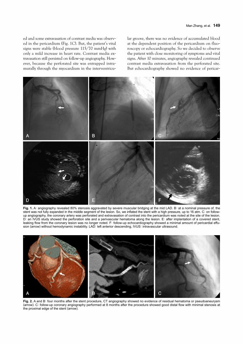
tors, presented with exertional chest pain for several weeks. The chest pain was typical for angina pectoris and depressed ST segments were noted at the exercise test. Echocardiography revealed normal left ventricular (LV) systolic function {ejection fraction (EF)=72%} with-out any regional wall motion abnormality. We perform-ed coronary angiography which showed significant ste-nosis (up to 80%) aggravated by severe myocardial bridg-ing at the mid-portion of the left anterior descending (LAD) artery (Fig. 1A) without significant stenotic le-sions at other coronary arteries. The pain was not re-lieved by optimal medical treatment for 2 weeks. So we decided to do a percutaneous coronary intervention (PCI) at the LAD lesion. Through a 7 Fr Judkins guid-ing catheter, the lesion was easily crossed with a 0.014 Choice floppy guidewire. Predilatation was performed with a maverick balloon catheter (2.5×15 mm, Boston Scientific, Natick, MA, USA) at 10 atmospheres for 20 seconds. We deployed a Taxus stent (3.5×16 mm, Bos-ton Scientific, Natick, MA, USA) according to the size of the predilated balloon catheter. The stent was inflat-ed up to a nominal pressure 8 atmospheres. But the middle segment of the lesion was not compliant, so the stent was not fully expanded with nominal pressure. We gradually inflated the stent using 16 atmospheres (Fig. 1B). In the mean time, the coronary artery was perforat-
Received: August 11, 2009
Revision Received: September 17, 2009 Accepted: September 30, 2009 Correspondence: Woong Chol Kang, MD, Department of Cardiology, Gil
Medical Center, Gachon University, 1198 Guwol-dong, Namdong-gu,
Incheon 405-760, Korea
Tel: 82-32-460-3046, Fax: 82-32-460-3117
E-mail: [email protected] ○ ○○ cc This is an Open Access article distributed under the terms of the CreativeCommons Attribution Non-Commercial License (http://creativecommons.org/licenses/by-nc/3.0) which permits unrestricted non-commercial use,distribution, and reproduction in any medium, provided the original work isproperly cited.
Man Zhang, et al.·149
ed and some extravasation of contrast media was observ- ed in the pericardium (Fig. 1C). But, the patient’s vital signs were stable (blood pressure 115/70 mmHg) with only a mild increase in heart rate. Contrast media ex-travasation still persisted on follow-up angiography. How-ever, because the perforated site was entrapped intra-murally through the myocardium in the interventricu-
lar groove, there was no evidence of accumulated blood at the dependent position of the pericardium on fluo-roscopy or echocardiography. So we decided to observe the patient with close monitoring of symptoms and vital signs. After 10 minutes, angiography revealed continued contrast media extravasation from the perforated site. But echocardiography showed no evidence of pericar-
A B C
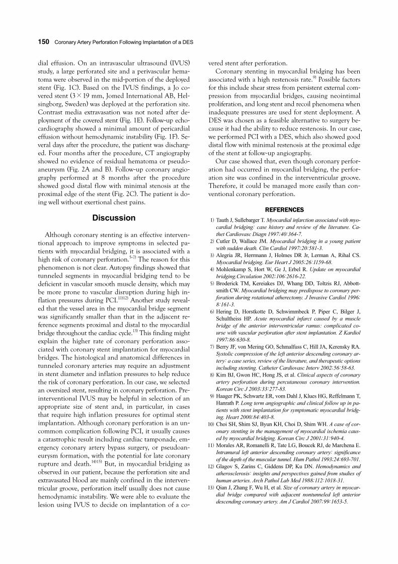
Fig. 2. A and B: four months after the stent procedure, CT angiography showed no evidence of residual hematoma or pseudoaneurysm(arrow). C: follow-up coronary angiography performed at 8 months after the procedure showed good distal flow with minimal stenosis atthe proximal edge of the stent (arrow).
A B C
D E
Fig. 1. A: angiography revealed 80% stenosis aggravated by severe muscular bridging at the mid LAD. B: at a nominal pressure of, the stent was not fully expanded in the middle segment of the lesion. So, we inflated the stent with a high pressure, up to 16 atm. C: on follow-up angiography, the coronary artery was perforated and extravasation of contrast into the pericardium was noted at the site of the lesion.D: an IVUS study showed the perforation site and a perivascular hematoma along the lesion. E: after implantation of a covered stent, leaking flow from the coronary lesion was no longer noted. F: follow-up echocardiography showed a minimal amount of pericardial effu-sion (arrow) without hemodynamic instability. LAD: left anterior descending, IVUS: intravascular ultrasound.
F
150·Coronary Artery Perforation Following Implantation of a DES
dial effusion. On an intravascular ultrasound (IVUS) study, a large perforated site and a perivascular hema-toma were observed in the mid-portion of the deployed stent (Fig. 1C). Based on the IVUS findings, a Jo co-vered stent (3×19 mm, Jomed International AB, Hel-singborg, Sweden) was deployed at the perforation site. Contrast media extravasation was not noted after de-ployment of the covered stent (Fig. 1E). Follow-up echo-cardiography showed a minimal amount of pericardial effusion without hemodynamic instability (Fig. 1F). Se-veral days after the procedure, the patient was discharg-ed. Four months after the procedure, CT angiography showed no evidence of residual hematoma or pseudo-aneurysm (Fig. 2A and B). Follow-up coronary angio-graphy performed at 8 months after the procedure showed good distal flow with minimal stenosis at the proximal edge of the stent (Fig. 2C). The patient is do-ing well without exertional chest pains.
Discussion
Although coronary stenting is an effective interven-
tional approach to improve symptoms in selected pa-tients with myocardial bridging, it is associated with a high risk of coronary perforation.5-7) The reason for this phenomenon is not clear. Autopsy findings showed that tunneled segments in myocardial bridging tend to be deficient in vascular smooth muscle density, which may be more prone to vascular disruption during high in-flation pressures during PCI.11)12) Another study reveal-ed that the vessel area in the myocardial bridge segment was significantly smaller than that in the adjacent re-ference segments proximal and distal to the myocardial bridge throughout the cardiac cycle.13) This finding might explain the higher rate of coronary perforation asso-ciated with coronary stent implantation for myocardial bridges. The histological and anatomical differences in tunneled coronary arteries may require an adjustment in stent diameter and inflation pressures to help reduce the risk of coronary perforation. In our case, we selected an oversized stent, resulting in coronary perforation. Pre-interventional IVUS may be helpful in selection of an appropriate size of stent and, in particular, in cases that require high inflation pressures for optimal stent implantation. Although coronary perforation is an un-common complication following PCI, it usually causes a catastrophic result including cardiac tamponade, em-ergency coronary artery bypass surgery, or pseudoan-eurysm formation, with the potential for late coronary rupture and death.14)15) But, in myocardial bridging as observed in our patient, because the perforation site and extravasated blood are mainly confined in the interven-tricular groove, perforation itself usually does not cause hemodynamic instability. We were able to evaluate the lesion using IVUS to decide on implantation of a co-
vered stent after perforation. Coronary stenting in myocardial bridging has been
associated with a high restenosis rate.9) Possible factors for this include shear stress from persistent external com-pression from myocardial bridges, causing neointimal proliferation, and long stent and recoil phenomena when inadequate pressures are used for stent deployment. A DES was chosen as a feasible alternative to surgery be-cause it had the ability to reduce restenosis. In our case, we performed PCI with a DES, which also showed good distal flow with minimal restenosis at the proximal edge of the stent at follow-up angiography.
Our case showed that, even though coronary perfor-ation had occurred in myocardial bridging, the perfor-ation site was confined in the interventricular groove. Therefore, it could be managed more easily than con-ventional coronary perforation.
REFERENCES 1) Tauth J, Sullebarger T. Myocardial infarction associated with myo-
cardial bridging: case history and review of the literature. Ca-thet Cardiovasc Diagn 1997;40:364-7.
2) Cutler D, Wallace JM. Myocardial bridging in a young patient with sudden death. Clin Cardiol 1997;20:581-3.
3) Alegria JR, Herrmann J, Holmes DR Jr, Lerman A, Rihal CS. Myocardial bridging. Eur Heart J 2005;26:1159-68.
4) Mohlenkamp S, Hort W, Ge J, Erbel R. Update on myocardial bridging.Circulation 2002;106:2616-22.
5) Broderick TM, Kereiakes DJ, Whang DD, Toltzis RJ, Abbott-smith CW. Myocardial bridging may predispose to coronary per-foration during rotational atherectomy. J Invasive Cardiol 1996; 8:161-3.
6) Hering D, Horstkotte D, Schwimmbeck P, Piper C, Bilger J, Schultheiss HP. Acute myocardial infarct caused by a muscle bridge of the anterior interventricular ramus: complicated co-urse with vascular perforation after stent implantation. Z Kardiol 1997;86:630-8.
7) Berry JF, von Mering GO, Schmalfuss C, Hill JA, Kerensky RA. Systolic compression of the left anterior descending coronary ar-tery: a case series, review of the literature, and therapeutic options including stenting. Catheter Cardiovasc Interv 2002;56:58-63.
8) Kim BJ, Gwon HC, Hong JS, et al. Clinical aspects of coronary artery perforation during percutaneous coronary intervention. Korean Circ J 2003;33:277-83.
9) Haager PK, Schwartz ER, vom Dahl J, Klues HG, Reffelmann T, Hanrath P. Long term angiographic and clinical follow up in pa-tients with stent implantation for symptomatic myocardial bridg-ing. Heart 2000;84:403-8.
10) Choi SH, Shim SJ, Byun KH, Choi D, Shim WH. A case of cor-onary stenting in the management of myocardial ischemia caus-ed by myocardial bridging. Korean Circ J 2001;31:940-4.
11) Morales AR, Romanelli R, Tate LG, Boucek RJ, de Marchena E. Intramural left anterior descending coronary artery: significance of the depth of the muscular tunnel. Hum Pathol 1993;24:693-701.
12) Glagov S, Zarins C, Giddens DP, Ku DN. Hemodynamics and atherosclerosis: insights and perspectives gained from studies of human arteries. Arch Pathol Lab Med 1988;112:1018-31.
13) Qian J, Zhang F, Wu H, et al. Size of coronary artery in myocar-dial bridge compared with adjacent nontunneled left anterior descending coronary artery. Am J Cardiol 2007;99:1653-5.

Man Zhang, et al.·151
14) Ajluni SC, Glazier S, Blankenship L, O’Neill WW, Safian RD. Perforations after percutaneous coronary interventions: clinical, angiographic, and therapeutic observations. Cathet Cardiovasc Diagn 1994;32:206-12.
15) Gruberg L, Pinnow E, Flood R, et al. Incidence, management and outcome of coronary artery perforation during percutaneous cor-onary intervention. Am J Cardiol 2000;86:680-2.