open access anti-cd105 inhibits primary cancer growth and ... · rahmawati minhajat, li hou, keita...
TRANSCRIPT

Vascular Disease Prevention, 2009, 6, 91-96 91
1567-2700/09 2009 Bentham Open
Open Access
Anti-CD105 Inhibits Primary Cancer Growth and Secondary Hematoge-nous Metastasis in a Xenograft Model
Rahmawati Minhajat, Li Hou, Keita Kai, Yukari Takase, Daisuke Mori and Osamu Tokunaga*
Department of Pathology and Biodefense, Faculty of Medicine, Saga University, Japan
Abstract: CD105, a receptor for TGF-b1 and -b-3 modulates TGF-b signaling by interacting with TGF-bRI and/or II. It is
now emerging as a prime vascular target of antiangiogenetic cancer therapy.
In the present study, we investigate the efficacy of CD105 in colorectal cancer models. We found decreases in tumor size
and in the number of metastatic foci to lung in a xenograft cancer model in scid mouse that was treated with Anti-CD105
antibody. The necrotic area in the tumor was greater in the anti-CD105 antibody group than that of the control group. The number of metastatic foci and the area of metastasis were also lesser in the anti-CD105 group than those of the con-
trol group. A direct effect of anti-CD105 antibody to the cultured colon cancer cell line was not detected in respect to
morphology and proliferation. Decreased vasculature by an antiangiogenic effect of anti-CD105 antibody was confirmed
by an immunohistochemical assessment of CD105 expression on frozen tumor tissue.
These findings demonstrated that CD105 was specifically expressed in vascular endothelial cells in a xenograft colon can-
cer model, and anti-CD-105 antibody inhibited both tumor growth and hematogenous metastasis by blocking the vascular
network. CD105 is a useful target and anti-CD105 antibody is a candidate for antiangiogenic colorectal cancer therapy.
Keywords: CD105, endoglin, angiogenesis, anti-angiogenesis therapy, scid mouse, colon cancer.
INTRODUCTION
Angiogenesis is an important step in the process of can-cer growth [1]. It is a multistep process and promotes metas-tasis involving the spread from the primary tumor through blood and lymphatic vessels and subsequent growth of the secondary tumor at the anchored distant organs. The estab-lished theory on the most potent mechanism of angiogenesis is based on the interaction between vascular endothelial growth factor (VEGF) and the receptor (VEGF-R). To block the VEGF activity, the first generation drugs that were de-signed to bind to VEGF have been developed [2] and the new second generation drugs were designed to block the VEGF action through inactivation of the tyrosine kinases of VEGF-R [3,4]. These drugs have shown a promising result in cancer growth inhibition and in a reduced death rate in some cancer types. Because the drug’s targeting is tyrosine kinase of VEGF-R, which distributes in the vasculature of any tissue, it will require additional careful and long term follow up study on the side effects.
CD105, a receptor for TGF-b1 and -b3, modulates TGF-b signaling by interacting with TGF-bRI and/or II [5]. CD105 is predominantly expressed on activated endothelial cells and its promoter is strongly and selectively active in endothelial cells [6, 7]. Elevated levels of CD105 expression were con-sistently detected on human microvascular endothelium and
*Address correspondence to this author at the Department of Pathology and Biodefense, Saga University, Faculty of Medicine, 5-1-1 Nabeshima, Saga
849-8501, Japan; Tel: 81-952-34-2230; Fax: 81-952-34-2055; E-mail: [email protected]
on vascular endothelial cells in tissues undergoing active angiogenesis, such as inflamed tissues and tumors. We pre-viously reported that CD105 was expressed exclusively in the de novo blood vessels of colon cancer, and its expression is related to the depth of invasion and lymph node metastasis [8]. Furthermore, our previous studies have shown that CD105 was expressed specifically in the tumor angiogenesis of brain, lung, breast, stomach, and colon cancers [9]. Taka-hashi N. et al. reported that the systemic administration of naked antihuman Endoglin mAb can suppress established tumors, and the efficacy is markedly enhanced by combining a chemotherapeutic drug using an antiangiogenic schedule of drug dosing [10].
In the present study, we investigated the efficacy of CD105 to the colon cancer model in the SCID mouse.
MATERIALS AND METHODS:
Cells and Proliferation Assay
The cancer cell line, WiDr, was obtained from the American type culture collection (ATCC, Rockville). The cancer cells were grown in RPMI-1640 medium (Nissui Pharmaceutical Co., Tokyo), supplemented with 5% fetal bovine serum (FBS, Life Technologies, Gaithersburg), 2 mM L-glutamine (GIBCO, Invitrogen Co., NY) and 1.5 g/L So-dium Bicarbonate (Wako Pure Chemical Industries Ltd., Osaka), 50mg/ml gentamicin sulfate (Schering Plough, Osaka) and maintained in an incubator with saturated humid-ity and 5 % CO2 at 37°C. The cells were seeded and grown in a monolayer in 25 cm2 culture flasks (Iwaki Glass, Chiba). The cells were harvested from the exponential

92 Vascular Disease Prevention, 2009, Volume 6 Minhajat et al.
growth phase by a brief trypsin treatment. The cell concen-tration was determined by a hemocytometer.
For the proliferation assay, the cancer cells were cultured in a chamber slide
TM (Nalgen Nunc International, NY) for 6
h, and then incubated with anti-CD105 antibody for 24 h. After fixation with cold acetone, the cells were reacted with a monoclonal mouse anti-human Ki67 antigen (MIB-1, Da-koCytomation, Denmark) at a dilution 1:50. The sections were incubated overnight at 4°C, washed, and then incubated with the secondary antibody goat anti-mouse IgG (EnVision, DakoCytomation) for 30 min at room temperature. The posi-tive staining of Ki67 in the nuclei was counted from 10 high power field areas.
Cancer Lesion Development in Back Skin and Lung of the SCID Mouse
Twenty three 5-week-old male SCID mice C.B-17/Icr (20-25 g) purchased from Clea Co. (Tokyo) were used in this study. All mice were maintained under specific pathogen-free conditions at the Center for Animal Experimentation, Saga University School of Medicine (Saga, Japan), in accor-dance with the Guide for Animal Experimentation, Saga University School of Medicine, and Japanese Governmental Law 105, which requires us to undertake experiments using the minimal number of animals. All mice were given a stan-dard chow and water ad libitum. The cancer cells were pre-pared from exponentially growing cells that were harvested with brief treatment of 0.25% tryspin-0.01 EDTA solution and resuspended in PBS at a final concentration of 3 10
7cells per ml. 1 10
7cells per mouse was injected subcu-
tis in the back (SC group, n=13) and tail vein (Hematoge-nous group, n=11). The anti-angiogenesis experiment was initiated 4 weeks after the cancer cell implantation when the tumors developed to a size of 20-25 mm in diameter. Treat-ment for the SC group was subdivided into the following two groups: untreated control group (n=3) and intra peritoneal injection group with anti-mouse CD105 antibody (MJ7/18, monoclonal, (Santa Cruz Biotechnology, CA, USA) (n=4) at 50μg/every 2 days. The treatment of lung metastatic tumor were the untreated control group (n=3) and the anti-CD105 antibody group (n=8). The injected doses were the same as those of the subcutaneous tumor group. During the treat-ment, the mice were monitored daily for tumor growth and morbidity. The body weight and the tumor size were meas-ured once a week. The tumor size was calculated using the following formula: Size=length x width, and expressed as mm
2. For evaluation of necrotic areas in sc group and of tu-
mor areas in the lung metastasis, HE stained tissue sections were scanned and then analyzed using the NIH image soft-wareprogram. They each were counted using the following formula and expressed as %: combined necrotic areas / total section area, and the tumor areas/whole section areas of lung, respectively.
Immunohistochemical Staining of CD-105 Positive Vas-culature in Cancer
The subcutaneous tumor tissue was frozen immediately after the sacrifice. It was cut with a cryostat at 6μ thickness, and fixed with cold acetone. The sections were incubated with primary antibody Rat-anti mouse CD105 monoclonal antibody diluted at 1:100 (Chemicon International,
CA,USA), overnight at 4°C, washed, and then incubated with secondary antibody Rabbit Anti-Rat IgG (DakoCytoma-tion), for 30 min at room temperature. The CD105 positive blood vessels were counted and expressed as the average from 5 areas of hot spots, expressed in the average, and ex-pressed as the microvessel density (MVD).
Statistic Analysis
The significance of the differences in tumor size, body weight, necrotic areas in xenograft tissue, tumor area in lung metastasis, the degree of positive expression of Ki67, and the vascular endothelial expression of CD105 were examined by Student’s t- test. P<0.05 was considered significant.
RESULTS
No Effect on Proliferation of Colon Cancer Cell Line by
Anti-CD105 Antibody
The colon cancer cell line WiDr was originally estab-lished from moderately differentiated adenocarcinoma (ATCC). They grow in a monolayer when cultured in a dish. The effect of Anti-CD105 antibody on cell proliferation was immunocytochemically determined by the expression of Ki67. A slight decrease in Ki-67 positive cells was seen, but no significant difference was found between the control and the Anti-CD105 antibody group, 153.8 vs 128.6. We decided to use the WiDr cell line for the xenograft colon cancer model in SCID mice.
Inhibitory Effect on Xenograft Cancer Model
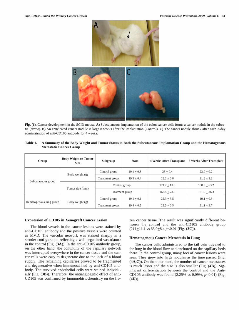
Four weeks after the cultured colon cancer cells were implanted in the subcutis of the back, the tumor developed nodules (Fig. (1A)) and the size increased with time (Fig. (1B)). We compared the tumor size after treatment with anti-CD105. The size of tumor declined from 163.6 mm to 131.6 mm after treatment for 2 weeks with anti-CD105 antibody (Fig. (1C)), and it remained unchanged for 1 week thereafter. The tumor size, however, was unchanged or even gradually regrew until sacrifice despite the same treatment regimen with anti-CD105 antibody as before. Whereas, the tumor size of the control group increased from 171.2+13.6 mm
2 to
180.5+63.2 mm2, the size continued growing up to 4 weeks
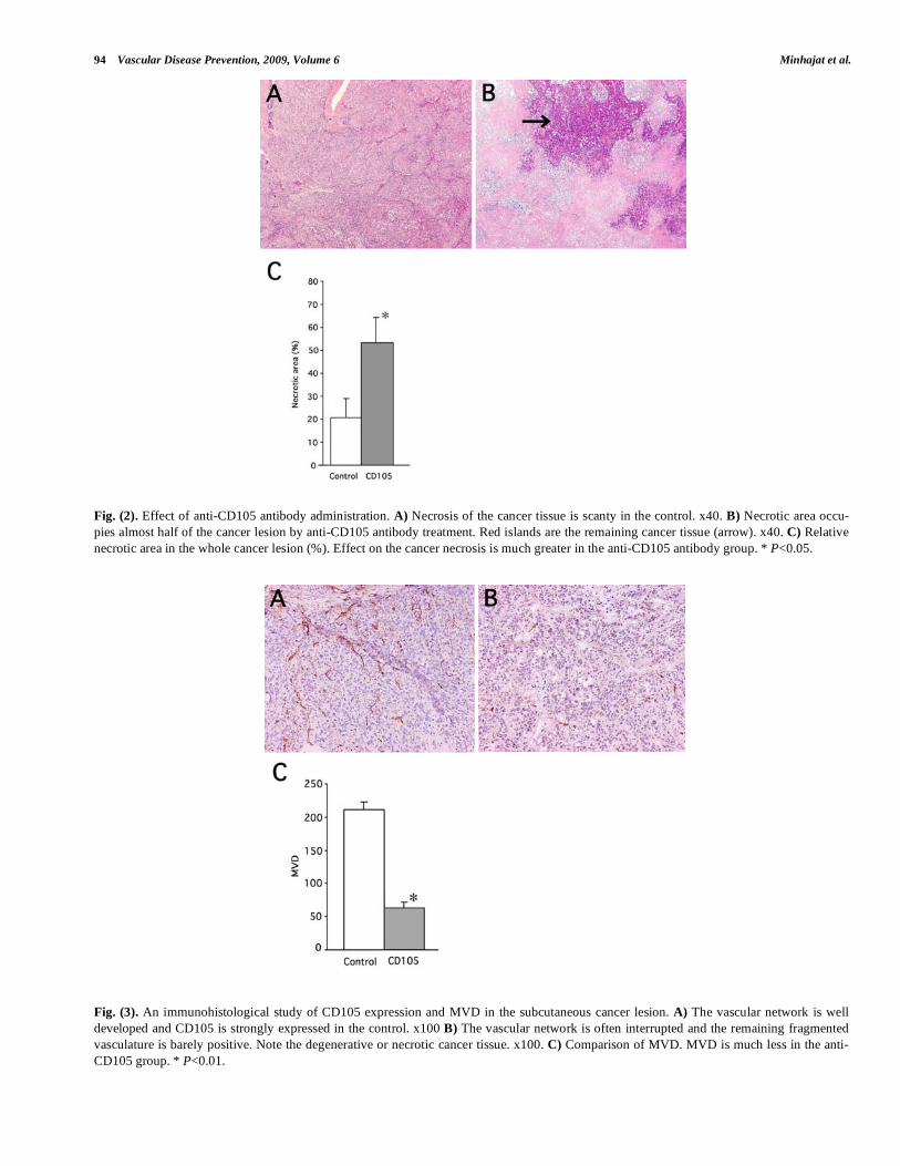
of sacrifice (Fig. (1B)). The body weight of the mice with the s.c. xenograft and the lung metastasis group decreased from an average of 23.2+0.8 and 22.3+3.5 grams to 21.8 +2.8 and 21.1+3.7 grams, respectively, after treatment with Anti-CD105. The body weight was unchanged and even decreased after 4 weeks of implantation (Table 1). This resulted from the cachexia of the individual mouse bearing cancer in the back or lung. The necrotic areas of the colon cancer xenograft are shown in Fig. (2A). Necrosis tended to occur in the center, but the margin of the cancer lesion was well preserved. In the control mouse, the cancer lesion grew in solid and necrosis was hard to see. But as the cancer lesion grew, a patchy distribution of necrotic foci was observed at the end of the experiment. Each focus was smaller than that of the anti-CD105 antibody group (Fig. (2B)). A significant difference between the control and the Anti-CD105 group was found (20.8+8.4% vs. 53.5+11.0%, p<0.05) (Fig. (2C)).
Anti-CD105 Inhibit the Primary Cancer Growth Vascular Disease Prevention, 2009, Volume 6 93
Fig. (1). Cancer development in the SCID mouse. A) Subcutaneous implantation of the colon cancer cells forms a cancer nodule in the subcu-
tis (arrow). B) An enucleated cancer nodule is large 8 weeks after the implantation (Control). C) The cancer nodule shrunk after each 2-day
administration of anti-CD105 antibody for 4 weeks.
Table 1. A Summary of the Body Weight and Tumor Status in Both the Subcutaneous Implantation Group and the Hematogenous
Metastatic Cancer Group
Group Body Weight or Tumor
Size Subgroup Start 4 Weeks After Transplant 8 Weeks After Transplant
Control group 19.1 + 0.3 23 + 0.4 23.0 + 0.2 Body weight (g)
Treatment group 19.3 + 0.4 23.2 + 0.8 21.8 + 2.8
Control group 171.2 + 13.6 180.5 + 63.2
Subcutaneous group
Tumor size (mm) Treatment group 163.5 + 23.0 131.6 + 36.3
Control group 19.1 + 0.1 22.3 + 3.5 19.1 + 0.3 Hematogenous lung group Body weight (g)
Treatment group 19.4 + 0.5 22.3 + 0.5 21.1 + 3.7
Expression of CD105 in Xenograft Cancer Lesion
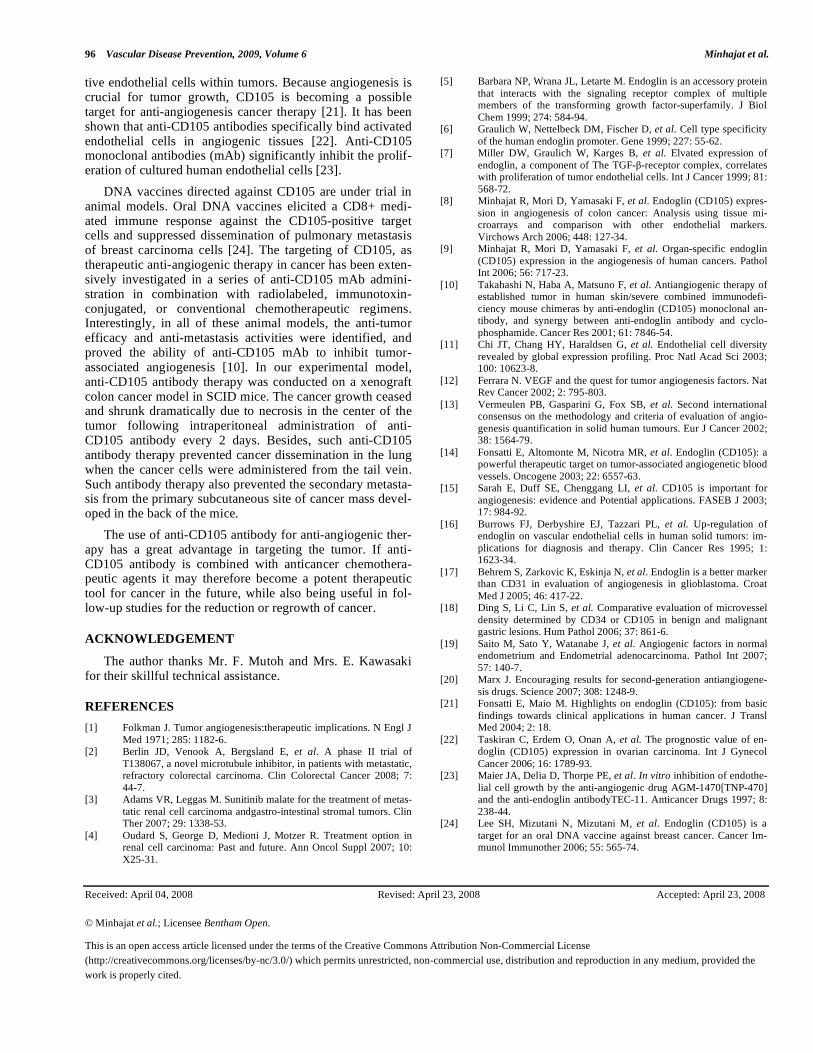
The blood vessels in the cancer lesions were stained by anti-CD105 antibody and the positive vessels were counted as MVD. The vascular network was stained sharply in a slender configuration reflecting a well organized vasculature in the control (Fig. (3A)). In the anti-CD105 antibody group, on the other hand, the continuity of the capillary network was interrupted everywhere in the cancer tissue and the can-cer cells were easy to degenerate due to the lack of a blood supply. The remaining capillaries proved to be fragmented and degenerative when immunostained by anti-CD105 anti-body. The survived endothelial cells were stained individu-ally (Fig. (3B)). Therefore, the antiangiogenic effect of anti-CD105 was confirmed by immunohistochemistry on the fro-
zen cancer tissue. The result was significantly different be-tween the control and the anti-CD105 antibody group (211+11.1 vs 63.0+8.4 p<0.01 (Fig. (3C)).
Hematogenous Cancer Metastasis in Lung
The cancer cells administered to the tail vein traveled to the lung in the blood flow and anchored on the capillary beds there. In the control group, many foci of cancer lesions were seen. They grew into large nodules as the time passed (Fig. (4A,C)). On the other hand, the number of cancer metastases is much lesser and the size is also smaller (Fig. (4B)). Sig-nificant differentiation between the control and the Anti-CD105 antibody was found (2.25% vs 0.09%, p<0.01) (Fig. (4D)).
94 Vascular Disease Prevention, 2009, Volume 6 Minhajat et al.
Fig. (2). Effect of anti-CD105 antibody administration. A) Necrosis of the cancer tissue is scanty in the control. x40. B) Necrotic area occu-
pies almost half of the cancer lesion by anti-CD105 antibody treatment. Red islands are the remaining cancer tissue (arrow). x40. C) Relative
necrotic area in the whole cancer lesion (%). Effect on the cancer necrosis is much greater in the anti-CD105 antibody group. * P<0.05.
Fig. (3). An immunohistological study of CD105 expression and MVD in the subcutaneous cancer lesion. A) The vascular network is well
developed and CD105 is strongly expressed in the control. x100 B) The vascular network is often interrupted and the remaining fragmented
vasculature is barely positive. Note the degenerative or necrotic cancer tissue. x100. C) Comparison of MVD. MVD is much less in the anti-
CD105 group. * P<0.01.

Anti-CD105 Inhibit the Primary Cancer Growth Vascular Disease Prevention, 2009, Volume 6 95
Fig. (4). A comparison of hematogenous metastasis to lung. A) Many foci of metastatic lung cancer are seen in the control group. x40. B) On
the other hand, the number of cancer metastases is much smaller and the size is also smaller. x100. C) Higher magnification of A. Most of the
cancer lesion is large. x200. D) A comparison of the cancer metastasis. The number of cancer foci is much less in the anti-CD105 group.
* P<0.01.
DISCUSSION
Angiogenesis is an important step in the process of can-cer growth [1]. It promotes metastatic spread by providing the means for cells to detach from the primary tumor site, travel in the blood stream, and anchor at the distant metas-tatic site.
The vascular endothelium of tumor and normal tissue is heterogeneous [11]. Recent advances in immunohistological and biochemical technologies have made it possible to detect the heterogeneous endothelium. Among those markers, vas-cular endothelial growth factor (VEGF) is a potent mitogen in angiogenesis. It plays a central role in both normal vascu-lar development and tumor neovascularization [12]. All of the VEGF-A isoforms bind to both VEGFR-1 and VEGFR-2.
von Willebrand Factor (vWF) is the most familiar and reliable marker, and has been used for a long time as a con-ventional endothelial marker, although it is more strongly expressed in larger blood vessels than in capillary endothe-lial cells. vWF is also expressed in bone marrow megakaryo-cytes and platelets.
Other more specific reagents, CD31 and CD34, are now considered to be optimal pan-endothelial markers [13]. Though vWF, CD34, and CD31 are used as pan-endothelial markers, they react not only with newly formed blood ves-sels but also with mature blood vessels. Therefore, the ques-tion arises as to whether these pan-endothelial markers are ideal for the evaluation of tumor angiogenesis.
CD105, a receptor for TGF-b1 and -b-3, modulates TGF-b signaling by interacting with TGF-bRI and/or II, and it is emerging as a prime vascular target of antiangiogenetic can-cer therapy [14,15].
CD105 has been shown to be a proliferation-associated and hypoxia-inducible protein, and is preferentially ex-pressed in the activated endothelial cells participating in neoangiogenesis [6], and is undetectable or weakly ex-pressed in the vessels of normal tissues [8], with a marked tissue specificity [16].
Because the interruption of the blood supply to the tumor results in a reduction of the tumor size, necrosis, or shrink-age of the tumor, CD105 is therefore considered to be a prime vascular target of antiangiogenetic cancer therapy [14].
CD105 has been demonstrated to be a more specific and sensitive marker for tumor angiogenesis than CD31 [17] and CD34 in gastric cancer [18, 19].
Anti-angiogenic drugs have been developed for cancer treatment targeting VEGF, VEGF receptors, and PDGF re-ceptors. Specifically, agents that prevent VEGF-A binding to its receptors and antibodies that directly block VEGFR-2 achieved reduction in tumor sizes and improvement in pro-gression-free survival [20]. However, side effects are also observed to occur because such molecules are universally distributed in the human vascular system.
Based on the recent advances in CD105 studies, it is the most specific and sensitive molecule in activated prolifera-
96 Vascular Disease Prevention, 2009, Volume 6 Minhajat et al.
tive endothelial cells within tumors. Because angiogenesis is crucial for tumor growth, CD105 is becoming a possible target for anti-angiogenesis cancer therapy [21]. It has been shown that anti-CD105 antibodies specifically bind activated endothelial cells in angiogenic tissues [22]. Anti-CD105 monoclonal antibodies (mAb) significantly inhibit the prolif-eration of cultured human endothelial cells [23].
DNA vaccines directed against CD105 are under trial in animal models. Oral DNA vaccines elicited a CD8+ medi-ated immune response against the CD105-positive target cells and suppressed dissemination of pulmonary metastasis of breast carcinoma cells [24]. The targeting of CD105, as therapeutic anti-angiogenic therapy in cancer has been exten-sively investigated in a series of anti-CD105 mAb admini-stration in combination with radiolabeled, immunotoxin-conjugated, or conventional chemotherapeutic regimens. Interestingly, in all of these animal models, the anti-tumor efficacy and anti-metastasis activities were identified, and proved the ability of anti-CD105 mAb to inhibit tumor-associated angiogenesis [10]. In our experimental model, anti-CD105 antibody therapy was conducted on a xenograft colon cancer model in SCID mice. The cancer growth ceased and shrunk dramatically due to necrosis in the center of the tumor following intraperitoneal administration of anti-CD105 antibody every 2 days. Besides, such anti-CD105 antibody therapy prevented cancer dissemination in the lung when the cancer cells were administered from the tail vein. Such antibody therapy also prevented the secondary metasta-sis from the primary subcutaneous site of cancer mass devel-oped in the back of the mice.
The use of anti-CD105 antibody for anti-angiogenic ther-apy has a great advantage in targeting the tumor. If anti-CD105 antibody is combined with anticancer chemothera-peutic agents it may therefore become a potent therapeutic tool for cancer in the future, while also being useful in fol-low-up studies for the reduction or regrowth of cancer.
ACKNOWLEDGEMENT
The author thanks Mr. F. Mutoh and Mrs. E. Kawasaki for their skillful technical assistance.
REFERENCES
[1] Folkman J. Tumor angiogenesis:therapeutic implications. N Engl J
Med 1971; 285: 1182-6. [2] Berlin JD, Venook A, Bergsland E, et al. A phase II trial of
T138067, a novel microtubule inhibitor, in patients with metastatic, refractory colorectal carcinoma. Clin Colorectal Cancer 2008; 7:
44-7. [3] Adams VR, Leggas M. Sunitinib malate for the treatment of metas-
tatic renal cell carcinoma andgastro-intestinal stromal tumors. Clin Ther 2007; 29: 1338-53.
[4] Oudard S, George D, Medioni J, Motzer R. Treatment option in renal cell carcinoma: Past and future. Ann Oncol Suppl 2007; 10:
X25-31.
[5] Barbara NP, Wrana JL, Letarte M. Endoglin is an accessory protein
that interacts with the signaling receptor complex of multiple members of the transforming growth factor-superfamily. J Biol
Chem 1999; 274: 584-94. [6] Graulich W, Nettelbeck DM, Fischer D, et al. Cell type specificity
of the human endoglin promoter. Gene 1999; 227: 55-62. [7] Miller DW, Graulich W, Karges B, et al. Elvated expression of
endoglin, a component of The TGF- -receptor complex, correlates with proliferation of tumor endothelial cells. Int J Cancer 1999; 81:
568-72. [8] Minhajat R, Mori D, Yamasaki F, et al. Endoglin (CD105) expres-
sion in angiogenesis of colon cancer: Analysis using tissue mi-croarrays and comparison with other endothelial markers.
Virchows Arch 2006; 448: 127-34. [9] Minhajat R, Mori D, Yamasaki F, et al. Organ-specific endoglin
(CD105) expression in the angiogenesis of human cancers. Pathol Int 2006; 56: 717-23.
[10] Takahashi N, Haba A, Matsuno F, et al. Antiangiogenic therapy of established tumor in human skin/severe combined immunodefi-
ciency mouse chimeras by anti-endoglin (CD105) monoclonal an-tibody, and synergy between anti-endoglin antibody and cyclo-
phosphamide. Cancer Res 2001; 61: 7846-54. [11] Chi JT, Chang HY, Haraldsen G, et al. Endothelial cell diversity
revealed by global expression profiling. Proc Natl Acad Sci 2003; 100: 10623-8.
[12] Ferrara N. VEGF and the quest for tumor angiogenesis factors. Nat Rev Cancer 2002; 2: 795-803.
[13] Vermeulen PB, Gasparini G, Fox SB, et al. Second international consensus on the methodology and criteria of evaluation of angio-
genesis quantification in solid human tumours. Eur J Cancer 2002; 38: 1564-79.
[14] Fonsatti E, Altomonte M, Nicotra MR, et al. Endoglin (CD105): a powerful therapeutic target on tumor-associated angiogenetic blood
vessels. Oncogene 2003; 22: 6557-63. [15] Sarah E, Duff SE, Chenggang LI, et al. CD105 is important for
angiogenesis: evidence and Potential applications. FASEB J 2003; 17: 984-92.
[16] Burrows FJ, Derbyshire EJ, Tazzari PL, et al. Up-regulation of endoglin on vascular endothelial cells in human solid tumors: im-
plications for diagnosis and therapy. Clin Cancer Res 1995; 1: 1623-34.
[17] Behrem S, Zarkovic K, Eskinja N, et al. Endoglin is a better marker than CD31 in evaluation of angiogenesis in glioblastoma. Croat
Med J 2005; 46: 417-22. [18] Ding S, Li C, Lin S, et al. Comparative evaluation of microvessel
density determined by CD34 or CD105 in benign and malignant gastric lesions. Hum Pathol 2006; 37: 861-6.
[19] Saito M, Sato Y, Watanabe J, et al. Angiogenic factors in normal endometrium and Endometrial adenocarcinoma. Pathol Int 2007;
57: 140-7. [20] Marx J. Encouraging results for second-generation antiangiogene-
sis drugs. Science 2007; 308: 1248-9. [21] Fonsatti E, Maio M. Highlights on endoglin (CD105): from basic
findings towards clinical applications in human cancer. J Transl Med 2004; 2: 18.
[22] Taskiran C, Erdem O, Onan A, et al. The prognostic value of en-doglin (CD105) expression in ovarian carcinoma. Int J Gynecol
Cancer 2006; 16: 1789-93. [23] Maier JA, Delia D, Thorpe PE, et al. In vitro inhibition of endothe-
lial cell growth by the anti-angiogenic drug AGM-1470[TNP-470] and the anti-endoglin antibodyTEC-11. Anticancer Drugs 1997; 8:
238-44. [24] Lee SH, Mizutani N, Mizutani M, et al. Endoglin (CD105) is a
target for an oral DNA vaccine against breast cancer. Cancer Im-munol Immunother 2006; 55: 565-74.
Received: April 04, 2008 Revised: April 23, 2008 Accepted: April 23, 2008
© Minhajat et al.; Licensee Bentham Open.
This is an open access article licensed under the terms of the Creative Commons Attribution Non-Commercial License
(http://creativecommons.org/licenses/by-nc/3.0/) which permits unrestricted, non-commercial use, distribution and reproduction in any medium, provided the
work is properly cited.