oocytes and early embryos of xenopus laevis contain...
TRANSCRIPT
J. Embryol. exp. Morph. 83, 169-187 (1984)Printed in Great Britain © The Company of Biologists Limited 1984
Oocytes and early embryos of Xenopus laeviscontain intermediate filaments which react with
anti-mammalian vimentin antibodies
By SUSAN F. GODSAVE, BRIAN H. ANDERTON,JANET HEASMAN AND CHRISTOPHER C. WYLIE*
Departments of Anatomy and Immunology, St. George's Hospital MedicalSchool, Cranmer Terrace, Tooting, London, SW17 ORE, U.K.
SUMMARY
Previous studies have shown that Xenopus oocytes possess a cortical shell, which includesactin-containing microfilaments and cytokeratin-containing intermediate filaments. In thispaper we show that oocytes of Xenopus laevis also contain filaments throughout theircytoplasm which are stained by several anti-vimentin antibody preparations. We also showthat dramatic changes in pattern of these filaments occur during oocyte differentiation, firstduring vitellogenesis, and then during maturation of the oocyte to form an egg.
INTRODUCTION
It is now becoming clear from immunocytochemical data that intermediatefilament proteins collectively represent a large and complex multigene family,only certain members of which appear in different cell lineages (Anderton, 1981;Lazarides, 1980,1982; Osborn & Weber, 1982). It is therefore of importance tostudy the ontogeny of intermediate filament proteins, in particular the spatialand temporal sequence of appearance of the five protein classes in early em-bryos. A certain amount of data have appeared recently in this respect, prin-cipally from mammalian and avian species.
In mouse embryos, two cytokeratin polypeptides have been found in theearliest cleavage stages (Lehtonen, Lehto, Paasivuo & Virtanen, 1983a) as wellas at later blastula stages (Jackson et al. 1980,1981; Paulin, Babinet, Weber &Osborn, 1980; Brulet, Babinet, Kemler & Jacob, 1980; Kemler et al. 1981).These proteins are followed by vimentin which appears in the primary mesen-chyme cells during gastrulation, and then desmin is found in the embryo at a laterstage (Ffanke et al. 1982a, 19826). In the developing nervous system, vimentinis found in both presumptive glial and neural cells, and the tissue-specificneurofilament proteins and glial fibrillary acidic protein (GFAP) appear later indevelopment (Schnitzer, Franke & Schachner, 1981; Raju, Bignami & Dahl,1981; Bignami, Raju & Dahl, 1982).
* To whom reprint requests should be addressed.

170 S. F. GODSAVE AND OTHERS
In chick embryos, studies of intermediate filament proteins have been largelyconfined to neurogenesis and myogenesis. Here the pattern of expression issimilar to that found in mammalian embryos. Vimentin is found before eitherneurofilament proteins or GFAP in the developing nervous system, but is gradu-ally replaced by these proteins in the neuron and glial lineages respectively(Tapscott etal. 1981; Holtzer etal. 1982; Bignami & Dahl, 1975; Bignami, Dahl& Seiler, 1980). Vimentin also precedes desmin expression in chick myogenesis(Bennett, Fellini, Toyama & Holtzer, 1979; Holtzer et al. 1982; Lazarides et al.1982; Gard & Lazarides, 1980).
For some time it was thought that differentiating gametes (i.e. the germ line)did not contain intermediate filaments. However, recent reports from severallaboratories have now demonstrated the presence of cytokeratin-containingintermediate filaments in both mouse (Lehtonen et al. 19836) and frog (Gall,Picheral & Gounon, 1983; Franz et al. 1983) oocytes. It is therefore of greatinterest to determine whether other intermediate filament proteins are present,whether they are stored for use during early postfertilization development,whether they play a role in oocyte function, and the temporal and spatial patternby which they are replaced by embryonic lineage-specific intermediate filamentproteins.
The female frog germ line is an attractive system to study in this respect. Theovary contains many hundreds of oocytes, at all stages of oogenesis, representinga very large tissue mass for routine biochemistry. The events of oogenesis arewell-characterized biochemically (Gurdon, 1974; De Robertis, Zeller, Carrasco&Mattaj, 1983; Woodland etal. 1983). Furthermore, the fully grown frog oocyteis a single very large cell (1-2-1-4 mm in diameter) with a complex and charac-teristic cytoarchitecture (Wischnitzer, 1966; Dumont, 1972; Mohun, Lane, Col-man & Wylie, 1981). This can be disrupted by cytoskeleton-disrupting agents,which also cause assay able biochemical effects on protein translation, secretionand turnover (Colman etal. 1981). Frog oocytes therefore represent a potentiallyuseful model system in which to study the role of intermediate filaments in themaintenance of cytoarchitecture and other aspects of cell physiology. We reporthere that oocytes of Xenopus laevis contain intermediate filaments from earlyprevitellogenic stages to the fully grown state. These are identifiable by electronmicroscopy, immunoblotting and immunofluorescence cytochemistry. We showthat vimentin is a major intermediate filament protein in these cells and demon-strate that the differentiation of the oocyte and its maturation to form the egg areaccompanied by major changes, in the pattern of vimentin-containing filaments.
MATERIALS AND METHODS
Preparation for electron microscopy
Pieces of ovary were excised from adult Xenopus laevis in normal saline andfixed in 1 % w/v glutaraldehyde in 0-lM-phosphate buffer for 3h. In order to

Vimentin in Xenopus oocytes and embryos 111
improve the fixation of cytoskeletal elements, 0-5mg/ml saponin and 2mg/mltannic acid were added to the fixative after the method of Maupin & Pollard(1983). Stage-4 and -5 oocytes were cut in half to allow complete penetration.After washing in O-lM-phosphate buffer, samples were postfixed in 1 % w/vOsO4 in 0-1 M-phosphate buffer for 2h. They were then dehydrated through anethanol series and embedded in Araldite. Blocks were sectioned on a SorvallUltramicrotome, stained with aqueous uranyl acetate and lead citrate and silversections were viewed in a Philips 301 electron microscope.
Immunocytochemistry
Adult Xenopus laevis ovaries were dissected into small fragments in Gurdon-modified Barth's solution (Barth-x, Gurdon, 1968). They were then fixed in 2 %w/v trichloroacetic acid, transferred to absolute ethanol and then topolyethyleneglycol 400 distearate wax (BDH) containing 1 % w/v cetyl alcohol,in which they were embedded (Dr P. Hausen, personal communication). Sec-tions were cut at 16-18 °C, mounted on glass slides and stored desiccated. Sec-tions were dewaxed in an acetone series and treated with the following sequenceof solutions: 3M-urea (lmin), phosphate-buffered saline (PBS) wash(2x5min), a blocking buffer consisting of PBS containing 1% w/v BSA(10 min), primary antibody solution (20 min), three washes in PBS plus 1 % BSA(5 min each), second antibody solution: a 1:100 dilution of fluorescein-coupledgoat anti-rabbit immunoglobulin (Nordic, 20min), PBS (2x5min washes);0-004 % w/v eryochrome black (5 min) and finally one further wash with PBS.Stained sections were mounted in an aqueous u.v.-free mounting medium (DifcoLtd.) and examined under a Zeiss photomicroscope III using epifluorescence.Three different anti-vimentin antibody preparations were used by this methodto stain Xenopus oocytes: a) a rabbit antiserum raised against hamster vimentin(kindly donated by Dr R. O. Hynes, See Hynes & Destree, 1978, for details),a purified IgG preparation from this antiserum was used in some of the studiesreported here, b) a rabbit antiserum raised against calf lens vimentin (kindlydonated by Dr F. Ramaekers, See Ramaekers et al. 1982), c) a monoclonalantibody raised against human vimentin (donated by Dr I. Virtanen, See Vir-tanen et al. 1981). All gave essentially the same staining pattern.
Preparation of triton-insoluble proteins from 3T3 cells and rat brain
Triton-insoluble proteins were prepared by the methods described by Pruss etal. (1981) using lmM-phenylmethylsulphonylfluoride and 1 mM-p-chloro-mercuribenzoate to inhibit proteolysis.
Production of eggs and embryos
Synchronously developing batches of embryos were produced by artificialfertilization of eggs as described by Gurdon & Woodland (1975). Embryos were

172 S. F. GODSAVE AND OTHERS
classified by developmental stage as described by Nieuwkoop & Faber (1956).Unfertilized eggs were collected from females prior to addition of sperm.
Preparation of defolliculated oocytes
Follicle cells were removed from the surface of oocytes by collagenase treat-ment. The efficiency of this procedure was monitored by scanning electronmicroscopy (see Mohun et al. 1981 for full description).
Immunoblotting
Cellular proteins were separated electrophoretically in 10% w/v SDS-polyacrylamide slab gels, transferred onto nitrocellulose sheets and stained withantibodies as described previously (Pruss et al. 1981). Anti-IFA was used at a1:10 dilution and was detected using a 125I-labelled rabbit anti-(mouse Ig) anti-body. The rabbit anti-vimentin antisera were used at 1:100 dilution and the anti-hamster vimentin IgG preparation was used at a concentration of 12/ig/ml.Rabbit antibody binding was detected using 125I-labelled goat anti-(rabbit Ig)antibody.
RESULTS
1. Identification of intermediate filaments in oocytes by electron microscopy
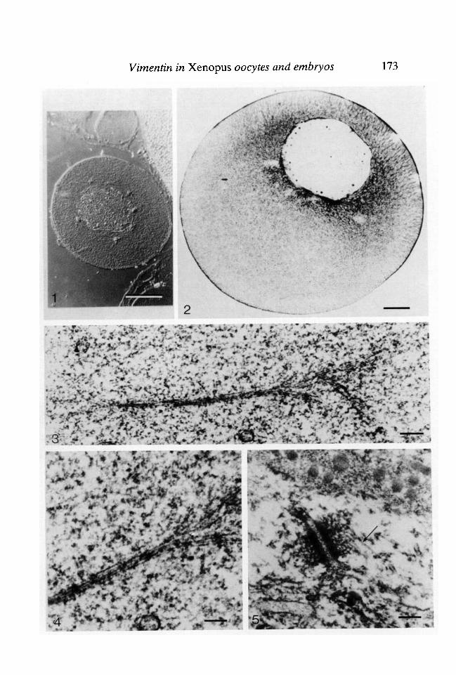
The adult Xenopus ovary contains oocytes of many different developmentalstages ranging from small previtellogenic cells to full-grown oocytes(approximately 1-2-1-4mm diameter). This enormous increase in volume takessome weeks and is largely due to the appearance and storage of yolk platelets(vitellogenesis), although other organelles, e.g. the nucleus, also increase in size.The general morphology of the previtellogenic oocyte is seen in Fig. 1. Thenucleus is more or less central and the cytoplasm is homogeneous, with theexception of large phase-dense perinuclear masses which are particularly striking
Fig. 1. Low-power view of previtellogenic oocyte. Oocyte from 1 pim thick sectionthrough polyester-wax-embedded ovary viewed with Nomarski optics. Bar 100 (im.Fig. 2. Low-power view of stage-6 oocyte. Vertical section, 7[im thick, throughpolyester-wax-embedded stage-6 oocyte, stained with 0-1 % toluidine blue in 0-1 %borax and viewed with bright-field optics. Bar 100/im.Fig. 3. Electron micrograph showing a branching bundle of intermediate filamentsin a previtellogenic oocyte. Bar 0-2 jitm.Fig. 4. High-power electron micrograph of intermediate-sized filaments in aprevitellogenic oocyte. Micrograph used for measurement of filament size. In-dividual filaments measure 15 nm in diameter. Bar 0-Fig. 5. Electron micrograph of intermediate filaments in follicle cells. The diametersof intermediate-sized filaments from oocytes were compared with intermediate-sizedfilaments (arrow) from follicle cells surrounding oocytes. Intermediate filamentsfrom both cell types measured 15 nm diameter. Bar 0
Vimentin in Xenopus oocytes and embryos 173
174 S. F. GODSAVE AND OTHERS
in interference images. These are connected to the mitochondrial cloud, acharacteristic feature of early germ-line cells (Billett, 1979). By the end ofvitellogenesis, the oocyte has an elaborate cytoarchitecture (Fig. 2). The animalhemisphere, which contains the nucleus, is characterized by a layer of pigmentimmediately beneath the surface membrane and by the presence of small yolkplatelets. These are arranged in large islands passing radially from just beneaththe cell surface inwards towards the nucleus. Between these islands are yolk-freetracts of 'active' cytoplasm i.e. endoplasmic reticulum, Golgi, mitochondria andother non-yolk constituents. The vegetal pole is not so obviously organized andconsists largely of a mass of yolk platelets larger than those in the animal pole(the 'vegetal yolk mass').
Examination of detergent-extracted and tannic-acid-treated previtellogenicoocytes at the electron microscopic level shows the presence of intermediatefilament bundles crossing the cytoplasm in various directions (Fig. 3). Thesebundles are of variable size and are sometimes seen to split into smaller struc-tures following divergent paths. Each filament of a bundle measuresapproximately 15 nm in diameter in saponin-/tannic-acid-fixed material (Fig. 4).These figures are in agreement with those of Maupin & Pollard (1983) for inter-mediate filament sizes. In order to compare the size of oocyte intermediatefilaments with that of known Xenopus intermediate filaments, photographs weretaken, at the same magnification, of desmosome-associated filaments foundbetween follicle cells surrounding the oocyte (Fig. 5). The intermediate fila-ments seen associated with these desmosomes are of the same diameter as thoseseen in the oocyte cytoplasm.
Examination of sections of previtellogenic oocytes by light microscopy showsthem to contain characteristic phase-dense masses in the cytoplasm arrangedaround the nucleus (Fig. 1). When these oocytes are fixed with glutaraldehydein the presence of saponin and tannic acid, and examined under the electronmicroscope the masses are found to contain mitochondria, microtubules, nuage(a generic name given to undefined fibrillogranular cytoplasmic masses in germ-line cells), and intermediate filament bundles (Figs 6 & 7).
In vitellogenic stages, the cytoplasm becomes much more crowded with or-ganelles and cytoskeletal elements are difficult to see. However, in cortical areasof full-grown oocytes, intermediate filaments are visible in close association withmitochondria (Fig. 8), although their organization cannot be deduced byelectron microscopy.
2. Identification of intermediate filament proteins in oocytes and embryos byimmunob lotting
The ontogeny of intermediate filament polypeptides from oocytes through toswimming tadpoles was followed by immunoblotting using a monoclonal anti-intermediate filament antibody (anti-IFA, Pruss etal. 1981). This antibody hasbeen shown to cross react with most (possibly all) intermediate filament

Vimentin in Xenopus oocytes and embryos 175
$ 'Fig. 6. Low-power electron micrograph of a mitochondrial cloud in a previtellogenicoocyte. Intermediate-sized filaments (arrow) can be seen in this mitochondria (m)-rich region. Bar 0-5 jum.Fig. 7. High-power electron micrograph of part of a mitochondrial cloud. An inter-mediate filament bundle is indicated (arrow). Bar 01 //m.Fig. 8. Intermediate filaments in a vitellogenic oocyte. Intermediate filament bundle(arrow) in a yolk free area of the animal pole of a full-grown oocyte. Many mitochon-dria (m) are also present in this area. Bar 0-5 jum.
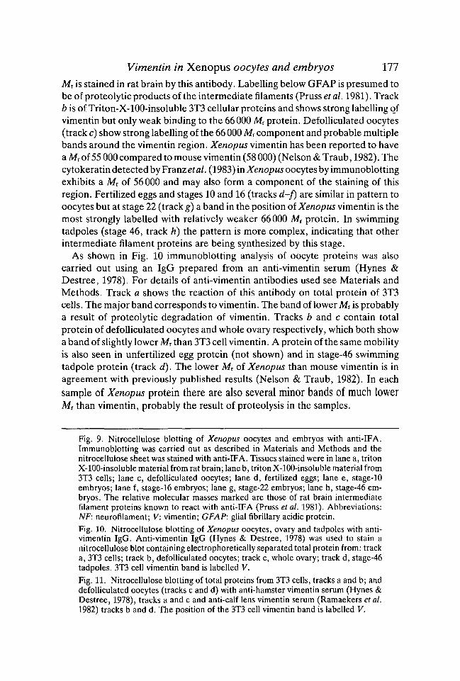
polypeptides from a wide variety of cell types and species. It therefore representsa useful initial probe with which to study Xenopus oocytes and embryos to seeif any recognizable intermediate filament polypeptides are present. The varioussamples were run on a single SDS-PAGE slab gel and transferred tonitrocellulose and then processed with antibodies (Fig. 9). Track A illustrates thepattern of reactivity of anti-IFA on Triton-X-100-insoluble protein from ratbrain. This tissue is enriched in GFAP, neuron"laments and vimentin. These areall labelled. Anti-IFA has been found to stain a protein of 66000 relativemolecular mass (Mr) in a number of tissues (Pruss et al. 1981) and a band of this

176
MrX10"3
NF200
NF 150
S. F. GODSAVE AND OTHERS
MrX10,-3
NF6866
V 58GFAP50
t
V58
9 • • 10a b c d e f g h a b e d
V-< 1
11a b e d
Vimentin in Xenopus oocytes and embryos 111
Mr is stained in rat brain by this antibody. Labelling below GFAP is presumed tobe of proteolytic products of the intermediate filaments (Pruss et al. 1981). Trackb is of Triton-X-100-insoluble 3T3 cellular proteins and shows strong labelling ofvimentin but only weak binding to the 66000 Mr protein. Defolliculated oocytes(track c) show strong labelling of the 66 000 Mr component and probable multiplebands around the vimentin region. Xenopus vimentin has been reported to havea Mr of 55 000 compared to mouse vimentin (58 000) (Nelson & Traub, 1982). Thecytokeratin detected by Franz etal. (1983) in Xenopus oocytes by immunoblottingexhibits a Mr of 56000 and may also form a component of the staining of thisregion. Fertilized eggs and stages 10 and 16 (tracks d-f) are similar in pattern tooocytes but at stage 22 (track g) a band in the position of Xenopus vimentin is themost strongly labelled with relatively weaker 66000 Mr protein. In swimmingtadpoles (stage 46, track h) the pattern is more complex, indicating that otherintermediate filament proteins are being synthesized by this stage.
As shown in Fig. 10 immunoblotting analysis of oocyte proteins was alsocarried out using an IgG prepared from an anti-vimentin serum (Hynes &Destree, 1978). For details of anti-vimentin antibodies used see Materials andMethods. Track a shows the reaction of this antibody on total protein of 3T3cells. The major band corresponds to vimentin. The band of lower Mr is probablya result of proteolytic degradation of vimentin. Tracks b and c contain totalprotein of defolliculated oocytes and whole ovary respectively, which both showa band of slightly lower Mr than 3T3 cell vimentin. A protein of the same mobilityis also seen in unfertilized egg protein (not shown) and in stage-46 swimmingtadpole protein (track d). The lower Mr of Xenopus than mouse vimentin is inagreement with previously published results (Nelson & Traub, 1982). In eachsample of Xenopus protein there are also several minor bands of much lowerMr than vimentin, probably the result of proteolysis in the samples.
Fig. 9. Nitrocellulose blotting of Xenopus oocytes and embryos with anti-IFA.Immunoblotting was carried out as described in Materials and Methods and thenitrocellulose sheet was stained with anti-IFA. Tissues stained were in lane a, tritonX-100-insoluble material from rat brain; lane b, triton X-100-insoluble material from3T3 cells; lane c, defolliculated oocytes; lane d, fertilized eggs; lane e, stage-10embryos; lane f, stage-16 embryos; lane g, stage-22 embryos; lane h, stage-46 em-bryos. The relative molecular masses marked are those of rat brain intermediatefilament proteins known to react with anti-IFA (Pruss et al. 1981). Abbreviations:NF: neurofilament; V: vimentin; GFAP: glial fibrillary acidic protein.Fig. 10. Nitrocellulose blotting of Xenopus oocytes, ovary and tadpoles with anti-vimentin IgG. Anti-vimentin IgG (Hynes & Destree, 1978) was used to stain anitrocellulose blot containing electrophoretically separated total protein from: tracka, 3T3 cells; track b, defolliculated oocytes; track c, whole ovary; track d, stage-46tadpoles. 3T3 cell vimentin band is labelled V.
Fig. 11. Nitrocellulose blotting of total proteins from 3T3 cells, tracks a and b; anddefolliculated oocytes (tracks c and d) with anti-hamster vimentin serum (Hynes &Destree, 1978), tracks a and c and anti-calf lens vimentin serum (Ramaekers et al.1982) tracks b and d. The position of the 3T3 cell vimentin band is labelled V.
178 S. F. GODSAVE AND OTHERS
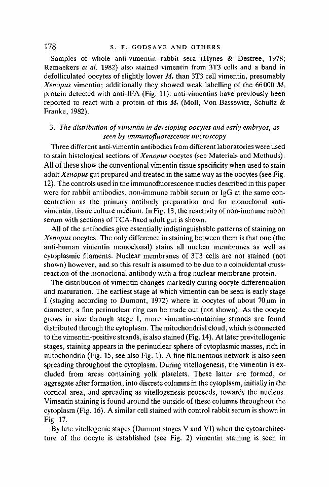
Samples of whole anti-vimentin rabbit sera (Hynes & Destree, 1978;Ramaekers et al. 1982) also stained vimentin from 3T3 cells and a band indefolliculated oocytes of slightly lower Mr than 3T3 cell vimentin, presumablyXenopus vimentin; additionally they showed weak labelling of the 66000 MT
protein detected with anti-IFA (Fig. 11): anti-vimentins have previously beenreported to react with a protein of this MT (Moll, Von Bassewitz, Schultz &Franke, 1982).
3. The distribution of vimentin in developing oocytes and early embryos, asseen by immunofluorescence microscopy
Three different anti-vimentin antibodies from different laboratories were usedto stain histological sections of Xenopus oocytes (see Materials and Methods).All of these show the conventional vimentin tissue specificity when used to stainadult Xenopus gut prepared and treated in the same way as the oocytes (see Fig.12). The controls used in the immunofluorescence studies described in this paperwere for rabbit antibodies, non-immune rabbit serum or IgG at the same con-centration as the primary antibody preparation and for monoclonal anti-vimentin, tissue culture medium. In Fig. 13, the reactivity of non-immune rabbitserum with sections of TCA-fixed adult gut is shown.
All of the antibodies give essentially indistinguishable patterns of staining onXenopus oocytes. The only difference in staining between them is that one (theanti-human vimentin monoclonal) stains all nuclear membranes as well ascytoplasmic filaments. Nuclear membranes of 3T3 cells are not stained (notshown) however, and so this result is assumed to be due to a coincidental cross-reaction of the monoclonal antibody with a frog nuclear membrane protein.
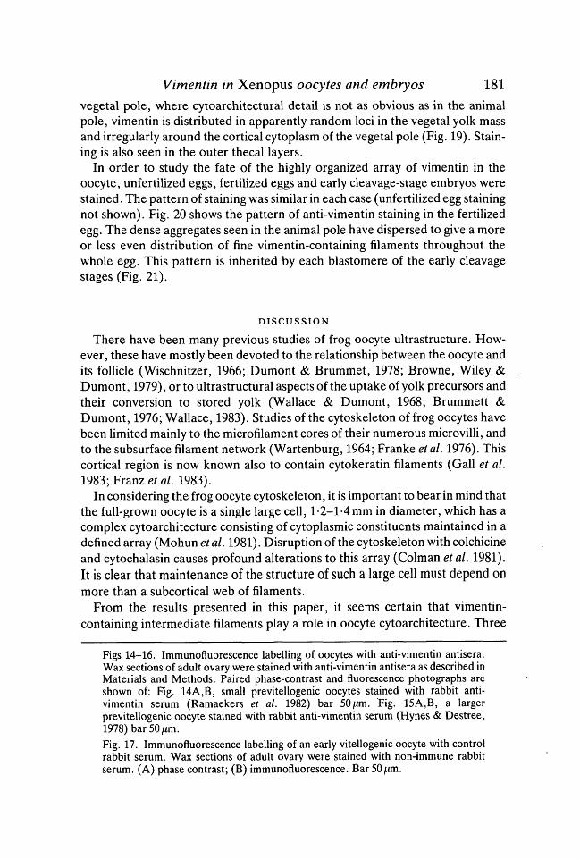
The distribution of vimentin changes markedly during oocyte differentiationand maturation. The earliest stage at which vimentin can be seen is early stageI (staging according to Dumont, 1972) where in oocytes of about 70jum indiameter, a fine perinuclear ring can be made out (not shown). As the oocytegrows in size through stage I, more vimentin-containing strands are founddistributed through the cytoplasm. The mitochondrial cloud, which is connectedto the vimentin-positive strands, is also stained (Fig. 14). At later previtellogenicstages, staining appears in the perinuclear sphere of cytoplasmic masses, rich inmitochondria (Fig. 15, see also Fig. 1). A fine filamentous network is also seenspreading throughout the cytoplasm. During vitellogenesis, the vimentin is ex-cluded from areas containing yolk platelets. These latter are formed, oraggregate after formation, into discrete columns in the cytoplasm, initially in thecortical area, and spreading as vitellogenesis proceeds, towards the nucleus.Vimentin staining is found around the outside of these columns throughout thecytoplasm (Fig. 16). A similar cell stained with control rabbit serum is shown inFig. 17.
By late vitellogenic stages (Dumont stages V and VI) when the cytoarchitec-ture of the oocyte is established (see Fig. 2) vimentin staining is seen in

Vimentin in Xenopus oocytes and embryos 179
/
. '
13BFig. 12. Immunofluorescence labelling of adult Xenopus gut with anti-vimentinSerum. Wax sections were Stained with rabbit anti-vimentin antiserum (Ramaekerset al. 1982) as described in Materials and Methods. Paired phase contrast (A) andfluorescence (B) photographs are shown. The epithelium is marked (e). Only theunderlying connective tissue is stained. Bar 50//m.Fig. 13. Immunofluorescence labelling of adult Xenopus gut with control serum.Wax sections of gut were stained with non-immune rabbit serum. (A) phase contrast(B) immunofluorescence. Bar 50/mi.
asymmetric fashion between animal and vegetal poles. In the animal pole (Fig.18) vimentin is confined to the yolk-free areas of cytoplasm which divide the yolkplatelets into columns. The thick strands of vimentin-containing cytoplasm donot extend right to the animal pole surface. A cortical layer containing pigmentcan be seen separating them from the surface membrane of the oocyte. In the

180 S. F. GODSAVE AND OTHERS
14A
i
15A 15 B
Si . - ,
•16A 16B
17A 17 B
Vimentin in Xenopus oocytes and embryos 181
vegetal pole, where cytoarchitectural detail is not as obvious as in the animalpole, vimentin is distributed in apparently random loci in the vegetal yolk massand irregularly around the cortical cytoplasm of the vegetal pole (Fig. 19). Stain-ing is also seen in the outer thecal layers.
In order to study the fate of the highly organized array of vimentin in theoocyte, unfertilized eggs, fertilized eggs and early cleavage-stage embryos werestained. The pattern of staining was similar in each case (unfertilized egg stainingnot shown). Fig. 20 shows the pattern of anti-vimentin staining in the fertilizedegg. The dense aggregates seen in the animal pole have dispersed to give a moreor less even distribution of fine vimentin-containing filaments throughout thewhole egg. This pattern is inherited by each blastomere of the early cleavagestages (Fig. 21).
DISCUSSION
There have been many previous studies of frog oocyte ultrastructure. How-ever, these have mostly been devoted to the relationship between the oocyte andits follicle (Wischnitzer, 1966; Dumont & Brummet, 1978; Browne, Wiley &Dumont, 1979), or to ultrastructural aspects of the uptake of yolk precursors andtheir conversion to stored yolk (Wallace & Dumont, 1968; Brummett &Dumont, 1976; Wallace, 1983). Studies of the cytoskeleton of frog oocytes havebeen limited mainly to the microfilament cores of their numerous microvilli, andto the subsurface filament network (Wartenburg, 1964; Franke etal. 1976). Thiscortical region is now known also to contain cytokeratin filaments (Gall et al.1983; Franz et al. 1983).
In considering the frog oocyte cytoskeleton, it is important to bear in mind thatthe full-grown oocyte is a single large cell, 1-2-1-4 mm in diameter, which has acomplex cytoarchitecture consisting of cytoplasmic constituents maintained in adefined array (Mohun etal. 1981). Disruption of the cytoskeleton with colchicineand cytochalasin causes profound alterations to this array (Colman etal. 1981).It is clear that maintenance of the structure of such a large cell must depend onmore than a subcortical web of filaments.
From the results presented in this paper, it seems certain that vimentin-containing intermediate filaments play a role in oocyte cytoarchitecture. Three
Figs 14-16. Immunofluorescence labelling of oocytes with anti-vimentin antisera.Wax sections of adult ovary were stained with anti-vimentin antisera as described inMaterials and Methods. Paired phase-contrast and fluorescence photographs areshown of: Fig. 14A,B, small previtellogenic oocytes stained with rabbit anti-vimentin serum (Ramaekers et al. 1982) bar 50/an. Fig. 15A,B, a largerprevitellogenic oocyte stained with rabbit anti-vimentin serum (Hynes & Destree,1978) bar 50 ̂ m.Fig. 17. Immunofluorescence labelling of an early vitellogenic oocyte with controlrabbit serum. Wax sections of adult ovary were stained with non-immune rabbitserum. (A) phase contrast; (B) immunofluorescence. Bar 50jUm.

182 S. F. GODSAVE AND OTHERS
18A 18B
* . * • , % # •
• • ' > ' - ; : ' • / .
19A 19B
20B
1A ;21B

Vimentin in Xenopus oocytes and embryos 183well-characterized anti-vimentin antibody preparations (Hynes & Destree,1978; Ramaekers etal. 1982; Virtanen etal. 1981) were used to demonstrate thepresence of this intermediate filament protein in oocytes. Also, anti-IFA stainedmaterial of the MT of Xenopus vimentin on Western blots. In addition, it wasfound to bind to a 66000 Mr protein; probably another important intermediatefilament-associated protein in oocytes (Pachter, Moraru & Liem, 1983). Manycell types contain a protein of Mr 66 000 which cross reacts with anti-IFA, thoughits role remains unknown.
In the previtellogenic oocyte, vimentin is found distributed throughout thecytoplasm as a network of filaments. The majority is associated with largecytoplasmic masses surrounding the nucleus. One interesting point to note is thatother authors (Franz et al. 1983) have not seen staining of the oocyte sectionswith different anti-vimentin-antisera. The explanation of this probably lies in themethods used. In the work cited above, unfixed tissue was used. In our studieswe found only a small degree of staining with anti-vimentin in unfixed tissue orafter fixation in aldehyde or with absolute ethanol. The very bright stainingshown in the results section here was found after fixation with TCA, a strongdenaturing agent. The continued tissue specificity seen with all the antibodiesused with this fixation method, as well as the blotting data, leave little doubt thatvimentin is present in oocytes. However, the requirement for a particular fixa-tion method is puzzling. One possible explanation is that the determinants withwhich the anti-vimentins cross react are masked in some way in Xenopus oocytes,and only exposed by the denaturation of themselves, or adjacent molecules.Masking of antigenic determinants has previously been suggested as an explana-tion for unexpected patterns of reactivity of several anti-intermediate filamentprotein antibodies (Woodcock-Mitchell, Eichner, Nelson & Sun, 1982;Lazarides et al. 1982) and it has been demonstrated that antigen masking is thecause of an anti-cytokeratin monoclonal antibody staining only at certain stagesof the cell cycle in PtK2 cells (Franke et al. 1983).
We cannot at the moment exclude the possibility that some of the immuno-fluorescence staining of oocytes seen is due to the 66 x 103 Mr protein recognizedby a-IFA. However, this polypeptide is not recognized on immunoblots by the
Figs 18-21. Immunofluorescence staining of late vitellogenic oocytes and early em-bryos with anti-vimentin antibodies. Wax sections of adult ovary, fertilized eggs and4-cell embryos were stained with anti-vimentin antibodies as described in Materialsand Methods. Fig. 18 shows fluorescence photographs of the animal pole of (A) anoocyte at an advanced stage of vitellogenesis stained with the IgG preparation fromanti-hamster vimentin (Hynes & Destree, 1978) and (B) a fully grown oocyte stainedwith rabbit anti-vimentin serum (Hynes & Destree, 1978). Fig. 19A,B, show phase-contrast and fluorescence exposures of the vegetal pole of a fully grown oocytestained with the IgG preparation of a-hamster vimentin serum; Fig. 20A,B, show thevegetal pole of a fertilized egg stained with rabbit anti-vimentin serum (Hynes &Destree, 1978). Fig. 21A,B> show the cleavage furrow in the animal hemisphere ofa 4-cell embryo stained with rabbit anti-vimentin serum (Hynes & Destree, 1978).Bar 50/im.

184 S. F. GODSAVE AND OTHERS
IgG preparation of a-hamster vimentin under the conditions used. The 66 x 103
Mr protein has recently been reported to be present in intermediate filaments ina variety of cell types and may bind to intermediate filament polypeptides ofseveral classes (Pachter et al. 1983).
It is known that Xenopus oocytes contain pools of stored protein for use duringearly development e.g. non-polymerized tubulin (Pestell, 1975) and actin(Franke et al. 1976; Sturgess et al. 1980). We are currently studying the state ofvimentin present in oocytes to see if there is a large soluble fraction.
The fact that the mitochondrial-rich masses are foci of vimentin and that theirdispersal coincides with the appearance of a vimentin-containing web of fila-ments during early vitellogenesis, suggests that the cytoskeleton and particularlyintermediate filaments play a major role in the developing cytoarchitecture ofthe oocyte. Intermediate filaments appear to be codistributed with mitochon-dria, a finding in agreement with previous studies in Other cell types (Lee, Mor-gan & Wooding, 1979; Toh, Lolait, Mathy & Baum, 1980; Mose-Larsen et al.1982; Summerhayes, Wong & Chen, 1983).
Following maturation of the oocyte to an egg and its subsequent fertilization,there is a profound change in the organization of vimentin, which presumablyindicates equally important changes in other components of the cytoskeleton. Incontrast to the fully grown oocyte, the egg contains a fine network of vimentinfilaments throughout the cytoplasm and this pattern is inherited by the blas-tomeres formed by its first cleavages. This change in pattern during oocytematuration is presumably due to redistribution of vimentin, although de novosynthesis is not ruled out by our results. Thus the changes in cytoarchitecturerequired to distribute the cytoplasm of the oocyte into the egg are paralleled bya change in distribution of vimentin. To what extent the former is dependentupon the latter will have to await physiological experiments.
In conclusion therefore, it seems most likely from the changing pattern ofvimentin-containing filaments seen during oogenesis, fertilization and earlycleavage, that intermediate filaments play a role in the maintenance of the highlyasymmetrical cytoarchitecture of the egg and early embryo. Whether theyinitiate this architecture is unknown.
We are grateful to the Science and Engineering Research Council and the Wellcome Trustfor their financial support for this work.
REFERENCESANDERTON, B. H. (1981). Intermediate filaments: a family of homologous structures. /. Mus-
cle Res. Cell Motil. 2, 141-166.BENNET, G. S., FELLINI, S. A., TOYAMA, Y. & HOLTZER, H. (1979). Redistribution of inter-
mediate filament subunits during skeletal myogenesis and maturation in vitro. J. Cell Biol.82, 577-584.
BIGNAMI, A. & DAHL, D. (1975). Astroglial protein in the developing spinal cord of the chickembryo. Devi Biol. 44, 204-209.
Vimentin in Xenopus oocytes and embryos 185BIGNAMI, A., DAHL, D. & SEILER, M. W. (1980). Neurofilaments in the chick embryo during
early development. I. Immunofluorescent study with antisera to neurofilament protein.Devi Neurosci. 3, 151-161.
BIGNAMI, A., RAJU, T. & DAHL, D. (1982). Localization of vimentin, the non-specific inter-mediate filament protein, in early differentiating neurons. Devi Biol. 91, 286-295.
BILLETT, F. S. (1979). Oocyte mitochondria. In Maternal Effects in Development, (eds D. R.Newth & M. Balls), 4th Symposium of the British Society for Developmental Biology, pp.147-166. Cambridge: Cambridge University Press.
BROWNE, C. L., WILEY, H. S. & DUMONT, J. N. (1979). Oocyte-follicle cell gap junctions andthe effects of gonadotropin on their permeability. Science 203, 182-183.
BRULET, P., BABINET, C , KEMLER, R. & JACOB, F. (1980). Monoclonal antibodies againsttrophectoderm-specific markers during mouse blastocyst formation. Proc. natn. Acad. Sci.,U.S.A. 77, 4113-4117.
BRUMMETT, A. R. & DUMONT, J. N. (1976). Oogenesis in Xenopus laevis (Daudin). III.Localization of negative charges on the surface of developing oocytes. /. Ultrastruct. Res.55, 4-16.
COLMAN, A., MORSER, J., LANE, C , BESLEY, J., WYLIE, C. & VALLE, G. (1981). Fate ofsecretory proteins trapped in oocytes of Xenopus laevis by disruption of the cytoskeletonor by imbalanced subunit synthesis. /. Cell Biol. 91, 770-780.
DE ROBERTIS, E. M., ZELLER, R., CARRASCO, A. & MATTAJ, I. (1983). Nucleocytoplasmictransport of macromolecules in frog oocytes and embryos. In British society for Develop-mental Biology Symposium, (eds C. C. Wylie & A. McLaren). Cambridge: CambridgeUniversity Press (In press).
DUMONT, J. N. (1972). Oogenesis in Xenopus laevis (Daudin). I. Stages of oocyte develop-ment in laboratory maintained animals. /. Morph. 136, 153-180.
DUMONT, J. N. & BRUMMETT, A. R. (1978). Oogenesis in Xenopus laevis (Daudin). V. Relation-ships between developing oocytes and their investing follicular tissues. J. Morph. 155,73-97.
FRANKE, W. W., RATHKE, P. C , SEIB, E., TRENDELENBURG, M. F., OSBORN, M. & WEBER, K.(1976). Distribution and mode of arrangement of microfilamentous structures and actin inthe cortex of the amphibian oocyte. Cytobiologie 14, 111-130.
FRANKE, W. W., GRUND, C , KUHN, C , JACKSON, B. W. &ILLMENSEE, K. (1982a). Formationof cytoskeletal elements during mouse embryogenesis. III. Primary mesenchymal cells andthe first appearance of vimentin filaments. Differentiation 23, 43-59.
FRANKE, W. W., SCHMID, E., SCHILLER, D. L., WINTER, S., JARASCH,E. D.,MOLL, R.,DENK,H., JACKSON, B. W. & ILLMENSEE, K. (1982ft). Differentiation-related patterns of ex-pression of proteins of intermediate-sized filaments in tissues and cultured cells. Cold SpringHarb. Symp. quant. Biol. 46, 431-453.
FRANKE, W. W., SCHMID, E., WELLSTEED, J., GRUND, C , GIGI, O. & GEIGER, B. (1983).Change of cytokeratin filament organisation during the cell cycle: selective masking of animmunologic determinant in interphase PtK2 cells. /. Cell Biol. 97, 1255-1260.
FRANZ, J. K., GALL, L., WILLIAMS, M. A., PICHERAL, B. & FRANKE, W. W. (1983).Intermediate-size filaments in a germ cell: expression of cytokeratins in oocytes and eggs ofthe frog Xenopus. Proc. natn. Acad. Sci., U.S.A. 80, 6254-6258.
GALL, L., PICHERAL, B.&GOUNON, P. (1983). Cytochemical evidence for the presence of inter-mediate filaments and microfilaments in the egg of Xenopus laevis. Biol. Cell. 47,331-342.
GARD, D. L. & LAZARIDES, E. (1980). The synthesis and distribution of desmin and vimentinduring myogenesis in vitro. Cell 19, 263-275.
GURDON, J. B. (1968). Changes in somatic cell nuclei inserted into growing and maturingamphibian oocytes. J. Embryol. exp. Morph. 20, 401-414.
GURDON, J. B. (1974). The control of gene expression in animal development. Oxford andHarvard University Presses.
GURDON, J. B. & WOODLAND, H. R. (1975). In Handbook of Genetics, 4, (ed. R. C. King),pp. 35-50. New York: Plenum.
HOLTZER, H., BENNET, G. S., TAPSCOTT, S. J., CROOP, J. M. & TOYAMA, Y. (1982).Intermediate-sized filaments: changes in synthesis and distribution in cells of the myogenicand neurogenic lineages. Cold Spring Harbor Symp. quant. Biol. 46, 317-329.

186 S. F. GODSAVE AND OTHERS
HYNES, R. O. & DESTREE, A. T. (1978). 10 nm filaments in normal and transformed cells. Cell13, 151-163.
JACKSON,B. W., GRUND, C , SCHMID,E.,BURKI, K.,FRANKE, W. W. &ILLMENSEE, K. (1980).Formation of cytoskeletal elements during mouse embryogenesis. Intermediate filamentsof the cytokeratin type and desmosomes in preimplantation embryos. Differentiation 17,161-179.
JACKSON, B. W., GRUND, C , WINTER, S., FRANKE, W. W. & ILLMENSEE, K. (1981). Formationof cytoskeletal elements during mouse embryogenesis II. Epithelial differentiation andintermediate-sized filaments in early postimplantation embryos. Differentiation 20,203-216.
KEMLER, R., BRULET, P., SCHNEBELEN, M.-T., GAILLARD, J. & JACOB, F. (1981). Reactivityof monoclonal antibodies against intermediate filament proteins during embryonic develop-ment. J. Embryol. exp. Morph. 64, 45-60.
LANE, E. B., HOGAN, B. L. M., KURKINEN, M. & GARRELS, J. I. (1983). Coexpression ofvimentin and cytokeratins in parietal endoderm cells of early mouse embryo. Nature 303,701-704.
LAZARIDES, E. (1980). Intermediate filaments as mechanical integrators of cellular space.Nature 283, 249-256.
LAZARIDES, E. (1982). Intermediate filaments: a chemically heterogeneous, developmentallyregulated class of proteins. Ann. Rev. Biochem. 51, 219-250.
LAZARIDES, E., GRANGER, B. L., GARD, D. L., O'CONNOR, C. M., BRECKLER, J., PRICE, M.& DANT, S. I. (1982). Desmin and vimentin-containing filaments and their role in theassembly of the Z disk in muscle cells. Cold Spring Harbor Symp. quant. Biol. 46, 351-378.
LEE, C. S., MORGAN, G. & WOODING, F. B. P. (1979). Mitochondria and mitochondria-tonofilament-desmosomal associations in the mammary gland secretory epithelium of lac-tating cows. /. Cell Sci. 38, 125-135.
LEHTONEN, E., LEHTO, V.-P., PAASIVUO, R. & VIRTANEN, I. (1983a). Parietal and visceralendoderm differ in their expression of intermediate filaments. EMBO J. 2, 1023-1028.
LEHTONEN, E., LEHTO, V.-P., VARTIO, T., BADLEY, R. A. & VIRTANEN, I. (19836). Expressionof cytokeratin polypeptides in mouse oocytes and preimplantation embryos. Devi Biol. 100,158-165.
MAUPIN, P. & POLLARD, T. D. (1983). Improved preservation and staining of HeLa cell actinfilaments, clathrin-coated membranes and other cytoplasmic structures by tannic acid-glutaraldehyde-saponin fixation. /. Cell Biol. 96, 51-62.
MOHUN, T. J., LANE, C. D., COLMAN, A. & WYLIE, C. C. (1981). The secretion of proteinsin vitro from Xenopus oocytes and their accessory cells: a biochemical and morphologicalstudy. /. Embryol. exp. Morph. 61, 367-383.
MOLL, R., VON BASSEWTTZ, D. B., SCHULTZ, U. & FRANKE, W. W. (1982). An unusual typeof cytokeratin filament in cells of a human cloacogenic carcinoma derived from the anorec-tal transition zone. Differentiation 22, 25-40.
MOSE-LARSEN, P., BRAVO, R., FEY, S. J., SMALL, J. V. & CELIS, J. E. (1982). Putativeassociation of mitochondria with a subpopulation of intermediate-sized filaments in cul-tured human fibroblasts. Cell 31, 681-692.
NELSON, W. J. & TRAUB, P. (1982). Intermediate (10nm) filament proteins and the Ca+-activated proteinase specific for vimentin and desmin in the cells from fish to man: anexample of evolutionary conservation. /. Cell Sci. 57, 25-49.
NIEUWKOOP, P. D. & FABER, J. (1956). Normal table of Xenopus laevis (Daudin). NorthHolland: Amsterdam.
OSBORN, M. & WEBER, K. (1982). Intermediate filaments: cell-type-specific markers in dif-ferentiation and pathology. Cell 31, 303-306.
PACHTER, J. S., MORARU, E. & LIEM, R. K. H. (1983). Characterization of a 66kd cytoskeletonassociated protein. /. Cell Biol. 97, 225a.
PAULIN, D., BABINET, C , WEBER, K. & OSBORN, M. (1980). Antibodies as probes of cellulardifferentiation and cytoskeletal organization in the mouse blastocyst. Expl Cell Res. 130,297-304.
PESTELL, R. Q. W. (1975). Microtubule protein synthesis during oogenesis and embryogenesisin Xenopus laevis. Biochem. J. 145, 527-534.

Vimentin in Xenopus oocytes and embryos 187PRUSS, R. M., MIRSKY, R., RAFF, M. C , THORPE, R., DOWDING, A. J. & ANDERTON, B. H.
(1981). AH classes of intermediate filaments share a common antigenic determinant definedby a monoclonal antibody. Cell 27, 419-428.
RAJU, T., BIGNAMI, A. & DAHL, D. (1981). In vivo and in vitro differentiation of neurons andastrocytes in the rat embryo. Immunofluorescence study with neurofilament and glial fila-ment antisera. Devi Biol. 85, 344-357.
RAMAEKERS, F. C. S., PUTTS, J. J. G., KANT, A., MOESKER, O., JAP, P. H. K. & Voous, G.P. (1982). Use of antibodies to intermediate filaments in the characterization of humantumours. Cold Spring Harb. Symp. quant. Biol. 46, 331-339.
SCHNITZER, J., FRANKE, W. W. & SCHACHNER, M. (1981). Immunocytochemical demonstra-tion of vimentin in astrocytes and ependymal cells of developing and adult mouse nervoussystem. J. Cell Biol. 90, 435-447.
STURGESS, E. A., BALLANTINE, J. E. M., WOODLAND, H. R., MOHUN, P. R., LANE, C. D. &DIMITRIADIS, G. J. (1980). Actin synthesis during the early development of Xenopus laevis.J. Embryol. exp. Morph. 58, 303-320.
SUMMERHAYES, I. C , WONG, D. & CHEN, L. B. (1983). Effect of microtubules and inter-mediate filaments on mitochondrial distribution. J. Cell Sci. 61, 87-105.
TAPSCOTT, S. J., BENNETT, G. S., TOYAMA, Y., KLEINBART, F. & HOLTZER, H. (1981). Inter-mediate filament proteins in developing chick spinal cord. Devi Biol. 86, 40-54.
TOH, B. H., LOLAIT, S. J., MATHY, J. P. & BAUM, R. (1980). Association of mitochondria withintermediate filaments and of polyribosomes with cytoplasmic actin. Cell Tissue Res. 211,163-169.
VIRTANEN, I., LEHTO, V.-P., LEHTONEN, E., VARTIO, T., STENMAN, S., KURKI, P., WAGER, O.,SMALL, J. V., DAHL, D. & BADLEY, R. A. (1981). Expression of intermediate filaments incultured cells. /. Cell Sci. 50, 45-63.
WALLACE, R. A. (1983). Interactions between somatic cells and the growing oocyte ofXenopus laevis. In British Society for Developmental Biology Symposium, (eds C. C. Wylie& A. McLaren). Cambridge: Cambridge University Press (In press).
WALLACE, R. A. & DUMONT, J. N. (1968). The induced synthesis and transport of yolkproteins and their accumulation by the oocyte in Xenopus laevis. J. Cell Physiol. 72, Suppl.1, 73-89.
WARTENBURG, H. (1964). Experimentelle Untersuchungen uber die stoffaufnahme durchpinocytose wahrend der vitellogenese des amphibienoocyten. Z. Zellforsch. Microsk. Anat.63, 1004-1019.
WEBER, K. & GEISLER, N. (1982). The structural relation between intermediate filamentproteins in living cells and the cr-keratins of sheep wool. EMBO J. 1, 1155-1160.
WISCHNITZER, S. (1966). The ultrastructure of the cytoplasm of the developing amphibian egg.Adv. Morphogen. 5, 131-179.
WOODCOCK-MITCHELL, J., EICHNER, R., NELSON, W. G. & SUN, T.-T. (1982). Immuno-localization of keratin polypeptides in human epidermis using monoclonal antibodies. /.Cell Biol. 95, 580-588.
WOODLAND, H. R., OLD, R. W., STURGESS, E. A., BALLANTINE, J. E. M., ALDRIDGE, T. C.& TURNER, P. C. (1983). In The Strategy of Histone Gene Expression in the Developmentof Xenopus, (eds C. C. Wylie & A. McLaren). Cambridge: Cambridge University Press.
(Accepted 26 April 1984)
