ontogeny of heart-rate controls in hamster, rat, and...
TRANSCRIPT

~\M~~~K:AN JOURNAL OF PI~YSIOLOGY
\‘()I. 220, No. 6, June 1 971. F'rinted
Ontogeny of heart-rate controls in hamster,
rat, and guinea pig
E. F. ADOLPH
Department of Physiology, University of Rochester, Rochester, New York l4620
ADOLPH, E. F. Ontogeny of heart-rate controls in hamster, rut, and
(G~rn pi+ Am. J. Physiol. 220(6): 1886-1902. ,\ 1971.-The aim
was to ascertain to what extent the resting heart rate at various ages was subject to autonomic influences. Three drugs were
agents for blocking or for enhancing these influences. Fetuses in
utero were drug injected while the mother was under anesthesia
and fetal electrocardiograms were being recorded. Infants and
adults were unanesthetized and restrained; all were kept at 37 C
body temperature. Isoproterenol (a catecholamine) accelerated
the heart in fetal stages. However, a certain fraction of individuals
did not respond in fetus and early infant. Propranolol (an adrener-
gic blocker) decelerated the heart in all stages tested. Atropine (a chclinergic blocker) accelerated the heart in most postnatal stages
(hamster, rat) and also in late prenatal stages of guinea pig.
Amounts of acceleration usually increased with age, but in rat
decreased in adult stages. In hamster the catecholamine effect was
suppressed in early infancy. In general, adrenergic influences upon
heart rates were present at earlier ages than cholinergic influences.
autonomic regulation; fetus, heart rate; infant, heart rate; cate-
c-hulamine; atropine; propranolol
HEART RATES can be recorded electrically in fetal mammals
at early stages of development. Initially the heart is not
innervated. In the rat, at least, catecholamines do not then influence the rate (1).
At what stages can the heart rate be acutely changed?
14t what stages can steady (tonic) influences upon it be demonstrated? In what degree do the cardiac responses change with age? Are the ages of change similar in species with short gestation and in species with long gestation?
To answer these questions, I measured at various fetal and postnatal ages the acceleration produced by catecho- lamine, the deceleration produced by the adrenergic beta-
blocker propranolol, and the acceleration produced by the cholinergic blocker atropine.
Onsets of these influences serve to identify stages in the ontogeny of physiological regulations. The cardiac controls in action represent autonomic nervous activities, phar- 1 t iacological receptions, and hormone liberations.
Three species of rodents were compared: golden hamster (gestation period 16 days), white rat (21.5 days), and guinea
pig (67 days). While all three species respond to the drugs, the ainourrts of response differ with species and with age.
METHOD
Body temperatures needed to be uniform in all tests. Fetuses were studied in mothers anesthetized with pento-
barbital (hamster 65 mg/kg, rat 42, guinea pig 35). The mother lay on a jacket containing resistance wires for heat-
ing; heat was supplied by d-c current whenever the colonic probe cooled below 37.3 C. Steady heat was also supplied from an overhead lamp. The abdomen was opened and two
wires, from which electrocardiogram was recorded, were inserted in the chest region of a fetus within the uterus.
Several fetuses served successively in each mother. After
each fetus had been used, a coarse needle was thrust into the fetus and an enclosed thermistor probe was pushed into
the region of the heart as the needle was withdrawn in order
to determine the final temperature, which was 34-36 C. Along with the fetal electrocardiogram, the maternal
electrocardiogram was recorded continuously to make sure that the wires in the fetus were not merely picking up the
mother’s heart beats. One fetal electrode was usually
grounded, but neither of the maternal electrodes was grounded.
Infants (unanesthetized) also required maintenance of body temperature. Each had the four legs taped to card-
board. Fine wires, first dipped in procaine solution, were placed subcutaneously in the arm pits and led to the
electrocardiograph. The animal, thus restrained, rested
in a deep jar sunk in water of a predetermined temperature, chosen so that a fine thermistor retained in the colon
recorded 36.5-37.5 C. Adult hamsters and rats and postnatal guinea pigs were
likewise not anesthetized. Each rested in a restraint cage
which stood in an open-glass chamber surrounded by water usually at 30 C. Subcutaneous electrodes led to the electrocardiograph and sometimes a thermistor probe was
retained in the colon. Ink-written R waves were recorded continuously. Heart
rates were counted and plotted against time to show the complete sequence of changes.
Variations of rate were usually related to muscular
movements, which were most frequent in hamsters 10 days
or more of postnatal age. A few tests of adult hamsters had
to be discarded because rates varied greatly and unpredict-
ably even in control periods.
Animals were mated on known dates by the supplier.
When the females were used in fetal studies, the fetal body weights were checked against the age standards for hamster
(13), rat (7), and guinea pig (9). Postnatal animals were
dated from the hour or the night of birth, those l-day old -
having been born at times between 1 and 24 hr, etc.
The three drugs used were I-isopropylnorepinephrine
1896

ONTOGENY OF HE:\RT-R;lTE CONTROLS
bitartrate dehydrate or isoproterenol (Winthrop), dcsig-
nated C, usual dose 0.04 mgjkg; propranolol (Ayerst)
designated P, usual dose 0.3 mg/kg; atropine sulfate, desig- nated A, usual dose 0.1 mg/kg. Chemical equivalents of 1
mg/kg are 2 X 10 +j M of C, 3.3 X 1 O-6 M of P, and 3
X l& n/I of A. In fetuses the doses were double those
given to infants, since in fetuses the drug spread through larger fluid compartments.
i>rugs in 0.15 M NaCl solution were injected, usually
intraperitoneally, but sometimes intramuscularly or sub-
cutaneously, from microliter syringes. For fetuses and
small infants the shaft of a no. 30 needle was connected
to the syringe by a long plastic tube. Any muscular move- ments and heart rate changes aroused by insertion into the
body subsided in a fraction of a minute, or in older infants
and adults, in 2-3 min. The increment of heart rate due to the injection of drug
was computed at 10 min, as percent of the rate prevailing
just before injection. Standard errors were estimated
among replicate tests, and t tests were applied. However, t
tests in theory pertain only to unimodal distributions, whereas in fetuses and infants some of the increments were
bimodal; SOIIIC tests showed less than I+3 % change while
other similar tests showed much larger changes. Therefore, some individuals were unresponsive to the drug, other in-
dividuals responded. For this reason I indicate at each
point in the graphs the fraction of the tests that showed
heart-rate increments greater than &3 %. This fraction
did not depend upon the sites of injection.
RESULTS
Control Heart Rates
These rates are here termed resting rates in spite of the
restraints imposed on the animals. They reflect in part the
intrinsic rhythm of the cardiac pacemaker and in part
the extrinsic controls that play steadily upon the
maker.
pace-
All three species gradually increased their resting heart c rates during fetal lift. They increased thern further after
birth, reaching peak rates 18-34 days thereafter (Fig. 1).
1897
Finally, they decreased the rates to those characteristic of the adults.
The changes with age thus follow similar patterns in the
three species, though at very different ages; and the absolute rates and the peak rates differ. Clearly the program of
resting rates is a resultant of multiple controls; dissection
of some of these controls will be indicated later.
Tz’me Courses
These courses of the acute changes of heart rate after
drug injections were as follows. When C was injected the rate increased within l-2 min, reaching a peak at 2-5
min. Thereafter the rate gradually diminished, reaching
halfway to the starting rate in 15-20 min if the standard dose had been given, and sooner if a smaller dose. In fetuses
the pattern was similar, but recovery toward the starting
rate was earlier, presumably due to losses of drug to other fetal fluids and through the placenta.
When P was injected the heart rate decreased within 2
min, reaching a steady rate at 10 min. No recovery (in- crease) was seen within the 20-30 rnin of observation.
When A was injected the rate increased more slowly than
after C, usually requiring 6-8 min to reach a peak. The peak rate in most instances was maintained for more than
20 min. Graphs of time course in every test assured one that
spontaneous influences did not account for the increment to be seen in a single reading of heart rate.
They also showed that the drug effects were not divided
into two successive portions, possibly a direct effect followed
by a reflex response. Occasionally injections of saline (placebo) were given,
without significant change of heart rate. Volumes of injecta lay between 0.02 and 0.50 YO of the body weight.
Sbecijc Influences of Each Drug
Catecholamine (C). C markedly accelerated the hearts of fetal hamsters; the mean increment was +22 %, and 9
out of 10 tests showed more than +3 Yo. At birth the heart
became less responsive; only 30 YO of tests were positive (Fig. 2). Even at 6 days of postnatal age only a minority
FIG. 1. Resting heart rates
ages. B = birth, A = adult. in 3 species at various
DAYS AFTER CONCEPTION

0
I I I I 99
hamster
I I I I
20 40
DAYS AFTER CONCEPTION FIG. 2. Percent changes of heart rate in hamsters of various ages
when drugs were injected; mean & SE. C, .05 mg/kg isoproterenol. A, .I mg/kg atropine or 4. mg/kg. P, 3 mg/kg propranolol or 12 mg/kg. B = birth. Fractions indicate how many tests showed significant in- crernents.
significantly accelerated the heart. At later ages nearly all
individuals responded and responded with large accelera-
tions (+49% in adult). Dose-response relations were typified in hamsters of 9-
12 postnatal days. Doses of C of .OO 1 and .003 mg/kg elicited appreciable accelerations; doses of .05 evoked maximal
increments.
Rats showed an entirely different age pattern. C evoked a few small responses that were judged insignificant at 15
days of fetal age, as previously shown (1). Responses became more positive at 19 days of age (Fig. 3). At birth responses became great and were seen in all tests. Influences were
maximal (+41 70) at 7 days after birth; they were less (+ 19 %) in adults.
Guinea pigs accelerated the heart as early as 31 days of fetal age; appreciable numbers of positive tests were ob- tained at 36 days (Fig. 4). Throughout fetal life the accelera- tions became greater both in fractions of individuals re- sponding and in the amounts of acceleration exhibited by each individual. The prograrn of mounting responses during fetal life resembles the program for hamsters during post-
natal life. Propranobol (P). P decelerated the hearts at all postnatal
ages in all three species, with doses as small as 0.3 mg/kg.
This dose is believed to block adrencrgic influences on the heart (4). A large dose of 12 mg/kg decelerated the heart
somewhat more. This dose may act directly upon the
E. F. ADOLPH
heart’s pacemaker, in addition to blocking. P decreased the variability of heart rates from minute to minute, from which one may infer that most variations represented un-
steady activities of the adrenergic system.
+40
rat
\ ’ 5/5
+20 1
5/9 &!- 4 9/1 0
/
‘“0 \
0 --- i- w
, o\o
-20
- 0 20 40
DAYS AFTER CONCEPTION
FIG. 3. Percent changes of heart rate in rats of various ages. Sym- bols as in Fig. 2.
+40-
+20-
O-
-2o-
/ 1 I 1 I I 1
guinea pig % 1 ‘7
CO8 A
DAYS AFTER CONCEPTION
bl’ I I I I I I
20 40 60
FIG. 4. Percent changes of heart rate in guinea pigs of various ages. Symbols as in Fig. 2.
ONTOGENY OF HE;\RT-Ri\TE CONTROLS 1899
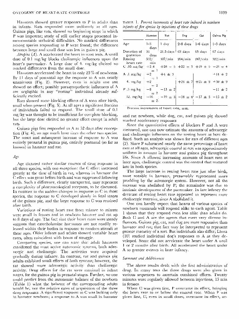
P was impotent; study of still earlier stages presented in- surmountable technical difficulties. No marked differences among species responding to P were found; the difference between large and small dose was less in guinea. pig.
Atropine (A). A accclcrated the heart in most tests. A small dose of 0.1 mg/kg blocks cholinergic influences upon the heart’s pacemaker. A large dose of 4. mg/kg elicited no marked differences from the small dose.
Hamsters accclcrated the heart in only 23 7’0 of newborns. By 11 days of postnatal age the response to A was nearly unanimous (Fig. 2). However, even in adults one test showed no effect; possibly parasympathetic influences of A are negligible in any ‘Cresting” individual already suf- ficiently excited.
Rats showed some blocking effects of A soon after birth,
Age
Duration of
gestation
Resting heart rate
C .05 mg/kg
A .l mg/kg
4. mg/kg
P .3 mg/kg
12. mg/kg
Hamster
1-3
days
16
days
332/
min
+2.4
*l
+2 Ztl
+2 *2
-8
-+I2 -16
zk2
Rat Dog Cat
1 day 2-8 days l-6 days
21.5 days 63 days 65 days
307/min 1 204/min 1 247/min
+28 ziz 5
+4 rt 3 /
+24 rt 7 +25 zt 9
-13 zk 2
-19 * 3 -14 zt 4 -17 rt 3
of individuals failed to respond. The small dose of 0.1 rercenr: increments of heart rate, &SE.
“gjkg was thought to be insufficient for complete blocking, but the large dose elicited no greater effect except in adult rats.
Guinea pigs first responded to A at 52 days after concep- tion (Fig. 4), an age much later than the other two species. The onset and subsequent increase of response to A was entirely prenatal in guinea pig, entirely postnatal (so far as known) in hamster and rat.
Age
Age dictated rather similar courses of drug response in all three species, with one exception: the C effect increased greatly at the time of birth in rat, whereas in hamster the C effect was great before birth and was suppressed following birth. Such a difference, clearly unexpected, rnay represent a complexity of pharmacological receptors, to bc discussed. In contrast to the sudden changes in response to C in those spccics, the response to C developed slowly in fetal stages of the guinea pig, and the large response to C was retained after birth.
Va ria tions of resting heart rate fro1 n minute wcrc small in fctuscs and in new born hamster a
to minute nd rat up
to 8 days of age. The fact that their heart rates were steady suggests that catccholamine hormones are not readily re- leased within their bodies in response to random stimuli at these ages. Older infants and adults showed variable heart rates, often coincident with bouts of struggle.
Comparing species, one can state that adult hamsters manifested the most active autonomic systems, both adre- nergic and cholinergic. The activities were acquired gradually during infancy. In contrast, rat and guinea pig adults exhibited‘small effects of both systems; however, the rat showed more adrenergic activity than cholinergic activity. Drug effects for the rat were maximal in infant stages, for the guinea pig in prenatal stages. Further, no one could pred ict fro (Tab1 .e 1) what
m the autonomi .c balance of the newborn the balance of the corresponding adults
would be, nor the relative rates of acquisition of the three When C was given first, P overcame its effect, bringing drug responses. A significant response to C was lacking only the heart rate to or below the control rate. When P was in hamster newborn; a response to A was small in hamster given first, C, even in small doses, overcame its effect, ac-
Guinea Pig
l-3 days
67 days
322/mi n
+23 zt 4
+14 zt 2
+30 * 9
-11 * 2
-15 rt 2
and rat newborn, while dog, cat, and guinea pig showed marked acceleratory responses.
Since the quantitative effects of blockers P and A were measured, one can now estimate the amounts of adrenergic and cholinergic influences on the resting heart as here de- fined. Such an analysis was shown for rats of different ages (2). Since P subtracted nearly the same percentage of heart rate at all ages, adrenergic control at rest was approximately uniform in amount in hamster and guinea pig throughout life. Since A allowed increasing amounts of heart rate at later ages, cholinergic control was the control that matured late in both species.
The large increase in resting heart rate just after birth, most notable in hamster, presumably represented more prodding by the adrenergic system. However, not all the increase was abolished by P; the remainder was due to intrinsic development of the pacemaker. In late infancy the decrease of resting heart rate appeared to represent more cholinergic restraint, since A abolished it.
One can hardly expect that hearts of various species of newborn mammals will respond alike to each agent. Table 1 shows that they respond even less alike than adults do. Both C and A are the agents that exert very diverse in- fluences. Guinea pig, cat, and dog are more responsive than hamster and rat; that fact may be interpreted to represent greater maturity of a sort. But individuals also differ; Lhota (10) studied individual dog’s responses to A as they de- veloped. Some did not accelerate the heart under A until 1 or 2 months after birth. All accelerated the heart under A to greater extents in later infancy.
Surmount and Additiueness
The above results dealt with the first administration of drug. In many tests the three drugs were also given in various sequences to ascertain combined effects. Twenty minutes were regularly allowed between injections, 15 min in fetuses.

1900 E. F. ADOLPH
c&rating the heart to or above the control rate. Complete titrations of the two drugs in various successive doses were not undcrtakcn.
When C was given first, A added to the heart’s accelera- tion an effect equal to that given by A alone. Also when A was given first, C added its usual amount of acceleration.
When A was given first, P decelerated the heart just as though the first drug had not been given, and vice versa.
Therefore, in all combinations of the moderate doses in- jected, the effects were additive, that is, independent of prcccding administrations.
If C effect is said to surmount P effect, then A effect also
surmounts P effect; and P effect surmounts A effect or C effect. However, the theory of surmount of receptors (4) can hardly be applied to a heart in situ, since such a heart
at the start of a test is not pharmacologically neutral, and extrinsic controls will work against varying backgrounds.
Body Temperatures
Infant animals given C, regularly warrned by 1.0-1.5 C.
The peak temperature was reached in about 15 min whereas heart rate had begun to decline earlier. Therefore
the rise of heart rate was not due merely to warming of the heart. The rise of body ternperature corresponds to the rise
of oxygen consumption after norepinephrine injection
measured by Moore and Underwood (1 1 ), a rise that ap- pears large in infants, especially of rats. In contrast, P and
A had 11; effect on colonic temperatures.
0 me ts of Extrinsic Influences
The three drugs here tested modified the heart rate in
most prenatal *and postnatal stages. Exceptions were: C had
no effect in earliest prenatal rat, possibly in earliest prenatal guinea pig and in early postnatal hamster. At one point in
the research I arrived at the conclusion that the hamster
was born without cardiac response to C. But when fetal hamsters were tested they proved markedly receptive to C.
If the fetal hamster passes through a stage unresponsive to
C, it is a very brief stage, for the heart first beats at 8 days and is accelerated by C at 13 days. In contrast, the fetal
rat shows no consistent response to C at 15 days and very little at 20 days. The hamster heart resembles the salamander
heart in which C accelerates the beat as early in life as the
heart beats at all (3). III the first week after birth, only 30 % of hamsters rc-
sponded to C, and those responses were smaller than in earlier or later stages. Previously one assumed that once
established, receptors would be perrnanent; now it seerns that receptors can effectively come and go.
However, at the same postnatal age when only 30 % of
hamster hearts responded to C, 90 % responded to P. In- deed, some of the same individuals were tested to both agents in succession. To C the heart rates did not change more than +4 %, but to P they changed -5 to - 12 %. The hearts behaved toward C as though they were already
saturated with adrenergic influences. Additionally, after an
injection of P had first decelerated hamster hearts, sub-
sequent C accelerated them by equal amounts. Whether the
reduced heart rate or the presence of P itself now increased the susceptibility to C cannot be decided.
,4ge EJec ts
Possibly the progressive increase in response to C as shown by the guinea pig is typical of ontogeny. In it the
proportions of individuals responding and the sizes of the responses increased progressively with prenatal age. In the end, however, the sudden changes shown by hamster and
by rat will probably reveal more physiological phenomena to a nyone who unravels thern.
Some extrinsic influences of the adrenergic system appear to play upon the mammalian heart at all stages of develop- ment after the early fetus. Thus, P in blocking dose de-
creased the heart rate at all ages, even before birth in rat and guinea pig. In contrast, A had a blocking effect later in life; in hamster its effect became significant at 22-27 days
after conception, in rat at about the same age, and in guinea pig at 50 days after conception. I infer that at these ages the - cholinergic system begins to transmit steady influences upon
the heart’s pacemaker. It is well recognized that vagal
efferent impulses when evoked will decelerate the heart at earlier stages when no natural impulses can be found. How-
ever, at various ages the blocking action of A is not observed in all individuals; tests that show no effect indicate either
that in different individuals the cholinergic influences begin
at various ages or that in a given individual these influences come and go.
That A accelerates the guinea pig heart shortly before
birth was previously reported (6); it is now shown not to accelerate it at much earlier ages. As the responses to C and
A increase in magnitude with age, probably more units,
such as receptors for C, and perhaps more efferent nerve fibers, come to participate in the heart’s control.
Though the age sequence of the drug effects is partly the
same in the three species, no division between prenatal and postnatal can be found. No one control is necessary to all
circumnatal animals. The guinea pig is far developed at
birth in its extrinsic controls of the heart as well as in other characteristics. Whether there is any advantage to this
species in its possession of cardiac controls before birth has
not been discerned. Present results demonstrate that susceptibilities to C, P,
and A develop independently of one another. In different species the ages of onset are movable in respect to the ages
at which birth occurs as well as to the ages from conception.
Drug Actions
P uniformly declerated the heart at all ages, presumably by blocking adrenergic influences. High doses of P de- celerated somewhat more than small blocking doses did, a
possible demonstration of a direct action upon the heart’s
pacemaker (4) at all the ages tested. No evidence of a direct action of A upon the pacemaker
was discovered. In all ages except the adult rat the large
dose had no greater effect than the small dose. However,
when small and large doses were injected in succession (in-
fant hamster and rat), added acceleration was found. It has

ONTOGENY OF HEART-RATE CONTROLS
been reported that A in large dose can block preganglionic (as well as postganglionic) autonomic nerve impulses of adult cats (5). But this action by itself might decrease heart rate rather than increase it, since both sides of the auto- nomic system would be blocked. In the present study, in- creased action of a large A dose suggests that the small A dose was too small to block cholincrgic influences completely.
Rating Heart Rn tes
,4 feature of heart rates common to the three species of rodents is that rates at rest continue to increase with age, and even after birth. This postnatal increase has been noted in rabbit (12) but not in large species (horse, bovine, dog, sheep). Its presence in guinea pig suggests that the postnatal increase is not merely a compensation for insufficient rnaturation before birth.
In absence of measurements here of heart rates in unre- strained infants, the rates in control periods may be pro- visionally regarded as representing the rates in resting individuals.
How much of the changes in resting heart rate with age arc due to extrinsic influences? The effects of blockers sug- gest that part of the postnatal increase of rate is furnished bv adrcnergic prodding, since P blocks part of the rate at all ages. However, the P effect, and hence the prodding, is nearly equal at all ages, even before birth, so that the changes of resting heart rate with age arc chiefly credited to intrinsic controls after all.
L4fter the peak heart rates have been reached, cholinergic influences increase, as shown by the greater blocking effect of A. Prcscnt data allow the possibility that all the declines to adult heart sates are due to cholincrgic restraint. This may be true even in man and dog where the declines of resting heart rate begin before birth.
Birth cannot bc ignored in the ontogeny of cardiac regulations, as shown by the responses to C. The responses to P and A, however, seem uninflucnccd by birth, at least in rat (for P) and guinea pig (for P and A).
Though the patterns of dcvcloprncnt of extrinsic cardiac influences of the three species are alike in many respects, they differ greatly in n) variabilities of the influences at one age in one spccics, b) intervals bctwccn the onsets of those influences, and c) extents of the influence found at each age. No simple transformation of coordinates will convert the program of influences from OIIC spccics to that of another c species.
To what extent these diversities plav a role in the lives
of individuals or of species can only be imagined. In fetal
stages the adrenergic control system is present, a fact sug-
gesting that it is more important to early survival than the
REFERENCES
1. ADOLPH, E. F. Capacities for regulation of heart rate in fetal, in- fant, and adult rats. Am. J. Physiol. 209 : 1095-l 105, 1965.
2. ADOLPH, E. F. Ranges of heart rate and their regulations at various ages (rat). Am. J. Physiol. 2 12 : 595-602, 1967.
3. ADOLPH. E. F., AND 1. M. FERRARI. Regulation of heart rate before
1901
cholinergic system. How, during infancy, the balance be- tween the two systems is determined, and wherein it differs with age and with species remains a puzzle. Even in the adults of the three species, the diverse balances between the two systcrw, here substantiated, has not yet been demon- strated to matter to the organism. The adult hamster has an outstanding predominance of adrenergic influences upon the heart, but this fact cannot yet be interpreted in terms of its daily life.
A famous concept, recorded by Aristotle, states that a young animal acquires first those functions it uses first. He irnplics that those functions arc earliest required for survival. Rigid tests of this concept would demand the experimental suspension of each function in order to observe whether the individual lives without it. However, the tests used here, namely, the blocking of autonomic influences, and those tests so far devised by other investigators, cover such brief times under such limited stresses that they can be counted as inadequate to the purpose.
Resume
I started out with the notion that drugs could furnish a sharp delineation of the development of cardiac controls in each species. Similarities and differences would be identified. Along the way I have found that cardiac controls also develop to diverse degrees among individuals of one species and age. Once established, a control may be suppressed for a considerable period of growth (hamster). These unex- pected features become part of any future account of the ontogeny of physiological regulations.
In gcncral, the onsets of cardiac controls appear earliest in the species (hamster) with shortest duration of gestation. For C, hamster precedes rat which precedes guinea pig. For A, hamster and rat dcvclop their responses at the same age from conception, guinea pig develops its much later. But guinea pig is the only one of the species here shown to have an A response before birth; the same has been found in hurnan, however (8). LNo program of onsets has been discerned for P; even in. small doses it decelerates the heart at all ages tested. But in all three species sympathetic in- fluences can exert controls before the time of birth.
An obvious conclusion from these cxpcriments is that regulation of heart rate is achieved step by step, not all at once. The qualitatively distinct steps arc those of autonomic controls; each begins small and increases in quantitative effect with age. 111 some later steps, controls diminish as the animal bccorncs adult. By choice of age and species, an animal can be employed for study that is endowed with any portion of its cardiac controls.
The National Science Foundation supported this research. Occa- sional aid was given by J. hl. Ferrari and S. R. Happ.
Received for publication 15 October 1970.
cardiac innervation in salamander larvae. Am. J. Physiol. 215: 753-756, 1968.
4. BLINKS, J. R. Evaluation of the cardiac effects of several beta adrenergic blocking agents. Ann. iV. Y. Acad. Sci. 139: 673-685, 1967.

1902
5.
6.
7.
8.
9.
BROWN, A. M. Cardiac sympathetic adrenergic pathways in which synaptic transmission is blocked by atropine sulphate. J. Physiol., London 19 1 : 271-288, 1967. 10. GREENFIELD, A. D. M., AND J. T. SHEPHERD. Cardiovascular re- sponses to asphyxia in the foetal guinea pig. J. Physiol., London 120 : 538-549, 1953. 11. HAMILTON, B., AND M. M. DEWAR. The relation between water and dry substance in the body of the rat, before and after birth. Growth 2: 13-23, 1938. HELLMAN, L. M., G. W. MORTON, E. E. WALLACH, W. E. TOLLES,
12.
AND L. P. FILLISTI. An analysis of the atropine test for placental transfer in 28 normal gravidas. Am. J. Obstet. Gynecol. 87 : 650-661, 1963. 13.
IBSEN, H. I,. Prenatal growth in guinea pigs with special reference
E. F. ADOLPH
to environmental factors affecting weight at birth. J. I%$&. Zool.
51: 51-94, 1928.
LHOTA, K. L. VON. Ueber die Ursachen der sinkenden Pulsfre- quenz bei wachsenden Hunden. Arch. Ges. Physiol. 141: 5 14-526, 1911.
MOORE, R. E., AND h/I. C, UNDERWOOD. The thermogenic effects of noradrenaline in new-born and infant kittens and other small
mammals. J. Physiol., London 168 : 290-3 17, 1963.
MOTT, J. C. Haemorrhage as a test of the function of cardiovascu-
lar system in rabbits of different ages. J. Physiol., London 181: 728-
752, 1965.
PURDY, D. M., AND H. H. HILLEMANN. Prenatal growth in the
golden hamster (Cricetus aura&s). Anat. Record 106 : 59 l-597, 1950.