one lung ventilation module (olv)
TRANSCRIPT

1
One Lung Ventilation Module (OLV)
A Thoracic Surgery Directors Association (TSDA)
Cardiothoracic Surgery Resident Boot Camp Syllabus
The ability to isolate one of the lungs is an essential skill set for the thoracic surgeons. One lung ventilation is required for most non-cardiac operations in the chest. It is also very useful in the treatment of massive hemoptysis, unilateral lung suppuration, and ventilation with significant unilateral broncho-cutaneuos fistula. It is very important that the thoracic surgeon be competent in the various methods of establishing one lung isolation and know how to troubleshoot any problems that may arise related to it. This syllabus will cover the information required for the one lung isolation practical module.
Techniques for establishing one lung isolation One lung isolation is established by the proper placement of a double lumen endotracheal tube (DLT) or by an endobronchial blocker (EBB). Proper placement s best accomplished by bronchoscopically directed placement. This can often be accomplished at the same time as the
intraoperative bronchoscopy prior to lung resection.
DOUBLE LUMEN ENDOTRACHEAL TUBES
Left Sided DLT Right Sided DLT with side hole for RUL Ventilation

2
The DLT can either be placed blindly into the airway with the bronchoscope used to confirm proper placement are placed under direct vision with the bronchoscope. The DLT comes in various sizes and in a left and right version. Guidelines for the proper size are listed below
Very small person 28F
Small person 35F
Medium size person 37Fr
Large size person 39Fr or 41Fr
Except in cases where there is a left mainstem tumor, a left bronchial sleeve is anticipated, or a previous left lower lobectomy has been performed (short left mainstem bronchus), left sided DLT’s are used since the proximal take-off of the right upper lobe bronchus makes the proper placement and stability of placement more difficult for right sided DLT’s.
To place a left sided DLT under direct vision using a bronchoscope, you must use a small bronchoscope not to exceed about 3.3mm in diameter since each lumen of a DLT is small. The patient is intubated in the usual manner with the tip of the DLT left in the trachea. The bronchoscope should be well lubricated and is passed into the bronchial or blue lumen through the provided connector. The proximal endotracheal balloon inflated. Ventilation continues during bronchoscopy.
Bronchoscopic Placement of Left DLT

3
1.) Bronchoscopic equipment is checked a. proper size scope b. scope and video equipment in working order
i. scope focused ii. light source working
c. scope lubricated (silicon spray preferred but water soluble lubricant acceptable) 2.) DLT checked
a. balloons checked (tracheal and bronchial) and b. lubricated with water soluble lubricant c. preformed over the stylet formed into a C shape similar to a regular endotracheal
tube d. connectors assembled
3.) Patient intubated with DLT so that end of bronchial lumen (blue) is in trachea. This usually means having the tracheal cuff (white) just below the cords.
4.) Placement in airway confirmed by presence of expiratory CO2 or bronchoscopy 5.) Bronchoscopy performed through the bronchial (blue) lumen. Patient can be ventilated
during bronchoscopy 6.) Bronchoscope passed distally down the left mainstem bronchus after inspection
bronchoscopy 7.) Tracheal cuff is deflated and tube is passed down over the bronchoscope into the left
mainstem bronchus. Moving the distally paced bronchoscope slightly back will confirm that the bronchial lumen (blue cuff) is in the left mainstem bronchus
8.) Bronchoscope is removed from the bronchial lumen , the bronchial lumen cap is closed, the tracheal lumen cap is opened and the bronchoscope is passed into the tracheal lumen
9.) At the end of the tracheal lumen, one should see the right mainstem orifice. The tube is then moved in or out so that the gray line on the bronchial lumen, which is just above the bronchial lumen cuff (blue) is at the carina. This insures that the tube is not too far down into the left mainstem bronchus leading to obstruction of the left upper lobe bronchus by the bronchial cuff when inflated. The blue cuff is inflated with a few cc’s of air to proper tension as is the tracheal cuff. The bronchial cuff should not herniate out of the left mainstem bronchus.
10.) The placement of the tube in cm’s at the teeth is noted and the bronchscope is removed.
11.) The endotracheal tube is secured to the face.

4
Bronchoscopic Placement of Right DLT

5
Steps 1 – 5 are the same as when pacing a left sided tube except that the tube should be confirmed as being a right sided tube
6.) The bronchoscope is place deep into the right mainstem bronchus
7.) The bronchoscope is backed out slowly until the side port within the blue cuff is visualized
8.) The tube and bronchoscope are then slowly withdrawn together until the side orifice in the blue cuff to the right upper lobe is visualized.
9.) The position of the tube is noted at the teeth and the bronchoscope
is removed
10.) The bronchoscope is passed down the tracheal lumen, the carina and bronchial limb is identified and both bronchial and tracheal cuffs are inflated with air to correct pressure
11.) The tube is secured to the face
If there is any question about endobronchial pathology, the tube should be placed under direct vision to avoid trauma to the endobronchial tumor
Bronchial Blockers
Bronchial blockers establish one lung ventilation by using a balloon catheter inflated to occlude the bronchus of the lung being operated upon. This method is useful when a DLT cannot not be placed such as in a small airway, if the patient is a difficult intubation, when the patient is already intubated and the risk of re-intubation is high, or when the patient has a tracheostomy. There are three methods of establishing OLV by a bronchial blocker. These are by using an Arndt Blocker, a Univent tube, or a Fogarty catheter.

6
ARNDT BLOCKER

7
The Arndt blocker is a balloon catheter that comes in a kit that includes a special adapter allowing the catheter to enter the ET tube separately and simultaneously with the pediatric bronchoscope. An ET tube of 7.5mm or larger is required. The tip of the blocker has a loop of suture through which the bronchoscope can be placed. This allows the scope to guide the blocker into the correct bronchus.

8
Steps in placing the Arndt Blocker
1.) Patient is intubated with a 7.5mm or larger standard endotracheal tube. If a smaller tube must be used, the pediatric Arndt blocker and an infant bronchoscope may be used.
2.) Bronchoscopy is performed using either a standard bronchoscope or the 3.2 mm scope. This is to insure that there is no unanticipated endobronchial pathology. It is important not to have the endotracheal tube positioned too close to the carina as this may inhibit directing the blocker into the correct
3.) The 3.2mm bronchoscope is lubricated and placed through the bronchoscopy adapter provided. The blocker is likewise placed through the appropriate port. Both are passed distal to the connector before the connector is placed in the circuit and the bronchoscope is passed through the loop at the end of the blocker.
4.) The ventilation circuit is then disconnected from the patient and the bronchoscope, passed through the blocker loop is passed into the endotracheal tube.
5.) The Arndt adaptor is connected to the ET tube and ventilation is resumed while bronchoscopy is performed.
6.) The bronchoscope is passed fairly deep into the bronchus of the lung being operated upon or isolated
7.) The blocker is then slid into the bronchus and the scope is partial withdrawn so that the blocker is visualized.
8.) The blocker is pulled back to the proximal mainstem bronchus and inflated with air under direct vision. A brief period of apnea is helpful at this point to allow the lung to deflate prior to obstruction. The blocker must be deep enough into the bronchus so that it doesn’t herniate into the trachea when the balloon is inflated.
9.) On the right side, the balloon should be placed at just below the take-off of the right upper lobe bronchus so that when the balloon is inflated, both the RUL bronchus and the Bronchus Intermedius are occluded.
10.) The suture loop can then be removed from the blocker and the blocker secured in position by tightening the connector where the blocker enters.
11.) Once the loop is removed, suction can be placed on the blocker which may help with lung collapse. This lumen can also be used for CPAP during the case should oxygen desaturation be a problem (see below).

9
FOGARTY CATHETER
One lung ventilation can be achieved by placing a Forgarty catheter into the airway outside of the endotracheal tube. This is an excellent way of overcoming problems related to intubation with a DLT or a large enough ET tube and when a pediatric bronchoscope is not available. A 8-14 or 8-22 Fogarty works best. The critical step in the use of a Fogarty catheter is to manually bend the very tip about 20 degrees before it is placed into the airway (see Figure 22-3 above). This allows for the catheter to be directed into the correct bronchus
Steps in the use of the Fogarty catheter as a bronchial blocker
1.)The Fogarty catheter (either 8-14 or 8-22 size) is removed from its packaging and the very tip is bent approx. 20 degrees at the balloon. It is advisable to have clean gloves on when doing this. The guide wire should be in place when this is done.
2.)The patient is then put to sleep and intubated with the blocker and the ET tube at one laryngoscopy. The ET tube is secured to the face ensuring it is no lower than the mid-portion of the trachea. The blocker should be placed just deep enough to be in the trachea and is not secured at this point. A connector that allows for ventilation to proceed during bronchoscopy is used and a full bronchoscopy of the airway is performed. If the blocker is already down one mainstem bronchus, it should be withdrawn into the trachea to allow full inspection of the airway
4.)With the scope at the end of the ET tube, the ET tube cuff is deflated and the blocker is placed into the bronchus of the lung to be isolated and operated upon. The catheter can be directed by twisting it and taking advantage of the angle at the tip.
5.)The balloon is positioned in the same position as with the Arndt blocker and a test inflation is performed to ensure that the balloon occludes the bronchus but does not herniate back into the trachea. The proper amount of air should be recorded.

10
6.) The ET tube cuff is then reinflated and the blocker is secured to the ET tube with tape. It is helpful to “tab” the tape so that it can be easily removed should repositioning be required.
7.) The guide wire is removed from the Fogarty but is not discarded in case the catheter becomes displaced and must be re-directed.
8.) One lung ventilation is achieved by having a short period of apnea and then inflating the catheter balloon.
UNIVENT TUBE
The Univent tube is a specially made endotracheal tube where the bronchial blocker is contained within a separate lumen within the wall of the tube.
Steps in placing the Univent Tube
1.) An adult or smaller bronchoscope is made available and in working order
2.) The patient is intubated with the Univent tube. The tube should be no lower than the mid-portion of the trachea
3.) Bronchoscopy of the airway is performed
4.) The bronchoscope is pull back to just distal to the end of the Univent tube and the balloon catheter is advanced into the desired mainstem bronchus. Positioning and inflation are the same as with the Arndt and Fogarty blockers

11
Troubleshooting
Unable to intubate with DLT
The DLT usually has a larger outer diameter than the normal endotracheal tube. Thus there are occasions when intubation with a DLT can be difficult. Lack of neck extension, a full complement of teeth, and limited mouth opening are usually cause for concern.
1.) Attempt to place DLT. Use stylet in bronchial lumen to curve DLT in similar way to the normal ET
2.) Intubate with appropriate size tube exchanger. Pass exchanger up bronchial lumen and pass tube over exchanger with laryngoscope in place. Twisted the tube as it goes in can help it to pass into the trachea
3.) Abandon DLT and use a bronchial blocker
Unable to get blocker down correct side
1.) Make sure endotracheal tube is well above carina to allow blocker access to each side 2.) If using Fogarty catheter as blocker, be sure just the tip is angled approx. 20 degrees. 3.) Turn patient’s head to the side opposite the side the blocker is supposed to go down 4.) Tighten the loop on the Arndt blocker where the bronchoscope flexes to allow it to direct
the blocker
Oxygen desaturation during the case
Most instances of oxygen desaturation are related to inadequate ventilation and can be corrected while still preserving OLV.
1.) Suction out ventilated lung
2.) Hyperventilate the ventilated lung with a few with a few large breaths to recruit lung parenchyma that may have become atelectatic during bronchoscopy or from inadequate tidal volumes
4.) Follow protocol for specific type of OLV being used
Left sided DLT/Left sided surgery
1.) Back tube out gradually since tube may be too far into the left mainstem bronchus thus inadequately ventilating the right lung through the tracheal lumen.
12
Left sided DLT/Right sided surgery
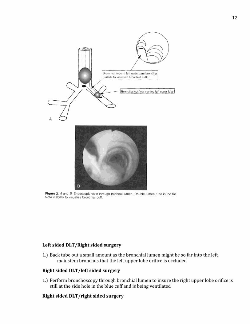
1.) Back tube out a small amount as the bronchial lumen might be so far into the left mainstem bronchus that the left upper lobe orifice is occluded
Right sided DLT/left sided surgery
1.) Perform bronchoscopy through bronchial lumen to insure the right upper lobe orifice is still at the side hole in the blue cuff and is being ventilated
Right sided DLT/right sided surgery

13
1.) Suction out left lung and hyperventilate
If these maneuvers do not rapidly correct the problem, two lung ventilation should be reinstituted and the oxygen saturation returned to acceptable range (usually greater than 95%).
Correct DLT position should reconfirmed via bronchoscopy
One lung ventilation should then be retried.
If one lung ventilation again fails, a small amount of CPAP on the non-ventilated lung should be tried.
If OLV is not compatible with adequate oxygenation, it should be abandoned and the case done with both lungs being ventilated
Left sided blocker/left sided surgery
1.) Other than secretions blocking the right sided bronchi, inadequate oxygenation does not occur as a result of left sided surgery being done with a left sided bronchial blocker
High airway pressures during the case
Other than the rare occasion where a pneumothorax occurs in the ventilated chest, high airway pressures result from a malposition of the DLT or bronchial blocker
Left sided DLT/Left sided surgery
Causes: tube backing out to a position where the inflated bronchial balloon is obstructing the distal trachea or tube slides in so far that the tracheal orifice in down the LMS bronchus and partially or completely obstructing

14
1.) Bronchial cuff is deflated and bronchial side is unclamped. This reestablishes at least left sided ventilation and possibly both lung ventilation depending on the cause
2.) Bronchoscope is placed down the distal (blue) lumen
3.) If carina is visualized indicating tube is too far proximal, the scope is passed into the LMS bronchus and with the tracheal cuff deflated, the tube is advanced back into the LMS bronchus
4.) Cuffs are re-inflated. Proper tube depth is confirmed by bronchoscopy through the tracheal lumen

15
5.) If it is unclear where the tip of the bronchial lumen is, the tracheal cuff is deflated and the tube withdrawn under direct bronchoscopic visualization until the carina is visualized.
6.) Repeat steps 3 and 4 to reposition tube and confirm proper placement
Left sided DLT/Right sided surgery
Cause: Tube is too far down the LMS bronchus thereby forcing the bronchial orifice into the segmental orifices of the left lower lobe
1.) Tube is backed out slowly while checking airway pressure until pressure normalizes
Right sided DLT/Left sided surgery
Causes: tube backing out to a position where the inflated bronchial balloon is obstructing the distal trachea or tube slides in so far that the tracheal orifice in down the RMS bronchus and partially or completely obstructing
1.) Bronchial cuff is deflated and bronchial side is unclamped. This reestablishes at least right sided ventilation and possibly both lung ventilation depending on the cause
2.) Bronchoscope is placed down the distal (blue) lumen
3.) If carina is visualized indicating tube is too far proximal, the scope is passed into the RMS bronchus and with the tracheal cuff deflated, the tube is advanced back into the RMS bronchus such that the side hole within the blue cuff is aligned with the RUL orifice
4.) Cuffs are re-inflated.
5.) If it is unclear where the tip of the bronchial lumen is, the tracheal cuff is deflated and the tube withdrawn under direct bronchoscopic visualization until the carina is visualized.
6.) Repeat steps 3 and 4 to reposition tube and confirm proper placement
Right sided DLT/Right sided surgery
Causes: tube backing out to a position where the inflated bronchial balloon is obstructing the distal trachea. If the tube slides in so far that the tracheal orifice is down the RMS bronchus, the RUL will be seen to re-inflate prior to high pressures occurring as the blue cuff passes distal to the RUL orifice
1.) Bronchial cuff is deflated and bronchial side is unclamped. This reestablishes at least right sided ventilation and possibly both lung ventilation depending on the cause
16
2.) Bronchoscope is placed down the distal (blue) lumen
3.) If carina is visualized indicating tube is too far proximal, the scope is passed into the RMS bronchus and with the tracheal cuff deflated, the tube is advanced back into the RMS bronchus such that the side hole within the blue cuff is aligned with the RUL orifice
4.) Cuffs are re-inflated.
Bronchial Blocker for either left or right sided surgery
Cause: Blocker has slipped back into the trachea and is obstructing it
1.) Blocker cuff is deflated re-establishing ventilation to both lungs
2.) Bronchoscopy is performed and blocker is repositioned in the appropriate bronchus
3.) Blocker is re-inflated under direct vision. A short period of apnea or manual pressure on the lung prior to re-inflation will allow the lung to deflate quicker.
Loss of one lung isolation during the case
Very often this is caused by inadequate inflation of the cuffs of the DLT or the bronchial blocker.
Small amounts of additional air should be added to see if this restores OLV.
Left sided DLT/Left sided surgery
Cause: Tube has backed out of the LMS bronchus and the bronchial orifice is in either the trachea or RMS bronchus. This occurs from traction on the operated lung during surgery. For this to occur, the bronchial cuff must not be inflated enough to block the distal trachea from being ventilated by the tracheal lumen.
1.) Try advancing the tube. The tube may still be partially in the LMS bronchus and advancing it will allow the blue cuff to occlude the LMS bronchus
2.) If Step 1 fails to re-establish OLV, the tube is withdrawn under direct bronchoscopic visualization through the distal lumen until the carina is visualized.
3.) The scope is passed into the LMS bronchus and with the tracheal cuff deflated, the tube is advanced back into the LMS bronchus
4.) Cuffs are re-inflated. Proper tube depth is confirmed by bronchoscopy through the tracheal lumen
HINT: The tip of the DLT can often be felt through the bronchus and proper position confirmed if the tube is down the side being operated upon.
Left sided DLT/Right sided surgery

17
Cause: Tube has backed out of the LMS bronchus and the bronchial orifice is in either the trachea or RMS bronchus. This occurs from traction on the operated lung during surgery.
1.) Under direct bronchoscopic visualization through the distal lumen tube is withdrawn until the carina is visualized.
3.) The scope is passed into the LMS bronchus and with the tracheal cuff deflated, the tube is advanced back into the LMS bronchus
4.) Cuffs are re-inflated. Proper tube depth is confirmed by bronchoscopy through the tracheal lumen
Right sided DLT/Left sided surgery
Cause: Tube has backed out of the RMS bronchus and the bronchial orifice is in either the trachea or LMS bronchus. This occurs from traction on the operated lung during surgery.
1.) Under direct bronchoscopic visualization through the distal lumen tube is withdrawn until the carina is visualized.
3.) The scope is passed into the RMS bronchus and with the tracheal cuff deflated, the tube is advanced back into the RMS bronchus until the side orifice is aligned with the RUL orifice
4.) Cuffs are re-inflated. Proper tube depth is confirmed by bronchoscopy through the tracheal lumen
Right sided DLT/Right sided surgery
Cause: Tube has backed out of the RMS bronchus and the bronchial orifice is in the LMS bronchus. This occurs from traction on the operated lung during surgery.
Cause: Tube has advanced into the Bronchus Intermedius such that the blue cuff no longer isolates the RUL orifice. This can be surmised if only the RUL re-inflates
1.) Tube is advanced back into the RMS bronchus with the bronchoscope in the distal lumen’s blue cuff at the level of the side orifice until the side orifice is aligned with the RUL orifice
Bronchial Blocker for either left or right sided surgery
Cause: Bronchial blocker slips out of the appropriate bronchus into either the trachea or the bronchus to the non-operated side. Bronchial blocker cuff cannot be inflated enough to obstruct the trachea
Lung fails to deflate

18
Lung deflation may be delayed or may not occur in the presence of bronchial secretions obstructing the airway or significant lung parenchymal disease. The lung should be suctioned as the first step in getting it to deflate
DLT (Left)
Cause: Bronchial cuff is occluding the upper lobe orifice
1.) Withdraw tube slowly until upper lobe can be easily compressed and deflated
DLT (Right)
Cause: Bronchial cuff can either be too far in or not far enough in the RMS bronchus
1.) Feel for tube in RMS bronchus and move in or out as appropriate
2.) Bronchoscope through the bronchial lumen.
3.) If the tube is well into the Bronchus Intermedius such that the lower and middle lobe bronchi are visualized, withdraw tube until the side orifice is aligned with the upper lobe bronchus.
4.) If the tube is at the orifice of the RMS bronchus, advance the tube until the side orifice is aligned with the upper lobe bronchus. Correct determination of the blue cuff is often times easier by bronchoscopically visualizing the cuff through the trachea lumen.
Bronchial Blockers
Cause: The lung will stay inflated after the bronchial blocker is inflated due to obstruction of the airway. Over time, atelectasis will occur and the lung will deflate. A period of apnea, suctioning of the lung before balloon inflation, and manual compression of the lung prior to balloon inflation will accelerated lung deflation.
JUNE 2011