on the evolution of primitive genetic codes - tbi · on the evolution of primitive genetic codes...
TRANSCRIPT

On the Evolution of Primitive Genetic Codes
Günter Weberndorfer, Ivo L. Hofacker and Peter F. StadlerInstitut für Theoretische Chemie und Molekulare StrukturbiologieUniversität Wien, Währingerstrasse 17, A-1090 Wien, Austria({gw,ivo,studla}@tbi.univie.ac.at)
2002/06/10
Abstract. The primordial genetic code probably has been a drastically simplified ancestor ofthe canonical code that is used by contemporary cells. In order to understand how the present-day code came about we first need to explain how the language of the building plan can changewithout destroying the encoded information. In this work we introduce a minimal organismmodel that is based on biophysically reasonable descriptions of RNA and protein, namelysecondary structure folding and knowledge based potentials. The evolution of a population ofsuch organism under competition for a common resource is simulated explicitly at the levelof individual replication events. Starting with very simple codes, and hence greatly reducedamino acid alphabets, we observe a diversification of the codes in most simulation runs. Thedriving force behind this effect is the possibility produce fitter proteins when the repertoire ofamino acids is enlarged.
1. Introduction
The evolution of the translation machinery still presents a great challenge toany theory of the Origin of Life. As far as we know, all extant life-formsuse protein enzymes and they all construct them in the same way by translat-ing an RNA message. Invariably, translation occurs in a highly complicatedRNA/protein complex, the ribosome, using tRNAs that are specifically loadedwith an amino acid. All organism use the same set of twenty amino acids(22 if we count selenocystein [41, 9] and the recently discovered pyrrolysine[70]). In all cases tRNA acts as an adapter that allows the transfer of an aminoacid to the growing chain if and only if the three consecutive nucleotides thatform the codon on the mRNA match the tree anticodon nucleotides of thetRNA. Aminoacyl-tRNA synthesis typically is performed by 20 aminoacyl-tRNA synthetases, each one specific for a single amino acid; but see [31] foran overview of an increasing collection of exception to this simple rule.
It is not hard to argue that such a complex mechanism should havedeveloped from a much simpler one. Unfortunately, because the translationmechanism is universal, there not too much evidence left from its earlierevolutionary stages. Even the code itself, i.e., the assignment of an amino acidto a codon is almost invariant. The most direct evidence for the evolution ofthe genetic code is the fact that the code is not quite “universal”. The firstdeviations from the standard code were observed in vertebrate mitochondria,soon many more were identified among different phyla, see Fig. 1.

2 Weberndorfer, Hofacker, Stadler
Yeast Nucl
CUGL−>S
Euplotid
UAGBlepharisma
L<−>Q
UCAS−>*
UAA*−>Q
Ciliate
ThraustochytriumInvertebrateMitochondria
MitochondriaYeast
AGA,AGG
L−>T
CUU,CUC,CUA,CUGR−>G
AscidianMitochondria
Mitocondria
MitochondriaScenedesmus
ChlorophyceanMitochondria
Mitochondria
AGA,AGG R−>*
Vertebrate
TrematodeMitochondria
MitochondriaEchinoderm
MitochondriaFlatworm
UAA*−>YAUA
I−>M
AGA,AGGR−>S
AAAK−>N*−
>C
UGA*−>W
L−>* UUA
MitochondriaMold
*−>Q*−>L
Figure 1. The genetic code shows variations among different species that can be representedas a tree-like graph. The black square marks the so-called universal or standard code. Thedefinitions of the code variants were obtained from the National Center for BiotechnologyInformation (NCBI) website http://www.ncbi.nlm.nih.gov/.
All known non-standard codes, however, appear to be secondarily de-rived [52]. Interestingly, some changes occur independently in related linagesimplying multiple changes within a short period of time during evolution.Several codons seem to be more easy changeable and were assigned to differ-ent amino acids. For instance AGG has been reassigned from Arg to Ser, Gly,and STOP. In particular, STOP-codons seem to be an evolutionary degree offreedom. Their neutrality may be achieved due to their rareness (they occuronce per gene) and the fact that transcriptional release factors are easy tochange [53].
Another factor that may make codon reassignment evolutionary feasibleis variations in codon frequencies. In fact, codon usage can vary dramati-cally between different species; see [16] for a recent review and [37] for adiscussion the context of the genetic code.
It has been argued repeatedly by different authors that the UniversalCode is optimal or near optimal in some sense. For example, Freeland et

Evolution of Genetic Codes 3
al. [21] show that the Universal Code is near optimal in terms of error min-imization, adaptation for double-strand coding is discussed in [39]. In [45]a balance of robustness and changeability is advocated, the approach in [1]focuses on amino acid properties.
While the idea that the genetic code evolves towards more robust codingproperties is compelling, it is by no means clear how such mutations areaccessible. Indeed, the rewired code must be at least neutral at the level ofthe proteins that it produces. The selection pressures towards robustness isweak: evolution towards robustness and evolvability is a second order effectthat can prevail only if the organizational changes do not cause immediatefitness losses [74, 75, 77].
Possible mechanisms of evolvability of genetic codes are reviewed in[36]: Code modifications can originate from changes in several componentsof the the translation apparatus, e.g.:
- Mutations of the identity elements of tRNA elements may change thespecificity of aminoacylation. The tRNA may then be loaded with adifferent amino acid or loading may become ambiguous
- Mutation of the anticodon of the tRNA will cause the incorporation of awrong amino acid (unless the anticodon is part of the identity elements,which is not always the case.
- Mutation of the Aminoacyl synthetase gene might lead to a change inthe loading specificity.
In general, however, such changes will be deleterious because every proteinthat contains the modified codon will be affected.
In recent years three mechanisms of codon changes especially in mi-tochondria were published and each of them predicts certain codon changesthat have not yet been observed.
(1) The Codon Capture Hypothesis [52] states that specific codons dis-appeared from the code by AT or GC pressure. Hence mutations in thetRNAs coding for these codons are neutral. If if the pressure is reducedthe codons reappear and may now code for a different amino acid. Sup-port for this theory comes from the mitochondrial codes, where genesare AT rich and small.
(2) Ambiguous Intermediate Hypothesis [83] proposes that codons un-dergo a period of ambiguity instead of disappearing when their meaningchanges. This idea is supported by that fact that RNA in some casesmis-pairs: G ·A and C ·A pairs may occur at the third codon positionsand G ·U pairs may even occur at the first codon position. Support alsocomes from yeast, where a mistranslation between Ser and Leu at theCUG has been reported.

4 Weberndorfer, Hofacker, Stadler
(3) The Genome Streamlining Hypothesis [3] assumes that the simplifi-cation of the translation apparatus is the driving force for codon reas-signment in mitochondria. Reduction of the genome size has a directselective advantage, and even the size of a single tRNA is significant forvery small genomes. This is the driving force for the loss of tRNAs andhence codons.
In this contribution we describe detailed mechanistic simulations of asimplified (proto)organism that show that the genetic code can indeed evolvein the presence of strong selection on the encoded polypeptides. This ap-proach differs from previous arguments for the adaptive nature of the code inthat we need not assume a direct selection pressure on higher order propertiessuch as evolvability. Indeed, or model is based on the reproductive success ofindividuals which depends only on the quality of the encoded proteins, not onthe code that they use. The evolution of the encoding is therefore an emergentproperty in our model.
2. The Minimal Organism Model of Genetic Code Evolution
The current implementation of the Neo-Darwinian framework in the form ofpopulation genetics or quantitative genetics in essence deals with selectionand is hence insufficient to describe features of phenotypic evolution such asinnovation [48]. The reason is that before selection can determine the fate of anew phenotype, that phenotype must first be produced, or accessed, by meansof variational mechanisms [17]. As far as we know, all heritable variationsof a phenotype must occur through genetic mutation. The accessibility ofa phenotype is therefore determined by the genotype-phenotype map whichdetermines how phenotypes vary with genotypes [43, 78, 20, 71].
A meaningful model of evolutionary innovation, and this includes anymodel evolutionary model of the genetic code, must therefore make explicitassumptions on the properties of the genotype-phenotype map. In fact, thegenotype-phenotype map must be modeled explicitly based on known princi-ples of physics, chemistry, and molecular biology in order to obtain a mean-ingful implementation of phenotypic accessibility.
This approach was tremendously successful in the case of RNA evo-lution. RNA folding from sequences to secondary structures can be used asa biophysically realistic, yet extremely simplified toy-model of a genotype-phenotype map. Simulated populations of replicating and mutating sequencesunder selection exhibit many phenomena known from organismal evolution:neutral drift, punctuated change, plasticity, environmental and genetic canal-ization, and the emergence of modularity, see e.g. [18, 62, 30, 20, 2]. Lab-oratory experiments [69, 42, 73] have generated phenomena consistent withthese patterns.

Evolution of Genetic Codes 5
Even a minimal model for the evolution of the genetic code is necessarilymuch more complex. It must deal with all the key players of the translationmachinery in order to provide a meaningful description of the accessibility ofvariant codes. In addition, it must include a biophysically reasonable fitnessfunction.
We base our model on the assumption of an RNA World [22, 7] as apredecessor of our present DNA/RNA/Protein biology. For a recent reviewof the arguments for and against an RNA World Era see [82]. We empha-size, however, that we make no claim as to whether RNA was the primordialbiopolymer or whether it was preceded by other, simpler molecules such asPNAs [38], that might be more plausible in terms of prebiotic synthesis [50].
The simulations presented here are motivated by a specific model organ-ism, Fig. 2 at a (very) late stage of the RNA world, just after tRNA-basedpeptide synthesis has been invented and the power of protein-enzyme cataly-sis is utilized for replication. The main features of our hypothetical primitivecell, which we interpret as a distant ancestor of the last universal commonancestor [54, 81] are the following:
(1) RNA genome. It is generally believed that RNA as a molecular carrierof genomic evolution was only later replaced by by DNA genomes. Apossible explanation for the advantage of DNA in larger genomes interms of the mechanism of homologous recombination is described in[63], although it the reason may simply be the greater chemical stabilityof DNA.
(2) RNA-ribosome. Evidence form both in vitro studies [35, 51] and theanalysis of the atomic structure [55] reveals that the ribosome is first andforemost a ribozyme. On the other hand, no isolated protein, or mixtureof proteins, has ever been shown to catalyze the peptidyl-transferasereaction [25]. Furthermore, even present-day ribosomes can deal witha wide variety of amino acids, as exemplified by the incorporation ofartificial amino acids by means of translation [44]. It seems reasonable,therefore, to assume that the ribosome performs its function independentof the amino acid alphabet that is used by the organism.
(3) tRNAs acted as crucial adaptors presumably even in the earliest versionsof the translation apparatus; they are mostly likely much older thanthe last common ancestor [13]. Each tRNA incorporates two codes: thecodon/anti-codon code that reads the information from the mRNA anda second operational code [11, 58, 61] that determines the amino acidwith which the tRNA is loaded. This second code is determined by theaminoacyl-synthetases.
(4) Ribozyme Aminoacyl synthetases. The RNA world hypothesis impliesthat present-day mechanism of coded protein synthesis evolved from

6 Weberndorfer, Hofacker, Stadler
ribozyme-catalyzed acyl-transfer reactions. The existence of specific a-minoacyl-tRNA synthetase ribozymes has been demonstrated by meansof in vitro evolution [40]. Furthermore, there is evidence that tRNAspredate their synthetases [56]. The present-day operational code is de-termined by an intricate pattern of sequence determinants that are rec-ognized by the aminoacyl-synthetases; in the late RNA world it mayhave been as simple as the complementary recognition of the ribozymedesigned by Lee et al.. There is ample evidence that amino acids mayhave acted as co-factors in the RNA world [59, 72]. It is plausible there-fore that specific amino acid recognition and aminoacyl-transferring ri-bozymes have evolved long before the onset of translation.
(5) Protein Replicase. Ribozymes with ligase-based replication activity [46]and true replicase activity [33] were recently obtained by in vitro evo-lution, lending additional credibility to the RNA world scenario. Oncereplication is protein dependent all modifications of the code have animmediate impact on survival. It is therefore sufficient in our model toconsider a polypeptide replicase as the only protein component.
(6) A ribozyme based metabolism is a convenient assumption in our settingbecause it need not be modeled explicitly. The wide range of chemicalreactions, including carbon bond formation, that can be catalyzed byribozymes [6, 67, 32] make this assumption even plausible.
Only a few of these components need to be modeled explicitly on thecomputer. We need a genomic sequence that has to be replicated, we needthe tRNAs and an implementation of the operational code relating a tRNAsequence to a (set of) amino acids with which it is loaded, and we need away of evaluating the replicase protein that is encoded on the genome. Wedon’t have to implement the details of the replication process, the action ofthe ribosome, and the metabolism. This is equivalent to assuming that (i) therate-limiting step in the “cell-cycle” of our model is the replication of thegenome.
We remark that our ansatz allows an alternative interpretation as well: ifwe assume that replication is still RNA based and that the rate limiting stepis a protein-enzyme based metabolism, we arrive at the same type of model.
3. Implementation of the Model Organism
The genome of our model organism consists of the mRNA for the replicaseprotein and a variable number of tRNA genes.
In order to model the structural requirements on a tRNA that are im-posed by the ribosome we require that each putative tRNA must fold into

Evolution of Genetic Codes 7
TRANSLATION
REPLICATION
LOADING
replicasegene tRNA genes RNA genome
tRNAs
loaded tRNAsreplicase
Figure 2. Model of a minimal organism with translation. It has a genome that carries genesfor a protein replicase and tRNAs as well as a primitive translation apparatus and a systemfor loading tRNAs with amino acids. Neither the proto-ribosome nor the aminoacyl trans-ferases are modeled in molecular detail. The protein sequence of the replicase determinesrate and accuracy of replication. Translation proceeds by the usual rule of codon/anti-codoncomplementarity. The loading of a tRNA with a certain amino acid depends on a sequence de-terminants on the tRNA. The replication rate of the organism is determined by the replicationrate of its genome.
the canonical cloverleaf structure that is characteristic for tRNAs, Fig. 3.RNA secondary structures can be predicted accurately and efficiently basedon thermodynamic rules [85]. We use the implementation of the minimumenergy folding from the Vienna RNA Package1 [28]. For the purpose ofour model, a functional tRNA is a sequence of length 76 whose secondarystructure matches the regular expression given in Fig. 3.
There is no generally accepted model for the affinity of individual amino-acids to RNA sequences. We therefore employ a rather arbitrary table ofamino acid assignments to the tRNAs that depends on the sequence of theanticodon loop and the two terminal nucleotides. The algorithm is describedin the lower panel of Fig. 3.
A codon of the message is translated to the amino acid of the tRNA inthe genome that has the anticodon sequence closest (in Hamming distance)to the complement of the codon. In case of equal hamming distance a matchat the 1st codon position is preferred over 2nd, and 2nd over 3rd. The codemay be ambiguous if two or more tRNAs match a codon equally well. In thiscase the assignment is done stochastically (but the assignment is then kept
1 http://www.tbi.univie.ac.at/RNA/

8 Weberndorfer, Hofacker, Stadler
Tψ C loop
G
Y
U R
Y RU Y C
Y
R
TGR
G
A
C
C
A
Y
R
Acceptor
35
30
20
10
55
40
5D loop
Variable loop
R
A
60
45
3’
5’
Anti codon
15
R Y
A
# 5’ acceptor stem (5-9 pairs)(^\({5,9}\.*# D arm (3-5 pairs)\({3,5}\.+\){3,5}# variable region\.*# anticodon arm (3-7 pairs)\({3,7}\.{2})(\.{3})(\.+\){3,7}# variable region (2-7 unpaired)\.{2,7}# T arm (3-6 pairs)\({3,6}\.+\){3,6}\.*# 3’ acceptor stem\){5,9}# trailing bases\.+)$
CGGGGUGGACACGCACUAGCAACGUGAUGCUUUCUACACAAGCAAUAGAACGGUCGGACCAACCGUCAUUCUGAUCA(((((((..((((.........)))).(((((.......))))).....(((((.......))))))))))))....
CACAA => 1100110000
11001 xor 10000 = 01001 ( = 9 )
UGU => [L]
Figure 3. The canonical clover leave structure of a tRNA. L.h.s.: conventional drawing withthe conserved nucleotides marked. The R.h.s. gives the perl-style regular expression thatdefined a tRNA for our purposes.Given a correctly folded tRNA sequence the amino acid with which is loaded is computedby the following algorithm: (i) The determinants are the nucleotides 1, 76, and the anticodonloop. (ii) These are translated to a binary code using A=00, U=01, G=10, and C=11. (iii) Thefirst and second five bits are combined using the “xor” operation to give a number between 0and 31. (iv) This number is interpreted as an amino acid from the alphabet N,P, Q, A, R, S, C,T, D, E, V, F, W, G, H, Y, I, K, L, M or as a STOP signal. In this example the anti-codon isACA, the corresponding codon is thus UGU, which is mapped to the leucine L.
fixed for the lifetime of the individual). The tRNAs that fold into the correctsecondary structure together with the sequence dependent loading algorithmdescribed in Fig.3 therefore determines the genetic code. The mRNA for thereplicase is translated into its amino acid sequence according to this code.
The evaluation of the resulting protein is based on its structure. Of coursewe do not attempt to solve the folding problem. Instead we determine howwell the amino acid sequence fits onto a target structure. We used the structure

Evolution of Genetic Codes 9
of the T7 RNA polymerase, for which an X-ray structure with a resolution of3.3Å, PDP file 4rnpA, is available [68], Fig. 4
Knowledge-based potential are well suited to discriminate between cor-rectly folded and mis-folded proteins [27, 65, 66], an approach that waspreviously used to explore the sequence-structure map of proteins [5, 4]. Forthe sake of computational efficiency we do not use M. Sippl’s PROSA-potentialhere. Instead we us a 4-point potential [80] that is based on Alexander Trop-sha’s Delauney tessellation potentials [49, 64, 84]. The idea of inverse folding[8] by means of knowledge-based potentials is to compare the energy W (x,ψ)of sequence x threaded onto structure ψ with the distribution of energiesobtained from threading x onto a large library of unrelated protein struc-tures. From W (x,ψ), the mean W (x) and the standard deviation σW (x) of thisdistribution one computes the z-score
z(x,ψ) =W (x,ψ)−W (x)
σW (x)(1)
which measure how well the sequence x fits onto structure ψ. It seems naturaltherefore to use z(x,4rnpA) as fitness function.
The replicase also determines the replication accuracy. Certain positionsat the active site are responsible for the identification of the template base, anddirect the recruitment of a nucleotide for elongation. We used the deviationof local folding energies from the values for the wild-type sequence for these21 amino acids. For the details we refer to the PhD dissertation of the firstauthor [79].
In summary, therefore, our model organism has a genome x that (via itstRNAs) defines its genetic code and (via properties of the protein resultingfrom this code) determines its replication rate Ax and its replication accuracy,as measured by the single-digit error rate µx.
4. Simulation in a Tank Reactor
The simplest experimental setup for observing a population over long periodsof time is serial transfer [69], where at fixed time interval a tiny fraction of thepopulation is transfered to a virgin growth medium. In chemical kinetics thechemostat (flow reactor) is preferred, where the population is fed a constantsupply of nutrients and the total volume is kept constant. An approximaterealization of an evolution reactor under constant organization is Husimi’scellstat [29]. From a theoretical point of view, serial transfer can be viewedas the discrete time version of the flow reactor; both lead to very similardynamical behavior [26].
Both models are rather easily implemented on the computer. Sophisti-cated version are based on Gillespie’s algorithm [23] that exactly simulates

10 Weberndorfer, Hofacker, Stadler
Figure 4. Delauney tessellation of the T7 RNA-polymerase structure 4rnpA. The red ballsindicate the Cα atoms. The energy W (x,4rnpA) is the sum of contributions Ui jkl for eachtetrahedron that depend on the aminoacids at corners and their relative location along thechain, and a surface term for each triangle on the surface of the molecule [80].
the stochastic reaction kinetics of mutation and fitness proportional selection[19]. In order to save computer resources we resort to a somewhat simplerapproximate scheme of tournament selection [24] where two individuals inthe population are picked at random, their fitness is compared, and the fitterone is replicated. In order to limit the population size, the child organismreplaces another randomly picked individual.
This reaction scheme in essence reproduces Eigen’s quasi-species model[12, 14]
dpx
dt= ∑
y{QxyAy py −QyxAx px} (2)
Here Ax is the replication rate of an organism with genome x and Qxy is themutation rate from y to x. If we consider only point mutations with a mutationprobability of µx at each position, we get
Qxy =
(
µy
α−1
)d(x,y)
(1−µy)n−d(x,y) (3)

Evolution of Genetic Codes 11
where d(x,y) is the Hamming distance between the parent and offspringgenome. Equ.(2) described replication and point mutation. In contrast to theusual quasi-species model the error rate µx is an explicit function of theparental genome. Nevertheless, the model behaves dynamically just like aclassical quasi-species: survival of the fittest leads to a predominant masterspecies that is surrounded by a “tail” of mutants. If a mutant becomes fitterthan the master, the population drifts toward this new species. The populationavoids the error-threshold phenomenon by adjusting the mutation rate.
Gene duplication still is an important mechanism of genomic evolution,see e.g. [76]. Hence we include the duplication of tRNA genes as macro-mutation events. Mutation may then act on the duplicate genes and lead todiversification of the code.
We assume that a rudimentary coding system is already in place, i.e., wedo not attempt to model the origin of coding itself. Thus an initial conditionmust be prepared consisting of a “primordial code” and a an associated genefor the replicase that leads to a non-zero replication rate.
It was shown in [5] by means of computer simulations that various smallsubsets of the amino acid alphabet can be used to design polypeptide se-quences with native-like z-scores for known proteins. Experimental evidenceis described e.g. in [10, 34, 60]. First we produce an inverse-folded proteinsequence for 4rnpA by means of adaptive walks with a restricted amino acidalphabet as described in [5], then we use the initial code to reverse-translateit into a mRNA. The tRNAs for the initial genome are produced by inverseRNA folding with prescribed nucleotides at the determinant positions usingthe program RNAinverse for the Vienna RNA Package [28]. The simula-tion is then started with the tank reactor filled with N identical copies of the“primordial organism”.
5. Results
5.1. EXPANSION OF TWO-AMINO-ACID ALPHABETS
The simplest conceivable initial alphabets distinguish only between one hy-drophilic and one hydrophobic amino acid. One of these simulations is dis-cussed in some details in Fig. 5. In some runs no new amino acid is incorpo-rated within some 107 replication events. In most simulation runs, however,we find 4-7 amino acids at the end of the simulation, often with one ortwo additional ones that were invented and managed to spread through thepopulation but were forgotten at later stage.
As a global indicator of evolutionary progress we consider the averagefitness F of the population as a function of time. The diversity of encodedamino acids in the population is conveniently measured by the “amino acid

12 Weberndorfer, Hofacker, Stadler
G
A
I
L
V
P
M
F
W
Y
C
N
T
Q
S
H
R
K
D
E
0 1000 2000 3000 4000 5000 6000 7000
0 1×10³
2×10³
3×10³
4×10³
5×10³
6×10³
7×10³
time [generations]
50
55
60
65
70
75
fitn
ess
GCG => T
AGU => K
CGU => R
AAU => N
GGG=>A|*
0 1×10³
2×10³
3×10³
4×10³
5×10³
6×10³
7×10³
time [generations]
1
2
3
entr
opy
SA
Sc
Figure 5. Extension of the LD amino acid alphabet as a function of simulation time. The upperplot shows the fraction of individuals in the population that use an amino acid (in gray scale).The lower panel displays the time evolution of (from top to bottom) the fitness, the codonusage entropy Sc, and the amino acid entropy SA. The jump in SA around t = 7000 occurswhen the AAU codon is reassigned from L to N. Only 16% of the simulation run is shown, butno further innovations occured.

Evolution of Genetic Codes 13
entropy”SA = −∑
afa log2 fa (4)
where fa is the fraction of amino acids a in an organism’s replicase. Anal-ogously, the frequencies of codon usage can be used to compute a “codonusage entropy” Sc. Both the average fitness and the entropy measures increasewith time. The increase of F is implicit in the model [12]; the increase of theentropy measures, on the other hand, describe the increase in the complexityof the evolving codes. We expect SA ≈ Sc if there is only one codon in usefor each amino acid. We observe, however, that Sc > SA, indicating that theredundancies in the code yield to diversification in codon usage. On the otherhand, the value of Sc ≈ 2.5bit at the end of the run in Fig. 5 is much smallerthan the theoretical maximum of 3× 2bit for a nucleotide triplets. The slowincrease in SA and Sc shows that amino acid innovation occur via rare codons,whose usage in the genome increases as a consequence of subsequent muta-tions. In some cases a codon that is already commonly used for a redundantlycoded amino acid is reassigned, i.e., the code is refined. Such an event can bedetected form a comparison of the two entropy curves: The codon entropy Sc
remains smooth while the amino acid entropy SA sharply increases becauseof the novel amino acid. An example of such a refinement event can be seenin Fig. 5.
Simulations that were started with small alphabets (e.g. LD) tend in afirst phase to reach “codon coverage”. By codon coverage we mean that eachgroup of codons (ANN, UNN, GNN, and CNN) is translated unambiguouslyto a different amino acid. Only in a later phase further refinements of the codeare observed. This is a consequence of the assignment of tRNAs to codonsdescribed in Section 3 which implies that the first codon position is moreimportant for the matching than the second and the third.
As soon as a modification of the alphabet is fixated in the population, afurther innovation becomes less likely because over the following thousandsof generations fitness advantages can be drawn rather easily from spreadingthe usage of the novel amino acid. As the number of innovations past codoncoverage is small we have not been able to extract a common pattern fromthe further expansion steps.
5.2. EXPERIMENTS WITH LARGER ALPHABETS
The amino acid alphabet AKGV, with codons of the form GNC was proposedas the primordial amino acid alphabet in [15], the alphabet ADLG is anothercandidate [47] for the primordial one; the restriction of inverse folding to thisalphabet was studied in some detail in [5]. Computations using knowledge-based potentials suggest that this alphabet allows inverse folding of a varietyof present day protein structures. A phage display experiment [57] resem-
14 Weberndorfer, Hofacker, Stadler
G
A
I
L
V
P
M
F
W
Y
C
N
T
Q
S
H
R
K
D
E
0 10000 20000 30000 40000 50000
G
A
I
L
V
P
M
F
W
Y
C
N
T
Q
S
H
R
K
D
E
0 10000 20000 30000 40000 50000
G
A
I
L
V
P
M
F
W
Y
C
N
T
Q
S
H
R
K
D
E
0 10000 20000
G
A
I
L
V
P
M
F
W
Y
C
N
T
Q
S
H
R
K
D
E
0 10000 20000 30000
G
A
I
L
V
P
M
F
W
Y
C
N
T
Q
S
H
R
K
D
E
0 10000 20000 30000
G
A
I
L
V
P
M
F
W
Y
C
N
T
Q
S
H
R
K
D
E
0 10000 20000 30000
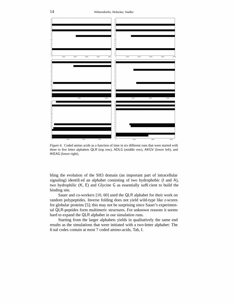
Figure 6. Coded amino acids as a function of time in six different runs that were started withthree to five letter alphabets QLR (top row), ADLG (middle row), AKGV (lower left), andIKEAG (lower right).
bling the evolution of the SH3 domain (an important part of intracellularsignaling) identified an alphabet consisting of two hydrophobic (I and A),two hydrophilic (K, E) and Glycine G as essentially sufficient to build thebinding site.
Sauer and co-workers [10, 60] used the QLR alphabet for their work onrandom polypeptides. Inverse folding does not yield wild-type like z-scoresfor globular proteins [5]; this may not be surprising since Sauer’s experimen-tal QLR-peptides form multimeric structures. For unknown reasons it seemshard to expand the QLR alphabet in our simulation runs.
Starting from the larger alphabets yields in qualitatively the same endresults as the simulations that were initiated with a two-letter alphabet: Thefinal codes contain at most 7 coded amino-acids, Tab, I.

Evolution of Genetic Codes 15
Table I. Summary of Simulation RunsRun E D K R H S Q T N C Y W F M P V L I A G
ADLG_pks05 � F F � � �
ADLG_prali � F F � � �
AGKV_pks04 � � � �
AGKV_pks07 F � F F � � �
IG_pks13 F ♦ F � �
IKEAG_pks04 � � � � �
IKEAG_pks13 ♣ � F F F � � �
LD_4_pks06 � F F F F � ♦
LD_3_pks06 � F F � ♦
LD_2_pks06 � �
LD_pks03 � F �
QLR_pks11 � � F �
QLR_pks12 � � F �
QLR_pks00 � � �
� kept from start, F invented, � lost, ♦ invented and lost again, ♣ lost and re-invented.
The model includes the possibility that the evolving organism fine-tunethe mutation rate. We observe that the mutation rate decreases with with timeso that the invention of additional amino acids become more and more un-likely. This can be understood by the fact that a reduction in mutation rateincreases the population fitness by reducing the number of detrimental off-spring. This self-adaptation of the mutation rate will require a more detailedinvestigation.
6. Concluding Remarks
We have described a mechanistic model of the evolution of simple geneticcodes. Our simulations show that the increase in fitness that can be achievedwith more diverse amino acid repertoires is sufficient to cause an increaseof the alphabet size from two to about six or seven. The small size of theprotein-coding part of our model genome (a single gene with only a fewhundred amino acids, Fig. 4) implies that a moderate diversity of the aminoacid alphabet is sufficient to produce very good sequences. We suspect thatthe inclusion of additional proteins in the fitness function will increase thepotential fitness effects of further amino acid innovations.
In the computational setting presented in this contribution, at least, wewere able to show that the genetic code can evolve. Our simulations tendto lead to codes that span the full range of polarities. We view this as anindication that the knowledge-based potentials underlying the evaluation ofthe protein’s fitness are at least qualitatively reasonable.
In principle, simulations of the type presented here allow to test hypothe-ses on the origin of the genetic code, such as whether a particular propertyis evolved or incidental. However, even for the minimal organism presented

16 Weberndorfer, Hofacker, Stadler
here, the simulations require considerable computational effort. The data thatwe have accumulated so far are, for example, insufficient to test hypothesesabout the optimality of the present-day code(s).
Further simulation with varied initial conditions may yield a realisticscenario for the expansion of the amino acid alphabet. Other questions will re-quire extensions of the model. One might argue, for example, that the present-day code is optimized to allow rapid adaptation of proteins. But in order tooptimize the code for “evolvability” our model would have to incorporate atime-dependent environment.
It will be interesting to see if extensions of the present models towardsa more sophisticated protein machinery will indeed lead to a full set aminoacids.
References
1. Aita, T., S. Urata, and H. Yuzuru: 2000, ‘From amino acid landscape to protein land-scape: analysis of genetic codes in terms of fitness landscape’. J. Mol. Evol. pp.313–323.
2. Ancel, L. and W. Fontana: 2000, ‘Plasticity, Evolvability and Modularity in RNA’. J. ofExp. Zoology (Molecular and Developmental Evolution) 288, 242–283.
3. Andersson, S. G. and C. G. Kurland: 1995, ‘Genomic evolution drives the evolution ofthe translation system’. Biochem. Cell Biol. 73, 775–787.
4. Babajide, A., R. Farber, I. L. Hofacker, J. Inman, A. S. Lapedes, , and P. F. Stadler: 2001,‘Exploring Protein Sequence Space Using Knowledge Based Potentials’. J. Theor. Biol.212, 35–46.
5. Babajide, A., I. L. Hofacker, M. J. Sippl, and P. F. Stadler: 1997, ‘Neutral Networks inProtein Space: A Computational Study Based on Knowledge-Based Potentials of MeanForce’. Folding & Design 2, 261–269.
6. Bartel, D. P. and P. J. Unrau: 1999, ‘Constructing an RNA world’. Trends Biochem. Sci.24, M9–M13.
7. Benner, S. A., A. D. Ellington, and A. Tauer: 1989, ‘Modern Metabolism as a palimpsestof the RNA world’. Proc. Natl. Acad. Sci. USA 86, 7054–7058.
8. Bowie, J. U., R. Luthy, and D. Eisenberg: 1991, ‘A Method to Identify Protein SequencesThat Fold into a Known Three-Dimensional Structure’. Science 253, 164–170.
9. Commans, S. and A. Böck: 1999, ‘Selenocysteine inserting tRNAs: an overview’. FEMSMicrobiology Reviews 23, 335–351.
10. Davidson, A. R. and R. T. Sauer: 1994, ‘Folded proteins occur frequently in libraries ofrandom amino acid sequences’. Proc. Natl. Acad. Sci. (USA) 91, 2146–2150.
11. de Duve, C.: 1988, ‘Transfer RNAs: the second genetic code’. Nature 333, 117–118.12. Eigen, M.: 1971, ‘Selforganization of Matter and the Evolution of Macromolecules’.
Naturwiss. 58, 465–523.13. Eigen, M., B. F. Lindemann, M. Tietze, R. Winkler-Oswatitsch, A. W. M. Dress, and
A. von Haeseler: 1989a, ‘How old is the genetic code? Statistical geometry of tRNAprovides an answer’. Science 244, 673–679.
14. Eigen, M., J. S. McCaskill, and P. Schuster: 1989b, ‘The Molecular Quasi-Species’. Adv.Chem. Phys. 75, 149–263.
15. Eigen, M. and P. Schuster: 1979, The Hypercycle. New York, Berlin: Springer-Verlag.

Evolution of Genetic Codes 17
16. Ermolaeva, M. D.: 2001, ‘Synonymous Codon Usage in Bacteria’. Curr. Issues Mol.Biol. 3, 91–97.
17. Fontana, W. and L. W. Buss: 1994, ‘”The Arrival of the Fittest”:Towards a Theory ofBiological Organisation’. Bull. Math. Biol. 56(1), 1–64.
18. Fontana, W., W. Schnabl, and P. Schuster: 1989, ‘Physical aspects of evolutionaryoptimization and adaption’. Phys. Rev. A 40, 3301–3321.
19. Fontana, W. and P. Schuster: 1987, ‘A computer model of evolutionary optimization’.Biophysical Chemistry 26, 123–147.
20. Fontana, W. and P. Schuster: 1998, ‘Continuity in Evolution: On the Nature ofTransitions’. Science 280, 1451–1455.
21. Freeland, S. J., R. D. Knight, L. F. Landweber, and L. D. Hurst: 2000, ‘Early Fixation ofan Optimal Genetic Code’. Mol. Biol. Evol. 17, 511–518.
22. Gilbert, W.: 1986, ‘The RNA World’. Nature 319, 618.23. Gillespie, D. T.: 1976, ‘A General Method for Numerically Simulating the Stochastic
Time Evolution of Coupled Chemical Reactions’. J. Comput. Phys. 22, 403.24. Goldberg, D. E. and K. Deb: 1991, ‘A Comparative Analysis of Selection Schemes Used
in Genetic Algorithms’. In: G. J. E. Rawlins (ed.): Foundations of Genetic Algorithms.San Mateo, CA, pp. 69–93.
25. Hampl, H., H. Schulze, and K. H. Nierhaus: 1981, ‘Ribosomal components from Es-cherichia coli 50S subunits involved in the reconstitution of peptidyltransferase activity.’.J. Biol. Chem. 256, 2284–2288.
26. Happel, R. and P. F. Stadler: 1999, ‘Autocatalytic Replication in a CSTR and ConstantOrganization’. J. Math. Biol. 38, 422–434.
27. Hendlich, M., P. Lackner, S. Weitckus, H. Floeckner, R. Froschauer, K. Gottsbacher, G.Casari, and M. J. Sippl: 1990, ‘Identification of Native Protein Folds Amongst a LargeNumber of Incorrect Models — The Calculation of Low Energy Conformations fromPotentials of Mean Force’. J. Mol. Biol. 216, 167–180.
28. Hofacker, I. L., W. Fontana, P. F. Stadler, L. S. Bonhoeffer, M. Tacker, and P. Schuster:1994, ‘Fast Folding and Comparison of RNA Secondary Structures’. Monatsh. Chem.125, 167–188.
29. Husimi, Y.: 1989, ‘Selection and Evolution in Cellstat’. Adv. Biophys. 25, 1–43.30. Huynen, M. A., P. F.Stadler, and W. Fontana: 1996, ‘Smoothness within Ruggedness:
The role of Neutrality in Adaptation’. Proc. Natl. Acad. Sci. USA 93, 397–401.31. Ibba, M. and D. Söll: 2001, ‘The renaissance of aminoacyl-tRNA synthesis’. EMBO
reports 2, 382–387.32. Jäschke, A.: 2001, ‘RNA-catalyzed carbon-carbon bond formation’. Biol. Chem. 382,
1321–1325.33. Johnston, W. K., P. J. Unrau, M. J. Lawrence, M. E. Glasner, and D. P. Bartel: 2001,
‘RNA-Catalyzed RNA Polymerization: Accurate and General RNA-Templated PrimerExtension’. Science 292, 1319–1325.
34. Kamtekar, S., J. M. Schiffer, H. Xiong, J. M. Babik, and M. H. Hecht: 1993, ‘Proteindesign by binary patterning of polar and nonpolar amino acids’. Science 262, 1680–1685.
35. Khaitovich, P., A. S. Mankin, R. Green, L. Lancaster, and H. F. Noller: 1999, ‘Charac-terization of functionally active subribosomal particles from Thermus aquaticus’. Proc.Natl. Acad. Sci. U.S.A. 96, 85–90.
36. Knight, R. D., S. J. Freeland, and L. F. Landweber: 2001a, ‘Rewiring the keyboard:evolvability of the genetic code.’. Nat. Rev. Genet 2, 49–58.
37. Knight, R. D., S. J. Freeland, and L. F. Landweber: 2001b, ‘A simple model based on mu-tation and selection explains trends in codon and amino-acid usage and GC compositionwithin and across genomes.’. Genome Biology 2, 1–13.

18 Weberndorfer, Hofacker, Stadler
38. Knight, R. D. and L. F. Landweber: 2000, ‘The Early Evolution of the Genetic Code’.Cell 101, 569–572.
39. Konecny, J., M. Eckert, M. Schöniger, and H. G. Ludwig: 1993, ‘Neutral adaptation ofthe genetic code to double-strand coding’. J. Mol. Evol. 36, 407–416.
40. Lee, N., Y. Bessho, K. Wei, J. W. Szostak, and H. Suga: 2000, ‘Ribozyme-catalyzedtRNA aminoacylation’. Nat. Struct. Biol 7, 28–33.
41. Leinfelder, W., E. Zehelein, M. A. Mandrand-Berthelot, and A. Bock: 1988, ‘Gene for anovel tRNA species that accepts L-serine and cotranslationally inserts selenocysteine.’.Nature 331, 723–725.
42. Lenski, R. E. and M. Travisano: 1994, ‘Dynamics of Adaptation and Diversification: A10,000-generation Experiment with Bacterial Populations’. Proc. Natl. Acad. Sci. USA91, 6808–6814.
43. Lewontin, R. C.: 1974, The Genetic Basis of Evolutionary Change. New York, NewYork: Columbia University Press.
44. Liu, D. R. and P. G. Schultz: 1999, ‘Progress toward the evolution of an organism withan expanded genetic code’. Proc. Natl. Acad. Sci. USA 96, 4780–4785.
45. Maeshiro, T. and M. Kimura: 1998, ‘The role of robustness and changeability on theorgin and evolution of genetic codes’. Proc. Natl. Acad. Sci. USA 95, 5088–5093.
46. McGinness, K. E. and G. F. Joyce: 2002, ‘RNA-Catalyzed RNA Ligation on an ExternalRNA Template’. Chem. Biol. 9, 297–307.
47. Miller, S. L. and L. E. Orgel: 1974, The Origin of Life on the Earth. Prentice Hall.48. Müller, G. B. and G. P. Wagner: 1991, ‘Novelty in Evolution: Restructuring the
Concept’. Annu. Rev. Ecol. Syst. 22, 229–256.49. Munson, P. J. and R. K. Singh: 1997, ‘Statistical significance of hierarchical multi-body
potentials based on Delauney tessellation and their application in sequence-structurealignment’. Protein Sci. 6, 1467–1481.
50. Nelson, K. E., M. Levy, and S. L. Miller: 2000, ‘Peptide nucleic acids rather than RNAmay have been the first genetic molecule’. Proc. Natl. Acad. Sci. USA. 97, 3868–3871.
51. Nitta, I., Y. Kamada, H. Noda, T. Ueda, and K. Watanabe: 1998, ‘Reconstitution ofPeptide Bond Formation with Escherichia coli 23S Ribosomal RNA Domains’. Science281, 666–669.
52. Osawa, S.: 1995, Evolution of the genetic code. Oxford: Oxford University Press.53. Osawa, S., T. H. Jukes, K. Watanabe, and A. Muto: 1992, ‘Recent evidence for evolution
of the genetic code’. Microbiol. Rev. 56, 229–264.54. Penny, D. and A. Poole: 1999, ‘The nature of the last common ancestor’. Curr. Opin.
Genet. Devel. 9, 672–699.55. Ramakrishnan, V. and P. B. Moore: 2001, ‘Atomic structures at last: the ribosome in
2000’. Curr. Opinions Struct. Biol. 11, 144–154.56. Ribas de Pouplana, L., R. J. Turner, B. A. Steer, and P. Schimmel: 1998, ‘Genetic Code
Origins: tRNAs older than their synthethases?’. Proc. Natl. Acad. Sci. USA 95, 11295–11300.
57. Riddle, D. S., J. V. Santiago, S. T. Bray-Hall, N. Doshi, V. P. Grantcharova, Q. Yi,and D. Baker: 1997, ‘Functional rapidly folding proteins from simplified amino acidsequences.’. Nat. Struct. Biol 10, 805–809.
58. Rodin, S. N. and S. Ohno: 1997, ‘Four primordial model of tRNA-synthase recognition,determined by the (G,C) operational code’. Proc. Natl. Acad. Sci. USA 94, 5183–5188.
59. Roth, A. and R. R. B. Breaker: 1998, ‘An amino acid as a cofactor for a catalyticpolynucleotide’. Proc. Natl. Acad. Sci. USA 95, 6027–6031.
60. Sauer, R. T.: 1996, ‘Protein folding from a combinatorial perspective’. Folding & Design1, R27–R29.

Evolution of Genetic Codes 19
61. Schimmel, P., R. Giegé, D. Moras, and S. Yokoyama: 1993, ‘An operational RNA codefor amino acids and possible relationship to genetic code’. Proc. Natl. Acad. Sci. USA90, 8763–8768.
62. Schuster, P., W. Fontana, P. F. Stadler, and I. L. Hofacker: 1994, ‘From Sequences toShapes and Back: A case study in RNA secondary structures’. Proc. Roy. Soc. Lond. B255, 279–284.
63. Shibata, T., T. Nishinaka, T. Mikawa, H. Aihara, H. Kurumizaka, S. Yokoyama, and Y.Ito: 2001, ‘Homologous genetic recombination as an intrinsic dynamic property of aDNA structure induced by RecA/Rad51-family proteins: A possible advantage of DNAover RNA as genomic material’. Proc. Natl. Acad. Sci. USA 98, 8425–8432.
64. Singh, R. K., A. Tropsha, and I. I. Vaisman: 1996, ‘Delauney Tessellation of Proteins:Four Body Nearest Neighbor Propensity of Amino Acid Residues’. J. Comp. Biol. 3,213–221.
65. Sippl, M. J.: 1990, ‘Calculation of Conformational Ensembles from Potentials of MeanForce — An Approach to the Knowledge-based Prediction of Local Structures inGlobular Proteins’. J. Mol. Biol. 213, 859–883.
66. Sippl, M. J.: 1993, ‘Recognition of Errors in Three-Dimensional Structures of Proteins’.Proteins 17, 355–362.
67. Soukup, G. A. and R. R. Breaker: 2000, ‘Allosteric nucleic acid catalysis’. Curr. Opin.Struct. Biol. 10, 318–325.
68. Sousa, R., Y. J. Chung, J. P. Rose, and B. C. Wang: 1993, ‘Crystal structure ofbacteriophage T7 RNA polymerase at 3.3Å resolution’. Nature 364, 593–599.
69. Spiegelman, S.: 1971, ‘An Approach to Experimental Analysis of Precellular Evolution’.Quart. Rev. Biophys. 4, 213–253.
70. Srinivasan, G., C. M. James, and J. A. Kryzcki: 2002, ‘Pyrrolysine Encoded by UAG inArchea: Charging of a UAG-decoding specialized tRNA’. Science 296, 1459–1462.
71. Stadler, B. M. R., P. F. Stadler, G. Wagner, and W. Fontana: 2001, ‘The topology of thepossible: Formal spaces underlying patterns of evolutionary change’. J. Theor. Biol. 213,241–274.
72. Szathmáry, E.: 1999, ‘The origin of the genetic code: amino acids as cofactors in theRNA world’. Trends Genet. 15, 223–229.
73. Szostak, J. W. and A. D. Ellington: 1993, ‘In Vitro Selection of Functional RNA Se-quences’. In: R. F. Gesteland and J. F. Atkins (eds.): The RNA World. Plainview, NY:Cold Spring Harbor Laboratory Press, pp. 511–533.
74. Wagner, A.: 1996, ‘Does evolutionary plasticity evolve?’. Evolution 50, 1008–1023.75. Wagner, A.: 1999, ‘Redundant gene functions and natural selection’. J. Evol. Biol. 12,
1–16.76. Wagner, A.: 2002, ‘Selection and gene duplication: a view from the genome’. Genome
Biology 3, 1012.1–1012.3.77. Wagner, A. and P. F. Stadler: 1999, ‘Viral RNA and Evolved Mutational Robustness’. J.
Exp. Zool./ MDE 285, 119–127. Santa Fe Institute preprint 99-02-010.78. Wagner, G. P. and L. Altenberg: 1996, ‘Complex adaptations and the evolution of
evolvability’. Evolution 50, 967–976.79. Weberndorfer, G.: 2002, ‘Computational Models ofthe Genetic Code Evolution Based
on Empirical Potentials’. Ph.D. thesis, Univ. of Vienna.80. Weberndorfer, G., I. L. Hofacker, and P. F. Stadler: 1999, ‘An Efficient Potential for
Protein Sequence Design’. In: Computer Science in Biology. Bielefeld, D, pp. 107–112.Proceedings of the GCB’99, Hannover, D.
81. Woese, C.: 1998, ‘The universal ancestor’. Proc. Natl. Acad. Sci. USA 95, 6854–6859.82. Yarus, M.: 1999, ‘Boundaries for an RNA World’. Curr. Opinions Chem. Biol. 3, 260–
267.

20 Weberndorfer, Hofacker, Stadler
83. Yarus, M. and D. Schultz: 1997, ‘Toward a theory of malleability in genetic coding.’. J.Mol. Evol. 45, 3–6.
84. Zheng, W., S. J. Cho, I. I. Vaisman, and A. Tropsha: 1996, ‘Statistical geometry analysisof proteins: implications for inverted structure prediction’. In: L. Hunter and T. Klein(eds.): Biocomputing: Proceedings of the 1996 Pacific Symposium. pp. 614–623.
85. Zuker, M.: 2000, ‘Calculating nucleic acid secondary structure’. Curr. Opin. Struct. Biol.10, 303–310.
Address for Offprints:P F StadlerInst. f. Theoretical Chemistry and Structural BiologyUniversity of ViennaWähringerstrasse 17A-1090 Vienna, AustriaPhone: +43 1 4277 52737, Fax: +43 1 4277 52793,