on the components of segregation distortion
TRANSCRIPT

ON THE COMPONENTS OF SEGREGATION DISTORTION I N DROSOPHILA MELANOGASTER1
BARRY GANETZKYZ
Department of Genetics, University of Washington, Seattle, Washington 98195
Manuscript received September 1,1976 Revised copy received January 31,1977
ABSTBACT
The segregation distorter ( S D ) cgmplex is a naturally occurring meiotic drive system with the property that males heterozygous for an SD-bearing chromosome 2 and an SD+-bearing homolog transmit the SD-bearing chromo- some almost exclusively. This distorted segregation is the consequence of an induced dysfunction of those sperm that receive the SD+ homolog. From pre- vious studies, two loci have been implicated in this phenomenon: the Sd locus which is required to produce distortion, and the Responder (Rsp) locus that is the site at which Sd acts. There are two allelic alternatives of Rsp-sensitive (Rspsens) and insensitive (&pins) j a chromosome carrying Rspins is not dis- torted by SD. In the present study, the function and location of each of these elements was examined by a genetic and cytological characterization of X-ray- induced mutations at each locus. The results indicate the following: (1) the Rsp locus is located in the proximal heterochromatin of 2R; ( 2 ) a deletion for the Rsp locus renders a chromosome insensitive to distortion; (3) the Sd locus is located to the left of pr (2-54.5), in the region from 37D2-D7 to 38A6-B2 of the salivary chromosome map; (4) an SD Chromosome deleted for Sd loses its ability to distort; (5) there is another important component of the SD sys- tem, E(SD), in o r near the proximal heterochromatin of 2L, that behaves as a strong enhancer of distwtion. The results of these studies allow a rein- terpretation of results from earlier analyses of the SD system and serve to limit the possible mechanisms to account for s-gregation distortion.
NORMALLY, an individual heterozygous for two allelic alternatives produces two kinds of gametes in equal frequencies. Those cases in which some aspect
of transmission has been altered, such that one gametic type is recovered in excess of the other, have been termed meiotic drive (ZIMMERING, SANDLER and NICO- LETTI 1970). Cases of meiotic drive have been reported in a variety of organisms including higher plants, mosquitoes, Drosophila, mice, and possibly humans (ZIMMERING, SANDLER and NICOLETTI 1970). One of the best characterized of these cases is the segregation distorter (SD) system in Drosophila melanogaster. Because an excellent recent reviem of segregation distortion has been written by HARTL and HIRAIZUMI (1976), only those features of the system which bear on the experiments to be described in this report will be discussed here.
Research sponsored jointly by grants GM09965 and GM00181, both from the U S Public Heal th Sen ice Present address Division of Biology, California Instltute of Technology, Pasadena, Califomia 91 125.
Genetics 86: 321-355 June. 1977
322 B. GANETZKY
SD, a locus on chromosome 2, was first discovered by Y. HIRAIZUMI in a natural population in Madison, Wisconsin (SANDLER, HIRAIZUMI and SANDLER 1959) ; subsequently SD-bearing chromosomes-2 have been recovered from many other natural populations throughout the world (HARTL and HIRAIZUMI 1976). The characteristic feature of SD is that males heterozygous for an SD-bearing second chromosome and most normal (SD+-bearing) homologs transmit a vast excess of the SD-bearing chromosome. In many cases, the proportion of SD-bearing prog- eny among the total offspring produced by such males (k) exceeds 99 percent.
Zygotic mortality as an explanation of these results was eliminated by the fact that the segregation ratio from heterozygous females is normal and, directly, by measuring egg hatch. Thus it was suggested that distortion resulted from the non- function of SD+-bearing gametes (SANDLER, HIRAIZUMI and SANDLER 1959). It has since been directly shown by several workers (NICOLETTI, TRIPPA and DE MARCO 1967; HARTL, HIRAIZUMI and CROW 1967) that in situations where the number of functional sperm is the limiting factor, the fecundity of heterozygous SD males is reduced. relative to controls, proportionally to the amount of dis- tortion. For example, males carrying an SD-bearing chromosome with a k value close to 1.00 were only half as fertile as control males. These data imply that gametes which received the SDf homolog are, in fact, rendered dysfunctional owing to the action of SD.
Although meiosis in heterozygous SD males is normal at the level of light microscopy (PEACOCK and ERICKSON 1965), anomalies in spermiogenesis con- sistent with the dysfunctional sperm hypothesis are observed in electron micro- scopic studies ( NXCOI.ETTI 1968; TOKUYASU, PEACOCK and HARDY 1976). For SD chromosomes that have k values close to 1.00, up to 32 of the 64 spermatid nuclei developing in the syncytial spermatid bundle appear abnormal in that the chromatin in these nuclei is incompletely condensed, often leading to subse- quent problems in spermatid maturation. The abnormal nuclei are presumably those that contain the homolog of SD, because SD males heterozygous for a homo- log that is insensitive to distortion show no aberrant sperm development (PEA- COCK, TOKUYASU and HARDY 1972).
Given that sperm dysfunction is the basis for segregation distortion, there are two distinct mechanisms which can be suggested to account for the gametic lethality. On the one hand. it could be that in SD males the presence of SD in the secondary spermatocyte is necessary for maturation of the resulting spermatid. Alternatively, SD could interact with its homolog and cause it to become a gametic lethal. SANDLER and CARPENTER (1972) showed that the latter was correct by demonstrating that nullo-2 sperm were recoverable from a heterozygous SD male, whereas diplo-2 sperm (carrying both SD and SD+) were not recovered.
The demonstration that SD in some way acts upon its homolog implies that there is a site on the homolog at which this interaction occurs. Recombinational dissections of the SD system have shown that this is indeed the case (SANDLER and HIRAIZUMI 1960b; HIRAIZUMI and NAKAZIMA 1967; HARTL 1974). The con- clusion from such studies is that there are two major, recombinationally separable, loci involved in segregation distortion. One locus is called Sd, and its presence is

M A P P I N G T H E SD REGION 323
necessary for distortion to occur; closely linked and to the right of Sd is a second locus called Responder (Rsp) . The responder locus exists in two forms-sensitive ( R S ~ ~ ~ ’ ~ ~ ) usually carried by the homolog of SD- and insensitive (RspiflS) carried by all highly distorting SD chromosomes recovered from the wild. The presence of RspSen8 is necessary for a chromosome to be sensitive to distortion. Rsp thus be- haves as the site at which the Sd locus exerts its effect, HARTL (1974) maps the entire complex to the base of the second chromosome between the markers Tft (2-53.2) and cn(2-57.5).
In addition to these major loci, a number of modifiers are known throughout the genome which alter the degree of distortion to varying extents (SANDLER 1962; KATAOKA 1967; HARTL 1970; MIKLOS 1972b; TRIPPA and LOVERRE 1975).
Except in the formal terms just discussed, the mechanism oi‘ action of SD is un- known. Based on complementation for male fertility among a collection of differ- ent SD chromosomes, HARTL (1 973) proposed a molecular model for distortion involving the binding of regulatory proteins at the Rsp locus. In their electron microscopic analysis, TOKUYASU, PEACOCK and HARDY (1976) noticed that the first anomalies seen in sperm development in SD-bearing males coincide closely in time with the transition from lysine-rich to arginine-rich histones-a transi- tion that occurs as a normal step in Drosophila spermiogenesis (DAs, KAUFMANN
and GAY 1964a,b). This observation, together with the localization (PARDUE, et al. 1972) of the histone structural genes by in situ hybridization to the base of 2L (the region containing the SD complex), raises the possibility that the mecha- nism of distortion may involve the histone transition (KETTANEH and HARTL 1976).
In this connection, it becomes important to determine the cytological location of Sd and Rsp, since, if one or the other were to correspond to the histone genes, it would lend considerable weight to this idea. In addition, in order to construct any model for the mechanism of distortion, or to test predictions of such models, it is important to know the behavior of null alleles in the system. For example, no available data bear on the question of whether a normal allele of Sd exists, as op- posed to the wild types being the absence of Sd ( SANDLER and CARPENTER 1972). To these ends, induced deletions for Sd and for Rsp were collected and character- ized genetically and cytologically.
A NOTE O N TERMINOLOGY
The nomenclature for the elements of the SD system, reviewed by HARTL and HIRAIZUMI (1976), has a long and confusing history. To clarify the nomenclature and to identify synonyms, a brief description of the name changes is given here.
The locus responsible for distortion was originally termed SD (SANDLER and HIRAIZUMI 1960b). Later, HARTL (1969) introduced the symbol Sd to refer to this locus, reserving SD to refer to the entire distorting chromosome. The distinction is a useful one and will be fol- lowed here. Sd+ is used by HARTL (1969) to indicate the allelic alternative of Sd. The same will be done here, but it should be pointed out that there is no evidence that Sd+ is something other than the absence of Sd.
The history of the locus concerned with sensitivity is more tangled. Originally, insensitivity was attributed to a chromosome aberration present on the SD chromosome, which is immune to the action of Sd (SANDLER and HIRAIZUMI 1960b). LEWIS (1962) showed that the aberration

324 B. GAlYETZKY
was a pericentric inversion present on the SD-72 chromosome used in the studies of SANDLER and HIRAIZUMI (1960b), but not present on other SD chromosomes. Thus, rather than the in- version itself being responsible for insensitivity, insensitivity was attributed to a locus within the inversion. This locus has been given a variety of designations.
HIRAIZUMI and NAKAZIMA (1 967) repeated the earlier recombinational analysis of SANDLER and HIRAIZUMI (1960b), but used an SD-5-type chromosome that lacks the pencentric inversion. These authors called the locus which conferred insensitivity on the SD chromosome Activator of SD [Ac(SD)] . This i s confusing because the term Activator was used first by SANDLER and HIRAIZUMI (1960b) to refer to a proposed locus, separable from the pericentric inversion, needed to activate the Sd locus; Ac(SD) was not, in their view, concerned with sensitivity. Nonethe- less, Ac(SD) has been used in subsequent literature to refer to the insensitivity locus
SANDLER and CARPENTER (1972) reinterpreted the earlier data of SANDLER and HIRAIZUMI (1960b), and reached conclusions consistent with the model of HIRAIZUMI and NAKAZIMA (1967). However, i s their analysis Ac(SD) is replaced by the name Receptor to indicate the site of action of Sd. Finally, the locus underwent yet another name change (HARTL 1973) to become known as Responder (Rsp) . Thc two alleles were referred to as Rsp (insensitive to Sd) and Rsp + (sensitive to Sd) .
Here we will follow Hartl’s nomenclature and use Rsp to indicate the site at which Sd acts. However, to indicate more simply the segregational properties of the two alleles, we will refer to them as Rspins (Responder-insensitive), for the allele not distorted by Sd, and Rspsens (Responder-sensitive), for the one that is. Rsp will be used to refer to the locus itself.
A new component of the SD system is reported in this paper. I t behaves as a strong positive enhancer of distortion and is therefore given the name E(SD). This locus is different from the Ac of SANDLER and HIRAIZUMI (1960b) in both its cytological location and effects on distor- tion, and the two should not be confused. As with Sd, the allelic form of E(SD) will be called E(SD)+, although again it is not clear that E(SD)+ is anything other than the absence of E(SD).
MATERIALS A N D PROCEDURES
Chromosomes: The following chromosomes were used in this study. For a complete descrip- tion of the markers, see LINDSLEY and GRELL (1968).
1. SD chromosomes (= Sd Rspins) : SD-72, recovered from natural populations in Madison, Wisconsin (SANDLER, HIRAIZUMI and SANDLER 1959), k > .99, carries a pericentric inversion, In(2LR)39-40; 42A and a paracentric inversion in 2R, In(2R)NS = In(2R)52A2-BI; 56F9-I3 (LEWIS 1962). SD-5, recovered in Madison (SANDLER, HIRAIZUMI and SANDLER 1959), k > .99, carries two nonoverlapping paracentric inversions in 2R, In(2R)45C-F; 49A and In(2R)NS (LEWIS 1962). SDR-’ recovered in Rome, Italy, (SANDLER et al. 1968), has no structural rear- rangements (NICOLETTI and TRIPPA 1967) ; IC = .97, kindly supplied by DRS. B. NICOLETTI and G. TRIPPA.
2. “Suicide chromosome” (= Sd Rspsens) : R(cn)-IO, a recombinant derivative from SD-36, where SD-36 is a chromosome identical to SD-5. This chromosome shows self-distortion when segregation is from an insensitive (Sd+ Rspnns) homolog (HARTL 1975). See below for further discussion of this “suicide behavior.” Constructed and kindly supplied by D. HARTL.
3. Tester chromosomes (= Sd+ Rspsens): (1 ) cn bw, structurally normal, standard tester chromosome in SD studies, highly sensitive to distortion and ( 2 ) pr”2 cn bw, carries a mutation of pr newly induced by X-rays in the standard cn bw chromosome; the sensitivity remains unaltered.
4. Heterochromatic deletions: Df(2R) M-SZ‘O, originally isolated by SCHULTZ, deleted for most of the centric heterochromatin on 2R (MORGAN et al. 1939), used in complementation analysis in these studies. Also used was a collection of heterochromatic deficiencies in 2L and 2R of varying extent. These were isolated as detachments from compound second chromosomes and generously supplied by A. HILLIKER. For a complete description of the isolation and char- acterization of these chromosomes see HILLIKER and HOLM (1975).
M A P P I N G THE SD REGION 325
5. Euchromatic deletions: A set of deletions that covers the entire euchromatic portion of the base of 2L has been isolated and cytologically characterized (WRIGHT, HODGETTS and SHERALD 1976). They were graciously supplied by R. HODGETTS. The deletions and their cytological breakpoints are shown in Figure 3; they were used in complementation tests.
Irradiation of chromosomes: Males 3-5 days old were irradiated with 5000 rad from a cobalt-60 source. Irradiated males were mated to appropriate females and discarded on the fourth day in order to sample only postmeiotically treated cells.
Tests for distortion: The ability of a chromosome to distort was measured by individually crossing 10-20 males heterozygous for the chromosome being tested and the cn bw chromosome to cn bw females. The segregation ratios are presented as k values: which are the proportion of SD-bearing progeny among the total offspring. When the chromosome being tested was itself marked with cn and bw, it was made heterozygous with a cn bw chromosome also carrying pr. These males were mated to pr cn mothers and k scored as the proportion of p r f progeny.
In many instances, the chromosome tested for distortion was recovered as a product of irradia- tion and was, therefore, likely to differ in viability from the cn bw chromosome. Because such viability differences will affect the estimate of k, the reciprocal cross with the female parent heterozygous was performed to evaluate deviations owing to viability differences between the homologs. With this estimate, the k values can be corrected.
Tests for sensitivity: The sensitivity of chromosomes to SD was measured by crossing 10-20 single males heterozygous for the chromosome being tested and an SD-5, SD-72, or SDR-l chromosome to cn bw females. As before, IC values were calculated as the proportion of SD- bearing progeny; a value close to 0.50 indicates a chromosome insensitive to distortion. In sev- eral cases, a chromosome to be tested for sensitivity did not carry either cn or bw. To test these, an SD derivative carrying an X-ray-induced bw mutation was used. These derivatives have the same distortion ability as the unirradiated SD chromosomes (k > .99).
As in the test for distortion, the k value measured in the male cross was affected, in several cases, by viability differences between the SD chromosomes and its homolog; in these cases, corrections were made as described above.
Tests for induction of self-distortion ("suicide behavior"): This test is based on the observa- tion by SANDLER and HIRAIZUMI (1960b) and by HARTL (1974, 1975) that recombinant SD chromosomes that carry Sd, but a sensitive responder (Sd Rspsens), will be recovered signifi- cantly less than 50 percent when heterozygous in a male with a homolog carrying Sd+, but Rspins (Sd Rspsens/Sd+ Rspins) . Both chromosomes are transmitted with equal frequency from an Sd Rspsens/Sd+ Rspsens male.
The special behavior of the Sd RspSens chromosome is used as an important diagnostic test for the presence of Rspins (HARTL 1975). A chromosome may be insensitive to distortion in two different ways: namely, because it carries Rspins, or because it carries a suppressor of Sd. If the chromosome carries &pins, it should induce the suicide behavior described above, with Sd acting at the Rspsens locus in coupling with itself. On the other hand, if a chromosome is insensi- tive because it carries a suppressor of Sd, then in males heterozygous for this chromosome and the Sd RspSens chromosome, Sd does not operate and both chromosomes are recovered equally frequently.
The test is to mate males heterozygous for the Sd RspsenS chromosome marked by cn and the chromosome being tested to cn bw females. The degree of self-distortion by the Sd RspsenS chromosome is measured by the proportion of cn-bearing offspring among the progeny. In this case, a k value significantly less than 50 percent jndicates suicide behavior.
THE RSp LOCUS
Isolation of responder mutants: T o devise a screen for mutations at the re- sponder locus, it is necessary to ask whether RspsenS is the functional allele and insensitivity the absence of function, or vice versa? If the former obtains, it should be possible to recover a mutation to insensitivity in a sensitive chromosome,

326 B. GANETZKY
whereas in the latter case, the opposite should be true. Because many wild-type stocks exhibit at least some degree of sensitivity to distortion (SANDLER, HIRAI- ZUMI and SANDLER 1959): it seems likely that Rspsens is the functional allele. Therefore, an attempt was made to recover X-ray-induced insensitive mutations on the sensitive cn bw chromosome.
Irradiated cn bu? chromosomes were made heterozygous in males with SD-72; these males were individually testcrossed with homozygous cn bw females, and each cross checked for a reduced k value. Because cncw progeny are white- eyed and SD-72 red-eyed, it was possible to screen these crosses without counting progeny by simply noting the presence of white-eyed flies. In this manner, 5,160 irradiated cn bw chromosomes were screened; among these, five were recovered that exhibit insensitivity to SD; these were designated Rspin8.
Characterization of RsplnS mutarits: Since numerous modifiers of SD that affect the degree of distortion are known, it is important to know whether the insensi- tive cn bw chromosomes recovered display all of the properties expected of a Rspin8 allele, rather than those of one of these modifiers.
The first question is whether all five insensitive cn bw chromosomes are mutant at the same site and, if so, is this site at the base of chromosome 2? It was found that along with the mutation to insensitivity, three of the RspinB cn bw chromo- somes (Rspins-l, -11, and -31) had acquired a recessive lethal. Complementation tests among these indicated that all three shared the same lethal mutation. Be- cause the RspZns cn bw chromosomes were of independent origin, and because the lethal is not present in the unirradiated C? bw stock, this result argues that the lelhal and the insensitivity are the result of a single X-ray-induced event and, therefore, that these three are all mutant at the same site.
The remaining two RspZns cn bw chromosomes ( Rspins-16, and -32) were via- ble as homozygotes and in all heterozygous combinations with each other and with the three lethal-bearing RspnnS cn bw chromosomes. Thus, complementation analysis is not informative. However, if the mutation that confers insensitivity on the viable-Rspins cn bw chromosomes were at a different site from that on the lethal-Rspins cn bw chromosomes, it should be possible, in viable/lethal heterozy- gotes, to recover recombinants sensitive to distortion. Accordingly, females of the genotype lethal-Rsptns (-1, -11, -31) cn bw/viaMe-Rspins (-16, -32) cn bw were mated to SDJcn bw males. Individual second chromosomes produced by the fe- males were recovered in SD-bearing male progeny, and the sensitivity of these chromosomes was tested by mating to cn bw females. Among 789 chromo- somes tested, none proved to be sensitive. This result indicates that if the lethal- and viable-Rsp*ns mutations represent two different sites, they can be no more than 0.38 map units apart (the upper limit of the 95% confidence interval based on a Poisson distribution, STEVENS 1942).
This experiment cannot definitely rule out the possibility of insensitivity re- sulting from a mutation at either of several sites which happen to be closely linked. Nonetheless, the failure to recover a sensitive recombinant, together with the indistinguishable behavior of lethal-insensitive and viable-insensitive

M A P P I N G THE SD REGION 327
chromosomes in the tests described below, suggests that all five insensitive chro- mosomes are mutant at the same site.
Assuming that the five insensitive cn bio chromosomes define a single site, we now ask if that site maps in the interval between p r and cn at the base of chromo- some 2, the region where the R S ~ " ~ carried by SD chromosomes is known to reside (SANDLER and HIRAIZUMI 1960b; HARTL 1974). That this is the case is shown by the following experiments.
Insensitivity was mapped with respect to the marker cn by making the Rspin8--l cn bw chromosome heterozygous with a wild-type chromosome in females and recovering cn-bearing chromosomes that were tested for sensitivity to distortion by SD-72. O€ 115 cn-bearing chromosomes tested, only one was sensitive, having resulted from a crossover between Rspins and cn. This places the R s p locus 0.87 map units from cn, but does not indicate whether it is to the left or right.
To examine this, p i cn recombinants were recovered from pr/RspinS--l cn bw females. Each recombinant was tested for the presence o€ the lethal carried by the RspZnY chromosome, and for sensitivity to distortion. The results were as follows: of 122 p r cn recombinations, 32 carried the lethal and 90 were lethal free; without exception, the lethal was inseparable from insensitivity. These data indicate that Rspins-l resides at a position to the right of p i and 32/122 = 0.26 of the distance from p r to cn. Since the map distance from pr to cn is about 1.2 units, RspinS maps 0.31 units to the right of p r and 0.89 to the left of cn. The latter result agrees well with the previous localization of Rspins-l at 0.87 units from cn.
This mapping places the mutationally induced insensitive mutation in the same region as the naturally occurring RspZnS alleles, and is consistent with the interpretation that these mutations affect the Rsp locus.
In addition to map location, HARTL (1975) has suggested three criteria for de- fining a chromosome that carries Sd+, but is Rspxns. These are: ( I ) the Rspin8 chromosome should not be a distorter; (2) it should be insensitive to dis- tortion by an SD chromosome (Sd Rspins) and, most importantly, (3) it should produce a suicide combination, i.e., a recovery of the Sd Rspsens chromosome sig- nificantly less than 50 percent (HARTL 1974), from males heterozygous for this chromosome. Each of these requirements was examined for each of the five induced insensitive cn bw chromosomes.
To test for distortion by the insensitive cn bw chromosomes, males of the geno- type R S ~ ' " ~ cn bw/pr cn bw were crossed to p r cn females. As a control, cn bw/ pr cn bw males were mated to the same females. The results of these crosses are given in Table 1 ; they demonstrate that the R S ~ " ~ chromosomes do not themselves distort a sensitive chromosome.
The requirement for insensitivity to distortion was tested by crossing SD Rsp%lkS-i cn bw males by cn bw females. Each Rspsns chromosomes was tested with three different SD chromosomes-SD-72, SD-5, and SDR-'. In each of these cases, the reciprocal cross, SD/Rsp"s-i cn bw females by cn bw males, was also per- formed; since SD is known to operate only in males, the expected k value i r ~ the female crosses is 0.50 and any deviations from this reflect relative viability dif-

328 B. GANETZKY
TABLE 1
Test for the ability of Rspins cn bw chromosomes to distort a sensitiue homolog. The cross is: Rspins-i cn bw/pr*Z cn bw 8 8 X pr cn Q P
~ ~~~~
Phenotype of progeny Chromosome tested cn pr cn k
RsptnS-1 686 660 0.51 Rsp rns -11 819 889 0.48 Rsprns-16 721 775 0.48 Rsptns-31 878 813 0.52 RsptnS-32 1160 1155 0.50 cn bw 1410 141 7 0.50
ferences. The k values from the male cross have been corrected for these viability differences.
The results of these crosses are shown in Table 2. It can be seen that the SD chromosomes strongly distort the unirradiated cn bw, but the Rspins cn bw chro- mosonies exhibit k values close to 0.50. The one exceptiolz to this is R ~ p ~ ~ ’ ~ - l l , which consistently has a k value somewhat closer to 0.60 than to 0.50. However, even in this case, there is no question but that the chromosome has become mark- edly less sensitive.
The third criterion for the demonstration of a Rspins allele, the production of suicide behavior, is tested by crossing Sd Rspsens/Sd+ Rspins-i cn bw males to cn bw females (Table 3). In each case, the recovery of the Sd-bearing chromosome is markedly less than 50 percent. The degree of self-distortion is similar to that ob- served by HARTL (1974; 1975) in tests of naturally occurring RspZns alleles. For comparison, the effect of one of these [the Rspins allele carried by In(2L+2R) Cy bw ( H A m L 1975)] is also shown.
That the depressed recovery of the Sd RspSens chromosome is the consequence of being heterozygous with a R s ~ ~ ~ ~ ~ chromosome in a male, rather than of some viability difference, for example, is demonstrated by the results of two control crosses. First, the recovery of the Sd RspSens chromosome from a male also carry- ing the original, unirradiated, cn bw chromosome (Sd Rspsens/Sd+ Rspsenscn bw) , is, if anything, slightly greater than of its homolog (k10.57). Secondly, when the Sd RspSens chromosome is segregating from an insensitive homolog [Sd RspsensjIn(2L+2R)Cy, CySd+ Rspins bw] in a female, where Sd does not operate, the two chromosomes are recovered equally frequently (k0 .48 ) .
One final point is that self-distortion of the Sd RspSens chromosome caused by Rspbn2-2 is higher than that produced by the other Aspins chromosomes. The rea- son for this is not clear. HARTL (1975) suggested that modifiers which alter the ability of a RspinS chromosome to induce self-distortion are segregating in various stocks. Whether or not such modifiers are present here is not known. The impor- tant point, however, is that in every case the insensitive cn bw chromosomes cause suicide behavior of the Sd RspSens chromosome, indicating that they all carry Rspins.

M A P P I N G T H E SD REGION
TABLE 2
Test for the sensitivity of Rspins cn bw chromosomes. The cross is: ( A ) SD-i/RspinS-j cn bw 6 6 x cn bw Q Q and ( B ) the reciprocal cross
329
Phenotype of progeny* Genotype of parent Type of cross + cn bw k t
SD-72/RspinS-l cn bw
SD-72/RspinS-l1 cn bw
SD-72/RspinS-16 cn bw
SD-72/RspinS-31 cn bw
SD-72/RspinS-32 cn bw
SD-72/cn bw
SD-S/Rspi*S-l cn bw
SD-S/RspinS-ll cn bw
SD-S/Rsp ins-16 cn bw
SD-S/Rspin8-31 cn bw
SD-S/RspinS-32 cn bw
SD-5/cn bw
SDR-'/Rspins-l cn bw
SDR-'/RspinS-ll cn bw
SDR-1/Rspifis-l6 cn bw
SDR-'/RspinS-31 cn bw
SDR-'/RspinS-32 cn bw
SDR-l/cn b w
A B A B A B A B A B A B A B A B A B A B A B A B A B A B A B A B A B A B
1629 1221 844
1212 1712 1294 1290 1243 792
1462 3525 1436 1740 1543 1259 1491 1675 1291 940
1351 1545 1346 2181 1465 1661 1172 1246 902
1554 1076 1299 843
1488 996
1573 708
1328 1063 49 1
1058 1397 1144 la29 1074 595
1131 0
1318 1715 1423 915
1498 1724 1394 938
1309 1485 1259
8 1358 1612 1069 864 774
1374 1098 1162
735 1000 734 4 8
603
0.52
0.61
0.52
0.52
0.51
1.00
0.4.8
0.58
0.51
0.49
0.49
0.93
0.48
0.55
0.54
0.501
0.52
0.96
* When SD-5 or SD-72 is heterozygous in the female parent, recombination between cn and bw is negligible because of the presence of inversions on 2R. SDR-1 is inversion free, and recombi- nation between cn and bw does occur (avg. map distance = 35). In this case, because cn and Rsp are tightly linked, the cn bw+ recombinants are added to the cn bw class and the c n f bw recom- binants to the + class in the Table above. + The k values in the A crosses are corrected for viability differences, measured in the B crosses, as follows: the observed number of cn bw progeny in the A cross is multiplied by the ratio ( f / c n bw) from the B cross to give a corrected number of cn bw progeny. k values are then computed in the usual way using the corrected number of cn bw progeny.

330 B. GANETZKY
TABLE 3
Test tor the ability of Rspins cn bw chromosomes to induce self-distortion of R(cnl-10 (=Sd Rspsens). The cross i s : ( A ) Rsp'nS-i cn bw/R (cn)-10 8 8 x cn bw 0 0 and
( B ) the reciprocal cross
Chromosome tested
Rsp i n s -1 Rspins-11 Rspins-16 Rspias-31 Rspins-32 cn bw I n (ZL+ZR)Cy bw
Type of cross Phenotype of progeny
cn cn bw (or CY bw)
A A A A A A A B
370 2248 805 1792 702 1539 764 1675 765 1499 960 711 478 1217 842 7 62
k
0.14 0.31 0.31 0.31 0.34 0.57 0.28 0.48
The evidence presented above, then. shows that both the map position and the three-fold operational test for RspZns are fulfilled by the behavior of the five X-ray-induced insensitive cn bw chromosomes which argues that all five carry a new mutation at the same site and that this site is the Rsp locus. Having estab- lished that all five insensitive crz tw chromosomes are mutant at the Rsp sit?, we now indicate their allelic status by writing their numeral designation as a superscript (i.e., Rspans-ll, etc.) ; henceforth they will be so represented.
Mapping the Rsp site: It now remains to localize the Rsp locus cytologically. The fact that three of the five RspZns cn bw chromosomes carried a newly induced recessive lethal genetically inseparable from insensitivity suggested that both phenotypes might be the consequence of a deletion at the base of chromosome 2: thus offering a means of localizing Rsp. However, an examination of the poly- tene chromosomes revealed no detectable chromosome aberrations. This indicated that any deletion present must be either too sinal1 to be seen or located in the heterochromatin. An alternative means of finding the cytological location cf Rsp, based on the proximity of the lethal and RspZns, was to use deletions in this region with known breakpoints to determine the location of the lethal.
Since the genetic mapping placed Rsp closer to pr than to cn, several deletions covcriiig the base of 2L proximal to pr were tested; none overlapped the lethal. A deficiency at the base of the right arm of chromosome 2, however, proved lethal in combination with all three lethal-Rsp3mS mutants. This deficiency, Df ( 2R)M-S210, is special in that salivary analysis reveals no euchromatic portion of the chromosome is missing, but 2R in mitotic metaphase chromosomes is only three-fourths its normal length (HILLIKER and HOLM 1975). Therefore, Df(2R)M-SB1O appears to be a deletion for most of the centric heterochromatin of 2R. Consequcntly, several other chromosomes containing deletions in this region were obtained from A. HILLIKER. who isolated them as detachments of compound autosomes (for a complete description of these deletions and their isolation, see HILLIKER and HoLhi 1975). Complementation analysis of the lethal-Rspins chromosomes using these heterochromatic deletions, as well as

M A P P I N G THE SD REGION 331
stw ap ---c---c-L-
FIGURE 1.-Complementation map of Rspin8 cn bw chromosomes with deletions and EMS- induced lethal mutations in ZR heterochromatin (after HILLIKER 1976, his Figure 1). The centromere is indicated by the open circle and the heterochromatin by the heavy black line. The extent of the various deficiencies is indicated by the horizontal solid lines; the dashed lines represent the regions where the breakpoint cannot be specified precisely. The vertical lines indicate the relative position of the EMS-induced lethal loci designated by the Roman numerals below. An overlap between two deficiencies, or between a deficiency and a lethal locus signifies that this heterozygous combination is lethal.
several EMS-induced lethal mutations in the region also recovered by HILLIKEB. yields the complementation map shown in Figure 1.
There are several features about this map that deserve notice. First, the lethal lesion for all three lethal-Rsp*ns chromosomes is contained in the heterochromatin. Second, RS~~ '~"-" and RspZns are themselves deletions because they overlap several loci. It cannot be established that Rspins-l is a deletion because it lacks only the TI+ locus. However, because the number of mutant sites in the hetero- chroma tin is small relative to the amount of DNA present, the distance between sites is likely large; thus Rspans-I may be a deletion for a sizeable amount of ma- terial, even though it encompasses only one knomn locus. Similarly, it is pos- sible, although difficult to prove, that RspanS--'O and Rspi7bs-sz are also deletions in the region, but which do not include a vital locus.
A related point is that the lethality of the lethal-Rspins chromosomes is prob- ably due ncit to mutation at the Rsp locus itself, but to the deletion of adjacent vital genes. The RspZns chromosomes that are viable as homozygotes suggest that alterations of the Rspsens function do not produce any detectable effects on the somatic phenotype. A third feature of the map is that the exact complementation patterns for the three RspanS chromosomes are different, indicating that they are, in fact, of independent origin, but, as expected, they all overlap in a common
332 E. GANETZKY
region-the interval between the Group I and the Group I11 lethal loci. These data suggest that this interval contains the Rsp locus.
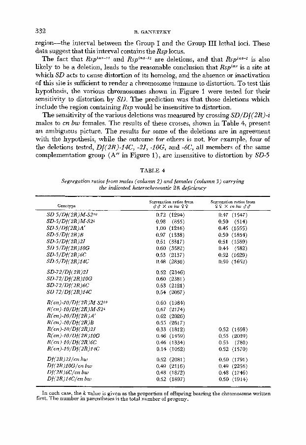
The fact that RspZ1@-" and Rspins-jl are deletions, and that Rspins-l is also likely to be a deletion, leads to the reasonable conclusion that RspZns is a site at which SD acts to cause distortion of its homolog, and the absence or inactivation of this site is sufficient to render a chromosome immune to distortion. To test this hypothesis, the various chromosomes shown in Figure 1 were tested for their sensitivity to distortion by SD. The prediction was that those deletions which include the region containing Rsp would be insensitive to distortion.
The sensitivity of the various deletions was measured by crossing SD/Df(2R)-z males to cn bw females. The results of these crosses, shown in Table 4, present an ambiguous picture. The results for some of the deletions are in agreement with the hypothesis, while the outcome for others is not. For example, four of the deletions tested, Df(2R)-Z4C, -2.7, -ZOG, and -6C, all members of the same complementation group (A" in Figure I), are insensitive to distortion by SD-5
TABLE 4
Segregation raiios from males (column 2) and females (column 3 ) carrying the indicated heterochromatic 2R deficiency
Segregation ratios from Segregation ratios from Genotype dd X cnbw 9 9 9 9 X c n b w d d
SD-S/Df(2R)M-S2'o SD-S/Df (2R)M-S24 SD-S/Df (2R)A' SD-S/Df (2R)B SD-S/Df (2R)ZJ SD-S/Df (2R)IOG SD-S/Df (2R)6C SD-S/Df (2R)I4C
SD-72/Df(2R)2J SD-72/Df(ZR)IOG SD-72/Df(2R)6C SD-721Df (2R)I4C
R(cn)-IO/Df(ZR)M-S21o R(cn)-IO/Df (2R) M-S24 R(cn)-iO/Df(ZR)A' R(cn) -1 O/Df(ZR) B R(cn)-IO/Df(2R)2J R(cn)-IO/Df (2R)IOG R (en) -I O/Df (ZR)6C R(cn)-IO/Df (2R)14C
Df(ZR)2J/cn bw Df(ZR)IOG/cn bw Df (2R) 6C/cn bw D f ( 2 R ) I4C/cn bw
0.72 (1234) 0.98 (855) 1.00 (1216) 0.97 (1338) 0.51 (3317) 0.60 (3582) 0.53 (2137) 0.48 (2830)
0.52 (2346) 0.60 (2381) 0.53 (2121) 0.54 (2067)
0.60 (1384) 0.67 (2174) 0.62 (2026) 0.55 (2617) 0.33 (1812) 0.46 (1459) 0.46 (1334) 0.14 (1052)
0.52 (2081) 0.49 (2116) 0.48 (1872) 0.52 (1897)
0.47 (1547) 0.50 (514) 0.45 (1555) 0.50 (1854) 0.51 (1589) 0.44 (582) 0.52 (1629) 0.50 (1652)
0.52 (1598) 0.55 (2039) 0.53 (780) 0.52 (1570)
0.50 (1791) 0.49 (2256) 0.48 (1746) 0.50 (1914)
In each case, the k value is given as the proportion of offspring bearing the chromo"ne written first. The number in parentheses is the total number of progeny.

MAPPING T H E SD REGION 333
and SD-72. To demonstrate that this insensitivity was the consequence of these chromosomes bearing Rspins, as opposed to carrying a modifier of SD, Sd RspsenE/ Df(2R)-i males were mated to cn bw females. As discussed before, the Sd Rspsens chromosome is expected to distort itself if its homolog carries Rspin8 but not otherwise. As shown, Df(2R)Z4C and Df(2R)2J induce the same degree of suicide behavior as did the Rspin8 cn bw chromosomes. This effect is less marked in the case of Df(2R)IOG and Df(2R)6C7 the k values corrected for via- bility differences being 0.41 and 0.43, respectively. The reason for the difference in strength is not clear, but here too the result suggests the presence of Rspins. Finally, i?one of these four deletions is itself a distorter as indicated by the normal segregation ratios from Df(BR)-i/cn bw males (Table 4).
The insensitivity was shown to be inseparable from the dcletioiis by backcross- ing to the sensitive cn bw stock and allowing three generations of free recom- bination between these chromosomes. The deletion was then reisolated and re- tested for sensitivity to distortion. In no case did this affect the insensitivity of the deletions. Thus, by all of the operational criterja used before to define a Rspins allele, these four deletions would be classified as Rspins mutants, in agreement with the hypothesis presented above.
These results, however, are tempered by the behavior of other deletions in Table 4, which do not fit easily with the simple hypothesis. For example, Df (2R)MS-21°, which spans the entire region, is partially sensitive to distortion, and Df(2R)A’, which should also delete the Rsp locus, is completely sensitive to distortion. In addition, HILLIKER (personal communication) has tested several other deletions covering this region, also isolated as detachments from compound autosomes, and found them to be sensitive to distortion. Why these deletions should remain sensitive to distortion while similar deletions are associated with insensitivity is not clear. Several possible explanations for this apparent conka- diction will be considered in the DISCUSSION. Suffice it to say here that, although the matter remains unsettled, the majority of the data strongly favor the view that Rsp is located in the heterochromatin of 2R and that a deficiency for it leads to insensitivity.
THE Sd LOCUS
Isolation of X-ray induced SD revertants: The same kinds of considerations discussed in respect of the isolation of RspiAS chromosomes apply to the isolation of SD revertants. Thus, if distortion results from the absence of some Sd+ product, or from the loss of an important site, deletion of Sd+ should turn a normal chro- mosome into a distorter. I n addition, it should not be possible to revert an SD chromosome by X rays. Conversely, if Sd is a neomorph, for example, an X-ray- induced deletion of the Sd locus would produce an SD revertant, whereas such a deletion in a normal chromosome would not affect segregation.
To examine these relations, X-ray-induced pr deletions in SD-72 and cn bw chromosomes were constructed and characterized on the assumption that some of these would include the Sd locus owing to the genetic proximity of Sd and pr (SANDLER and HIRAIZUMI 1960b). To isolate these deletions, SD-72/cn bw males

334 B. GANETZKY
and cn bw males were irradiated and crossed to pr cn females and their progeny screened for any pr-eyed off spring. Six p r mutations were recovered in the SD-72 chromosome and 26 in the cn bw chromosome. They were classified as deletions based on their lethality in combination with known deletions for pr. Because a deletion for Sd might be male-sterile, the pr deletions were recovered in both males and females to avoid selecting against a particular class of deletions.
The pr-bearing SD-72 and cn bw chromosomes were tested for their ability to distort a sensitive cn bw chromosome, and the pr-bearing cn bw chromosomes were also tested for their sensitivity to distortion by an unirradiated SD-72 chro- niosonie. These results are presented in Table 5 ; they indicate that none of the pr deletions affected the segegational properties of the chromosomes on which they were induced. The SD-72 chromosomes remain complete distorters, while the cn bw chromosomes remain sensitive to distortion and did not themselves become distorters.
It appears from these results that either Sd is not as close to pr physically as it is genetically, or that the pr deletions are limited ilz size. To distinguish between these two possibilities, the pr SD-72 chromosomes and some of the pr cn bw chro- mosomes were examined cytologically. The largest deletion among the pr SD-72 chromosomes extended from 38B to 39A-C (Figure 4c). All the remaining pr SD-72 chromosomes proved to carry cnly small deletions of several bands around pr, located at 38A8-38B6 on BRIDGES' (1942) revised salivary map (WRIGHT, HOUGETTS aiid SEIERALD 1976). These small deletions were all contained within the region 38A5,6 to 38C1,2. The pr cn bw chromosomes, on the other hand, in-
TABLE 5
Segregation ratios from males of the indicated genotypes crossed by cn bw females
p r deficiency tested'
AI A2 A3 A 4 A5 A6 A7 A8 A9
B5 B10 B11 Bl2 B14
k valuest when homolog is: cn bw SD-72
0.48 (2385) 0.50 (2827) 0.46 (7201) 0.48 (1594) 0.50 (1894) 0.49 (723) 0.48 (3164) 0.42 (3673) 0.43 (2222)
1.00 (1226) 1.00 (2750)
1.00 (2829) 1.00 (2615)
1.00 (1942)
1.00 (1099) 1.00 (1118) 1.00 (1382) 1.00 (1262) 1.00 (1317) 1.00 (13%) 1.00 (1436) 1.00 (1287) 1.00 (817)
The number in parentheses is the total number of progeny. * Lines AI-A9 are pr deficiencies induced in the cn bw chromosome and lines B5-BI4 are pr
t k values in the second column are given as the proportion of progeny carrying the pr deletion; deficiencies induced in the SD-72 chromosome.
in the last column as the proportion of SD-72 progeny.

MAPPING THE SD REGION 335
cluded some very large deficiencies extending almost two numbered sections. The reason for this nonrandom recovery of small deletions in the S D J 2 chromo- some is not known.
It has been argued (SANDLER and CARPENTER 1972) that Sd resides between 37B2 and 39-40. The collection of pr deletions in the cn bw chromosome spans the region 37B to 39C, yet none of these deletions shows any ability to1 distort. From this it may be concluded either that Sd does not result from a deletion of Sd+ G r that Sd does not reside within the region deleted. As will be shown below. tlie former alternative is probably correct.
Assuming this to be m e , the absence of a revertant among the prSD-72 chromosomes must be because the pr deletions induced in this chromosome do not include the Sd locus. The pr deletions in SD-72 span the region 38B to 39A-C. Therefore, Sd must reside outside these boundaries.
In a subsequent effort to induce deletions of the Sd locus, irradiated SD chro- mosomes were directly screened for alterations in their ability to distort. Males carrying SD-5 were irradiated and the irradiated SD-5 chromosome recovered in sons heterozygous with cn bw. These sons were individually mated to cn bw females and screened for a reduction in k value, evidenced by the production o€ white-eyed progeny. Among 4,000 chromosomes so screened, eight were recovered that had very reduced k values; these are designated SDRev chromosomes. Characterization of SD revertants: It is necessary to consider the types of X-
ray-induced events that could reduce the ability of an SD chromosome to distort. Among these are: deletion or inactivation of Sd itself; deletion or inactivation of an essential enhancer of distortion; induction of a suppressor of distortion; mutation of R s z ~ ~ ~ ~ to Rspsens (this would convert Sd RspinX to Sd Rspsens, which does not distort a sensitive homolog) ; and perhaps some chromosome rearrange- ments, several of which have been reported to eliminate distortion entirely or even reverse the direction of distortion (NOVITSKI and EHRLICH 1970).
Because the SD-5 chromosome already carries one or more recessive lethals, it is difficult in this case to ask whether any of the SD revertants had acquired a new lethal by performing complementation tests among the various SD revert- ants, as was done for the Rspins mutations. It was decided, therefore, to first char- acterize the eight SD revertants with respect to the properties of distortion, to determine whether any distinctions could be made among them, and whether any of these behaved as if it carried a deletion or inactivation of the Sd site.
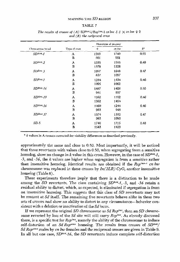
The first test was to measure the k value for each SD revertant to determine if the ability to distort had been partially reduced o r if it had been eliminated entirely. The results of this test and the reciprocal cross are shown in Table 6. The segregation ratio in the reciprocal cross measures viability differences be- tween the two chromosomes and thus allows computation of corrected k values. These corrected IC values reveal that the eight revertants €all into at least two classes: SDRev-2, -7, -16, -19 and -37 all with k values close to 0.50, and SDRev-I, -3 , and -36 with k values around 0.70.
This distinction depends upon Ihe observation of a segregation ratio signifi- cantly less than 0.50 for SD"ev-l, -3, and -36 in the reciprocal cross and the in-

336 E. GANETZKY
TABLE 6
Test for the ability of SDReV chromosomes to distort a sensitive homolog. The cross is: ( A ) SDReV-i/cn bw 3 8 x cn bw 0 0 and ( B ) the reciprocal cross
Phenotype of progeny
Chromosome tested Type of cross + cn bw k'
SDReV-1 A 1813 1389 0.73
SDRev-2 A 745 716 0.48
SDReV-3 A 1202 1371 0.64
SDRev-7 A 1463 1474 0.51
SDReV-I 6 A 623 571 0.50
SDReV-19 A 75 1 680 0.52
SDRev-36 A 2920 1741 0.71
SDReV-37 A 525 695 0.48
SD-5 A 2181 8 0.99
B 773 1584
B 698 61 3
B 839 1712
B 141 1 1486
B 738 688
B 623 630
B 659 983
B 51 1 633
B 1466 1359
* k values in A crosses corrected for viability differences as explained in Table 2.
terpretation that this reflects a reduced viability of these reverted-SD chromo- somes. If this assumption is incorrect-if, €or example, the reduced k values sig- nified instead some type of distorted segregation in the female-the corrected k values for these three revertants would be spuriously high.
In order to test the validity of this assumption, we may consider the expected results for the segregation of the SD revertants from an insensitive homolog. Because, in this case, distortion in the male should be eliminated, the observed k values in the male cross and the reciprocal cross should be comparable to one another, and, further, the corrected k values for the male cross should be close to 0.50. For this reason, the k values were measured in crosses of SDRev/RspinS-' cn bw males by cn bw females and also in the reciprocal cross. The Rspins-l chromosome has already been shown (Table 2) to be completely insensitive to distortion by a normal SD chromosome.
The results of this cross are shown in Table 7. Several p i n t s of interest emerge from these data. First, consistent with the expectation, the observed segregation ratios in the male cross and the reciprocal cross are now similar for all the SD revertants, whereas in the previous set of crosses (Table 6), the observed segre- gation ratios for SDRev-I, 3 and -36 were consistently much higher in the male cross than the reciprocal cross. Secondly, if observed k values in the male cross are corrected in the same way as before, the k values for all the SD revertants are
M A P P I N G THE SD REGION
TABLE 7
The results of crosses of ( A ) SDRev-i/RspinB-l cn bw 8 8 X cn bw 0 P and ( B ) the reciprocal cross
337
Phenotype of progeny
Chromosome tested Type of cross + cn bw k*
A B A B A B A B A B A B A B A B A B
1269 561
1585 1378 1097 83 7
1284 1095 1487 941
1362 1362 1060 963
1074 963
174.0 1543
1740 95 1
1593 1328 1648 1097 1434 1 (E62 1 428 897
1702 1464 1244 948
1392 1 093 1715 1423
0.55
0.49
0.47
0.46
0.50
0.46
0.46
0.47
0.48
* k values in A crosses corrected for viability differences as described previously.
approximately the same and close to 0.50. Most importantly, it will be noticed that those revertants with values close to 0.50, when segregating from a sensitive homolog, show no change in k value in this cross. However, in the case of SDRev-I, -3, and -36, the k values are higher when segregation is from a sensitive rather than insensitive homolog. Identical results are obtained if the Rsp"+-' cn bw chromosome was replaced in these crosses by In(2LR) CyO, another insensitive homolog (Table 8).
These experiments therefore imply that there is a distinction to be made among the SD revertants. The class containing SDRev-l, -3, and -36 retain a residual ability to distort, which, as expected, is eliminated if segregation is from an insensitive homolog. This suggests that this class of SD revertants may not be mutant at Sd itself. The remaining five revertants behave alike in these two sets of crosses and show no ability to distort in any circumstance-behavior con- sistent with a deletion or inactivation of the Sd locus.
If we represent the original SD chromosome as Sd RspinS, then an SD chromo- some reverted by loss of the Sd site will still carry Rspins. As already discussed there, is a specific test for Rspins, namely the ability of the chromosome to induce self-distortion of an Sd RspSens homolog. The results from crosses of SDRev/ Sd Rspsens males by cn bw females and the reciprocal crosses are given in Table 9. In all but one case, SDRCv-16, the SD revertants induce complete self-distortion

338 B. GANETZKY
TABLE 8
Results of crosses of ( A ) SDRev-i/In (2LR) Cy0 8 8 x cn bw 0 0 and ( B ) the reciprocal cross
Phenotype of progeny
Cbromoscme tested Type of cross + CY k*
A B A B A B A B A B A B A B A B A B
558 616
12010 817 756 878
1061 1071 1147 861
1109 647 45 8
1020 990 688
1086 812
1161 1094 1684 1090 1520 1529 1684 1455 1772 1110 1846 896 918
1616 1693 1250 1433 1248
0.46
0.49
0.46
0.46
0.45
0.45
0.44
0.52
0.53
* k values in A crosses corrected for viability differences as described previously.
of the Sd RspRrnr chromosome. 'The amount of self-distortion in these crosses is much greater than that induced by the lispin' mutations described earlier, even though the Sd RspSens chromosome is the same in both crosses. This difference does not necessarily reflect a difference in behavior of the induced RspZns muta- tions and the Rspins carried by the reverted-SD chromosomes, hut more likely is the result of differences in other genes carried on the chromosomes. Thus, for SDReV-l, -3, and -36, which retain some ability to distort and may, in fact, still carry Sd, here would be two Sd loci in SDEev/Sd RspSCnns males, both acting a t the sensitive-Rsp locus of the Sd Rspsens chromosome. The other SD revertants, which may have lost the Sd locus, should still carry the complete set of enhancers of distortion present on an SD chromosome (SANDLER and HIRAIZUMI 1960a; MIKLOS 1972a) and partly lacking on the suicide chromosome since it is an ex- change product (HARTL 1975). The addition of these enhancers should enable the Sd locus carried on the suicide chromosome to operate at full capacity. The out- come in either of these cases is complete self-distortion of the suicide chromosome. The RspZnS mutations induced in the cn bw chromosome are derived from non- distorters and lack enhancers of distortion, so that they should be unable to produce complete self-distortion.

MAPPING THE SD REGION 339
TABLE 9
Test for the ability of SDRev chromosomes to induce self-distortion of R(cn)-IO (= Sd Rspsens). The cross is: ( A ) SDRev-i/R(cn)-lO 8' 8 x cn bw Q 0 and ( B ) the reciprocal cross
Chromosome tested Type of cross
A B A B A B A B A B A B A B A B A B
Phenotype of progeny
cn + k'
10 959 0.004
0 1783 0.00
7 2223 0.002
6 2157 0.002
2055 942
IO65 958
1655 1117
940 704
736 677
676 5 72
2063 1411
469 388
311 352
1494 1153 0.54
1 2254 0.0004
4 757 0.004
6 1670 0.003
0 486 0.00
* k values are given as the proportion of R(cn)-iO bearing progeny. Corrections made for viability differences in A crosses as before.
In any case, whatever the cause of this difference between the R S ~ " ~ cn bw and the SDRev chromosomes, the important point here is that all of the revertants except SDRe'-16 induce suicide behavior, demonstrating that they still carry R . Y ~ % ~ ~ .
SDRev-16 behaves differently from the others and thus defines a third class of revertant. In this case, the suicide chromosome is recovered at least as often as the SD revertant. This outcome would be explained if SDRev-16 carried a newly induced suppressor of distortion, or mutation from R s ~ * ~ ~ to Rspsens. In the latter case, the original Sd RspinS chromosome would now be Sd Rspsens, which is itself a suicide combination of genes. If SDRev"-16 were such a chromosome, it should have distorted itself in the cross of SDReV-l6/Rspins--' cn bw males by cn bw fe- males and given a k value under 0.50, which it did not. Another test of this pos- sibility is to ask whether SDRev-16, but not the other revertants, can be distorted by another SD chromosome. The results from crosses of SD-72 bw/SDRev males to cn bw females, and the reciprocal crosses are shown in Table IO. It is evident that the k value in each case is close to 0.50, indicating that all of the revertants, including SDReu-l 6, are insensitive to distortion. This experiment rules out the possibility that SDReu-16 carries a sensitive responder and suggests rather, that it carries a suppressor of distortion. SDRe2-16 has been tested further, in preliminary
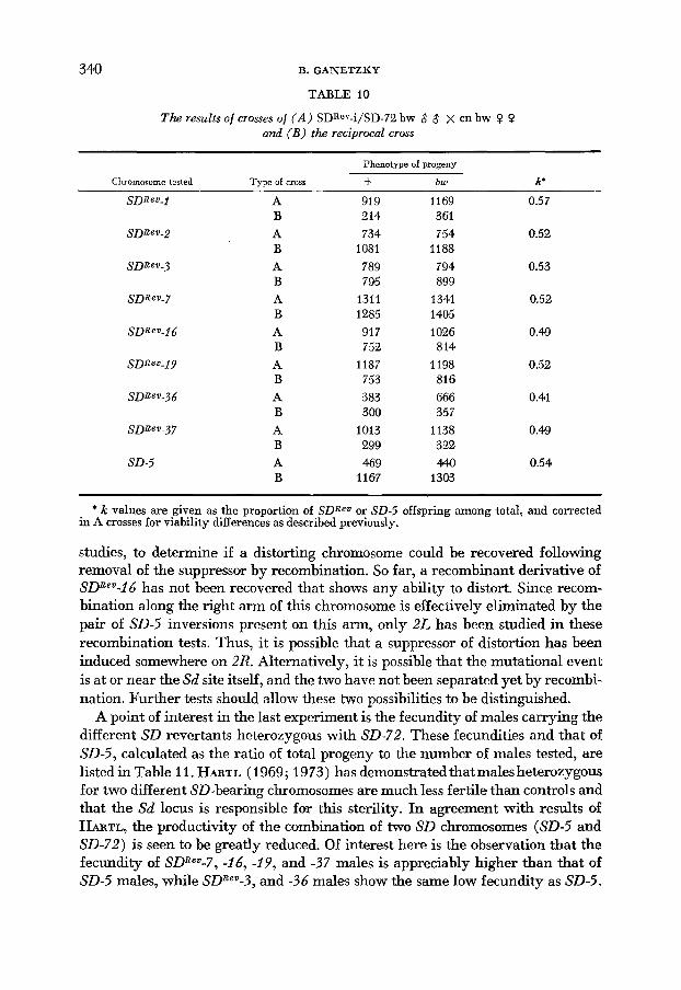
340 B. GANETZKY
TABLE 10
The results of crosses of ( A ) SDRe''-i/SD-72 bw 8 8 X cn bw 0 0 and ( B ) the reciprocal cross
~ ~
Phenotype of progeny
Chromosome tested Type of cross -I- bw k'
A B A B A B A B A B A B A B A B A B
919 214 734
1081 789 795
1311 1285 917 752
1187 753 383 300
1013 299 4 9
1167
1169 361 754
1188 794 899
1341 1405 1026 814
1198 816 666 357
1138 322 440
1303
0.57
0.52
0.53
0.52
0.49
0.52
0.41
0.49
0.54
* k values are given as the proportion of SDRev or SD-5 offspring among total, and corrected in A crosses for viability differences as described previously.
studies, to determine if a distorting chromosome could be recovered following removal of the suppressor by recombination. So far, a recombinant derivative of SDRev-16 has not been recovered that shows any ability to distort. Since recom- bination along the right arm of this chromosome is effectively eliminated by the pair of SD-5 inversions present on this arm, only 2L has been studied in these recombination tests. Thus, it is possible that a suppressor of distortion has been induced somewhere on 2R. Alternatively, it is possible that the mutational event is at or near the Sd site itself, and the two have not been separated yet by recombi- nation. Further tests should allow these two possibilities to be distinguished.
A point of interest in the last experiment is the fecundity of males carrying the different SD revertants heterozygous with SD-72. These fecundities and that of SD-5, calculated as the ratio of total progeny to the number of males tested, are listed in Table 11. HARTL (1969; 1973) has demonstratedthatmales heterozygous for two different SD-bearing chromosomes are much less fertile than controls and that the Sd locus is responsible for this sterility. In agreement with results of HARTL, the productivity of the combination of two SD chromosomes (SD-5 and SD-72) is seen to be greatly reduced. Of interest here is the observation that the fecundity of SDRev-7, -26, -19, and -37 males is appreciably higher than that of SD-5 males, while SDRev-3, and -36 males show the same low fecundity as SD-5.

MAPPING THE SD REGION
TABLE 11
Fecundity* of SDReV and SD-5 males when heterozygous with the indicated homolog
34 1
Number of progeny per male when homolog is:
Chromosome tested SD-72 Rsp'La-I cn bw
SDReV-I 116 201 S D R e V - 2 106 212 SDReu-3 61 183 SDRev-7 176 181 SDRev-16 134 194 SDReV-I9 159 2w SDRev-36 48 177 SDReV-37 I 43 164 SD-5 34 181
* These fecundities are calculated by dividing the total number of progeny (results shown in Tables 7 and IO) by the number of fertile males.
SDRev-I, and -2 males exhibit intermediate fecundity. Those revertants that are most fertile are those that behaved as deletions or inactivations of Sd, while those that have the same low fertility as SD-5 are those that show residual dis- tortion and therefore likely retain the Sd locus. The revertants with intermediate levels of fertility are ambiguous in that one of these, SDRev-I, behaves as an incompletely reverted SD, while the other, SDReu-2, is indistinguishable from the completely reverted SD chromosomes. Nonetheless, the overall pattern pro- vides additional evidence in support of the division of the SD revertants into different classes.
It should be pointed out that the data presented above were not derived from experiments specifically designed to measure male fertility as were the more precise experiments of HARTL (1969,1973). However, there are several reasons to believe that these data accurately measure male fecundity. First, the numbers correspond well with those of HARTL for similar chromosome combinations. Sec- md, the numbers are reproducible. For example, in the cross involving SDRev/Sd RspSens males, a similar pattern of fecundities as that shown in Table 11 is ob- served. Finally, the differences in fecundity are specifically related to the SD sys- tem, because the fecundity of the incompletely reverted SD chromosomes in- creases when segregation is from an Sd+ Rspins homolog, a situation where no dis- tortion OCCUTS. (Male fecundities for this cross shown in Table 11 demonstrate that all the revertants have the same level of fertility.)
In summary, the eight SD revertants can be divided into at least three classes: (1) SDRev-I, -3, and -36, which retain some ability to distort and thus presumably still carry Sd; (2) SDReu-16, which shows no distortion but fails to induce suicide behavior, suggesting it carries a suppressor of Sd; and ( 3 ) SdRev-2, -7, -19, and -37, which show no distortion and whose behavior in a variety of crosses is consistent with a deletion or inactivation of Sd.
Having divided the SD revertants into these categories it remains to determine the location of any associated chromosomal lesion. Of particular interest are those
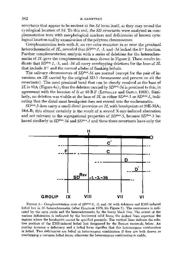
342 B. GANETZKY
revertants that appear to be mutant at the Sd locus itself, as they may reveal the cytological location of Sd. To this end, the SD revertants were analyzed in com- plementation tests with morphological markers and deficiencies of known cyto- logical location and by examination o i the polytene chromosomes.
Complementation tests with It, an eye color mutation in or near the proximal heterochromatin of 2L, revealed that SDReu-l, -3, and -36 lacked the It + function. Further complementation analysis with a series of deletions for the heterochro- matin of 2L gave the complementation map shown in Figure 2. These results in- dicate that SDReu-l, -3, and -36 all carry overlapping deletions for the base of 2L that include It+ and the normal alleles of flanking lethals.
The salivary chromosomes of SDRev-36 are normal (except for the pair of in- versions on 2R carried by the original SD-5 chromosome and present on all the revertants). The most proximal band that can be clearly resolved at the base of 2L is 40A (Figure 4a) ; thus the deletion carried by SDRe"-36 is proximal to this, in agreement with the location of I t at 40 B-F (LINDSLEY and GRELL 1968). Simi- larly, no deletion was visible at the base of 2L in either SDReu-3 or SDReu-1, indi- cating that the distal-most breakpoint does not extend into the euchromatin.
SDBeu-3 does carry a small distal inversion on 2L with breakpoints at 29E-30A; 34A-B; this almost certainly is the result of a second X-ray-induced aberration and not relevant to the segregational properties of SDReu-3, because SDReu-3 be- haved similarly to SDRe"-36 and SDReu-I and these three revertants have only the
FIGURE 2.-Complementation map of SDRev-I, -3, and -36 with deletions and EMS-induced lethal loci in 2L heterochromatin (after HILLIKER 1976, his Figure 5). The centromere is indi- cated by the open circle and the heterochromatin by the heavy black line. The extent of the various deficiencies is indicated by the horizontal solid lines; the dashed lines represent the regions where the breakpoint cannot be specified precisely. The vertical lines indicate the rela- tive position of the EMS-induced lethal loci designated by the Roman numerals below. An overlap between a deficiency and a lethal locus signifies that this heterozygous combination is lethal. Two deficiencies are lethal in heterozygous combination if they are both drawn as overlapping a common lethal locus; otherwise the heterozygous combination is viable.

M A P P I N G TIEE SD REGION 343
deletion near It in common. SDRev-1 is involved in a translocation with the tip of 3R translocated to the base of the right arm of the second chromosome. Transloca- tion tests confirm that SDRev-l, but none of the other revertants, carries a translo- cation. This translocation too is surely the result of a second X-ray-induced event and, for the same reasons as in the case of SDRev-3, does not influence the segrega- tional behavior of SDRev-1.
The conclusion from the genetic and cytological examination of SDRev-l, -3, and -36 is that there is a locus carried by SD chromosomes at the base of 2L near It, which behaves as a strong enhancer of distortion; we name it E(SD). This locus is deleted in SDRev-l, -3, and -36; it is the absence of E(SD) that accounts for the reduced, but not entirely eliminated, ability to distort.
The other SD revertants were not mutant at It, nor did they carry newly in- duced lethals contained in any of the heterochromatic 2L cieficiencies tested. To determine whether any of these revertants carried detectable X-ray-induced le- sions elsewhere on the base of 2L, they were examined in complementation tests with a series of deletions that cover the base of 2L from 36F to 40A (Figure 3) . SDRe"-2, -7, -16, and -19 all proved fully viable in combination with each of the deficiencies, implying the absence of any newly induced lethals in this region. The salivary chromosomes of these revertants confirmed this result; there were no
FIGURE 3.-Diagra"atic representation of the base of 2L (after WRIGHT, HODGETTS and SHERALD 1976, their Figure 2) showing the cytological extent of the deficiencies used in this study. The location of several genes is also shown above the chromosome. Those deletions which are indicated as overlapping are lethal in heterozygous combination. Deletions marked with a star were kindly supplied by DR. R. HODGETTS; the others were recovered in the present study.

r"
FIGURE 4.--Salivary gland preparations of some of the chromosomes analyzed in these studies: (a) SDRW-36. This chromosome illustrates the normal banding pattern at the base of 2L. There
is no cytologically detectable lesion associated with this chromosome, although it carries a proximal deletion as explained in the text.
(b) SDReV-37 = Df(2L)37D2-7; 38A6-B2. Segment between arrows is deleted in the deficient homolog.
(c) Df(2L)prB1PSD-72 = Df(2L)38A-B; 39A-D. Segment between black arrows is deleted in deficient homolog. The bracket indicates that the breakpoint in this region is uncertain. Also shown is the pericentric inversion associated with SD-72. The left breakpoint is between 39D and 39E (upper double arrow) and the right breakpoint is at 42A (bottom double arrow).

MAPPING THE SD REGION 345
detectable lesions at the base of 2 L (or elsewhere in the chromosome comple- ment). In the case of these revertants, the event that caused the loss of the ability to distort is not associated with any cytologically detectable chromosomal altera, tion. Thus, SDRev-2, -7, and -19 all behave as deletions or inactivations of Sd (Sdz~ev-lh prcbably carries a suppressor of SD; see above) , yet none is associated with a mutational lesion ai the base of 2L that is detectable either cytologically or in complementation tests. It is possible, therefore, that these represent "point" mutations at the Sd locus, but it is not yet possible to eliminate alternative explanations.
The remaining revertant, SDRev-37, is more informative. The results of comple- mentation tests with SDRe"-37 are shown in Figure 3. These tests indicate that SDRev-37 is a deletion, since it overlaps two deletions which do not themselves overlap. Examination of the polytene chromosomes (Figure 4b) shows that this was indeed the case; the breakpoints of the deletion are 38A6-B2 on the right and 37D2-7 on the left. Genetically, this deletion is from just to the left of pr to just to the right of Tft. SDRev-37 was characterized on genetic grounds as a revertant with properties consistent with a deletion o r inactivation of Sd. Together with the cyto- logical demonstration of a deletion at the base of 2L, this strongly argues that both are the consquence of the same event and therefore that the cytological location of the Sd locus is in the region spanned by the SDRev"-37 deletion.
DISCUSSION
The Rsp locus: Five chromosomes that carried X-ray-induced mutations to in- sensitivity to distortion were recovered in these studies. These were classified as mutations at the Rsp locus, resulting in a change from Rspsens to Rspins, based on the following evidence. The mutation conferring insensitivity, in each case: (1 ) is located between pr and cn; (2) does not cause distortion of a sensitive homo- log; (3) results in insensitivity to distortion by three different SD chromosomes; and (4) is capable of inducing the suicide response in an Sd Rspsen8 homolog. Com- plementation tests with three of the Rspins cn bw chromosomes that were homo- zygous lethal indicated that all three shared a common lethal in the centric heterochromatin of 2R and that at least two of these chromosomes are deleted in this region. From this it is inferred that the Rsp locus is in the centric heterochro- matin of 2R, and that a deletion for the Rsp locus results in a Rspins phenotype. This agrees with, and extends, previous information on the location of the Rsp site. The first evidence on this point came from the recombination studies of SANDLER and HIRAIZUMI (1960b) who showed that the locus of insensitivity was between pr and cn, although they could not determine whether it mapped to 2L or 2R. Moreover, SANDLER and HIRAIZUMI were unaware that the SD chromo- some that they used carried a pericentric inversion, though they did realize that there was a chromosome aberration in the region. causing a ten-fold reduction in map distance between pr and cn. They imagined that it was the aberration itself that produced insensitivity because insensitivity was genetically inseparable from the reduction in the pr-cn map distance. After LEWIS (1962) reported the

346 B. GANETZKY
presence of a pericentric inversion on that SD chromosome but not on other S D chromosomes, the data were reinterpreted to mean that insensitivity was due to a locus within the pericentric inversion, rather than to the aberration itself (HIRAIZUMI and NAKAZIMA 1967; SANDLER and CARPENTER 1972).
Because the breakpoints of the pericentric inversion are at 39-40 and 42A (LEWIS 1962), the Rsp locus must be located within these limits. SANDLER and CARPENTER (1972) showed that the RF,D site was not located ia salivary regions 41 to 43A, because a Y chromosome into which this material was inserted was not distorted, while the deficient second chromosome from which the piece was removed was distorted. Thus. the best information available had mrrowed the location of the Rsp site to between 39-40 and 41, which agrees exactly with the location of Rsp reported here.
The analysis of the Rspin* cn bw chromosomes in these studies indicates that the location of Rsp is in the heterochromatin of 2R between HILLIKER’S (1976) Group I and Group I11 lethal loci. One difficulty with this placement of the Rsp locus is that some chromosomes, including Df (2R)M-S21° and Df(2R)A’, ap- parently deleted for the region (Figure 1 ) , are sensitive to distortion (Table 4) . There are several possible explanations of this apparent inconsistency. One is that the Rsp site is not located in exactly the same place in chromosomes of different origin. For example, a rearrangement entirely limited to the heterochromatin would normally be indetectable, but could change the location of the Rsp locus. The possibility that Rsp has the capacity to remove itself from the chromosome and reintegrate at another nearby locus also canpot be eliminated; indeed, MINA- MORI (1970) has reported behavior of this type for the extrachromosomal ele- ment, delta, another distorting system. Another possibility is that the Rsp site actually occupies a location different from that shown on the complementation map in Figure 1 (between the Group I and Group I11 lethal loci). The critical chromosome in this analysis is RsptnS--’, which is represented in Figure 1 as a de- letion contained in this region. However, because Rspans-ll and R s ~ ~ ~ ~ - ~ ~ are dele- tions which extend through the most proximal h o w n locus on 2R, it may be that Rsp is actually located in the most proximal portion of the 2R heterochromatin, and that RspZnS-’ is not a deletion but a double “hit” of some type. with one “hit” at rl and a more proximal one at the Rsp locus. producing insensitivity.
If this notion is correct, Rsp may be located in the region just adjacent to the centromere such that a break between the centromere and Rsp is expected to be rare. As a consequence, in the construction of a compound second chromosome both C(2L) and C(2R) would carry Rsp, and only rarely would a heterochro- matic deletion isolated as a detachment from these compounds be deleted for Rsp, thus explaining the sensitivity of these deletions. Because the breakpoints of a deletion proximal to the Group I locus (Figure 1 ) cannot be specified with pre- cision, it may be the case that those deletions that are insensitive extend more proximally than those that are sensitive, accounting for the differences seen among the deletions tested.
At this point it is not possible to decide in favor of any of these possibilities. However, the analysis of the X-ray-induced Rspin8 mutations themselves is self-

MAPPING THE SD REGION 347
consistent, and the data clearly suggest that the Rsp site is located somewhere in the heterochromatin of 2R, and that a deletion for this locus renders the chromo- some insensitive to distortion. How general this is for second chromosomes differ- ent from the cn bw chromosome can be determined only by mapping X-ray- induced mutations to insensitivity on such chromosomes.
The mapping of Rsp to the heterochromatin, a region sparse in structural genes, is consistent with the views of SANDLER and CARPPNTE~ (1972) and HARTI- (1973), who picture Rsp as a regulatory sile rather than a structural gene. The question that arises is what is the function of the Rsp site? As HARTL (1973) has pointed out, the facts that transcription ceases after meiosis (OLIVIERI and OLI- VIERL 1965; HENNIG 1967; GOULD-SOMERO and HOLLAND 1974) and that sperm almost devoid of chromosomes complete development and function normally (MULLER and SETTLES 1927; MCCLOSICEY 1966; LINDSLFY and GRELL 1969), argue against the idea that the effect of SD can be to cause the SD+ chromosome to fail to produce a normal product, which failure results in sperm lethality. On2 is forced to the opposite view, namely. that the inactivation of genes during sper- miogenesis is necessary for the completion of the process and that the action of SD is to cause genes on its homolog to remain active; it is this unscheduled activity that causes the affected sperm to be inviable. The electron microscopic studies of TOKUYASU, PEACOCK and HARDY (1 976), showing the failure of the chromatin tc; condense in the SD+ nuclei, is corrsistent with this idea. Moreover. this would explain the dominance of sensitivity apparent in the dysfunction 01 SD/SD+ gametes ( SANDLER and CARPENTER 1972).
If these ideas are correct, the Rsp locus is a site of considerable importance, upon whose activity or inactivity the state of the rest of the chromatin in the sperm nu- cleus depends. The evidence presented here indicates that a deletion of the Rsp site is equivalent to Rspins, which in respect of the ideas discussed above, suggests that the complete absence of Rsp prevents the unscheduled activity. Formally, the Rsp locus has some similarity to an operator or promoter region.
This leads to a consideration of what the “normal” function of the RspSens IOCUF might be. The only effect that can be definitely attributed to RspSCns is that it causes a chromosome carrying it to be distorted by SD, a peculiar reason to exist. HARTL (1973) has argued that R S ~ ~ ” ‘ ~ ~ must be part of a normal regulatory mecha- nism operating during spermiogenesis, and that Sd and RspCns are mutations in this system. This is quite appealing, but direct evidence indicating a function for the RspsPns locus in the absence of Sd is lacking. The recovery of X-ray-induced RspZnS mutations that are homozygous viable and fertile seems to indicate that the RspSens function is dispensable. In view G€ this, it is puzzling why any second chromosome isolated from nature would show sensitivity to distortion, rather than having disposed of the RspSens locus completely. One possibility is that the RspZns mutants that are liomozygous viable and ferhle are not completely deleted for the Rsp locus, but represent changes in base sequence or partial deletions of the Rsp locus. This might affect the interaction of Rsp with Sd but still allow it to perform its indispensible functions.
The E (SD) locus: Based on the following criteria, three of the eight SD-rever-

348 B. GANETZKY
tants define a distinct class, not mutant at the Sd locus, but with reduced distor- tion: (1) a reduced, but not entirely eliminated, ability to distort; (2) reduced fecundity in combination with another SD chromosome, a characteristic of homo- zygosity for Sd; (3) a newly induced deletion that includes the It+ locus and ex- tends into the centric heterochromatin o i 2L.
It seems clear that these revertants are not mutant at the Sd locus, not only from their segregational properties, but also because of the location of the muta- tional event. SANDLER and HIRAIZUMI (1960b) demonstrated ihat the Sd locus is distal to the leftmost breakpoint of the pericentric inversion carried by SD-72. LEWIS (1962) reported that this breakpoint is at 39-40. I have confirmed this and narrowed the location of the break to 39D-E (Figure 4c). Because It is proximal to this (LINDSLEY and GRELL 1968), deletions extending proximal to It do not in- clude Sd. Direct cytological analysis of these revertants indicate that the deletion defined by complementation tests must be proximal to 40A. Thus, these rever- tants define a new component of the SD system, located in or near the centric heterochromatin of 2L, whose behavior is that of strong enhancer of Sd; i t has been given the name E(SD).
This E(SD) would not have been evident in the previous studies of the SD re- gion by SANDLER and HIRAIZUMI (1960b) and HIRAIZUMI and NAKAZIMA (1967), because their analyses dealt with specifying whether a recombinant product of an SD and SD+ chromosome pair could distort a sensitive homoiog and whether it was sensitive to distortion. These criteria are insufficient to recognize the pres- ence of E(SD) because any exchange that separates Sd from E(SD) also separates Sd from RspXns, producing a chromosome of the genotype Sd E(SD)+ Rspsens, while an exchange to the other side of E(SD) produces an Sd E(SD) RspSens bearing recombinant. Both of these recombinants would be sensitive to distortion, but neither would distort a sensitive homolog, owing to the fact that both carry RspSens (HARTL 1974; 1975). MIKLOS (19724 has proposed the existence of posi- tive modifiers of distortion at the base of the second chromosome; these too must differ from E(SD) because MIKLOS deduces the existence of these modifiers from a re-examination of the data of SANDLER and HIRAIZUMI (1960b) and HIRAIZUMI and NAKAZIMA (1967), which, as just discussed, are insufficient to reveal the presence of E(SD).
The presence of E(SD) does, however, suggest itself in HARTL’S (1975) work. The Sd Rspsens chromosomes that he recovered fall into two distinct classes with respect to suicide behavior-those that are complete self-distorters (k = 0.00) and those that self-distort less strongly (k = 0.15 - 0.20). The magnitude of the difference in k for these chi-omosomes corresponds quite closely to the reduction in k observed in the present study for those SD revertants missing E(SD). More- over, HARTL (1975) specifically reports that those Sd RspSens recombinants re- covered from an exchange distal to p r [that of necessity separates Sd from E(SD) because, as discussed in the next section, Sd is located to the left of p r ] are always in the class that are incomplete self-distorters, while Sd RspSens recombinants from ail exchange between p r and crz may show either complete or partial self-distor- tion. HARTL notes that these results suggest the existence of an enhancer of dis-

M A P P I N G THE SD REGION 349
tortion. located in the centrometric region, so that an exchange between Sd and Rsp%ns is almost always between Sd and the enhancer. It is likely that this en- hancer of distortion corresponds to the locus defined by SDRev-I, -3, and -36 in the present studies.
Further evidence on the existence and location of E(SD) has been provided by SHARP and HOLM (personal communication), who screened for X-ray-induced It deletions in SD-5 and found that several of these had reduced k values.
An examination of HARTL'S data (1 975, his Table 3) suggests that E(SD) en- hances distortion in repulsion as well as in coupling with the Sd locus. It was men- tioned above that, owing to the location of E(SD), most Sd RspSCnS recombinants will not carry E(SD); the complementary product, on the other hand, should therefore usually carry both E(SD) and Rspin8. If E(SD) is capable of acting in repulsion, we would expect Sd E(SD)+ Rspsens/Sd+ E(SD) Rspins heterozygotes to show the same amount of self-distortion as Sd E(SD) RspSensJSd+ E(SD)+ Rspins heterozygotes. Males of the first genotype are represented by the more weakly self-distorting Sd RspSens recombinants heterozygous with their comple- mentary recombinant product. Males of the second genotype are constructed by making a completely self-distorting Sd RspSens chromosome heterozygous with a homolog carrying Rspins but not E(SD). In(2L + 2R)Cy bw is a chromosome of the latter type, carrying a naturally occurring Rspins (HARTL 1975) but, because it is not derived from an SD chromosome, in all probability not carrying E(SD). The k value in these males is about 0.1 7, which corresponds closely to the k values of 0.15 - 0.20 observed for males of the first type.
Finally, based on the analysis above, males which may be represented as Sd E(SD) Rspsens/Sd+ E(SD) Rspins are shown by Hart1 to have a k value of 0.00 (complete self-distortion). Males represented by Sd E(SD) + Rspsens/Sd+ E(SD) R S P ~ " ~ show very little, if any, distortion. Thus, there is a dosage effect of E (SO); Sd in the presence of two copies of E(SD) distorts more strongly than In the presence of one copy, whether this copy is in coupling or repulsion with Sd, and this in turn results in more distortion than the absence o€ E(SD).
The Sd locus: Of the eight X-ray-induced nondistorting SD-5 chromosomes re- covered in these studies, four appear to be mutations at the Sd locus itself. The evidence for this conclusion is that these four revertants exhibit: (1 ) complete loss of ability to distort; (2) insensitivity to distortion by another SD chromo- some; and (3) increased fecundity, relative to an unirradiated SD-5 chromo- some, when heterozygous with SD-72.
Of these four, only one (SDRe"-37) was associated with a detectable lesion; the lesion is a deficiency with breakpoints at 37D2-7 and 38A6-B2. It seems most reasonable to conclude that the Sd locus is between these breakpoints. Because only one of the SD revertants has a detectable lesion in this region, the possibility that the deficiency associated with SDRev-37 is unrelated to the reversion of the S D phenotype cannot be eliminated. However, this placement of Sd is in agree- ment with eviderxe from other sources and helps explain heretofore puzzling results from earlier recombination studies of SD chromosomes.
The important new piece of information inherent in this placement is that Sd
350 B. GANETZKY
is very close to, but to the left of pr, rather than to the right as previously proposed (SANDLER and HIRAIZUMI 1960b; HIRAIZUMI and NAKAZIMA 1967; SANDLER and CARPENTER 1972). This is suggested not oniy by the deficiency associated with SDRev-37, but also by the recovery of a pr ddetim in the SD-72 chromosome (Figure 4c) that deletes almost all of the rnaterial between pr and the pericentric inversion, but does not affect the SD phenotype. Other evidence supports the contention that Sd is to the left of pr. HARTL (1975) reports the recovery of Sd R S ~ ~ ~ “ ’ (suicide) chromosomes as recombinants from R - l / I n (2L)Cy, C y b pr cn females, where R-I (which stands for “recombinant-1” and is unrelated to SDR-‘ = SD-Rona) is an SD chromosome of the SD-5 type, carrying both 2R in- versions bui no pericentric inversior?, and In(2L)Cy. Cy b p i cn is a chromosome sensitive to distortion. An Sd RspSe’lS combination is produced by an exchange be- tween the Sd and RspZnS loci carried on R-l . What is significant is that some of the suicide chromosomes have picked up the markers pr and cn. The simplest ex- planation of this result is that Sd is between b and pr, and that these recombinants result from a single exchange between Sd and pr, picking up RspSens and cn in the process. If Sd were proximal to pr, a triple crossover is required to produce recombinants of this type. TANZARELLA et al. (1972) report that SDR-’ maps genetically at 52.9 on the second chromosome. Although the data on which this mapping is based are not reported, their location is consistent with Sd being to the left of pr.
HARTL’S (1974) recent recombination experiments using Tft, a marker 1.3 map units to the left of pr, demonstrated that Tf t is distal to Sd, and therefore that Sd is located between Tf t and pr . The cytological location of T f t is between 36F10 and 37B7, and pr is at 38A8-38BG (WRIGHT, HODGETTS and SHERALD 1976). This corresponds closely to the cytological location of Sd proposed above, based on the limits of the deficiency associated with SDReu-37. Thus, the evidence strongly favors the view that SDRer-37 is a deletion of the Sd locus.
Models of segregation distortion: The analysis of SD revertants makes i t POS-
sibie to rule out one recept suggestion concerning the SD system. Based on simi- larities between SD and the sc4sc8 drive system. FEACOCI~ and MIKLOS (1973) suggested that, “the different Sd alleles could be deficiencies of varying extents.” The fact that AD is X-ray revertable, while SD+ chromosomes in which deletions along the entire base of the second chromosome have been induced show no tefidency whatever to distort, makes this notion untenable.
The possibility that the SD system involves an abnormality in the transition from lysine-rich to arginine-rich histones that occurs in Drosophila spermio- genesis has been raised (TOKUYASU, PEACOCK and HARDY 1976). The localiza- tions of Sd and Rsp reported here show that neither is at the site of the histone genes, which are at 39D-E (PARDUE et al. 1972). This does not, however, necessa- rily invalidate the notion because, in the first place, the SD system could be in- volved in the regulation of the histone transition event, though neither Sd nor Rsp code for histones, and, in the second place, only the somatic histone genes have been localized by in situ hybridization; structural genes for the sperm histones may be different and located elsewhere. It may in this context be worth noting

MAPPING THE SD REGION 35 1
that the Sd region is shown on BRIDGES' (1935) salivary map to pair ectopically with the region containing the histone genes. Direct evidence lending further support to the idea of a connection between SD and the histones has been pro- vided recently by KETTANEH and HARTL (1 976), who have shown by cytochem- ical techniques that the histone transition does not occur in males homozygous f o r SD.
The results described here also bear on an important prediction of HARTL'S (1973) model of segregation distortion. Briefly, HARTL'S model is that Sd codes for a multimeric regulatory protein that must bind with the Rsp region in order for normal spermiogenesis to occur. Regulatory Sd+/Sd+ homomultimers can bind to both Rspsens and Rspins, resulting in neither distortien nor sperm dys- function; Sd+/Sd heteromultimers bind only at Rspins, resulting in the gametic lethality of Rspsenn"-bearing spermatids; Sd/Sd homomultimers bind neither at RspSens nor' Rspins. This model accounts for distortion in Sd Rspins/Sd+ RspSen' males, suicide behavior in Sd Rspsens/Sd + RspZns males, the absence of distortion in Sd Rspzvs/Sd+ Rspins males, and the near sterility of homozygous SD males.
A prediction from this model is that malm carrying Sd and a deficiency for Sd would be sterile or nearly so, because only Sd/Sd homoniultimers can be pro- duced. However, it was observed in these studies that the various deletions at the base of 2L shown in Figure 3 in combination with an SD chromosome are male Iertile.
Even mwe revealing is the finding that SDRet'-37, which we have argued is a deletion of Sd, has greater fecundity in combination with SD-72 than does the unirradiated SD-5 chromosome (Table 1 1 ) . This implies that the sterility of homozygous SD males is not the consequence of a failure of an Sd/Sd homomul- timer to carry out a required function, but suggests. rather, that each SD-bearing chromosome is actively doing something deleterious to the other, causing both to become gametic lethals. A deletion of Sd on one chromosome would then restore partial Iertility.
This leads to a model of distortion quite similar to that of HARTL'S, but with the following revision: rather than the binding of an Sd product at the Rsp locus being a necessary step in spermiogenesis. it is this binding that causes sperm dys- function (perhaps by preventing the chromosomal inactivation which would otherwise occur). A function is not ascribed to the Sd+ locus in this model; indeed, Sd+ may simply be the absence of Sd. Finally, it is assumed that the Sd product binds more readily to Rspsens than to AspavS and that the amount of Sd product is limiting with respect to the number of binding sites available. In the simplest case, each Sd locus may be thought of as producing a single molecule of product and each Rsp locus capable of binding one such molecule. For simplicity we will not concern ourselves here with the possible role of E(SD), o r other enhancers or suppressors of SD, except to note that they might well influence the degree to which the Sa! product binds at Rsp (HARTI. 1973).
The model accounts, in a straighforward way, for distortion and suicide be- havior. In addition, it can explain several other observations. Thus. if the Sd product, when not competed for by a RspSens locus, can bind to a site; ir,

352 B. GANETZKY
Sd Rsptns/Sd+ Rspins males there should be gametic dysfunction of either one homolog or the other but not both from any one spermatocyte. This would result in a k value of 0.50, but in a reduction in fecundity of one-half; this is the result reported by HARTL (1969). In homozygous SD maies, two copies of the Sd procl- duct would be produced, both of which are free to attach at an unbound Rsp site, resuIiiiig in the near sterility seen in these males. Those cases reported by HARTL (1973) that show partial complementation for fertility are understandable if the a€finity of an Sd product for the Rspins site varies depending upon the particular Sd and Rspinns in question.
If the Sd locus were deleted or inactivated, a homozygous SD (i.e., Sdi Rspin8/ Sd, RspZnS) combination would be converted to Sd Rspsn8/ [Sd] RspZns, which in this model is the equivalent of Sd Rspins/Sd+ Rspins. The fertility of these males should be only half that of normal males, but should be much higher than that of homogygous SD males. Thus, the model explains the increased fertility of SD-72/SDRev-37 males compared to SD-72/SD-5 males.
It will be noticed that many features of the proposed model bear a strong resem- blaiice to a recent model of mammalian X-chromosome inactivation (BROWN and CHANDRA 1973). That model also invokes the existence of a locus which produces a single “informational entity” (corresponding to the Sd product in the present nicdel) that binds to a specific locus (equivalent to Rsp) , on either of the two X chromosomes present in the nucleus causing that X chromosome to remain active \vhiie its homolog is inactivated. These ideas of BROWN and CHANDRA formed the basis of the revision OI the H-~RTL (1973) model suggested here.
It is apparent that segregation distortion and X-chromosome inactivation share many similarities. In both systems, two homologs, initially equivalent, have dif- ferent fates; one of the homologs remains active while the other becomes inactive. In both cases, the state of the entire chromosome appears to depend ultimately on a primary eff eci at a single regulatory site ( CATTANACH, POLLARD and PEREZ 1970; RUSSELL and MONTGOMERY 1970). There are even alleles of a controlling element in mice with the property that an X chromosome carrying such an allele is more likely than its homolog to be inactivated (CATTANACH 1975). The ap- parent similarities between the two systems may not be lortuitous. LIFSCHYTZ and LINDSLEY (1972) have pointed out the significance of X-chromosome inacti- vation as a normal step in spermiogefiesis in male heterogametic organisms, and suggest that the inactivation of X chromosomes in &e somatic cells of female mammals evolved from this system. It is not unreasonable to imagine that. a can - trolling system also exists to bring about the inactivation of the autosomes for the completion of spermiogenesis, and that SD represents an anomaly in this system.
I would like to thank LARRY SANDLER for his advice, encouragement and enthusiasm through- out the course of these studies. I am indebted to DAN LINDSLEY and Ross HODGETTS for teaching me salivary chromosome analysis and lo DAN HARTL, ART HILLIKER and Ross HODGETTS for supplying many of the chromosomes used in this study. BRUCE BAKER, ADELAIDE CARPENTER, JIM MASON, JEFF HAEMER, IAN DUNCAN, JIM FLANAGAN and TERRY LYTTLE all contributed helpful comments during the progress of this study, and DAN LINDSLEY, DAN HARTL, Ross HODGETTS and DAVE HOLM provided useful comments on the manuscript. A special acknowledge- ment is due MARSHA MCCLANAHAN who initiated this project for her Master’s Degree and thus helped to shape the direction of my own studies.

M A P P I N G THE SD REGION 353
LITERATURE CITED
BRIDGES, C. B., 1935
BRIDGES, P. N., 1942 A new map of the salivary gland 2Lchromosome of Drosophila melano- gaster. 1. Heredity 33 : 403-408.
BROWN, S. W., and H. S. CHANDRA, 1973 Inactivation system of the mammalian X chromo- some. Proc. Natl. Acad. Sci. U. S. 70: 195-199.
CATTANACH, B. M., 1975
CATTANACH, B. M., J. M. PEREZ and C. E. POLLARD, 1970
DAS, C. C., B. P. KAUFMANN and H. GAY, 1964a
Salivary chromosome maps. J. Heredity 26: 60-64.
Control of chromosome inactivation. Ann. Rev. Genetics 9: 1-18.
Controlling elements in the mouse
Histone-protein transition in Drosophila melanogaster. Exp. Cell Res. 35: 607-514. -, 1964b Autoradiographic evidence of synthesis of an arginine-rich histone during spermiogenesis in Drosophila melanogaster. Nature 20.4: 1008-1009.
On the timing of RNA synthesis for spermiogenesis in organ cultures of Drosophila melanogaster testes. Wilhelm Roux Archiv. 174: 133-148.
Dysfunctimal sperm production in Drosophila melanogaster males homo- zygous for the segregation distorter elements. Proc. Natl. Acad. Sci. U. S. 63: 782-789. -, 1970 Meiotic drive in natural populations of Drosophila melanogaster. IX. SUP- pressors of segregation distorter in wild populations. Can. J. Genet. Cytol. 12: 594-600. --, 1973 Complementation analysis of male fertility among the segregation distorter chromosomes in Drosophila melanogaster. Genetics 73 : 613-629. - , 1974 Genetic dissection of segregation distortion. I. Suicide Combinations of S D genes. Genetics 76: 477- 486. -, 1975 Genetic dissection of segregation distortion. 11. Mechanism of suppres- sion of distortion by certain inversions. Genetics 80: 539-547.
Segregation distortion after fifteen years. pp. 615-666. In: The Genetics of Drosophila melanogaster, Vol. lb. Edited by E. NOVITSKI and M. ASHBURNER. Academic Press, New York.
HARTL, D. L., Y. HIRAIZUM~ and J. F. CROW, 1967 Evidence for sperm dysfunction as the mech- anism of segregation distortion in Drosophila melanogaster. Proc. Natl. Acad. Sci. U. S. 58 : 2240-2245.
Untersuchen zur Structur und Funktion des Lampbursten-Y-chromosomen in der Spermatogenese von Drosophila. Chromosoma (Berl.) 22 : 294-357.
Genetic analysis of the centromeric heterochromatin of chromosome 2 of Drosophila melanogaster: Deficiency mapping of EMS-induced lethal complementation groups. Genetics 83 : 765-782.
HILLIKER, A. J. and D. G. HOLM, 1975 Genetic analysis of the proximal region of chromosome 2 of Drosophila melanogaster. I. Detachment products of compound autosomes. Genetics 81: 705-721.
Deviant sex ratio associated with segregation distortion
A genetic system modifying segregation distortion in a natural population
Histone transition during spermiogenesis is absent
Salivary gland chromosomes analysis of segregation distorter lines. Dro-
The role of X-chromosome inactivation during sper-
X-chromosome. 11. Location in the linkage map. Genet. Res. 15: 183-195.
GOULD-SOMERO, M. and L. HOLLAND, 1974
HARTL, D. L., 1969
HARTL, D. L. and Y. HIRAIZUMI, 1976
HENNIG, W., 1967
HILLIKER, A. J., 1976
HIRAIZUMI, Y. and K. NAKAZIMA, 1967
KATAOKA, Y., 1967
KETTANEH, N. P. and D. L. HARTL, 1976
LEWIS, E. B., 1962
LIFSCHYTZ, E. and D. L. LINDSLEY, 1972
in Drosophila melanogaster. Genetics 55: 681-697.
of Drosophila melanogaster in Japan. Jap J. Genet. 42: 327-337.
in segregation distorter males of Drosophila melanogaster. Science 193 : 1020-1021.
sophila Inform. Serv. 36: 87.
matogenesis. Proc. Natl. Acad. Sci. U. S. 69: 182-186.

354 B. GANETZKY
LINDsLEY, D. L. and E. H. GRELL, 1968 Genetic variations of Drosophila mekznogaster. Car- negie Inst. Wash. Publ. No. 627. __ , 1969 Spermiogenesis without chromosomes in Drosophila melanogaster. Genetics 61, Suppl. 1 : 69-78.
The problem of gene activity in the sperm of Drosophila melanogaster. Am. Naturalist 100: 211-218.
An investigation of the components of segregation distorter systems in Drosophila melanogaster. Genetics 70: 405-418. --, 1972b The genetic structure of chromosomes carrying segregation distorter. Can. J. Genet. Cytol. 14: 235-243.
Extrachromosomal element delta in Drosophila melanogaster. 111. Induc- tion of one-sided gamete recovery from male and female heterozygotes. Genetics 66: 505-515.
Investigations on the constitu- tion of the germinal material in relation to heredity. Carnegie Inst. Wash. Yearbook 38: 273-277.
SETTLES, 1927 The non-functioning of the genes in spermatozoa. Z. Induktive Abstammungs-Verebungslehre 43: 285-312.
MCCLOSKEY, J. D., 1966
MIKLOS, G. L. G., 1972a
MINAMORI, S., 1970
MORGAN, T. H., J. SCHULTZ, C. B. BRIDGES and V. CURRY, 1939
MULLER, H. J. and F.
NICOLETTI, B., 1968 NICOLETTI, B. and G. TRIPPA, 1967 Osservazioni citogenetiche su d i un muovo cas0 di “Segrega-
tion Distortion’’ ( S D ) in una populazione naturale di Drosophila melanogaster. Atti ASSOC. Genet. Ital. 12: 361-365.
NICOLETTI, B., G. TRIPPA and A. DEMARCO, 1967 Reduced fertility in S D males and its bearing on segregation distortion in Drosophila melanogaster. Atti Acad. Naz. Lincei 43 : 383-392.
NOVITSKI, E. and E. EHRLICH, 1970 Suppression of SD by Y; autosome translocations. Dro- sophila Inform. Serv. 45: 102.
OLIVIERI, G. and A. OLIVIERI, 1965 Autoradiographic study of nucleic acid synthesis during spermatogenesis in Drosophila melanogaster. Mutation Res. 2 : 366-380.
PARDUE, M. L., E. WEINBERG, L. KEDES and M. BIRNSTIEL, 1972 Localization of sequences coding for histone messenger RNA in the chromosomes of D. melanogaster. J. Cell Biol. 55: 199a.
Segregation-distortion and regularly nonfunctional products of spermatogenesis in Drosophila melanogilster. Genetics 51 : 313-328.
Meiotic drive in Drosophila: new interpretations of the Segregation Distorter and sex chromosome systems. Advan. Genet. 17: 361-409.
Spermiogenesis and meiotic drive in Drosophila. pp. 247-268. In: Edinburgh Symposium on the Genetics of the Spermatozoon. Edited by R. A. BEATTY and S. GLUECKSOHN-WAELSCH. Bogtrykkeriet Forum-Copenhagen.
RUSSELL, L B. and C. S. MONTGOMERY, 1970 Comparative studies on X-autosome translocations in the mouse. 11. Inactivation of autosomal loci, segregation, and mapping of the autosomal
breakpoints in five T(X;l)’s. Genetics 64: 281-312. A directed, permanent, genetic change involving the segregation-distorter
system in Drosophila melanogaster. Am. Naturalist 94: 161-165. A note on the chromosomal site of action of SD in
Drcsophila melanogaster. pp. 233-246. In: Edinburgh Symposium on the Genetics of the Spermatozoon. Edited by R. A. BEATTY and S. GLUECKSOHN-WAELSCH. Bogtrykkeriet
I1 controllo genetic0 della meiosi. Atti Assoc. Genet. Ital. 13: 1-71.
PEACOCK, W. J. and J. ERICKSON, 1965
PEACOCK, W. J. and G. L. G. MIKLOS: 1973
PEACOCK, W. J., K. T. TOKUYASU and R. W. HARDY, 1972
SANDLER, L., 1962
SANDLER, L. and A. T. C. CARPENTER, 1972
Forum-Copenhagen. SANDLER, L. and Y. HIRAIZUMI, 1960a Meiotic drive in natural populations of Drosophila
melanogaster. IV. Instability at the segregation-distorter locus. Genetics 45: 1269-1284. ___ , Meiotic drive in natural populations of Drosophila melanogaster. V. On the nature of the SD region. Genetics 45: 1671-1689.
1960b

MAPPING THE SD REGION 355 SANDLER, L. Y. HIRAIZUMI and I. SANDLER, 1959 Meiotic drive in natural populations of
DrosophJa melanogaster. I. The cytogenetic basis of segregation-distortion. Genetics 44: 232-25 0.
Mutants affecting meiosis SANDLER, L., D. L. LINDSLEY, B. NICOLETTI and G. TRIPPA, 1968
STEVENS, W. L., 1942 TANZARELLA, C., V. SPANO, A. MICHELI and B. NICOLETTI, 1972 Localizzazione genetica e
caratteristiche funzionali del fattore SD-RI in Drosophila melanogaster. Atti Assoc. Genet. Ital. 17: 108-109.
Dynamics of spermiogenesis in Drosophila melanogaster. VII. Effects of Segregation Distorter ( S D ) chromosome. J. Ultra- struct. Res. 58: 96-107.
TRIPPA, G. and A. LOVERRE, 1975 A factor on a wild third chromosome that modifies the Segre- gation Distortion phenomenon in Drosophila melnnogwsier. Genet. Res. 26: 113-125.
WRIGHT, T. R. F., R. B. HODCETTS and A. F. SHER~LD, 1976 The genetics of dopa decarboxylase in Drosophila melanogaster. I. Isolation and characterization of deficiencies that delete the dopa decarboxylase dosage sensitive region and the a-methyl dopa hypersensitive locus. Genetics a: 267-285.
ZIMMERING, S., L. SANDLER and B. NICOLETTI, 1970 Mechanisms of meiotic drive. Ann. Rev. Genet. 4: 409-436.
Corresponding editor: B. H. JUDD
in natural populations of Drosophila melanogaster. Genetics 60 : 525-558. Accuracy of mutation rates. J. Genet. 43: 301-307.
T o ~ u u a s u , K. T., W. J. PEACOCK and R. W. HARDY, 1976