on some specifi cities of seed formation in salvia nemorosa
TRANSCRIPT
79PHYTOLOGIA BALCANICA 10 (1): 79–84, Sofi a, 2004PHYTOLOGIA BALCANICA 10 (1): 79–84, Sofi a, 2004
On some specifi cities of seed formation in Salvia nemorosa (Lamiaceae)
Tzvetana Daskalova
Institute of Botany, Bulgarian Academy of Sciences, Acad. G. Bonchev St., bl. 23, 1113 Sofi a, e-mail: [email protected]
Received: December 4, 2002 ▷ Accepted: January 20, 2003
Abstract. Th e embryological processes leading to seed formation in S. nemorosa are comprehensively studied for the fi rst time. A number of embryological specifi cities have been established, typical of the malesterile plants. Th ey show that the investigated species manifests a tendency for transition from hermaphrodite to the evolutionally more advanced unisexual fl owers in Magnoliophyta.
Key words: development of the female gametophyte, embryo, endosperm, Lamiaceae, macrosporogenesis, Salvia, seed
IntroductionIntroduction
Salvia nemorosa L. is an essential-oil plant widely dis-tributed on grassy and shrub terrains in the foremoun-tains, up to 2000 m a.s.l., all over Bulgaria. It yields some 0.01– 0.04 % of essential oil during blossoming, with pleasant aroma resembling that of S. sclareawith pleasant aroma resembling that of S. sclareawith pleasant aroma resembling that of L.
In the Bulgarian folk medicine S. nemorosa is used chiefl y to treat stomach ache, diarrhoea, haemorrhag-es, furuncles, etc.
We have found no data on the embryology of S. nemorosa in the world literature. Along with oth-er medicinal plants, it has been object of compara-tive anatomical research of the spermoderm (Ryding 1995; Wojcechowska 1966). Furthermore, male steril-ity was also established in S. nemorosa (Linnert 1955; Mohan & Kans 1990), and in the Bulgarian represent-atives of the species only a tendency for transition to functionally female fl owers (Daskalova 1999). All this calls for a more thorough study of the processes in the female generative sphere of the fl ower, in order to trace out how much it has been infl uenced by the evolved sterilisation processes.
Material and methodsMaterial and methods
Th e studied material (fl ower buds, fl owers in diff er-ent stages of development and seeds) was collected in the period 1993–1994 from two natural habitats of S. nemorosa: the fi rst near Bistritsa village (Vitosha re-gion) and the second near Novo Selo village, Veliko Turnovo district (the Forebalkan). Th e material was fi xed with Navashin’s mixture and treated according to the classical paraffi n methods. Th e staining was made with Heidenhain’s hematoxylin. Eight to twelve µm thick sections were cut with Minot rotation micro-tome and the observations were made with Amplival light microscope. Th e microphotos were made with MF-matik.
Results and discussionResults and discussion
Similarly to all representatives of family Lamiaceae(Junell 1934; Kamelina & Dzevaltovsky 1987; Ryding 1995), the ovary in S. nemorosa is in upper position, 4-locular. In each loculus, an anatropous, tenuinucellate
80 Daskalova, Ts. • Seed formation in Daskalova, Ts. • Seed formation in Salvia nemorosa
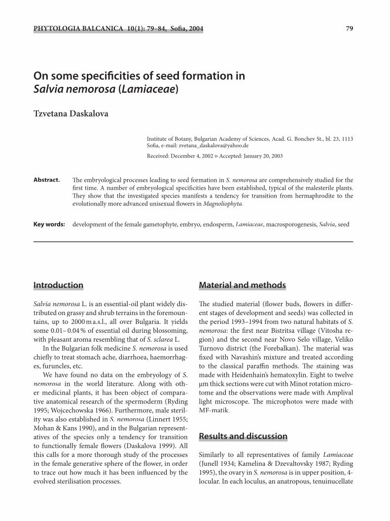
unitegmic ovule forms and develops (Plate I, Fig. 1). Already in the comparatively young ovule, only one archesporial cell forms subepidermally, contrary to S. pratensis L. (Daskalova 2002), S. offi cinalis L. and S. sclarea (Kamelina & Dzevaltovsky 1987), in which occasionally two archesporium cells form. A multi-cellular female archesporium consisting of eight cells that simultaneously set into meiosis was found out in Stachys trinervis (Kamelina & Dzevaltovsky 1987).
Th e archesporium cell diff ers distinctly from the remaining nucellar cells by its larger size and charac-teristic orthogonal to polygonal shape. It does not di-vide further and directly transforms into a macrospore mother cell (MMC).
Th e type of the ovules and the only one-celled arche-sporium formed in each, without cover cells, testify to a high level of development of the female generative sphere of the fl ower, judging by these embryological characteristics. Th e same was found out in other in-vestigated representatives of genus Salvia (Daskalova 1997) too, as well as in the evolutionally most ad-vanced taxa of family Lamiaceae (Dzevaltovsky 1979; Poddubnaya-Arnoldi 1982; Kamelina & Dzevaltovsky 1987).
Macrosporogenesis in MMC runs normally, re-sulting in a linear macrospore tetrad. Th e chalazal cell becomes the embryo sac mother cell (Plate I, Fig. 2). Th e remaining macrospores degenerate comparatively slowly in acropetal succession, occasionally lingering until the one-celled embryo sac stage (Plate I, Fig. 2). Development of the four macrospores in the tetrad was also observed in S. offi cinalis, as well as forma-tion of a greater number of tetrads in the ovule, sub-sequently with a greater number of embryo sacs in Stachys trinervis (Kamelina & Dzevaltovsky 1987).
Development of the female gametophyte fol-lows the Polygonum–type, the only type identifi ed so far within the entire family Lamiaceae (Davis 1966, Kamelina & Dzevaltovsky 1987) and in most of the in-vestigated species of genus Salvia (Carlson & Stuart 1936), particularly in those referred to the so-called S. mellifera–type.
Aft er the end of nuclear division in the one-celled embryo sac, the formed central vacuole pushes the two resulting nuclei out towards the poles of the sac. Th e subsequent mitotic division and polarisation of the nuclei in the 2–4 celled embryo sacs respectively run normally.
In the 8-nuclear embryo sac, fi rst diff erentiate the elements structuring the egg apparatus, which in their
morphology do not diff er from those described for oth-er investigated species of genus Salvia (Dzevaltovsky1979; Daskalova 1997).
In the initial development stages of the embryo sac the egg cell is almost equal in size to the two synergids. In most of the observed embryo sacs it does not occu-py a central position, but is slightly laterally shift ed, most oft en overlaying one of the two synergids. Th e egg cell is pyriform, with a big nucleus and distinct vacuole. Most oft en it maintains direct contact with the two polar nuclei, respectively at a later stage of de-velopment of the embryo sac with the secondary nu-cleus, which aft er fertilization moves to the chalazal end of the embryo sac.
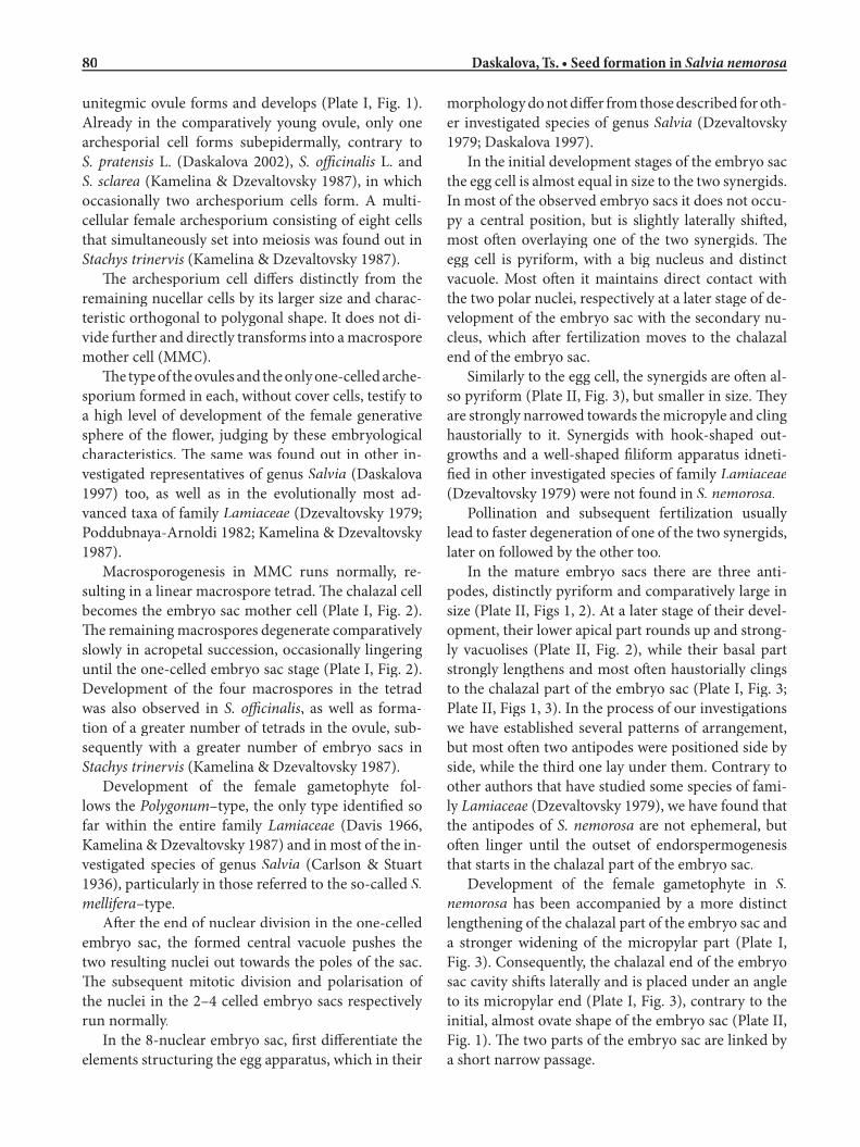
Similarly to the egg cell, the synergids are oft en al-so pyriform (Plate II, Fig. 3), but smaller in size. Th ey are strongly narrowed towards the micropyle and cling haustorially to it. Synergids with hook-shaped out-growths and a well-shaped fi liform apparatus idneti-fi ed in other investigated species of family Lamiaceae(Dzevaltovsky 1979) were not found in S. nemorosa.
Pollination and subsequent fertilization usually lead to faster degeneration of one of the two synergids, later on followed by the other too.
In the mature embryo sacs there are three anti-podes, distinctly pyriform and comparatively large in size (Plate II, Figs 1, 2). At a later stage of their devel-opment, their lo wer apical part rounds up and strong-ly vacuolises (Plate II, Fig. 2), while their basal part strongly lengthens and most oft en haustorially clings to the chalazal part of the embryo sac (Plate I, Fig. 3; Plate II, Figs 1, 3). In the process of our investigations we have established several patterns of arrangement, but most oft en two antipodes were positioned side by side, while the third one lay under them. Contrary to other authors that have studied some species of fami-ly Lamiaceae (Dzevaltovsky 1979), we have found that the antipodes of S. nemorosa are not ephemeral, but oft en linger until the outset of endorspermogenesis that starts in the chalazal part of the embryo sac.
Development of the female gametophyte in S. nemorosa has been accompanied by a more distinct lengthening of the chalazal part of the embryo sac and a stronger widening of the micropylar part (Plate I, Fig. 3). Consequently, the chalazal end of the embryo sac cavity shift s laterally and is placed under an angle to its micropylar end (Plate I, Fig. 3), contrary to the initial, almost ovate shape of the embryo sac (Plate II, Fig. 1). Th e two parts of the embryo sac are linked by a short narrow passage.
81Phytol. Balcan. 10(1) • Sofi a • 2004 Phytol. Balcan. 10(1) • Sofi a • 2004
Plate I
Fig. 1-3. Female gametophyte in Salvia nemorosa.1, tenuinucellate unitegmic ovule (× 160); 2, one-celled embryo sac with degenerating macrospore above (×500); 3, mature embryo sac with two antipodes (× 250).
11 22
33
11 • Phytol. Balcan. 10(1) Phytol. Balcan. 10(1) • 2004
82 Daskalova, Ts. • Seed formation in Daskalova, Ts. • Seed formation in Salvia nemorosa
Plate II
Fig. 1-4. Female gametophyte, embryo- and embryogenesis.1, the ovate in shape embryo sac (× 350); 2, antipodes with vacuolising cytoplasm (× 600); 3, synergids (× 300); 4, two-celled proem-bryo and cellular endosperm (× 300).
11
33
44
22

83Phytol. Balcan. 10(1) • Sofi a • 2004 Phytol. Balcan. 10(1) • Sofi a • 2004
Diff erentiation of the integumental tapetum of-ten runs at the stage of mature embryo sac. Most of-ten and similarly to all investigated representatives of Lamiaceae (Wunderlich 1967), soon aft er fertilization it occurs only in the upper and chalazal part (Plate II, Fig. 4), as well as in the passage which connects it with the micropylar part. Th us it obstructs the quick growth of the embryo sac in its chalazal part, contrary to lateral development in the micropylar part, which subsequently determines the shape of the mature em-bryo sac. According to the reports of some other au-thors (Wunderlich 1967), against the background of the constant embryological characteristics in the fe-male sphere of the fl ower in family Lamiaceae, this shape is specifi c for the diff erent investigated species.
Aft er the end of double fertilization processes in the embryo sac, division of the primary endosperm nucle-us considerably precedes that of the fertilized egg cell. In S. nemorosa, similarly to all investigated species in family Lamiaceae (Davis 1966), the endosperm is cel-lular. Its development begins in the chalazal part of the embryo sac, where the fertilized secondary nucleus has moved (Plate II, Fig. 4). Its fi rst division is accompa-nied by the formation of a transverse wall, with subse-quent formation of a primary micropylar and chalazal cell. Th e latter consequently turns into a two-nucleate chalazal haustorium. Th e second division takes place by the formation of a transverse wall in the formed micropylar cell. Th e cell situated towards the micro-pyle turns into a two-nucleate micropylar haustorium, better developed in the later stages of development, as compared to the chalazal one. Th e further running of endospermogenesis and the development of the two terminal haustoria have shown that endospermogen-esis follows the widest spread Lamiaceae Prunella–type (Wunderlich 1967), to which all species of genus Salvia have been referred to and especially those of the S. mellifera-type.
Th e quickly developing endosperm gradually dis-integrates the remnants of the ovule integument, leav-ing only its outer epidermis in the mature seed to form the seed coat. Endosperm in mature seed is complete-ly disintegrated by the developing embryo.
Aft er the end of porogamous fertilization, contra-ry to the central cell, the zygote envelopes into a thick coat and usually considerably increases in size, the vacuole disappears, and it fi lls in with homogeneous cytoplasm.
Our observations of the embryogenesis in S. nemo-rosa have shown that it follows the Onagrad-type,
widely spread in family Lamiaceae (Johansen 1950). Th e fi rst division of the zygote starts by formation of a transverse wall, usually aft er the endosperm has con-siderably advanced in its development (Plate II, Fig. 4). Th us the formed apical cell also divides transversely, while the formed basal cell does not participate in the further formation of the embryo.
Th e embryo grows comparatively slowly and al-ready in the globular and torpedo stage acquires a long uniseriate, multicellular suspensor to penetrate deep into the endospermal tissue. Th e basal cell of the suspensor is strongly elongated towards the micropyle and probably has a trophic function.
Th e embryo in the mature seed is straight, with well-formed cotyledons, each with an outgrowth tight-ly enveloping the hypocotil and the radicle. On the strength of this it could be referred to the evolution-ary most advanced Investing-type in Lamiacea family (Wunderlich 1967) found in all so-far studied species of genus Salvia (Daskalova 1993, 1997).
In the course of macrosporogenesis, macrogame-togenesis and development of the female gameto-phyte, we have observed some very distinct degenera-tion processes aff ecting single macrospore tetrads and elements of the embryo sac (most frequently the egg cell and the mature embryo sacs). Consequently, we have observed a rather high percentage of empty and sterile seeds in S. nemorosa (about 25 %).
Th e distinct, above-described degeneration pro-cesses are probably a consequence of the trend towards transition into functionally female fl owers established by us (Daskalova 1999). Th ese, in turn, furnish an-other proof in support of the existing positive corre-lation of the processes between the male and female generative sphere of the fl owers in malesterile plants (Laroche 1964).
ConclusionConclusion
During the detailed study of processes leading to seed formation in S. nemorosa conducted by us, some em-bryological structures have been identifi ed, already known for other investigated representatives of genus Salvia. Along with this, the species diff ered from each other both in single insignifi cant variations of some embryological characteristics (morphological features of the elements forming the mature embryo sac and the time of their functioning, as well as the shape of the mature embryo sac), and in a number of essen-
84 Daskalova, Ts. • Seed formation in Daskalova, Ts. • Seed formation in Salvia nemorosa
tial characteristics of proven evolutionary importance within the framework of Magnoliophyta (the type of formation of the female archesporium and the degree of transition to malesterile plants).
Th e formation of one-celled female archespori-um in the ovule of S. nemorosa, the Prunella-type en-dospermogenesis, the formation of an Investing-type mature embryo, and of functionally female fl owers, all evidence the high degree of evolutionary development of this species, both within the framework of the ge-nus and within the entire family Lamiaceae.
ReferencesReferences
Carlson, E. M. & Stuart, B .G. 1936. Development and gametophytes in certain new world species of Salvia. – New Phytol., 35(1): 68-91.
Daskalova, Tz. 1993. A cytoembriological study of malesterile forms in Salvia sclarea L. III. Microsporogenesis. – Fitologiya, 44: 56-60.
Daskalova, C. 1997. Seed production in malesterile plants in Salvia scalrea L. (Lamiaceae) – In: Chlodwig, F. & al. (eds), Essential Oil Symp. Proc. Vienna 1996, pp. 87-89. Allured Publ. Corp., Carol Stream.
Daskalova, T. C. 1999. Cytoembryological study on the male generative organs in Salvia nemorosa L. (Lamiaceae). – Abstracts. 30th Int. Symp. Essential Oils, September 5-8, 1999, p. 15. Leipzig & Miltitz.
Daskalova, T. C. 2002. Macrosporogenesis and development of the female gametophyte in Salvia pratensis L. – Phytol. Balcan., 8(2): 205-209.
Davis, G. L. 1966. Systematic Embryology of the Angiosperms. Acad. Press, New York, London, Sydney.
Dzevaltovsky, A. 1979. Embryology of the mint family (Labiatae Juss.). – Bot. Zhurn. (Leningrad), 64(1): 100-111 (in Russian).
Johansen, D. A. 1950. Plant Embryology. Publ. Chron. Bot. Waltham, Massachusetts.
Junell, S. 1934. Zur Gynäceummorphologie und Systematik der Verbenaceen und Labiaten. – Symb. Bot. Upsal., 4: 1-129.
Kamelina, O. & Dzevaltovsky, A. 1987. Lamiaceae. – In: Batygina, T. B. & Yakovlev, M. S. (eds), Comparative Embryology of Flowering Plants. Vol. 4, pp. 225-236. Nauka, Leningrad (in Russian).
Laroche, M. J. 1964. Étude de la megasporogenese et de la megagametogenese chez l’Asparagus offi cinalis L. – Rev. Gén. Bot., 71: 385-395.
Linnert, B. 1955. Zytologische Grundlagen für Sterilisationserscheinungen in der Gattung Salvia. – Züchter, 25(7-9): 237-240.
Mohan, L. & Kans, H. 1990. Male sterility in higher plants. – In: Frankel, K. & al. (eds), Monogr. Th eor. Appl. Genet. Springer Verlag, Berlin & Heidelberg.
Poddubnaya–Arnoldi, V. 1982. Characteristics of the Angiosperms families by cytoembriologycal features. Nauka, Moscow (in Russian).
Ryding, O. 1995. Pericarp structure and phylogeny of the Lamiaceae–Verbenaceae-complex. – Pl. Syst. Evol., 198(1-2): 101-141.
Wojciechowska, B. 1966. Morphology and anatomy of fruits and seeds in the family Labiatae, particularly in respect to medicinal species. – Monogr. Bot., 21: 1-244 (in Polish).
Wunderlich, R. 1967. Ein Vorschlag zu einer natürlichen Gleiderung der Labiaten auf Grund der Pollenkörner, der Samenentwicklung und des reifen Samens. – Oesterr. Bot. Z., 114(4-5): 380-483.