on physical properties of tetraether lipid membranes: effects of
TRANSCRIPT
Hindawi Publishing CorporationArchaeaVolume 2012, Article ID 138439, 11 pagesdoi:10.1155/2012/138439
Review Article
On Physical Properties of Tetraether Lipid Membranes:Effects of Cyclopentane Rings
Parkson Lee-Gau Chong, Umme Ayesa, Varsha Prakash Daswani, and Ellah Chay Hur
Department of Biochemistry, Temple University School of Medicine, 3420 North Broad Street, Philadelphia, PA 19140, USA
Correspondence should be addressed to Parkson Lee-Gau Chong, [email protected]
Received 6 July 2012; Accepted 8 August 2012
Academic Editor: Yosuke Koga
Copyright © 2012 Parkson Lee-Gau Chong et al. This is an open access article distributed under the Creative CommonsAttribution License, which permits unrestricted use, distribution, and reproduction in any medium, provided the original work isproperly cited.
This paper reviews the recent findings related to the physical properties of tetraether lipid membranes, with special attention tothe effects of the number, position, and configuration of cyclopentane rings on membrane properties. We discuss the findingsobtained from liposomes and monolayers, composed of naturally occurring archaeal tetraether lipids and synthetic tetraethers aswell as the results from computer simulations. It appears that the number, position, and stereochemistry of cyclopentane ringsin the dibiphytanyl chains of tetraether lipids have significant influence on packing tightness, lipid conformation, membranethickness and organization, and headgroup hydration/orientation.
1. Introduction
Archaea are subdivided into two kingdoms: euryarchaeotaand crenarchaeota [1]. Euryarchaeota include methanogensand halophiles, whereas crenarchaeota are traditionally com-prised of thermophilic or hyperthermophilic archaea [1].Halophiles and some methanogens are found mostly in highsalt water or hypersaline systems such as natural brines,alkaline salt lakes, and salt rocks; while thermophilic andhyperthermophilic archaea are found in very high temper-ature environments [2]. In recent years, crenarchaeota havealso been found in nonextreme environments such as soiland pelagic areas [3, 4].
The plasma membranes of archaea are rich in tetraetherlipids (TLs) and diphytanylglycerol diethers, also knownas archaeols (reviewed in [11–13]). TLs are the domi-nating lipid species in crenarchaeota, particularly in ther-moacidophilic archaea (∼90–95%). They are also found inmethanogens (0–50%) but are virtually absent in halophiles.Archaeal TLs contain either a caldarchaeol (GDGT) ora calditoglycerocaldarchaeol (GDNT) hydrophobic core(Figure 1) [13–17]. GDGT has two glycerols at both endsof the hydrophobic core. GDNT has a glycerol backbone atone end of the hydrophobic core and the calditol group at
the other end. Typically, TLs in methanogens contain onlyGDGT, but TLs in thermoacidophiles, particularly in themembers of the order Sulfolobales, have both GDGT andGDNT components. The Metallosphaera sedula TA-2 strainfrom hot springs in Japan, which has only GDGT-basedlipids, is an exception [18]. TLs have been thought to playan important role in the thermoacidophile’s high stabilityagainst extreme growth conditions such as high temperatures(e.g., 65-90◦C) and acidic environments (e.g., pH 2-3) [19].However, more recent studies showed that GDGT-basedTLs are also abundant in nonextremophilic crenarchaeotapresent in marine environments, lakes, soils, peat bogs,and low temperature areas [20, 21]. The functional role oftetraether lipids in crenarchaeota is not fully understood.
The hydrophobic core of archaeal TLs is made ofdibiphytanyl hydrocarbon chains, which may contain upto 8 cyclopentane rings per molecule (reviewed in [13]).The number of cyclopentane rings increases as growthtemperature increases [22–25], but decreases with decreasingpH in growth media [26]. The presence of cyclopentane ringsis a structural feature unique for archaeal tetraether lipids.Therefore, it is of great interest to unravel its biological roles.
Various polar headgroups can be attached to the glyceroland calditol backbones and yield either monopolar or bipolar
2 Archaea
R1 O O
OO
O
O
O
P
H
H
R2
O−
(a)
R1 O
O
OO
O
O
O
O
O
O
O
O
O
O
P
O−
H H
H
HH
H
H
H H
H
H
H
H
H
H
HH
H
H
HH
H
H
H
H
OH
OH
OH
OH OH
OH
OH
OH
OH
OH
OH
OH
OH
OH
HO
HO
HO
HOHO
HO
R2
GDNT R2 = GDGT R2 =
R1 =
(b)
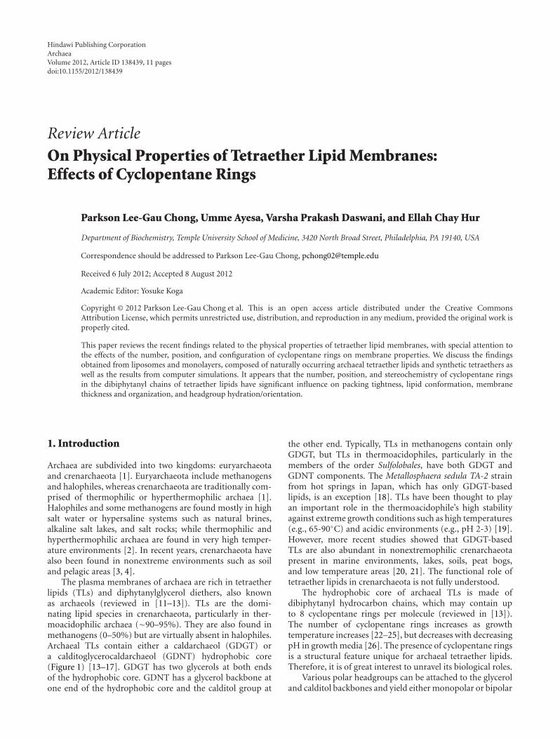
Figure 1: Illustrations of the molecular structures of the bipolar tetraether lipids in the polar lipid fraction E (PLFE) isolated from S.acidocaldarius. PLFE contains (a) GDGT (or caldarchaeol) and (b) GDNT (or calditolglycerocaldarchaeol ). The number of cyclopentanerings in each biphytanyl chain can vary from 0 to 4. The different head groups of GDNT and GDGT are presented at the bottom. GDG(N)T-0and GDG(N)T-4 contain 0 and 4 cyclopentane rings per molecule, respectively (taken from [5], reproduced with permission).
tetraether (BTL) lipids. Archaeal BTLs are glycolipids orphosphoglycolipids (illustrated in Figure 1). Liposomes thatare made of BTLs containing two or more sugar moietiesexhibit lower proton permeability than those containingonly one sugar molecule [26]. It has been proposed thatthermoacidophilic archaea cells adapt to low pH and hightemperature by increasing the number of sugar moietiesand cyclopentane rings [26, 27]. Increasing the number ofcyclopentane rings tightens membrane packing (discussedlater) [27]. Sugar moieties and the phosphate group in theBTL polar headgroup regions interact with each other toform a strong hydrogen bond network at the membranesurface [28].
BTLs are unique to archaea and cannot be biosyn-thesized by eukaryotic or bacterial cells. The ether for-mation from glycerol has been studied to a great extent([29] and references cited therein). The calditol moietyof GDNTs can be synthesized via an aldol condensationbetween dihydroxyacetone and fructose [30]. Calditol isthen reduced and alkylated to form GDNTs [30]. An invitro study showed that with the aid of 1L-myo-inositol1-phosphate synthase, archaetidylinositol phosphate (AIP)
synthase and AIP-phosphatase, archaeal inositol phospho-lipid (see Figure 1 e.g.) can be formed from CDP-archaeoland D-glucose-6-phosphate via myo-inositol-1-phosphateand AIP [31]. It has been proposed that the cyclopentanerings in BTLs of Sulfolobus are synthesized from glucose bya “cyclase” enzyme of the calditol carbocycle [32].
In this paper we focus on the recent findings related to thephysical properties of tetraether lipid membranes, with spe-cial attention on the effects of the number, position, and con-figuration of cyclopentane rings on membrane properties.We discuss the findings obtained from model membranescomposed of naturally occurring archaeal tetraether lipidsand synthetic tetraethers as well as the results from computersimulations.
2. Physical Properties of ModelMembranes Composed ofThermoacidophilic Tetraether Lipids
2.1. Membranes Made of Total Polar Lipid Extracts. Thestability and physical properties of liposomes made from
Archaea 3
the total polar lipids (TPLs) extracted from archaea havebeen studied extensively (reviewed in [11, 12, 33, 34]). TPLextracts contain both diether and tetraether lipids. The gen-eral trend shows that membranes become more stable as themole fraction of tetraether lipids increases. As an example,liposomes made of diether lipids such as Methanosarcinamazei TPL (0 wt% in caldarchaeols) were unstable againstsimulated human bile while those made of TPL fromMethanobacterium espanolae (65% in caldarchaeols) andThermoplasma acidophilum (90% in caldarchaeols) wererelatively more stable [35]. Solute and water permeabilityalso decrease as the content of tetraether lipids in membranesmade with archaeal TPLs increases [36].
Sprott et al. [37] demonstrated that liposomes made withTPL from the archaeon M. smithii AL1 can be highly fuso-genic when exposed to low pH and α- and β-glucosidases.It was suggested that, at low pH (4.8), the positively chargedglucosidases interact with the anionic phospholipids in M.smithii TPL, which in turn causes archaeosomes to rapidlyaggregate [37]. Aggregation is a prerequisite for membranefusion. This result is somewhat surprising because previousstudies showed that tetraether liposomes are resistant tofusogenic compounds [38–40]. Since TPL of M. smithiiAL1 contains a significant amount of diethers, in additionto caldarchaeols (∼40 wt %), it is possible that the strongfusogenic activity mentioned above comes from the diethercomponent.
2.2. Membranes Made of Partially Purified Tetraether LipidFractions. Since tetraethers are the dominating lipid speciesin thermoacidophiles, and the presence of diethers in thetotal polar lipid extracts makes the data interpretation moredifficult, it is of biophysical interest to study membranesmade only with tetraether lipids. The physical propertiesof lipid membranes made of partially purified polar lipidfractions from the archaeon Sulfolobus solfataricus havebeen reviewed [11, 34]. In this section, we focus on therecent studies of membranes made of partially purifiedpolar lipid fractions isolated from the archaeon Sulfolobusacidocaldarius.
2.2.1. PLFE. The polar lipid fraction E (PLFE) is oneof the major bipolar tetraether lipids (BTLs) found inthe thermoacidophilic archaeon S. acidocaldarius [41, 42].PLFE is a mixture of GDNT and GDGT (Figure 1). TheGDNT component (∼90% of total PLFE) contains phospho-myo-inositol on the glycerol end and β-glucose on thecalditol end, whereas the GDGT component (∼10% of totalPLFE) has phospho-myo-inositol attached to one glyceroland β-D-galactosyl-D-glucose to the other glycerol skeleton(Figure 1). The nonpolar regions of these lipids consist ofa pair of 40-carbon biphytanyl chains, each of which maycontain up to four cyclopentane rings [22].
2.2.2. PLFE Liposomes. PLFE lipids can form stable unil-amellar (∼60–800 nm in diameter), multilamellar, and giantunilamellar (∼10–150 μm) vesicles [40, 41, 43]. The lipids inthese vesicles span the entire lamellar structure, forming a
monomolecular thick membrane [44], which contrasts to thebilayer structure formed by monopolar diester (or diether)phospholipids. Compared to liposomes made of diesteror diether lipids, PLFE liposomes exhibit extraordinarymembrane properties (reviewed in [11, 12, 34]). PLFEliposomes exhibit low proton permeability and dye leakage[45, 46], high stability against autoclaving and Ca2+-inducedvesicle fusion [40, 47], tight and rigid membrane packing[43], and low enthalpy and volume changes associated withthe phase transitions [48, 49].
It is known that a decrease in archaeal cell growthtemperature (Tg) decreases the number of cyclopentane ringsin archaeal TLs [22]. In the case of S. acidocaldarius, theaverage number of cyclopentane rings per tetraether lipidmolecule decreases from 4.8 to 3.4 when Tg drops from82◦C to 65◦C [23]. Recent experimental work (see below)has addressed the effect of Tg , inferentially the number ofcyclopentane rings, on the physical properties of tetraetherlipid membranes.
2.2.3. Effect of Cyclopentane Rings on Phase Behavior ofPLFE Liposomes. The phase behavior of PLFE liposomes hasbeen characterized by small angle X-ray scattering, infraredand fluorescence spectroscopy, and differential scanningcalorimetry (DSC). PLFE liposomes exhibit two thermally-induced lamellar-to-lamellar phase transitions at ∼47–50◦Cand ∼60◦C [34, 43, 48, 49] and a lamellar-to-cubic phasetransition at ∼74–78◦C [48, 49] all of which involve smallor no volume changes as revealed by pressure perturbationcalorimetry (PPC) [49]. The calorimetry experiments alsosuggested that the number of cyclopentane rings in the dibi-phytanyl chains affect membrane packing in PLFE liposomesbecause the liposomes derived from different cell growthtemperatures showed different thermodynamic properties[49]. DSC allows us to determine the enthalpy change (ΔH)of the phase transition. PPC, on the other hand, allows us todetermine the relative volume change (ΔV/V) at the phasetransition and the thermal expansivity coefficient (α) at eachtemperature.
For PLFE liposomes derived from cells grown at 78◦C,the DSC heating scan exhibited an endothermic transi-tion at 46.7◦C, which can be attributed to a lamellar-to-lamellar phase transition and has an unusually low ΔH(3.5 kJ/mol), when compared to that for the main phasetransitions of saturated diacyl monopolar diester lipids (e.g.,1,2-dimyristoyl-sn-glycero-3-phosphocholine, DMPC). ThePPC scan revealed that, at this same phase transition, therelative volume change (ΔV/V) in the membrane is verysmall (∼0.1%) and much lower than the ΔV/V value 2.8%for the main phase transition of DMPC. The low ΔHand ΔV/V values may arise from the restricted transaucheconformational changes in the dibiphytanyl chain due tothe presence of cyclopentane rings, branched methyl groups,and to the spanning of the lipid molecules over the wholemembrane [49].
For PLFE liposomes derived from cells with growthtemperature of 65◦C, similar DSC and PPC profiles wereobtained. However, the lower cell growth temperature

4 Archaea
0
1
2
3
4
5
6
7
10 20 30 40 50 60 70 80 90
T (◦C)
PLFE-68PLFE-76
PLFE-81DPPC
ko s(1
0−10
mL
g−1
pas−
1)
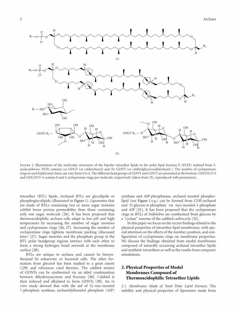
Figure 2: Adiabatic compressibilities (koS) of PLFE liposomesderived from cells grown at three different temperatures: 68◦C (darksquares), 76◦C (open circles), and 81◦C (open triangles). Solid line:DPPC liposomes for comparison (taken from [6], reproduced withpermission).
yielded a higher ΔV/V (∼0.25%) and ΔH (14 kJ/mol) valuefor the lamellar-to-lamellar phase transition measured atpH 2.1. The lower growth temperature also generated lessnegative temperature dependence of α. The changes inΔV/V,ΔH, and the temperature dependence of α can be attributedto the decrease in the number of cyclopentane rings in PLFEdue to the lower growth temperature [49]. A decrease in thenumber of cyclopentane rings makes the membrane less tightand less rigid; thus, a higher ΔV/V value is shown throughthe phase transition.
2.2.4. Effect of Cyclopentane Rings on Compressibility andMembrane Volume Fluctuations of PLFE Liposomes. Theisothermal and adiabatic compressibility and relative volumefluctuations of PLFE liposomes have been determined byusing calorimetry (DSC and PPC) and molecular acoustics(ultrasound velocimetry and densimetry) [50]. The com-pressibility values of PLFE liposomes were low, comparedto those found in a gel state of 1,2-dipalmitoyl-sn-glycero-3-phosphocholine (DPPC) [50]. Relative volume fluctuationsof PLFE liposomes at any given temperature examined were1.6–2.2 times more damped than those found in DPPCliposomes [50]. Volume fluctuations are closely related tosolute permeation across lipid membranes [51] and lateralmotion of membrane components [52]. Thus, the low valuesof relative volume fluctuations explain why PLFE liposomesexhibit unusually low proton permeation and dye leakage[45, 46] as well as limited lateral mobility, especially at lowtemperatures (e.g., <26◦C) [43, 53].
Zhai et al. [6] have used the growth temperature Tg
to alter the structure of PLFE lipids. They determinedthe compressibilities and volume fluctuations of PLFEliposomes derived from different cell growth temperatures
(Tg = 68, 76, and 81◦C). The compressibility and volumefluctuation values of PLFE liposomes exhibit small butsignificant differences with Tg . Figure 2 shows that adiabaticcompressibility (koS) of PLFE liposomes changes significantlywith Tg : koS(Tg = 68◦C) > koS(Tg = 81◦C) > koS(Tg = 76◦C). Forisothermal compressibility (koT), isothermal compressibilitycoefficient (βT) and relative volume fluctuations, a similar,but somewhat different, trend is seen: (Tg = 68◦C) >(Tg = 81◦C) ≥ or ≈ (Tg = 76◦C). These data indicatethat, among the three employed growth temperatures, thegrowth temperature 76◦C leads to the least compressible, andinferentially the most tightly packed PLFE lipid membranes.Note that 76◦C is in the temperature range for optimalgrowth of S. acidocaldarius (75–80◦C, [54, 55]). This findingsuggests that membrane packing in PLFE liposomes mayactually vary with the number of cyclopentane rings in anonlinear manner, reaching maximal tightness when thetetraether lipids are derived from cells grown at the optimalgrowth temperatures [6].
2.2.5. Future Studies of Physical Properties of Tetraether LipidMembranes. PLFE is a mixture of GDNT- and GDGT-derived BTLs with varying numbers of cyclopentane rings.Furthermore, at any given growth temperature, there isalways a broad distribution of the number of cyclopen-tane rings. In order to gain more insight into the effectof cyclopentane rings on compressibility and membranevolume fluctuations, it will be necessary to use purifiedarchaeal BTLs with a well-determined number and locationof cyclopentane rings. It has been reported that intactpolar lipids (archaeols (diethers) and caldarchaeols (GDGT))of the archaeon Thermoplasma acidophilum can be sep-arated with single cyclopentane ring resolution by high-performance liquid chromatography (HPLC) as detected byevaporative light-scattering detection [26, 56]. However, thestudy by Shimada et al. on T. acidophilum was limited toGDGT-based BTLs. To separate intact archaea BTLs at singlecyclopentane ring resolution when both GDNT- and GDGT-derived BTLs are present remains a major challenge.
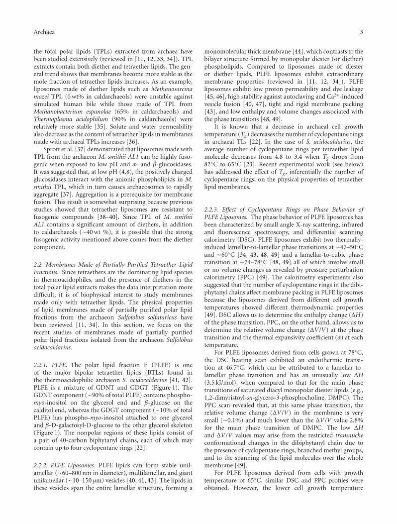
Hydrolyzed BTLs can also be separated with singlecyclopentane ring resolution using normal phase HPLCand positive ion atmospheric pressure chemical ionizationmass spectrometry [7]. Figure 3 shows the structures of thecyclopentane-containing GDGT hydrophobic cores previ-ously identified from the archaeon Sulfolobus solfataricus.These structures were determined by mass spectrometry.Compounds F′ and G′ (Figure 3) were reported as minorcomponents in S. solfataricus [7]. The relative distributionof these GDGT structures varies from species to species.The GDGT fraction of S. solfataricus is dominated by thosestructures with one (Structures E and G, Figure 3) or two(F) biphytanyl chains with two cyclopentane rings. Thedistribution of GDGTs in the extract of the archaeon M.sedula is somewhat different. In this case, the distribution isdominated by structures containing one or two biphytanylswith one cyclopentane ring. Physical properties of liposomesmade of hydrolyzed BTLs (without sugar and phosphate

Archaea 5
HO
OH
OH
OH
OH
OH
OH
OH
OH
OH
OH
HO
HO
HO
HO
HO
HO
HO
HO
HO
OOO
O
O
O
O
OO
O
O
O
O
O
O
OO
OO
OO
O
OO
OO
O
O
OO
O
O
O
O
OO
OO
OO
C
D
E
F
G
H
I
J
Ring number
(1, 0)
(1, 1)
(1, 2)
(2, 2)
(1, 3)
(3, 2)
(1, 4)
(3, 3)
(3, 4)
(4, 4)
1300
1298
1296
1294
1294
1292
1292
1290
1288
1286
[M + H]+
F
G
Figure 3: Structures of cyclopentane ring containing GDGTs previously reported to exist in archaea [7]. The number of cyclopentane ringsin the first and second hydrocarbon chains is indicated in the parentheses. The mass-to-charge ratio (m/z) of the protonated form [M+H]+
for each structure is also listed.
moieties) are not expected to be the same as those obtainedfrom the liposomes made of intact BTLs [47].
2.2.6. Disruption of PLFE Liposome Stability. While BTLliposomes (such as PLFE liposomes) exhibit remarkablestability against a number of chemical and physical stressorsas mentioned above, their stability can be attenuated or abol-ished under certain conditions. The most striking findingin this regard is that PLFE liposomes become excessivelydisrupted by the presence of two archaeal proteins, namely,CdvA and ESCRT-III (ESCRT: endosomal sorting complexrequired for transport) [57]. CdvA is a membrane interactingprotein that forms structures at mid-cell prior to nucleoidsegregation. CdvA recruits ESCRT-III to membranes in orderto aid in the final steps of cell division in some species
of archaea. Negative stain electron microscopy revealedextensive deformation of PLFE liposomes in the presenceof both CdvA and ESCRT-III together, but not individually[57]. The molecular mechanism underlying this disruptionis not clear.
PLFE liposomes are “autoclavable.” However, low pH(<4) and low salt concentrations (<50 mM) are unfavorablefor autoclaving PLFE-based liposomes [47]. PLFE liposomesand PLFE-based stealth liposomes (e.g., 95 mol% PLFE,3 mol% 1,2-distearoyl-sn-glycerol-3-phosphoethanolamine-polyethylene glycols (2000) (DSPE-PEG(2000)) and 2 mol%DSPE-PEG(2000)-maleimide) are extraordinarily stableagainst autoclaving between pH 4–10 [47]. These liposomesretained their particle size and morphology against multipleautoclaving cycles. One autoclaving cycle refers to theincubation of a sample for 20 min at 121◦C under a steam

6 Archaea
pressure of ∼18 psi. However, at pH 2-3, one or twoautoclaving cycles appeared to disrupt these liposomalmembranes, causing a significant increase in particle size[47]. PLFE liposomes were more resistant to dye leakage thanthe gel state of conventional diester liposomes under high saltand autoclaving conditions. As the salt concentration wasdecreased from 160 to 40 mM, the percent of dye moleculesthat leaked out from PLFE-based stealth liposomes after oneautoclaving cycle increased from 10.8% to 56.3% [47].
As expected, PLFE-based liposomes can also be disruptedby surfactants. The effect of the surfactant n-tetradecyl-β-D-maltoside (TDM) on unilamellar vesicles composedof PLFE and POPC (1-palmitoyl-2-oleoyl-sn-glycero-3-phosphocholine, a monopolar diester lipid) has been exam-ined [58]. TDM disrupts the POPC/cholesterol vesicleseffectively; however, higher concentrations (∼10 times) ofTDM were required to disrupt PLFE/POPC vesicles.
2.2.7. Structural and Packing Properties of PLFE MonolayerFilms Spread at the Interface between Air and Water. Effectsof cell growth temperature, subphase temperature and pH,and lateral film pressure on PLFE lipid monolayers at the air-water interface have been examined using X-ray reflectivity(XRR) and grazing incidence X-ray diffraction (GIXD)[5]. XRR and GIXD determine the vertical and horizontalstructure of the monolayers, respectively.
For PLFE derived from cells grown at 76◦C, a totalmonolayer thickness of ∼30 A was found in the XRR mea-surements for all monolayers studied. This finding suggeststhat both head groups of a U-shaped conformation of themolecules are in contact with the subphase and that asingle hydrocarbon chain region is protruded into the air.Similar U-shaped monolayer structures have been reportedin other tetraether lipid membranes [59]. However, someother studies [60, 61] suggest that the U-shaped and theupright conformations may coexist in the monolayer at thesame time or occur sequentially after spreading the TL lipidsat the water-air interface.
At the subphase temperatures 10◦C and 20◦C, large,highly crystalline domains were observed by GIXD; and thethickness of the crystalline part of the monolayer is slightlylarger than 30 A, which indicates a tight packing of the wholelipid monolayer, including both the hydrocarbon chain andthe head group regions. The area per hydrocarbon chainof PLFE (∼19.3 A2) found by GIXD is significantly smallerthan that of DPPC (∼23.2 A2) or 1,2-dipalmitoyl-sn-glycero-3-phosphoglycerol (DPPG) (∼22.6 A2). In fact, both the twohydrocarbon chains of a single PLFE lipid and the chainsof neighbouring lipid molecules adopted an extremely tightpacking.
For PLFE lipids derived from cells grown at highertemperatures, a slightly more rigid structure in the lipiddibiphytanyl chains was observed. However, the growthtemperature, inferentially the number of cyclopentane rings,does not affect the parameters of the unit cell in GIXDmeasurements. This suggests that there exists a nearlyidentical crystalline packing of all the PLFE lipids examinedand that, at high film pressures, membrane packing is
primarily governed by the lipid headgroup region [5]. It isinteresting to mention that the lack of cyclopentane ringsin the bipolar tetraether lipids from M. hungatei has beensuggested to be the cause of the U-shaped configurationadopted by these lipids in the monolayer film at the air-waterinterface [62]. Apparently, the presence of cyclopentane ringswould hinder the dibiphytanyl hydrocarbon chains frombending to form the U-shaped configuration.
3. Physical Properties of Membranes Made ofSynthetic Tetraether Lipids
The process of isolating well-defined archaeal tetraetherlipids can be difficult and time consuming. In addition,archaeal tetraether lipids have several structural featuresdistinctly different from conventional diester lipids. There-fore, it is rather difficult to elucidate the structure-activityrelationship for each of the individual structural featureswhen using native archaeal lipids. To resolve these problemsto some extent, synthetic tetraether lipid analogues have beenused [63–67].
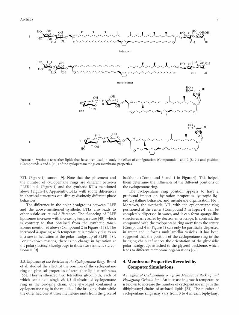
3.1. Importance of the Stereochemistry of the CyclopentaneRing. Jacquemet et al. were able to study the effect of thestereochemistry of the cyclopentane ring on BTL membraneproperties by using two synthetic tetraether lipids [8, 9](Compounds 1 and 2 in Figure 4). Both lipids have abridging hydrocarbon chain with a single 1,3-disubstitutedcyclopentane ring at the center. The substitutes on thering are ether-linked to C3 of the two opposite glycerolmoieties, while C2 of the glycerols is ether-linked to aphytanyl chain and C1 is linked to a lactosyl polar headgroup(Figure 4). The only difference between these two isomersis the configuration (cis or trans) of the 1,3-disubstitutedcyclopentane ring [8, 9].
The trans-isoform showed multilamellar vesicles whereasthe cis-counterpart led to nonspherical nanoparticles, asrevealed by cryo-transmission electron microscopy [8].Small angle X-ray scattering (SAXS) studies further showedthat the cis-isomer exhibited Lc-Lα-QII (cystal, lamellar, andbicontinuous cubic phase (Pn3 m), resp.) phase transitionswhereas the trans-isomer remained in Lα phase from 20to 100◦C. The electron density profiles calculated from theSAXS data were consistent with a stretched conformationof these synthetic BTLs within the Lα phase [9]. Thedifference in the phase behaviors was attributed to theconformation equilibrium of 1,3-disubstituted cyclopenatnerings. The dominant conformational motion in cyclopentaneis pseudorotational [68]. Pseudorotation is more restrictedfor the trans-isomer whereas several more orientations ofthe two substituents on the ring can be created for the cis-1,3-dialkyl cyclopentane ring [9, 68, 69]. Even though thisstudy shows that the stereochemistry at the cyclopentanering has a dramatic influence on membrane properties, morework is still required in order to explain why liposomesmade of PLFE, which naturally occurs and contains trans-1,3-disubstituted cyclopentyl rings, can undergo the Lα-to-QII phase transition [48, 49], while the synthetic trans
Archaea 7
HO
HO
HO
HO
HOHO
HO
HO
HO
HO
HO
HO
HO
HOHO
HO
HO HO
HO
HO
OH OH OH
OH
OH
OH
OH
OH
OH
OH
OH
OH
OH
OH
OH
OH
OH
OH
OH
OH OH
OHOH
OH
OH
OH
OH
OH
OH
OH
OO O O
O O
O
OO
O O
O
O
O
OO
O
O
OOOO
O
O
O OO O
O O
O
OO
O
O
O
O
O
O
O
O
OO
O
cis-isomer
1
trans-isomer
2
3
4
Figure 4: Synthetic tetraether lipids that have been used to study the effect of configuration (Compounds 1 and 2 [8, 9]) and position(Compounds 3 and 4 [10]) of the cyclopentane rings on membrane properties.
BTL (Figure 4) cannot [9]. Note that the placement andthe number of cyclopentane rings are different betweenPLFE lipids (Figure 1) and the synthetic BTLs mentionedabove (Figure 4). Apparently, BTLs with subtle differencesin chemical structures can display distinctly different phasebehaviors.
The difference in the polar headgroups between PLFEand the above-mentioned synthetic BTLs also leads toother subtle structural differences. The d-spacing of PLFEliposomes increases with increasing temperature [48], whichis contrary to that obtained from the synthetic trans-isomer mentioned above (Compound 2 in Figure 4) [9]. Theincreased d-spacing with temperature is probably due to anincrease in hydration at the polar headgroup of PLFE [48].For unknown reasons, there is no change in hydration atthe polar (lactosyl) headgroups in those two synthetic stereo-isomers [9].
3.2. Influence of the Position of the Cyclopentane Ring. Brardet al. studied the effect of the position of the cyclopentanering on physical properties of tetraether lipid membranes[66]. They synthesized two tetraether glycolipids, each ofwhich contains a single cis-1,3-disubstituted cyclopentanering in the bridging chain. One glycolipid contained acyclopentane ring in the middle of the bridging chain whilethe other had one at three methylene units from the glycerol
backbone (Compound 3 and 4 in Figure 4). This helpedthem determine the influences of the different positions ofthe cyclopentane ring.
The cyclopentane ring position appears to have aprofound impact on hydration properties, lyotropic liq-uid crystalline behavior, and membrane organization [66].Moreover, the synthetic BTL with the cyclopentane ringpositioned at the center (Compound 3 in Figure 4) can becompletely dispersed in water, and it can form sponge-likestructures as revealed by electron microscopy. In contrast, thecompound with the cyclopentane ring away from the center(Compound 4 in Figure 4) can only be partitially dispersedin water and it forms multilamellar vesicles. It has beensuggested that the position of the cyclopentane ring in thebridging chain influences the orientation of the glycosidicpolar headgroups attached to the glycerol backbone, whichleads to different membrane organizations [66].
4. Membrane Properties Revealed byComputer Simulations
4.1. Effect of Cyclopentane Rings on Membrane Packing andHeadgroup Orientation. An increase in growth temperatureis known to increase the number of cyclopentane rings in thedibiphytanyl chains of archaeal lipids [23]. The number ofcyclopentane rings may vary from 0 to 4 in each biphytanyl
8 Archaea
chain (i.e., 0 to 8 per dibiphytanyl unit). To evaluate howthe number of cyclopentane rings might affect membranepacking, Gabriel and Chong have conducted molecularmodeling studies on a membrane containing 4 × 4 GDNTmolecules (with sugar moieties, Figure 1) [27]. It was foundthat when 8 cyclopentane rings are contained, the headgroupof GDNT runs almost parallel to the membrane surface.However, without containing any rings, the headgroupis oriented perpendicular to the membrane surface. Themolecular modeling further showed that an increase in thenumber of cyclopentane rings in the dibiphytanyl chainsof GDNT from 0 to 8 made GDNT membrane packingtighter, more rigid, and more negative in interaction energy(−156.5 kcal/mol for 0 cyclopentane ring to −191.6 kcal/molwith 8 rings [27]). The resulting energy lowering effect isneither due to the decrease in polar headgroup separation,nor the change in the van der Waals interactions. Instead, itis due to the more favorable hydrogen bonding, and bondedinteractions including harmonic bending, theta expansionbond angle bending, dihedral angle torsion, and inversion[27].
4.2. Effect of Macrocyclic Linkage on Membrane Properties.Most archaeal BTLs are macrocyclic molecules with twobiphytanyl hydrocarbon chains linked to two oppositeglycerol or calditol backbones (illustrated in Figure 1 forthe case of PLFE). The effect of the macrocyclic linkageon membrane properties has been studied by moleculardynamics simulations [10, 70, 71]. For simplicity, coarsegraining approaches were employed and BTL moleculeswere modeled as di-monopolar lipids such as di-DPPC[10] and diphytanyl phosphatidylcholine (DPhPC) [70]. Inessence, two monopolar molecules were tethered togethereither at one pair of the hydrocarbon chains (acyclic di-DPPC or di-DPhPC) or at both pairs (cyclic di-DPPC ordi-DPhPC). The simulations showed that in the membranescomposed of macrocyclic BTL-like molecules, the uprightconfiguration gains favor over the U-shaped configuration[70]. The macrocyclic linkage also leads to a condensingeffect on the membrane surface, increases the order ofthe lipid hydrocarbon chains, slows lateral mobility in themembrane, and increases membrane thickness [10, 70, 71].Furthermore, the molecular dynamics simulations made bythe dissipative particle dynamics method [71] revealed theformation of two types of membrane pores. Hydrophobicpores are unstable and transient and exist at the lowtemperature. Hydrophilic pores are more stable with muchlonger lifetimes and are observed at high temperatures. Thesimulation data [71] suggested that hydrophilic pores canlead to the rupture of membrane vesicles. More intriguingly,it was proposed that hydrophobic pores, which occur at lowtemperatures, may result in the permeation of encapsulatedsmall molecules [71]. This implies that although BTLmembranes are extremely stable and tightly packed, somesmall leakage of entrapped molecules can still occur due toeither the formation of hydrophobic pores [71] or membranevolume fluctuations [6, 50] (discussed earlier).
5. Applications of Tetraether Lipid Membranes
The extraordinary stability of tetraether lipid membranesagainst a variety of physical and biochemical stressorshas provided the basis for using these lipids to developtechnological applications. BTLs can be used as a stable lipidmatrix for biosensors [72], a light harvesting device [73], andnanoparticles for targeted imaging and therapy (reviewed in[12, 74]).
It has been proposed that liposomes made of archaeallipids (also called archaeosomes) are taken up via a phago-cytosis receptor in the plasma membrane of the targetcells [75]. This uptake occurs in a liposomal composition-dependent manner [75]. Total polar lipids from thearchaeon, Halobacterium salinarum CECT 396, have beenused to make archaeosomes and archaeosomal hydrogelsas a possible topical delivery system for antioxidants [76].Compared to conventional liposomes, those archaeosomesand archaeosomal hydrogels showed better stability andmore sustained drug release [76]. It is of interest toextend their study from diethers (abundant in Halobacteriumsalinarum CECT 396) to tetraether lipids (e.g., PLFE lipidsisolated from thermoacidophiles). BTL-based liposomes aresuitable for oral delivery of therapeutic agents because BTLliposomes are stable against the harsh conditions (suchas bile salts, pancreatic enzymes, and low pH) in thegastrointestinal tract [77]. Tetraether lipid membranes havealso been tailored and evaluated as an intranasal peptidedelivery vehicle [78]. PEGylated tetraether lipids have beensynthesized and tested for their stability in test tubes and forliposomal encapsulation potential [79]. Knowledge gainedfrom the physical studies of cyclopentane rings, sugarmoieties, and macrocyclic structures should help to optimizethe numerous potential applications.
Abbreviations
AIP: archaetidylinositol phosphateBTL: bipolar tetraether lipidsDMPC: 1,2-dimyristoyl-sn-glycero-3-
phosphocholineDPhPC: 1,2-di-O-phytanyl-sn-glycero-3-
phosphocholineDPPC: 1,2-dipalmitoyl-sn-glycero-3-
phosphocholineDPPG: 1,2-dipalmitoyl-sn-glycero-3-
phosphoglycerolDSC: differential scanning calorimetryDSPE-PEG: 1,2-distearoyl-sn-glycerol-3-
phosphoethanolamine-polyethyleneglycols
ESCRT: endosomal sorting complex required fortransport
GDGT: caldarchaeolGDNT: calditoglycerocaldarchaeolGIXD: grazing incidence X-ray diffractionHPLC: high-performance liquid chromatographyPLFE: polar lipid fraction E

Archaea 9
POPC: 1-palmitoyl-2-oleoyl-sn-glycero-3-phosphocholine
PPC: pressure perturbation calorimetryTDM: n-tetradecyl-β-D-maltosideTL: tetraether lipidsTPL: total polar lipidsXRR: X-ray reflectivity.
Acknowledgment
Financial support from the NSF (DMR-1105277) is gratefullyacknowledged.
References
[1] C. R. Woese, O. Kandler, and M. L. Wheelis, “Towardsa natural system of organisms: proposal for the domainsArchaea, Bacteria, and Eucarya,” Proceedings of the NationalAcademy of Sciences of the United States of America, vol. 87, no.12, pp. 4576–4579, 1990.
[2] A. S. Andrei, H. L. Banciu, and A. Oren, “Living with salt:metabolic and phylogenetic diversity of archaea inhabitingsaline ecosystems,” FEMS Microbiology Letters, vol. 330, no. 1,pp. 1–9, 2012.
[3] L. A. Powers, J. P. Werne, T. C. Johnson, E. C. Hopmans, J. S.S. Damste, and S. Schouten, “Crenarchaeotal membrane lipidsin lake sediments: a new paleotemperature proxy continentalpaleoclimate reconstruction?” Geology, vol. 32, no. 7, pp. 613–616, 2004.
[4] M. B. Karner, E. F. Delong, and D. M. Karl, “Archaealdominance in the mesopelagic zone of the Pacific Ocean,”Nature, vol. 409, no. 6819, pp. 507–510, 2001.
[5] C. Jeworrek, F. Evers, M. Erlkamp et al., “Structure and phasebehavior of archaeal lipid monolayers,” Langmuir, vol. 27, no.21, pp. 13113–13121, 2011.
[6] Y. Zhai, P. L. G. Chong, L. J. Taylor et al., “Physical prop-erties of archaeal tetraether lipid membranes as revealed bydifferential scanning and pressure perturbation calorimetry,molecular acoustics, and neutron reflectometry: effects ofpressure and cell growth temperature,” Langmuir, vol. 28, no.11, pp. 5211–5217, 2012.
[7] E. C. Hopmans, S. Schouten, R. D. Pancost, M. T. van derMeer, and J. S. Sinninghe Damste, “Analysis of intact tetraetherlipids in archaeal cell material and sediments by high perfor-mance liquid chromatography/atmospheric pressure chemicalionization mass spectrometry,” Rapid Communications inMass Spectrometry, vol. 14, no. 7, pp. 585–589, 2000.
[8] A. Jacquemet, L. Lemiegre, O. Lambert, and T. Benvegnu,“How the stereochemistry of a central cyclopentyl ringinfluences the self-assembling properties of archaeal lipidanalogues: synthesis and cryoTEM observations,” Journal ofOrganic Chemistry, vol. 76, no. 23, pp. 9738–9747, 2011.
[9] A. Jacquemet, C. Meriadec, L. Lemiegre, F. Artzner, and T.Benvegnu, “Stereochemical effect revealed in self-assembliesbased on archaeal lipid analogues bearing a central five-membered carbocycle: a SAXS study,” Langmuir, vol. 28, no.20, pp. 7591–7597, 2012.
[10] M. Bulacu, X. Periole, and S. J. Marrink, “In silico design ofrobust bolalipid membranes,” Biomacromolecules, vol. 13, no.1, pp. 196–205, 2012.
[11] P. L.-G. Chong, “Physical properties of membranes composedof tetraether archaeal lipids, ? In,” in Thermophiles, F. Robb,
G. Antranikian, D. Grogan, and A. Driessen, Eds., pp. 73–95,CRC Press, Boca Raton, Fla, USA, 2008.
[12] P. L.-G. Chong, “Archaebacterial bipolar tetraether lipids:physico-chemical and membrane properties,” Chemistry andPhysics of Lipids, vol. 163, no. 3, pp. 253–265, 2010.
[13] Y. Koga and H. Morii, “Recent advances in structural researchon ether lipids from archaea including comparative and phys-iological aspects,” Bioscience, Biotechnology and Biochemistry,vol. 69, no. 11, pp. 2019–2034, 2005.
[14] A. Sugai, R. Sakuma, I. Fukuda et al., “The structure of thecore polyol of the ether lipids from Sulfolobus acidocaldarius,”Lipids, vol. 30, no. 4, pp. 339–344, 1995.
[15] E. Untersteller, B. Fritz, Y. Bleriot, and P. Sinay, “Thestructure of calditol isolated from the thermoacidophilicarchaebacterium Sulfolobus acidocaldarius,” Comptes Rendusde l’Academie des Sciences, vol. 2, no. 7-8, pp. 429–433, 1999.
[16] A. Gambacorta, G. Caracciolo, D. Trabasso, I. Izzo, A. Spinella,and G. Sodano, “Biosynthesis of calditol, the cyclopentanoidcontaining moiety of the membrane lipids of the archaeonSulfolobus solfataricus,” Tetrahedron Letters, vol. 43, no. 3, pp.451–453, 2002.
[17] Y. Bleriot, E. Untersteller, B. Fritz, and P. Sinay, “Total synthesisof calditol: structural clarification of this typical component ofArchaea order Sulfolobales,” Chemistry, vol. 8, no. 1, pp. 240–246, 2002.
[18] Y. H. Itoh, N. Kurosawa, I. Uda et al., “Metallosphaerasedula TA-2, a calditoglycerocaldarchaeol deletion strain of athermoacidophilic archaeon,” Extremophiles, vol. 5, no. 4, pp.241–245, 2001.
[19] T. A. Langworthy and J. L. Pond, “Membranes and lipidsof thermophiles,” in Thermophiles: General, Molecular, andApplied Microbiology, T. D. Brock, Ed., pp. 107–134, JohnWiley & Sons, New York, NY, USA, 1986.
[20] J. S. SinningheDamste, S. Schouten, E. C. Hopmans, A. C.T. Van Duin, and J. A. J. Geenevasen, “Crenarchaeol: thecharacteristic core glycerol dibiphytanyl glycerol tetraethermembrane lipid of cosmopolitan pelagic crenarchaeota,”Journal of Lipid Research, vol. 43, no. 10, pp. 1641–1651, 2002.
[21] J. W. H. Weijers, S. Schouten, O. C. Spaargaren, and J. S.Sinninghe Damste, “Occurrence and distribution of tetraethermembrane lipids in soils: implications for the use of theTEX86 proxy and the BIT index,” Organic Geochemistry, vol.37, no. 12, pp. 1680–1693, 2006.
[22] M. De Rosa, A. Gambacorta, B. Nicolaus, B. Chappe, andP. Albrecht, “Isoprenoid ethers; backbone of complex lipidsof the archaebacterium Sulfolobus solfataricus,” Biochimica etBiophysica Acta (BBA)/Lipids and Lipid Metabolism, vol. 753,no. 2, pp. 249–256, 1983.
[23] M. De Rosa, E. Esposito, A. Gambacorta, B. Nicolaus, and J. D.Bu’Lock, “Effects of temperature on ether lipid composition ofCaldariella acidophila,” Phytochemistry, vol. 19, no. 5, pp. 827–831, 1980.
[24] L. L. Yang and A. Haug, “Structure of membrane lipidsand physico-biochemical properties of the plasma membranefrom Thermoplasma acidophilum, adapted to growth at 37◦C,” Biochimica et Biophysica Acta (BBA)/Lipids and LipidMetabolism, vol. 573, no. 2, pp. 308–320, 1979.
[25] I. Uda, A. Sugai, Y. H. Itoh, and T. Itoh, “Variation in molecu-lar species of polar lipids from Thermoplasma acidophilumdepends on growth temperature,” Lipids, vol. 36, no. 1, pp.103–105, 2001.
[26] H. Shimada, N. Nemoto, Y. Shida, T. Oshima, and A. Yam-agishi, “Effects of pH and temperature on the composition of

10 Archaea
polar lipids in Thermoplasma acidophilum HO-62,” Journal ofBacteriology, vol. 190, no. 15, pp. 5404–5411, 2008.
[27] J. L. Gabriel and P. Lee Gau Chong, “Molecular modeling ofarchaebacterial bipolar tetraether lipid membranes,” Chem-istry and Physics of Lipids, vol. 105, no. 2, pp. 193–200, 2000.
[28] M. G. L. Elferink, J. G. de Wit, A. J. M. Driessen, and W.N. Konings, “Stability and proton-permeability of liposomescomposed of archaeal tetraether lipids,” Biochimica et Biophys-ica Acta, vol. 1193, no. 2, pp. 247–254, 1994.
[29] M. De Rosa, A. Gambacorta, B. Nicolaus, and S. Sodano,“Incorporation of labelled glycerols into ether lipids inCaldariella acidophila,” Phytochemistry, vol. 21, no. 3, pp. 595–599, 1982.
[30] B. Nicolaus, A. Trincone, E. Esposito, M. R. Vaccaro, A.Gambacorta, and M. De Rosa, “Calditol tetraether lipidsof the archaebacterium Sulfolobus solfataricus. Biosyntheticstudies,” Biochemical Journal, vol. 266, no. 3, pp. 785–791,1990.
[31] H. Morii, S. Kiyonari, Y. Ishino, and Y. Koga, “A novel biosyn-thetic pathway of archaetidyl-myo-inositol via archaetidyl-myo-inositol phosphate from CDP-archaeol and D-glucose 6-phosphate in methanoarchaeon Methanothermobacter ther-mautotrophicus cells,” Journal of Biological Chemistry, vol.284, no. 45, pp. 30766–30774, 2009.
[32] N. Yamauchi, N. Kamada, and H. Ueoka, “The possibility ofinvolvement of ”cyclase” enzyme of the calditol carbocyclewith broad substrate specificity in Sulfolobus acidcaldarius, atypical thermophilic archaea,” Chemistry Letters, vol. 35, no.11, pp. 1230–1231, 2006.
[33] L. Krishnan and G. D. Sprott, “Archaeosome adjuvants:immunological capabilities and mechanism(s) of action,”Vaccine, vol. 26, no. 17, pp. 2043–2055, 2008.
[34] A. Gliozzi, A. Relini, and P. L. G. Chong, “Structure and per-meability properties of biomimetic membranes of bolaformarchaeal tetraether lipids,” Journal of Membrane Science, vol.206, no. 1-2, pp. 131–147, 2002.
[35] G. B. Patel, B. J. Agnew, L. Deschatelets, L. P. Fleming, andG. D. Sprott, “In vitro assessment of archaeosome stabilityfor developing oral delivery systems,” International Journal ofPharmaceutics, vol. 194, no. 1, pp. 39–49, 2000.
[36] J. C. Mathai, G. D. Sprott, and M. L. Zeidel, “Molecular Mech-anisms of Water and Solute Transport across ArchaebacterialLipid Membranes,” Journal of Biological Chemistry, vol. 276,no. 29, pp. 27266–27271, 2001.
[37] G. D. Sprott, J. P. Cote, and H. C. Jarrell, “Glycosidase-inducedfusion of isoprenoid gentiobiosyl lipid membranes at acidicpH,” Glycobiology, vol. 19, no. 3, pp. 267–276, 2009.
[38] A. Relini, D. Cassinadri, Q. Fan et al., “Effect of physicalconstraints on the mechanisms of membrane fusion: bolaformlipid vesicles as model systems,” Biophysical Journal, vol. 71,no. 4, pp. 1789–1795, 1996.
[39] A. Relini, D. Cassinadri, Z. Mirghani et al., “Calcium-inducedinteraction and fusion of archaeobacterial lipid vesicles: afluorescence study,” Biochimica et Biophysica Acta, vol. 1194,no. 1, pp. 17–24, 1994.
[40] R. Kanichay, L. T. Boni, P. H. Cooke, T. K. Khan, and P. L.G. Chong, “Calcium-induced aggregation of archaeal bipolartetraether liposomes derived from the thermoacidophilicarchaeon Sulfolobus acidocaldarius,” Archaea, vol. 1, no. 3, pp.175–183, 2003.
[41] S. L. Lo and E. L. Chang, “Purification and characterizationof a liposomal-forming tetraether lipid fraction,” Biochemicaland Biophysical Research Communications, vol. 167, no. 1, pp.238–243, 1990.
[42] E. L. Chang and S. L. Lo, “Extraction and purification oftetraether lipids from Sulfolobus acidocaldarius,” in Protocolsfor Archaebacterial Research, E. M. Fleischmann, A. R. Place,R. T. Robb, and H. J. Schreier, Eds., pp. 2.3.1–2.3.14, MarylandBiotechnology Institute, Baltimore, Md, USA, 1991.
[43] L. Bagatolli, E. Gratton, T. K. Khan, and P. L. G. Chong, “Two-photon fluorescence microscopy studies of bipolar tetraethergiant liposomes from thermoacidophilic archaebacteria Sul-folobus acidocaldarius,” Biophysical Journal, vol. 79, no. 1, pp.416–425, 2000.
[44] M. G. L. Elferink, J. G. De Wit, R. Demel, A. J. M. Driessen,and W. N. Konings, “Functional reconstitution of membraneproteins in monolayer liposomes from bipolar lipids ofSulfolobus acidocaldarius,” Journal of Biological Chemistry,vol. 267, no. 2, pp. 1375–1381, 1992.
[45] H. Komatsu and P. L. G. Chong, “Low permeability ofliposomal membranes composed of bipolar tetraether lipidsfrom thermoacidophilic archaebacterium Sulfolobus acido-caldarius,” Biochemistry, vol. 37, no. 1, pp. 107–115, 1998.
[46] E. L. Chang, “Unusual thermal stability of liposomes madefrom bipolar tetraether lipids,” Biochemical and BiophysicalResearch Communications, vol. 202, no. 2, pp. 673–679, 1994.
[47] D. A. Brown, B. Venegas, P. H. Cooke, V. English, and P. L.G. Chong, “Bipolar tetraether archaeosomes exhibit unusualstability against autoclaving as studied by dynamic lightscattering and electron microscopy,” Chemistry and Physics ofLipids, vol. 159, no. 2, pp. 95–103, 2009.
[48] P. L.-G. Chong, M. Zein, T. K. Khan, and R. Winter, “Structureand conformation of bipolar tetraether lipid membranesderived from thermoacidophilic archaeon Sulfolobus acido-caldarius as revealed by small-angle X-ray scattering and high-pressure FT-IR spectroscopy,” Journal of Physical Chemistry B,vol. 107, no. 33, pp. 8694–8700, 2003.
[49] P. L. G. Chong, R. Ravindra, M. Khurana, V. English, andR. Winter, “Pressure perturbation and differential scanningcalorimetric studies of bipolar tetraether liposomes derivedfrom the thermoacidophilic archaeon Sulfolobus acidocaldar-ius,” Biophysical Journal, vol. 89, no. 3, pp. 1841–1849, 2005.
[50] P. L.-G. Chong, M. Sulc, and R. Winter, “Compressibilitiesand volume fluctuations of archaeal tetraether liposomes,”Biophysical Journal, vol. 99, no. 10, pp. 3319–3326, 2010.
[51] E. Falck, M. Patra, M. Karttunen, M. T. Hyvonen, and I.Vattulainen, “Impact of cholesterol on voids in phospholipidmembranes,” Journal of Chemical Physics, vol. 121, no. 24, pp.12676–12689, 2004.
[52] P. F. F. Almeida, W. L. C. Vaz, and T. E. Thompson, “Lateraldiffusion and percolation in two-phase, two-component lipidbilayers. topology of the solid-phase domains in-plane andacross the lipid bilayer,” Biochemistry, vol. 31, no. 31, pp. 7198–7210, 1992.
[53] Y. L. Kao, E. L. Chang, and P. L. G. Chong, “Unusualpressure dependence of the lateral motion of pyrene-labeledphosphatidylcholine in bipolar lipid vesicles,” Biochemical andBiophysical Research Communications, vol. 188, no. 3, pp.1241–1246, 1992.
[54] T. D. Brock, K. M. Brock, R. T. Belly, and R. L. Weiss,“Sulfolobus: a new genus of sulfur-oxidizing bacteria living atlow pH and high temperature,” Archiv fur Mikrobiologie, vol.84, no. 1, pp. 54–68, 1972.
[55] T. A. Langworthy, W. R. Mayberry, and P. F. Smith, “Longchain glycerol diether and polyol dialkyl glycerol triether lipidsof Sulfolobus acidocaldarius,” Journal of Bacteriology, vol. 119,no. 1, pp. 106–116, 1974.

Archaea 11
[56] H. Shimada, N. Nemoto, Y. Shida, T. Oshima, and A. Yam-agishi, “Complete polar lipid composition of Thermoplasmaacidophilum HO-62 determined by high-performance liquidchromatography with evaporative light-scattering detection,”Journal of Bacteriology, vol. 184, no. 2, pp. 556–563, 2002.
[57] R. Y. Samson, T. Obita, B. Hodgson et al., “Molecular andStructural Basis of ESCRT-III Recruitment to Membranesduring Archaeal Cell Division,” Molecular Cell, vol. 41, no. 2,pp. 186–196, 2011.
[58] A. Fafaj, J. Lam, L. Taylor, and P. L. G. Chong, “Unusualstability of archaeal tetraether liposomes against surfactants,”Biophysical Journal, vol. 100, no. 3, p. 329a, 2011.
[59] M. De Rosa, “Archaeal lipids: structural features andsupramolecular organization,” Thin Solid Films, vol. 284-285,pp. 13–17, 1996.
[60] U. Bakowsky, U. Rothe, E. Antonopoulos, T. Martini, L.Henkel, and H. J. Freisleben, “Monomolecular organization ofthe main tetraether lipid from Thermoplasma acidophilum atthe water-air interface,” Chemistry and Physics of Lipids, vol.105, no. 1, pp. 31–42, 2000.
[61] S. Vidawati, J. Sitterberg, U. Bakowsky, and U. Rothe, “AFMand ellipsometric studies on LB films of natural asymmetricand symmetric bolaamphiphilic archaebacterial tetraetherlipids on silicon wafers,” Colloids and Surfaces B, vol. 78, no.2, pp. 303–309, 2010.
[62] A. Gliozzi, A. Relini, R. Rolandi, S. Dante, and A. Gambacorta,“Organization of bipolar lipids in monolayers at the air-waterinterface,” Thin Solid Films, vol. 242, no. 1-2, pp. 208–212,1994.
[63] T. Benvegnu, M. Brard, and D. Plusquellec, “Archaeabacteriabipolar lipid analogues: structure, synthesis and lyotropicproperties,” Current Opinion in Colloid and Interface Science,vol. 8, no. 6, pp. 469–479, 2004.
[64] T. Benvegnu, G. Rethore, M. Brard, W. Richter, andD. Plusquellec, “Archaeosomes based on novel synthetictetraether-type lipids for the development of oral deliverysystems,” Chemical Communications, no. 44, pp. 5536–5538,2005.
[65] M. Brard, C. Laine, G. Rethore et al., “Synthesis of archaealbipolar lipid analogues: a way to versatile drug/gene deliverysystems,” Journal of Organic Chemistry, vol. 72, no. 22, pp.8267–8279, 2007.
[66] M. Brard, W. Richter, T. Benvegnu, and D. Plusquellec, “Syn-thesis and supramolecular assemblies of bipolar archaeal gly-colipid analogues containing a cis-1,3-disubstituted cyclopen-tane ring,” Journal of the American Chemical Society, vol. 126,no. 32, pp. 10003–10012, 2004.
[67] G. Lecollinet, R. Auzely-Velty, M. Danel et al., “Syntheticapproaches to novel archaeal tetraether glycolipid analogues,”Journal of Organic Chemistry, vol. 64, no. 9, pp. 3139–3150,1999.
[68] W. Cui, F. Li, and N. L. Allinger, “Simulation of conforma-tional dynamics with the MM3 force field: the pseudorotationof cyclopentane,” Journal of the American Chemical Society, vol.115, no. 7, pp. 2943–2951, 1993.
[69] O. R. de Ballesteros, L. Cavallo, F. Auriemma, and G.Guerra, “Conformational analysis of poly(methylene-1,3-cyclopentane) and chain conformation in the crystallinephase,” Macromolecules, vol. 28, no. 22, pp. 7355–7362, 1995.
[70] W. Shinoda, K. Shinoda, T. Baba, and M. Mikami, “Moleculardynamics study of bipolar tetraether lipid membranes,” Bio-physical Journal, vol. 89, no. 5, pp. 3195–3202, 2005.
[71] S. Li, F. Zheng, X. Zhang, and W. Wang, “Stability and ruptureof archaebacterial cell membrane: a model study,” Journal ofPhysical Chemistry B, vol. 113, no. 4, pp. 1143–1152, 2009.
[72] B. A. Cornell, V. L. B. Braach-Maksvytis, L. G. King et al., “Abiosensor that uses ion-channel switches,” Nature, vol. 387, no.6633, pp. 580–583, 1997.
[73] K. Iida, H. Kiriyama, A. Fukai, W. N. Konings, and M. Nango,“Two-dimensional self-organization of the light-harvestingpolypeptides/BChl a complex into a thermostable liposomalmembrane,” Langmuir, vol. 17, no. 9, pp. 2821–2827, 2001.
[74] T. Benvegnu, L. Lemiegre, and S. Cammas-Marion, “Archaeallipids: innovative materials for biotechnological applications,”European Journal of Organic Chemistry, no. 28, pp. 4725–4744,2008.
[75] G. D. Sprott, S. Sad, L. P. Fleming, C. J. Dicaire, G. B. Patel,and L. Krishnan, “Archaeosomes varying in lipid compositiondiffer in receptor-mediated endocytosis and differentiallyadjuvant immune responses to entrapped antigen,” Archaea,vol. 1, no. 3, pp. 151–164, 2003.
[76] A. Gonzalez-Paredes, B. Clares-Naveros, M. A. Ruiz-Martinez,J. J. Durban-Fornieles, A. Ramos-Cormenzana, and M.Monteoliva-Sanchez, “Delivery systems for natural antioxi-dant compounds: archaeosomes and archaeosomal hydrogelscharacterization and release study,” International Journal ofPharmaeutics, vol. 421, no. 2, pp. 321–331, 2011.
[77] J. Parmentier, B. Thewes, F. Gropp, and G. Fricker, “Oralpeptide delivery by tetraether lipid liposomes,” InternationalJournal of Pharmaceutics, vol. 415, no. 1-2, pp. 150–157, 2011.
[78] G. B. Pate, H. Zhou, A. Ponce, G. Harris, and W. Chen,“Intranasal immunization with an archaeal lipid mucosalvaccine adjuvant and delivery formulation protects against arespiratory pathogen challenge,” PLoS ONE, vol. 5, no. 12,Article ID e15574, 2010.
[79] J. Barbeau, S. Cammas-Marion, P. Auvray, and T. Benvegnu,“Preparation and characterization of stealth archaeosomesbased on a synthetic PEGylated archaeal tetraether lipid,”Journal of Drug Delivery, vol. 2011, Article ID 396068, 11pages, 2011.

Submit your manuscripts athttp://www.hindawi.com
Hindawi Publishing Corporation http://www.hindawi.com Volume 2013Hindawi Publishing Corporation http://www.hindawi.com Volume 2013
The Scientific World Journal
Hindawi Publishing Corporationhttp://www.hindawi.com
Nucleic AcidsJournal of
Volume 2013
ArchaeaHindawi Publishing Corporationhttp://www.hindawi.com Volume 2013
ISRN Biotechnology
Hindawi Publishing Corporationhttp://www.hindawi.com Volume 2013
Hindawi Publishing Corporationhttp://www.hindawi.com
GenomicsInternational Journal of
Volume 2013
Evolutionary BiologyInternational Journal of
Hindawi Publishing Corporationhttp://www.hindawi.com Volume 2013
Hindawi Publishing Corporationhttp://www.hindawi.com Volume 2013
Advances in
Virolog y
ISRN Microbiology
Hindawi Publishing Corporationhttp://www.hindawi.com Volume 2013
Marine BiologyJournal of
Hindawi Publishing Corporationhttp://www.hindawi.com Volume 2013
BioMed Research International
Hindawi Publishing Corporationhttp://www.hindawi.com Volume 2013
ISRN Zoology
Hindawi Publishing Corporationhttp://www.hindawi.com Volume 2013
Hindawi Publishing Corporationhttp://www.hindawi.com Volume 2013
Signal TransductionJournal of
ISRN Cell Biology
Hindawi Publishing Corporationhttp://www.hindawi.com Volume 2013
Hindawi Publishing Corporationhttp://www.hindawi.com Volume 2013
BioinformaticsAdvances in
PeptidesInternational Journal of
Hindawi Publishing Corporationhttp://www.hindawi.com Volume 2013
Hindawi Publishing Corporationhttp://www.hindawi.com Volume 2013
Enzyme Research
Hindawi Publishing Corporationhttp://www.hindawi.com Volume 2013
Biochemistry Research International
ISRN Molecular Biology
Hindawi Publishing Corporationhttp://www.hindawi.com Volume 2013
Stem CellsInternational
Hindawi Publishing Corporationhttp://www.hindawi.com Volume 2013