on call head and neck gladwin hui acknowledgement: special thanks to elissa price
TRANSCRIPT

On Call Head and Neck
Gladwin Hui
Acknowledgement: Special thanks to Elissa Price

CT Head and Neck
• Emergency
• Requests from Emerg or ENT
• Talk to referring physician to make sure airway is secured
• IV Contrast
• Neck vs. C-spine
CT Head and Neck
• Difficult studies
• Not very often (maybe once a month)
• Focus on the urgent issues (will take a long time to learn Head and Neck well)
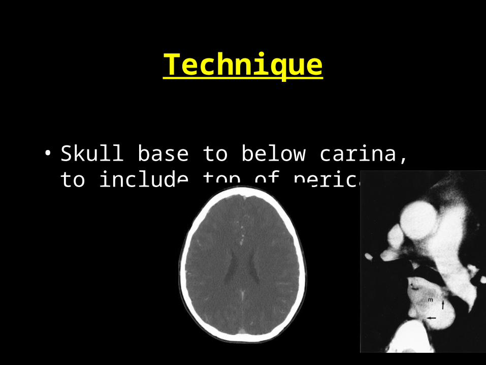
Technique
• Skull base to below carina, to include top of pericardium

My Approach to CT Neck1) Airway
- Nasopharynx, oropharynx and hypopharynx (whole airway)- Trachea and esophagus
2) Deep neck spaces- Parapharyngeal space- Retropharyngeal space- Masticator space- Carotid space- Perivertebral space- Anterior visceral space- Submandibular/sublingual space

Approach to CT Neck
3) Glands- Parotid- Submandibular- Thyroid
4) Vessels and lymph nodes5) Bones and Soft tissues6) Neuro
- Brain, orbits, paranasal sinuses, mastoid air cells7) Cord8) Chest
- Lung apices - Mediastinum, Pericardial region

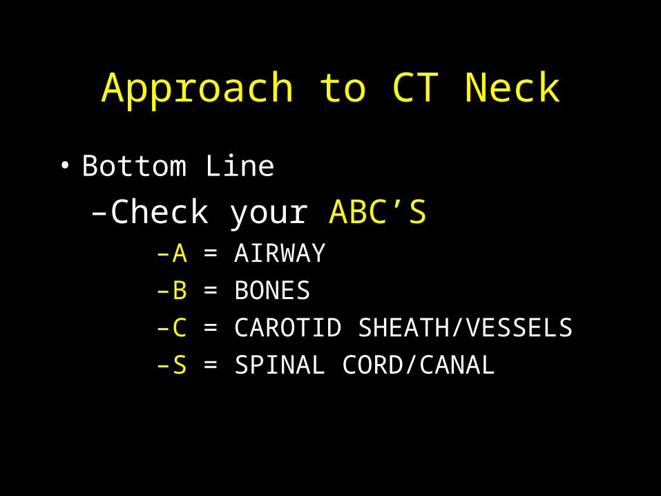
Approach to CT Neck
• Bottom Line
– Check your ABC’S– A = AIRWAY– B = BONES– C = CAROTID SHEATH/VESSELS– S = SPINAL CORD/CANAL

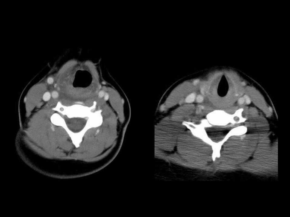
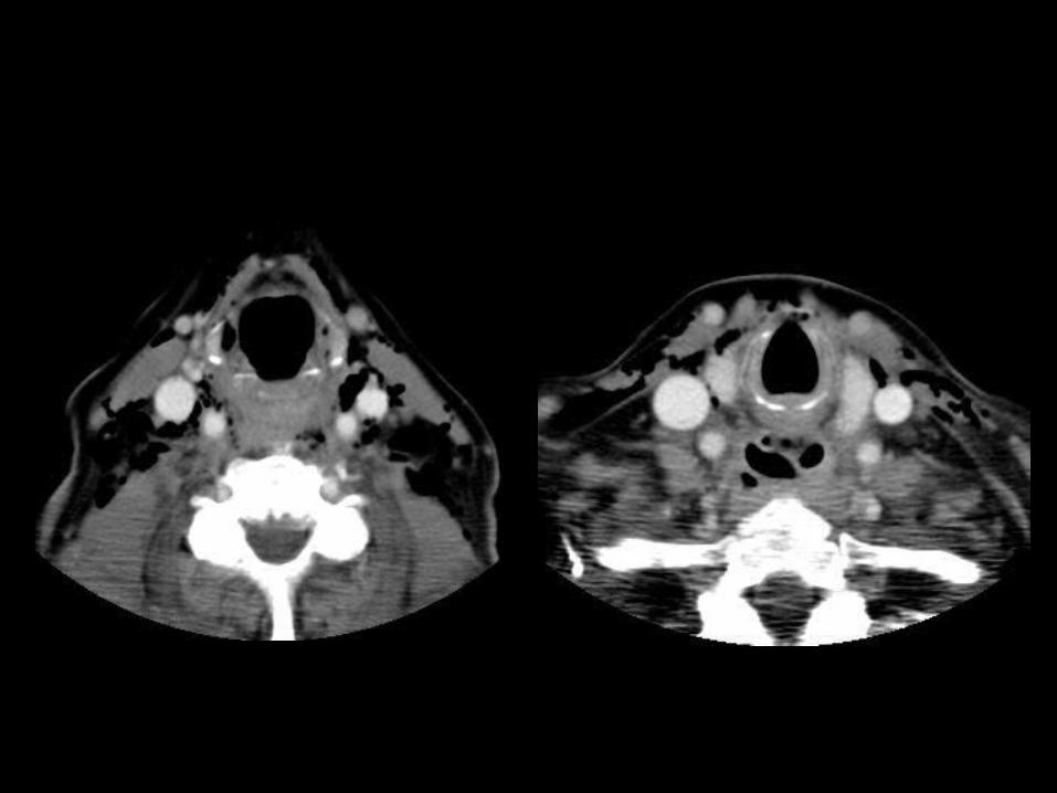
Anatomy: Fat Planes & Spaces
• Deep neck spaces
• - Parapharyngeal space
• - Retropharyngeal space
• - Masticator space
• - Carotid space
• - Perivertebral space
• - Anterior visceral space
• - Submandibular/sublingual space

Lateral pterygoid muscle
Masticator space
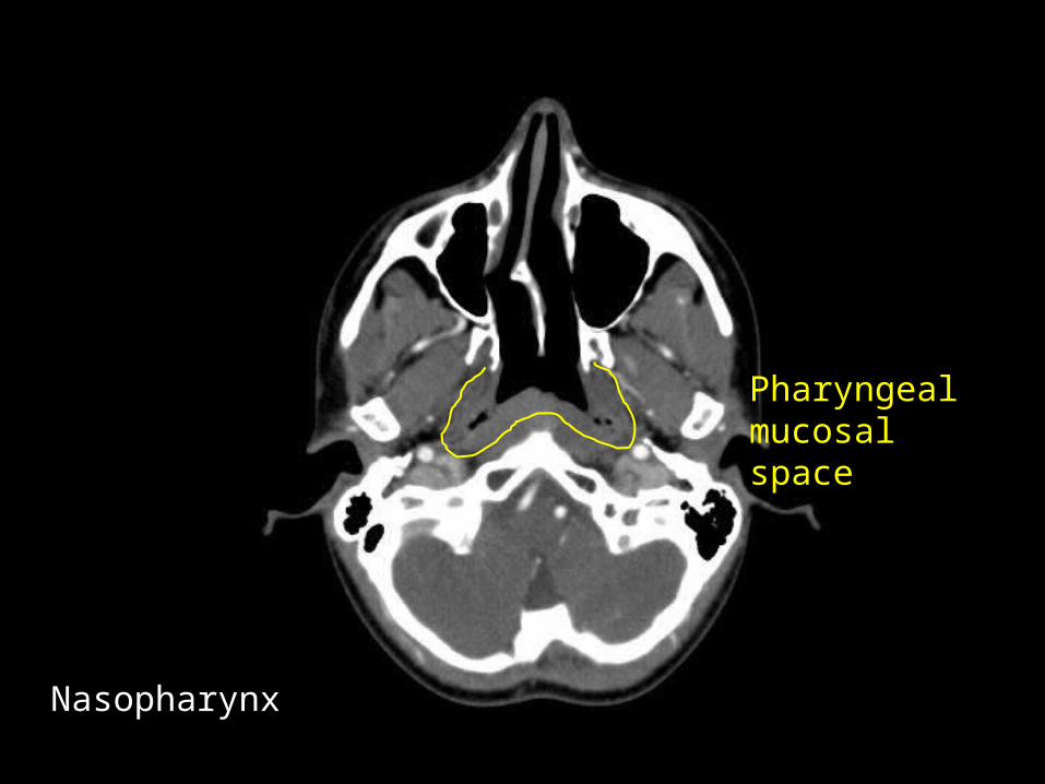
Pharyngealmucosal space
Nasopharynx

Medial PterygoidMuscle
Parotid

Styloid process
ECA
ICAInternal jugular vein
Parapharyngeal space
Carotid space

Uvula
Nasopharynx Oropharynx
Retropharyngeal space
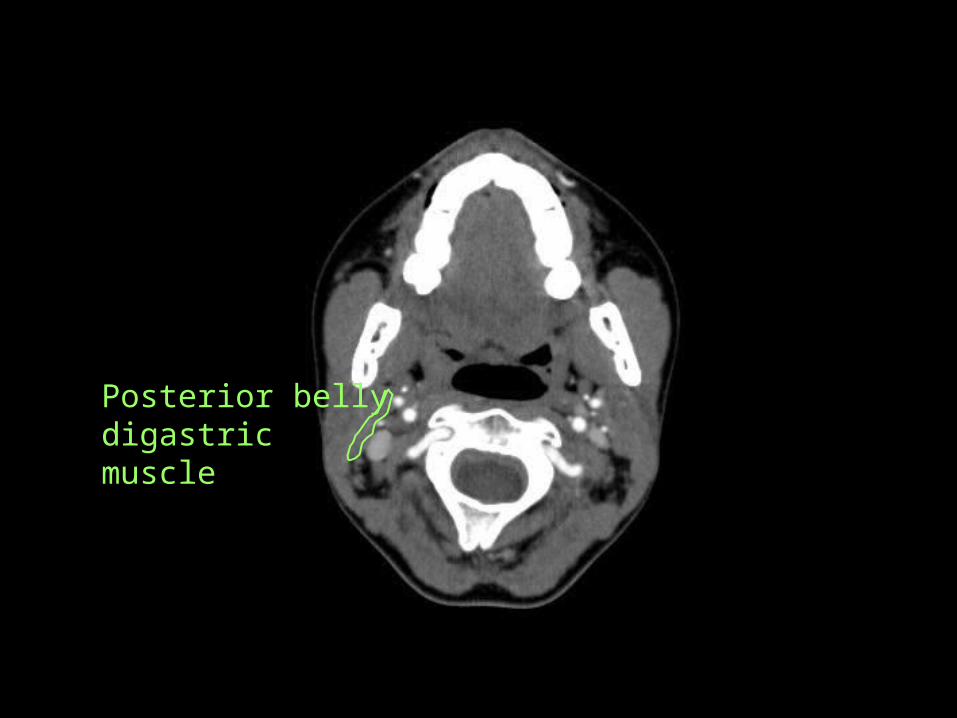
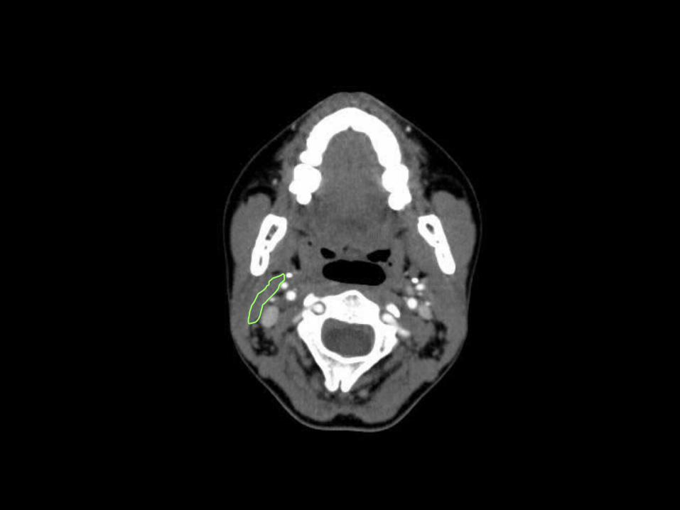
Posterior bellydigastricmuscle


Perivertebral space


Jugulodigastric lymph node </= 1.5-cm
Lt JDG node
Back edge submandibular gland
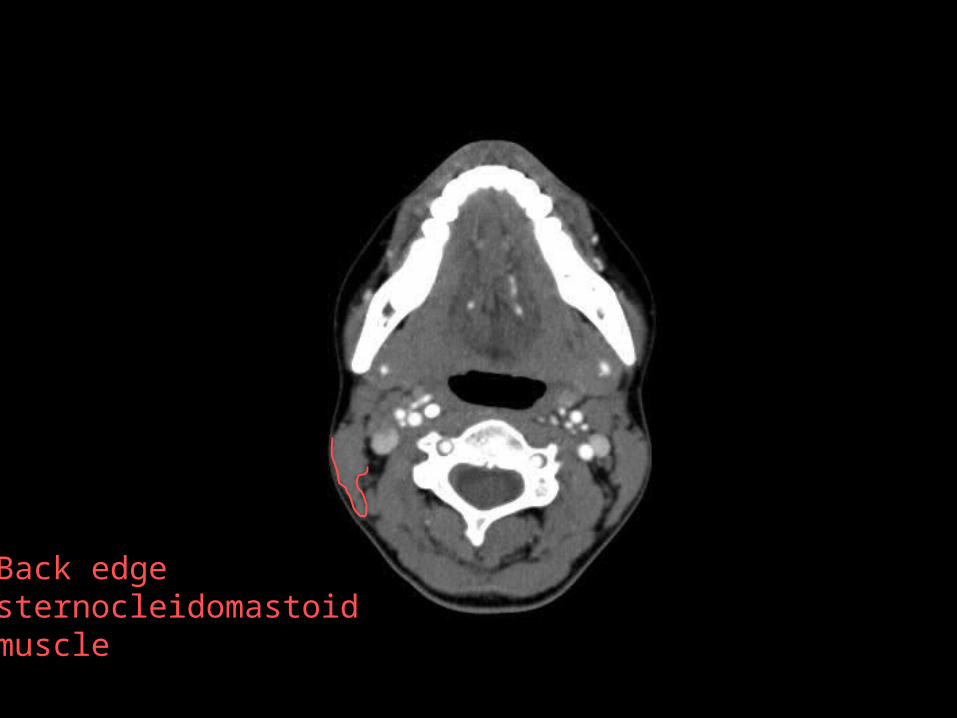
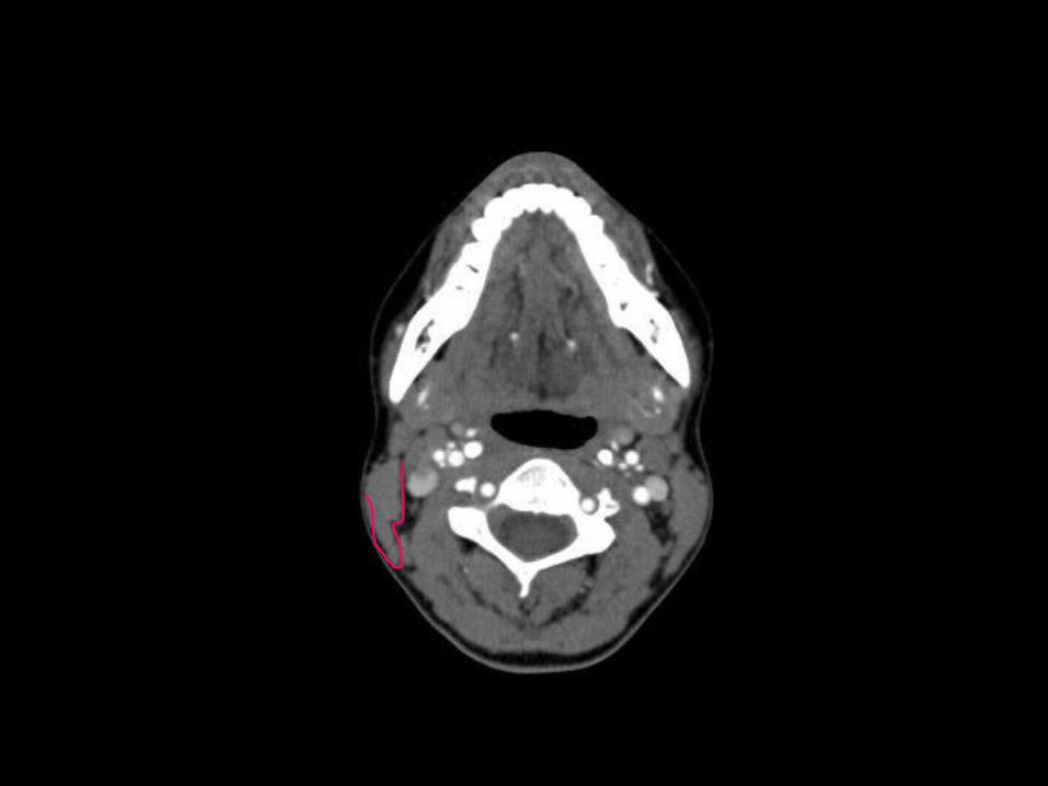
Back edge sternocleidomastoid muscle

mylohyoid


ad
ad= ant belly digastric muscle


Epiglottis
Oropharynx Hypopharynx
Vallecula
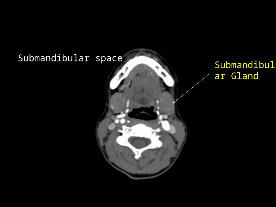
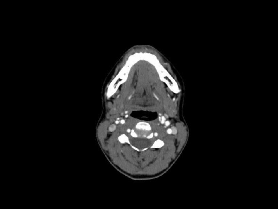
Submandibular Gland
Submandibular space

Hyoid bone

Hyoid bone


Hyoid bone

Aryepiglottic Folds
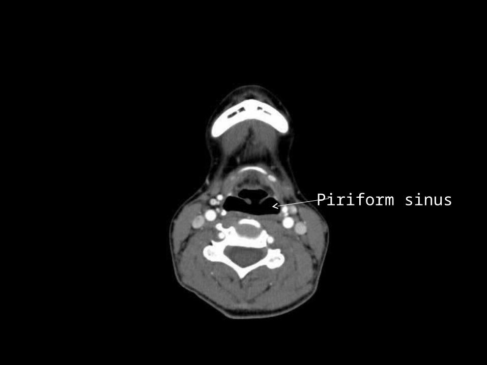
Piriform sinus


Cricoid cartilage


Cricoid cartilage


Cricoid cartilage
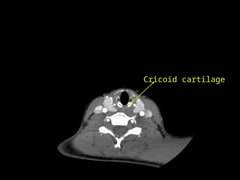
Cricoid cartilage

Cricoid cartilageThyroid

Anterior Visceral Space
• Extends from hyoid bone to anterior mediastinum
• Sling around the trachea, esophagus
• Contiguous with the retropharyngeal space
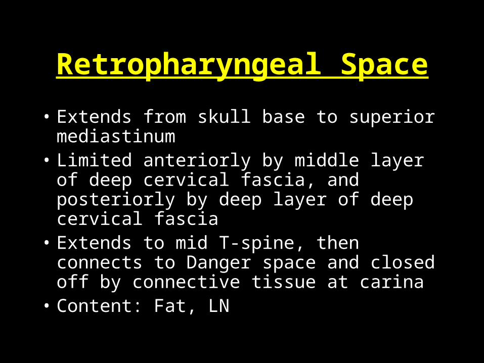
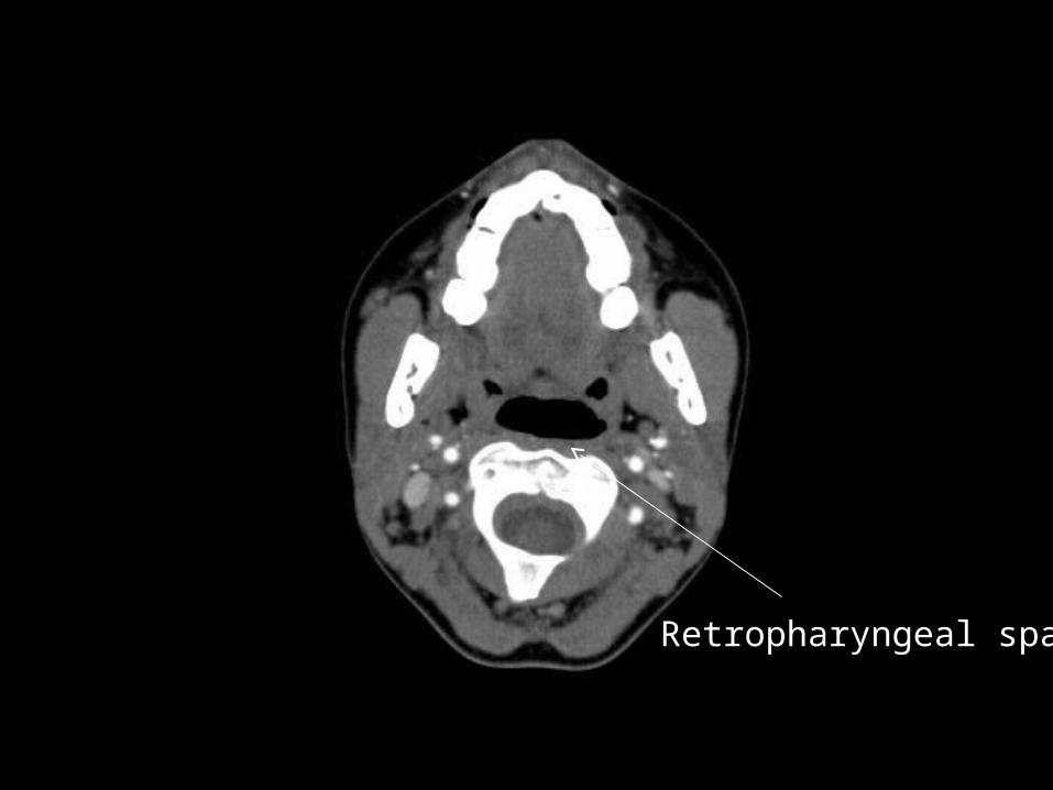
Retropharyngeal Space
• Extends from skull base to superior mediastinum
• Limited anteriorly by middle layer of deep cervical fascia, and posteriorly by deep layer of deep cervical fascia
• Extends to mid T-spine, then connects to Danger space and closed off by connective tissue at carina
• Content: Fat, LN

Danger Space
• Extends from skull base to diaphragm in the posterior mediastinum
• Posterior to retropharyngeal space
• Lies between the alar and prevertebral layers of the deep cervical fascia
• Spread of infection from neck to mediastinum

Carotid space – Neurovascular Bundle
• Extends from skull base to mediastinum
• CCA, IJV, Vagus– Dissection, narrowing, aneurysm, rupture– Thrombus– Mass

Parapharyngeal Space
• Key landmark – primarily fat-containing– How is it being effected by a process going on
in the region?

Retropharyngeal space
Parapharyngeal space
Carotid artery
Internal jugular vein
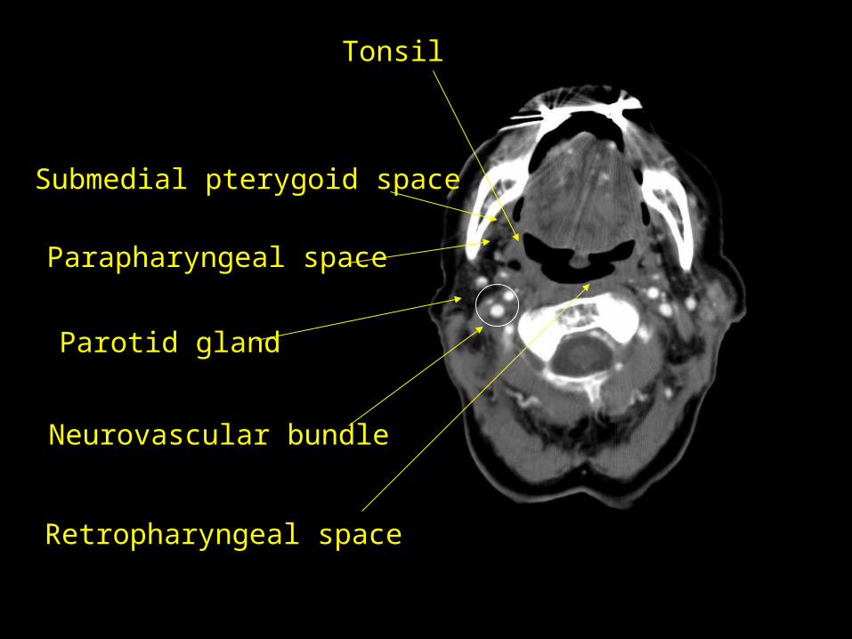
Tonsil
Submedial pterygoid space
Parapharyngeal space
Parotid gland
Neurovascular bundle
Retropharyngeal space

Retropharyngeal space
Neurovascular bundle
Anterior visceral space
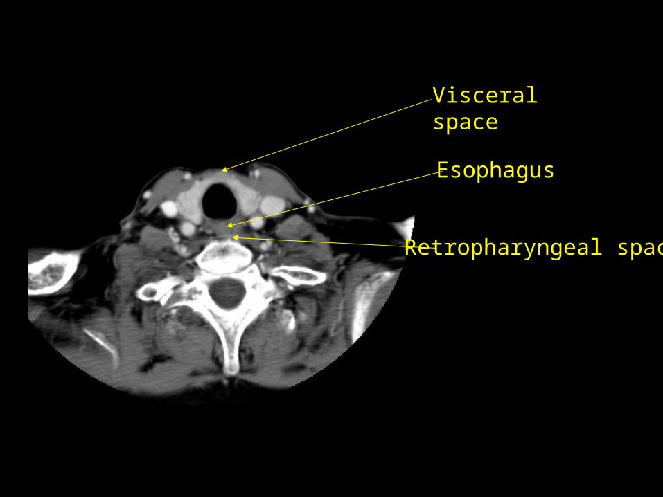
Esophagus
Retropharyngeal space
Visceral space

Tonsil
Parapharyngeal space
Submandibular gland
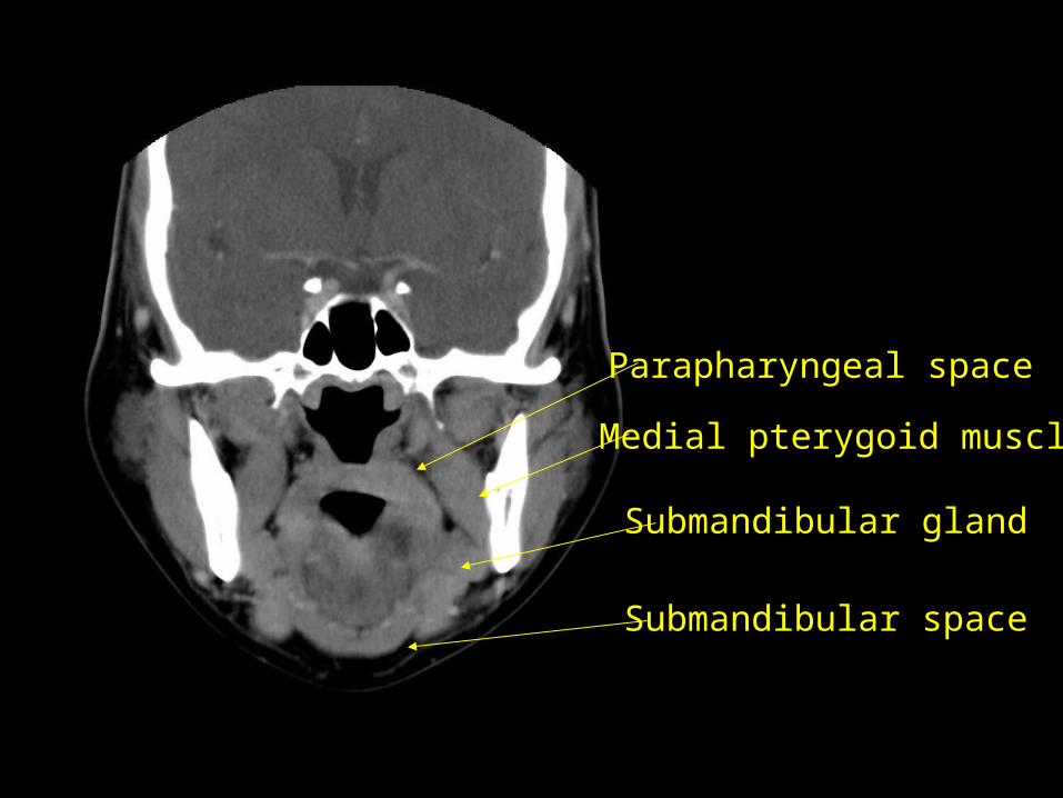
Parapharyngeal space
Submandibular gland
Submandibular space
Medial pterygoid muscle

Submandibular and Sublingual Spaces
• Important regions to evaluate for floor of mouth infections

Pathophysiology
• Cellulitis
• Phlegmon
• Fluid collections
• Abscess
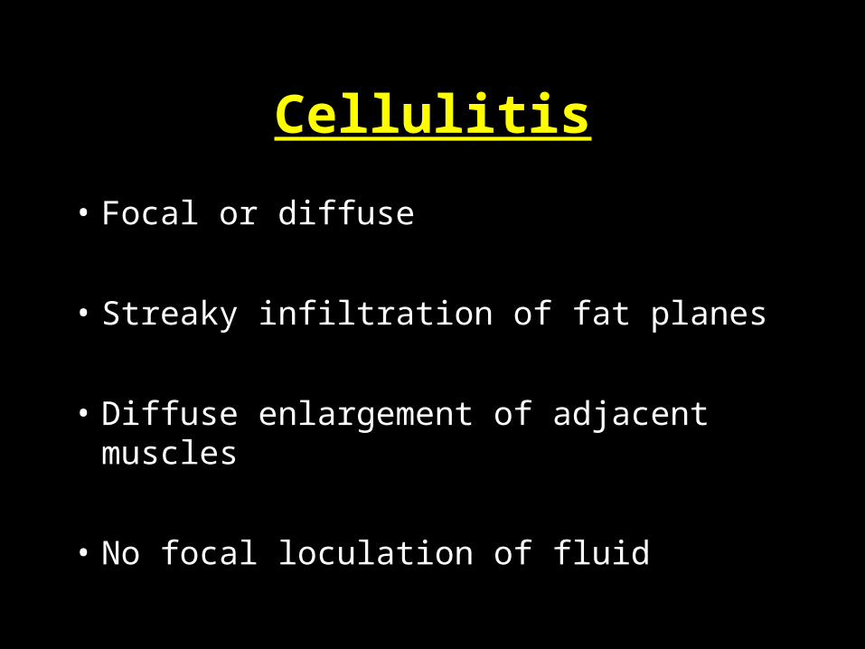
Cellulitis
• Focal or diffuse
• Streaky infiltration of fat planes
• Diffuse enlargement of adjacent muscles
• No focal loculation of fluid

Cellulitis

Phlegmon
• Slightly heterogeneous solid swelling
• May be minimal low density suggestive of fluid loculation developing
• Usually seen in tonsillar/peritonsillar or retropharyngeal locations

Phlegmon
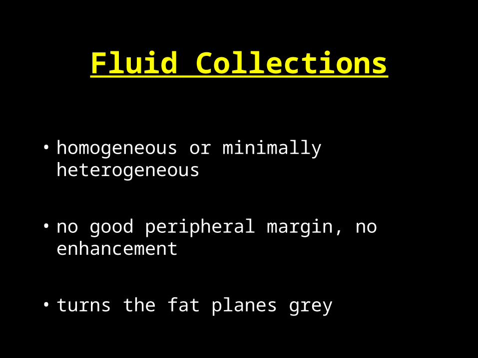
Fluid Collections
• homogeneous or minimally heterogeneous
• no good peripheral margin, no enhancement
• turns the fat planes grey
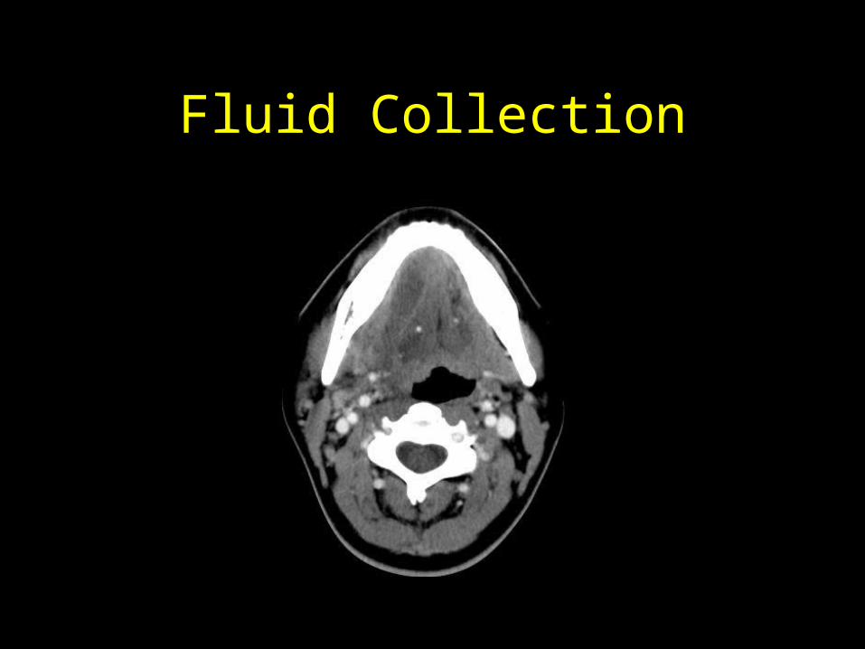
Fluid Collection
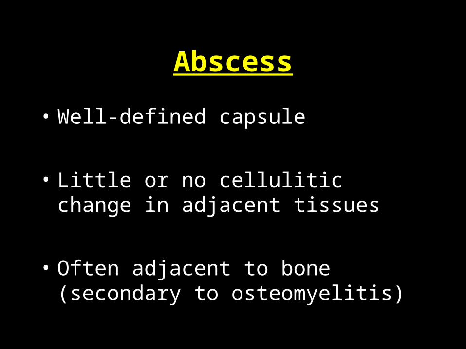
Abscess
• Well-defined capsule
• Little or no cellulitic change in adjacent tissues
• Often adjacent to bone (secondary to osteomyelitis)

Complications
ALWAYS CHECK FOR:• Airway obstruction
• Carotid pseudoaneurysm or rupture
• Internal jugular vein thrombosis
• Mediastinitis/fluid collection/abscess
• Pericarditis

Dental Infections
• Usually mandibular, usually molar
• Submedial pterygoid space
• Floor of mouth
• Anterior visceral space
• Parapharyngeal space
• Neurovascular bundle
• Retropharyngeal space


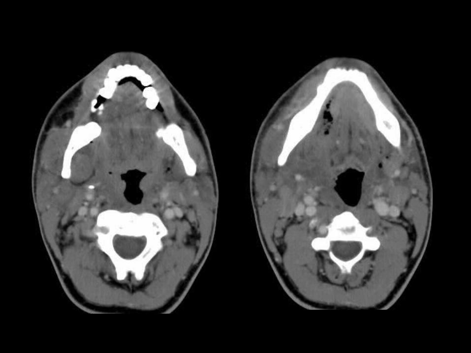
Ludwig’s Angina
• Cellulitis that involves inflammation of the tissues of the floor of the mouth, under the tongue
• Extremely dangerous• Early airway compromise• Extensive edema of tongue and floor of mouth• +/- Floor of mouth fluid/air• No abscess• Dental origin
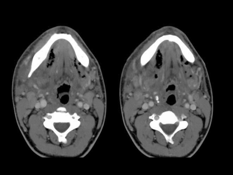
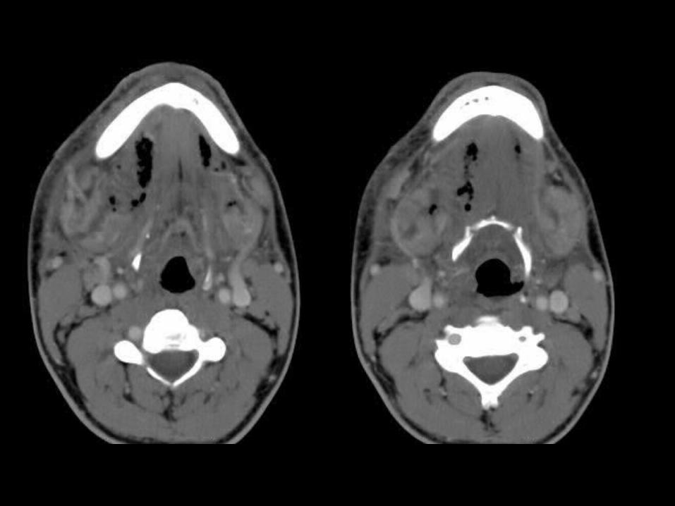

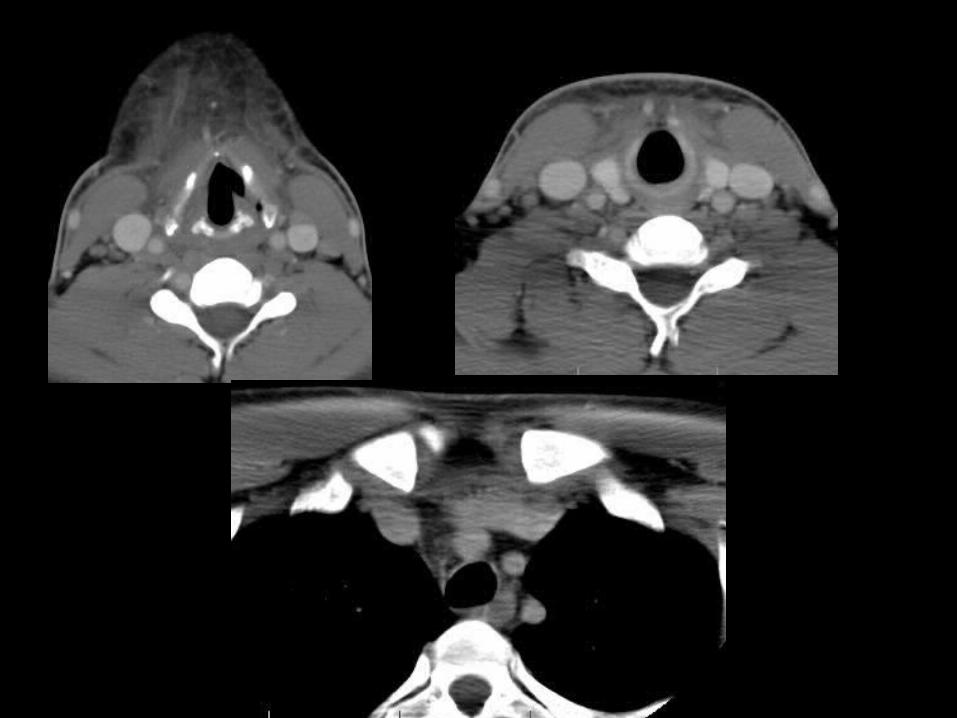
Tonsil
• Unilateral swollen tonsil
• Parapharyngeal space
• Floor of mouth
• Neurovascular bundle
• Retropharyngeal space


Iatrogenic
• Post-intubation
• Post-endoscopy

Pharyngeal/Esophageal Perforations
• Air in the fat planes
• Retropharyngeal space
• Neurovascular bundle
• Mediastinum


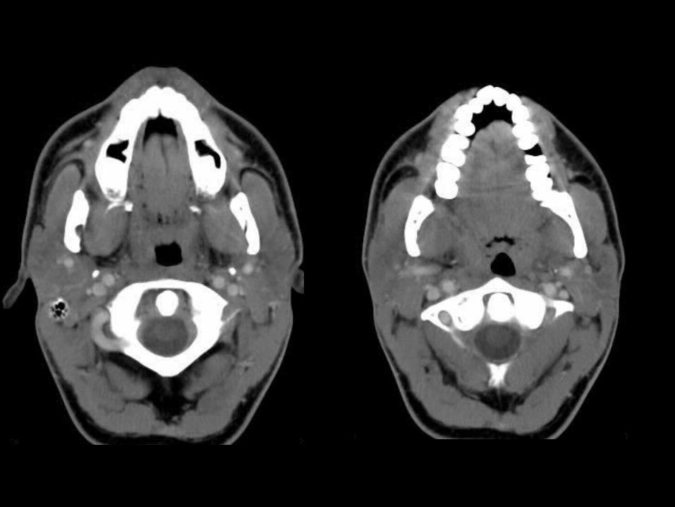
Salivary Gland Obstruction
• Parotid
• Submandibular


Courtesy: Learning Radiology
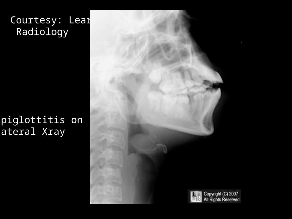
Courtesy: Learning Radiology
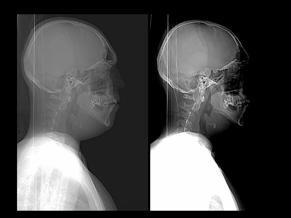
Epiglottitis on Lateral Xray

My Approach to CT Neck1) Airway
- Nasopharynx, oropharynx and hypopharynx (whole airway)- Trachea and esophagus
2) Deep neck spaces- Parapharyngeal space- Retropharyngeal space- Masticator space- Carotid space- Perivertebral space- Anterior visceral space- Submandibular/sublingual space

Approach to CT Neck
3) Glands- Parotid- Submandibular- Thyroid
4) Vessels and lymph nodes5) Bones and Soft tissues6) Neuro
- Brain, orbits, paranasal sinuses, mastoid air cells7) Cord8) Chest
- Lung apices - Mediastinum
Approach to CT Neck
• Bottom Line
– Check your ABC’S– A = AIRWAY– B = BONES– C = CAROTID SHEATH/VESSELS– S = SPINAL CORD/CANAL

Good resources
• Statdx• http://www.med.wayne.edu/diagRadiology/
Anatomy_Modules/axialpages/Overview.html

Thank you