old man with chronic chagasic myocarditis, sinus node...
TRANSCRIPT

Old man with chronic chagasic myocarditis, sinus node dysfunction, intraventricular conduction disturbances(RBBB/LAFB), symptomatic sustained monomorphic ventricular tachycardia and very low left
ventricle ejection fraction
Homem idoso portador de miocardite chagásica crônica, disfunção do nódulo sinusal, distúrbios intraventricular da condução, taquicardia ventricular monomórfica sustentada e fração de ejeção muito baixa
Raimundo Barbosa-Barros M.D. Nickname “ The fox”Coronary Center of the Hospital de Messejana Dr. Carlos Alberto Studart Gomes. Fortaleza/Brazil
•Specialist in Cardiology by the Brazilian Society of Cardiology (SBC).•Specialist in Intensive Care by the Sociedade Brasileira de Terapia Intensiva.•Chief of the Coronary Center of the Hospital de Messejana Dr. Carlos Alberto Studart Gomes. Fortaleza - Brazil.
Bom dia maestro: Gostaria de ouvir a opinião dos colegas do foro sobre este caso.Trata-se de um homem,77anos, portador de miocardite chagásica crônica. Há 2 anos foi implantado um marcapassos bicameral devido ao quadro de disfunção sinusal sintomática associada a conduçãoatrioventricular a AV alterada + BRD + BDASEAdmitido dia 15-09-2012 durante evento de TVMS TVMS associada à instabilidade hemodinâmica pelo quefor a submetido à cardioversão elétrica com sucesso. Atualmente em classe funcional II com terapiaotimizada, fração de ejeção do ventrículo esquerdo = 25%. Em uso de amiodarona, betabloqueador, enalapril e espironolactona. Foi indicado implante de CDI para prevenção secundária de morte súbita.Perguntas: Onde está o foco da TMMS? Existe indicação para ressincronização concomitante?Um abraçoRaimundo Barbosa-Barros
Good morning teacherI would like to hear the opinion of colleagues about this case.This is a man, 77 years of age, carrier chronic chagasic myocarditis. Two years ago was implanted a bicameral pacemaker due to symptomatic sinus node dysfunction associated with AV conduction disturbance: right bundle branch block and left anterior fascicular block.Admitted (15/09/2012) during event of symptomatic sustained monomorphic ventricular tachycardia (SMVT) associated with hemodynamic instability so is subjected to electrical cardioversion successfully.Currently in functional NYHA class II with optimized therapy, LVEF = 25%. In use of amiodarone, β-blockers, spironolactone and enalapril maleate.He was appointed an ICD for secondary prevention of sudden cardiac death.Questions: 1) Where is the focus location of S-MVT?2) Is there an indication for concomitant resynchronization?A hugRaimundo Barbosa-Barros M.D.
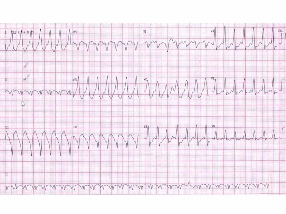


Estimado RaimundoMe impresina una TV originada del area infero-basal del VI, donde asientan algunos aneurismas chagásicos. Por supuesto es mas frecuente el aneurisma apical, pero en este caso como hay predominancia de Onda R en la cara anterior, las fuerzas se dirigen de la base al apex. Tiene eje infero-superior, indicando entonces la region infero-basal. por supuesto el prigen epicardico en esta region es dificil de estimar por ECG, epro no debiera descartarse a priori, sobre todo el si el paciente recurre con su CDI puesto y es considerado paraablacion.Respecto a resincronizar, la info en chagásicos es muy pobre pero extrapolando la informacion que tenemosen coronários, los subestudios de Zareba sobre el MADIT CRT demuestran que la resincronizacion en pacientes con bloqueos de rama derecha NO mejoran con CRT.Por lo cual le implantaria un CDI doble camara para usar los discriminadores del canal auricular (ademas de atender su disfunción sinusal).Un abrazo y escucho otras ideas
AB

Dear Raimundo: I think it is a VT originating from the infero-basal region of the left ventricle, In that
location, chagasic aneurysms are frequently placed. Of course, chagasic aneurysms are more frequent in the apex region of LV, but in this case, there is a predominance of R wave in the anterior wall indicating a basal to apex direction. The electrical axis has inferior-superior direction, indicating then that the origin focus is located on the infero-basal region of LV (and more septal than free wall).It is very difficult to estimate whether the focus is epicardial in origin by electrocardiogrphy or not. The epicardial source of the VT should be ruled out if recurrences occur, and the patient is considered for RF ablationRegarding CRT or not, the information that we have in chagasic patients is quite scarce, but extrapolatingthe scientific information that we have on coronary disease, the Zareba's sub studies (MADIT CRT) show that resynchronization (CRT) in patients with right bundle branch block do not improve clinical symptomsof LV parameters. Consequently, I would implant a dual chamber ICD to use atrial discriminators (besidesdealing with his sick sinus syndrome).
Let's hear from others
A hug and hear other ideasAdrian Baranchuk M.D. FACC FRCPC [email protected] Professor of Medicine and Biomolecular Sciences DepartmentDirector, EP Fellowship Training Program at Queen’s University; Kinston, Ontario, Canada
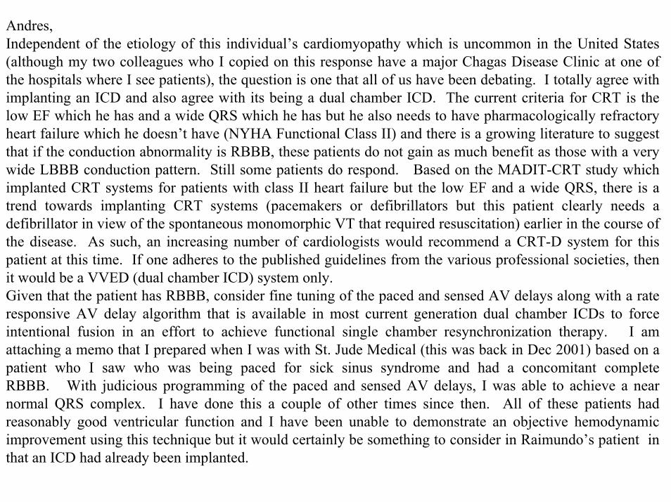
Andres,Independent of the etiology of this individual’s cardiomyopathy which is uncommon in the United States (although my two colleagues who I copied on this response have a major Chagas Disease Clinic at one of the hospitals where I see patients), the question is one that all of us have been debating. I totally agree with implanting an ICD and also agree with its being a dual chamber ICD. The current criteria for CRT is the low EF which he has and a wide QRS which he has but he also needs to have pharmacologically refractory heart failure which he doesn’t have (NYHA Functional Class II) and there is a growing literature to suggest that if the conduction abnormality is RBBB, these patients do not gain as much benefit as those with a very wide LBBB conduction pattern. Still some patients do respond. Based on the MADIT-CRT study which implanted CRT systems for patients with class II heart failure but the low EF and a wide QRS, there is a trend towards implanting CRT systems (pacemakers or defibrillators but this patient clearly needs a defibrillator in view of the spontaneous monomorphic VT that required resuscitation) earlier in the course of the disease. As such, an increasing number of cardiologists would recommend a CRT-D system for this patient at this time. If one adheres to the published guidelines from the various professional societies, then it would be a VVED (dual chamber ICD) system only.Given that the patient has RBBB, consider fine tuning of the paced and sensed AV delays along with a rate responsive AV delay algorithm that is available in most current generation dual chamber ICDs to force intentional fusion in an effort to achieve functional single chamber resynchronization therapy. I am attaching a memo that I prepared when I was with St. Jude Medical (this was back in Dec 2001) based on a patient who I saw who was being paced for sick sinus syndrome and had a concomitant complete RBBB. With judicious programming of the paced and sensed AV delays, I was able to achieve a near normal QRS complex. I have done this a couple of other times since then. All of these patients had reasonably good ventricular function and I have been unable to demonstrate an objective hemodynamicimprovement using this technique but it would certainly be something to consider in Raimundo’s patient in that an ICD had already been implanted.

Another consideration should episodes of VT recur on a frequent basis and they are always monomorphic, induce and map the VT and then perform catheter ablation of the identified VT focus rather than using more antiarrhythmic agents to minimize the recurrent VT episodes and need for a shock if antitachycardia pacing (ATP) is not successful.Paul Paul A. Levine MD, FHRS, FACC, CCDS25876 The Old Road #14Stevenson Ranch, CA 91381Cell: 661 565-5589Fax: 661 253-2144Email: [email protected]
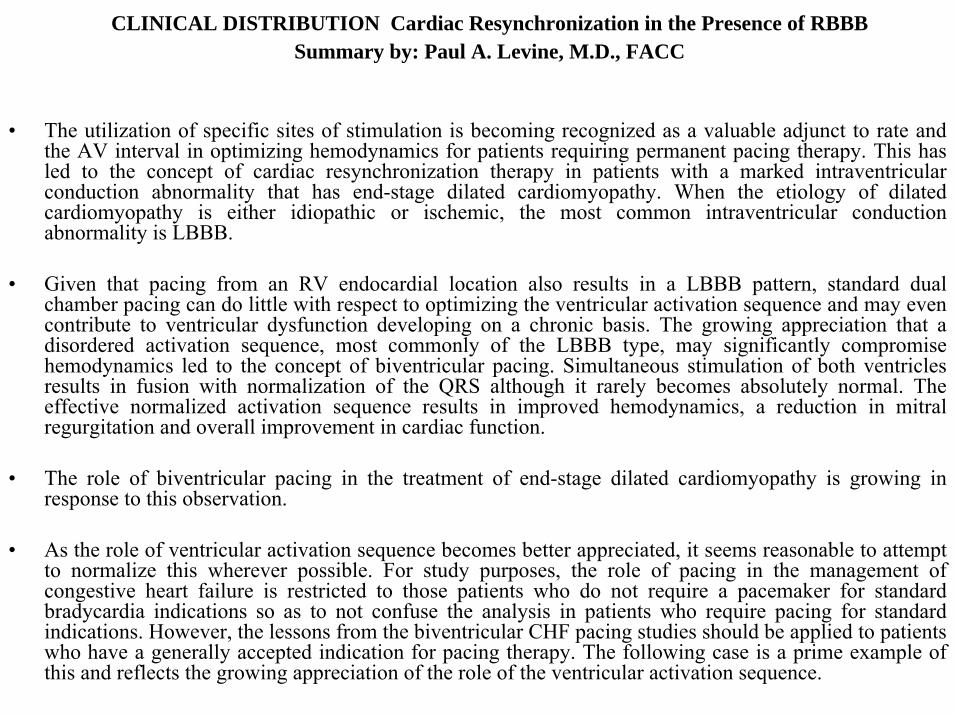
CLINICAL DISTRIBUTION Cardiac Resynchronization in the Presence of RBBB Summary by: Paul A. Levine, M.D., FACC
• The utilization of specific sites of stimulation is becoming recognized as a valuable adjunct to rate and the AV interval in optimizing hemodynamics for patients requiring permanent pacing therapy. This has led to the concept of cardiac resynchronization therapy in patients with a marked intraventricular conduction abnormality that has end-stage dilated cardiomyopathy. When the etiology of dilated cardiomyopathy is either idiopathic or ischemic, the most common intraventricular conduction abnormality is LBBB.
• Given that pacing from an RV endocardial location also results in a LBBB pattern, standard dual chamber pacing can do little with respect to optimizing the ventricular activation sequence and may even contribute to ventricular dysfunction developing on a chronic basis. The growing appreciation that a disordered activation sequence, most commonly of the LBBB type, may significantly compromise hemodynamics led to the concept of biventricular pacing. Simultaneous stimulation of both ventricles results in fusion with normalization of the QRS although it rarely becomes absolutely normal. The effective normalized activation sequence results in improved hemodynamics, a reduction in mitralregurgitation and overall improvement in cardiac function.
• The role of biventricular pacing in the treatment of end-stage dilated cardiomyopathy is growing in response to this observation.
• As the role of ventricular activation sequence becomes better appreciated, it seems reasonable to attempt to normalize this wherever possible. For study purposes, the role of pacing in the management of congestive heart failure is restricted to those patients who do not require a pacemaker for standard bradycardia indications so as to not confuse the analysis in patients who require pacing for standard indications. However, the lessons from the biventricular CHF pacing studies should be applied to patients who have a generally accepted indication for pacing therapy. The following case is a prime example of this and reflects the growing appreciation of the role of the ventricular activation sequence.
CLINICAL DISTRIBUTION Cardiac Resynchronization in the Presence of RBB
Summary by: Paul A. Levine, M.D., FACC
• The patient is a 60+ year old man whose pacemaker was implanted for symptomatic sinus node dysfunction. He had intact AV nodal conduction with a right bundle branch block ventricular activation pattern. His pacemaker, St. Jude Medical’s Trilogy DR+ ® model 2364 had been programmed to the DDDR mode with a long AV delay to allow for intact AV nodal conduction. This resulted in effective inhibition of the ventricular channel with the beneficial effect of reducing overall battery current drain and increasing device longevity. However, even more important than device longevity is optimization of ventricular function. On the most recent visit, the growing appreciation of the role of ventricular activation sequence was taken into consideration and the AV delay adjusted to force ventricular fusion.
• The patient is a 60+ year old man whose pacemaker was implanted for symptomatic sinus node dysfunction. He had intact AV nodal conduction with a right bundle branch block ventricular activation pattern. His pacemaker, St. Jude Medical’s Trilogy DR+ ® model 2364 had been programmed to the DDDR mode with a long AV delay to allow for intact AV nodal conduction. This resulted in effective inhibition of the ventricular channel with the beneficial effect of reducing overall battery current drain and increasing device longevity. However, even more important than device longevity is optimization of ventricular function. On the most recent visit, the growing appreciation of the role of ventricular activation sequence was taken into consideration and the AV delay adjusted to force ventricular fusion.
• The LBBB pattern associated with RV apical pacing combined with the patient’s RBBB ventricular activation pattern effecting a near-normal QRS pattern.

Intentional Fusion to Normalize Ventricular Activation
Figure 1
Figure 1 was recorded with the pacemaker programmed to the VVI mode and a low rate to allow AV conduction and a demonstration of his usual ventricular activation pattern. The PR interval is 160 ms and the QRS duration is 160-180 ms with a RBBB pattern. The monitor lead is a modified V1.
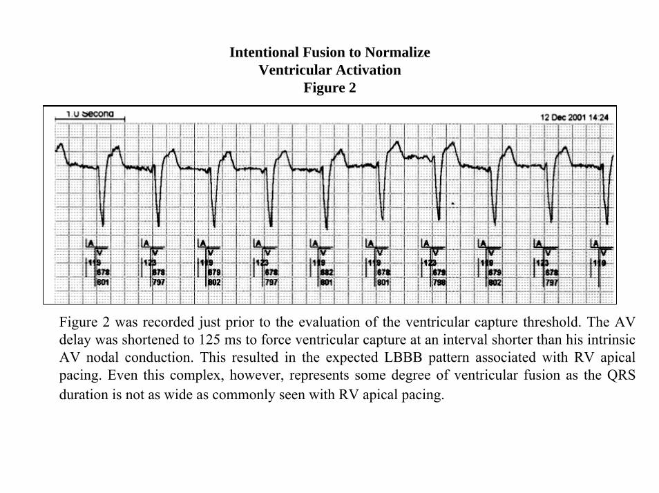
Intentional Fusion to Normalize Ventricular Activation
Figure 2
Figure 2 was recorded just prior to the evaluation of the ventricular capture threshold. The AV delay was shortened to 125 ms to force ventricular capture at an interval shorter than his intrinsic AV nodal conduction. This resulted in the expected LBBB pattern associated with RV apical pacing. Even this complex, however, represents some degree of ventricular fusion as the QRS duration is not as wide as commonly seen with RV apical pacing.

Intentional Fusion to Normalize Ventricular Activation
Figure 3
Figure 3 represents fusion between the native conduction pattern and RV apical pacing. The ECG was monitored while adjusting the AV delay. In this case, the AV delay that provided the narrowest QRS was 225 milliseconds.
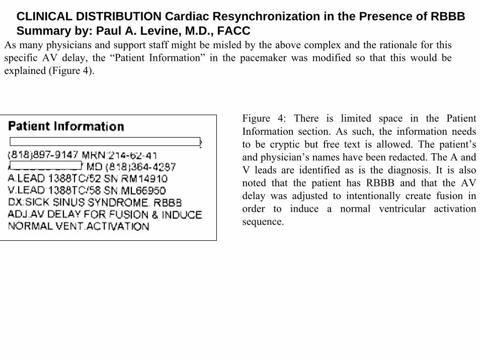
Figure 4: There is limited space in the Patient Information section. As such, the information needs to be cryptic but free text is allowed. The patient’s and physician’s names have been redacted. The A and V leads are identified as is the diagnosis. It is also noted that the patient has RBBB and that the AV delay was adjusted to intentionally create fusion in order to induce a normal ventricular activation sequence.
As many physicians and support staff might be misled by the above complex and the rationale for this specific AV delay, the “Patient Information” in the pacemaker was modified so that this would be explained (Figure 4).
CLINICAL DISTRIBUTION Cardiac Resynchronization in the Presence of RBBB Summary by: Paul A. Levine, M.D., FACC

Summary
• In addition to selecting the optimal resting base rate, the rate-modulated settings and the AV delay, one should begin to think about ventricular activation sequence. When there is normal ventricular function, a normal QRS complex and intact AV nodal conduction, a long AV delay (either fixed or preferably achieved with AV/PV Hysteresis [AutoIntrinsic Conduction Search]) is associated with optimal hemodynamics.
• When there is an intrinsic intraventricular conduction abnormality, careful attention to placement of the ventricular lead is being recognized as being of increasing value.
• In those patients who have a RBBB with intact AV nodal conduction, careful adjustment of the AV delay with a standard right ventricular endocardial lead can result in consistent fusion that effectively normalizes the ventricular activation sequence.
Independente da etiologia da cardiomiopatia, este paciente é portador de uma etiologia incomum de ver nos Estados Unidos da América (embora meus dois colegas que eu he copiado esta resposta tem uma Clínica de Doença de Chagas grande em um dos hospitais onde eu vejo pacientes), a questão é que todos vêm debatendo. Concordo totalmente com a implantação de um CDI e também estou de acordo com que tenha sido um CDI de câmara dupla. Nos critérios atuais de CRT é a FE baixa com QRS alargado também precisam ter insuficiência cardíaca farmacologicamente refratária que este paciente não tem (classe funcional II) Por outra parte, existem crescentes evidências que sugerem que se o distúrbio de condução é o BRD, a CRTnão terá um benefício, tanto quanto aqueles com um padrão de BCRE com QRS largo. Baseado no estudo MADIT-CRT que implantou CRT em pacientes em ICC classe II, associada a FE baixa e QRS largo, há uma tendência para a implantação de sistemas de CRT (este paciente necessita claramente de um CDI em vista da TVM espontânea de ressuscitação obrigatória mais cedo no curso da doença. Um número crescente de cardiologistas recomendariam um sistema CRT-D para o paciente no momento. Se um adere às orientações publicadas nas diversas sociedades profissionais, então seria um sistema (CDI de câmara dupla) VVED apenas.Tendo em conta que o paciente tem BRD, considerar o ajuste fino do ritmo e sentiu atrasos AV junto com um algoritmo que calcule la taxa de atraso da resposta AV disponível nas atuais gerações de CDIs de dupla câmara para forçar a fusão intencional com o intuito de alcançar a terapia de ressincronização funcional única câmara. Estou anexando um memorando que preparei em Dezembro de 2001, quando estava no St. Jude Medical. Dito memorando foi baseado em um paciente que estava evoluindo para a síndrome do Nósinusal doente e tinha um BCRD concomitante. Com uma programação criteriosa dos atrasos AV estimulados e detectados, consegui um complexo QRS próximo do normal. Desde então tenho feito isso algumas outras vezes. Todos esses pacientes tinham função ventricular razoavelmente boa porém, não consegui demonstrar uma melhora hemodinâmica objetiva, utilizando esta técnica. Não obstante certamente seria algo a ser considerado neste paciente de Raimundo em que o CDI já havia sido implantado.Se ocorrem episódios de TV freqüentes sempre monomórficos, penso que deveriamos mapear a TV a seguir realizar ablação por cateter do foco VT identificado em vez de usar os agentes antiarrítmicos com o intuito de minimizar o número de episódios recorrentes de TV e a necessidade de um choque se o marcapasseoantitaquicardia (ATP) não é bem sucedido.

Independientemente de la etiología de la miocardiopatía, este paciente tiene una etiología poco común de ver en los Estados Unidos (a pesar de que mis dos colegas a quienes le he copiado esta respuesta tienen una clínica para enfermedad de Chagas en un hospital grande donde veo pacientes). El punto es el que todos han estado debatiendo. Estoy totalmente de acuerdo con la implantación de un CDI y también estoy de acuerdo con que haya sido uno de doble cámara. En los actuales criterios de implante de CRT con FE baja y QRS ancho también necesitan la insuficiencia cardíaca farmacológicamente refractaria que este paciente no tiene (clase funcional II) Por otro lado, hay evidencia crecientes que sugieren que en el distúrbio dromótropo tipo BRD, el CRT no tendrá un beneficio tanto como aquellos con un patrón de BRI con QRS ancho. Basados en MADIT-CRT en pacientes en clase II, asociados con baja FE y QRS ancho, hay una tendencia para resincronización CRT (este paciente necesita claramente un ICD en vista de la TVMS espontánea ocurrida mas temprano en el curso de la enfermedad). Un creciente número de cardiólogos recomiendan un sistema CRT-D para el paciente en ese momento. Si uno adhiere a las directrices publicadas en varias sociedades profesionales, entonces sería un sistema (de doble cámara CIE) en VVED apenas.Tendo cuenta de que el paciente tiene BRD, deberiamos considerar el ajuste fino del ritmo y sentir los intervalos AV junto con un algoritmo que calcula que la tasa de respuesta retardada AV disponible en las generaciones actuales de CDI de doble cámara para forzar la fusión intencional con el fin de lograr la terapia de resincronización funcional cámara única. Estoy adjuntando un memorando que preparé en diciembre de 2001, cuando trabajaba en el St. Jude Medical. Dicho memorando se basó en un paciente que estaba evolucionando para el síndrome del nódulo sinusal enfermo y tenía una BCRD concomitante. Con unos retrasos AV en el programa cuidadosamente estimulados y detectados, obtuve complejos QRS casi normales. Desde entonces lo he hecho varias veces en algunos otros casos. Todos estos pacientes tenían una función ventricular razonablemente buena, no obstante no pude demostrar mejoría hemodinámica objetiva con esta técnica. Sin embargo, sería sin duda algo a ser considerado en este paciente de Raimundo ya que el CDI había sido implantado.Si tiene frecuentes episodios de TV monomórfica siempre, creo que deberiamos mapear la TV y luego realizar ablación con catéter en lugar de utilizar los fármacos antiarrítmicos con el fin de minimizar el número de episodios recurrentes de TV y la necesidad de repetidos shocks si el marcapasseo antitaquicardia (ATP) no tiene éxito.

Final comments Andrés Ricardo Pérez-Riera M.D. Ph.D.
Institution: Faculdade de Medicina do ABC Fundação do ABC. Santo André, São Paulo, Brazil.

QRSd = 195ms
Monophasic R in V2
F fusion beatThe presence of fusion beats- QRS complexes with
intermediary morphology is suggestive of VT
Sustained VT (S-VT): is defined as continuous VT lasting for >30s or that requires and intervention for termination (such cardioversion) with Structural Heart Disease and hemodynamic involvement. with “RBBB-like” pattern arise in the LV. It is a VT hemodynamically unstable: A VT that causes hemodinamically compromise requiring prompts termination

Monomorphic VT: has a similar QRS configuration from beat to beat. originating from a single focus with identical QRS complexes. Monomorphic VT results from a single abnormal focus or reentrant pathway and has regular, identical-appearing QRS complexes. Some variability in QRS morphology at initiations is not uncommon, followed by stabilization of the QRS morphology. RBBB and LBBB-like VT configurations are terms used to described the dominant deflection in V1, with a dominant R wave described as “RBBB-like”and dominant S wave as “LBBB-like” configuration.(1) While virtually all VT or PVCs with “RBBB-like”pattern arise in the LV only, VTs or PVCs with “LBBB-like” morphology can arise in either the LV or the RV. In the presence of prior infarction, VTs with “LBBB-like” pattern virtually always arise on or adjacent to the LV septum. In patients without structural heart disease, QRS complexes tend to be smooth and tall. With scarring of any etiology, the QRS complexes have lower amplitudes and are broader. Notching of the QRS is a sign of scar. QS complexes, other than in aVR, suggest the wave front is moving away form the recording site, but does not necessarily mean scar/infarct; however, qR or QR complexes in anatomically adjacent sites typically is a sign of infarction. Patients without structural heart disease usually exhibit a single-VT morphology, while in patient with significant structural heart disease multiple VTs are common.(2; 3)
1. Miller JM, Marchlinski FE, Buxton AE Josephson ME. Relationship between the 12-lead electrocardiogram during ventricular tachycardia and endocardial site of origin in patients with coronary artery disease. Circulation. 1988 Apr;77:759-766.
2. Josephson ME, Waxman HL, Cain ME, Gardner MJ, Buxton AE. Ventricular activation during ventricular endocardial pacing. II. Role of pace-mapping to localize origin of ventricular tachycardia. Am J Cardiol. 1982 Jul;50:11-22.
3. Josephson ME, Clinical Cardiac Electrophysiology: Technique and Interpretation Walters Kluwer/Lippicott Williams& Wilkins, 4th edn. Philadelphia, 2008.

CLASS I Asymptomatic or palpitations
CLASS II Dizziness, precordial pain and/or dyspnea.
CLASS III Presyncope or syncope.CLASS IV Cardiopulmonary arrest.
CLASSIFICATION ACCORDING TO CLINICAL CHARACTERISTICS OF VT
1. Hemodynamically unstable VT: is a VT that causes hemodinamically compromise requiring prompts termination( The present case)
2. Incessant VT: Incessant ventricular tachycardia (VT) is defined as hemodynamically stable VT continuing for hours. This is continuous sustained VT that recurs immediately despite repeated spontaneous or therapeutic termination
3. Repetitive monomorphic VT: is defined as continuously repeating episodes of self-terminating nonsustained VT
4. Unmappable VT is a VT that does not allow interrogation of multiple sites to define the activation seequence or preform entrainment mapping and may be caused of: hemodynamic intolerance that necessitates immediate VT termination, spontaneous or pacing-induced transitional to other morphologies of VT, or repeated termination during mapping
5. VT storm or electrical storm arrhythmic storm: is considered three or more separate episodes of sustained VT/VF within 24h, each requiring termination VT intervention it is defined as recurrent or multiple episodes of VF or VT: 20 or more per day, or 4 or more per hour. This type of event, with ominous meaning, may be observed in the acute phase of myocardial infarction and in Brugada Syndrome., refers to multiple recurrences of ventricular arrhythmias over a short period of time.
Classification of VT by the presence or not of underlying structural heart disease and the four classes of symptoms secondary to VT.

aVR aVL
I
IIIII aVF
X
Y
QRS axis
-40º
The exit is on the Inferior wall because
Superior axis.
The presence of QR complexes indicates activation is moving away from the site where the complex is registered.
Lead I R or Rs is suggestive of septal-parahissian origin
SÂQRS in the FP located in the right superior quadrant between – 90º and±180º: or extreme shift to the left such as the present case is suggestive of VT.

Ventriculographic image of LV aneurysm in infero-basal region of the left ventricle
III
aVL
I
QRS axis

V6
V1
V4
V5
V2 V3
X
Z
Precordial early transition
VTs that originate at the subepicardiumgenerally have slower QRS upstrokes in the precordial leads
Septal-parahisian origin

V4-V6
1. Berruezo A, Mont L, Nava S, Chueca E, Bartholomay E, Brugada J. Electrocardiographic recognition of the epicardial origin of ventricular tachycardias.Circulation. 2004 Apr 20;109:1842-1847.
2. Daniels DV, Lu YY, Morton JB et al. Circulation 2006; 2006; 113:1659-1666.
Berruezo et al(1) demonstrated that the epicardial origin of the ventricular activation can be recognized on the ECG by slurring of the initial part of the QRS complex (pseudodelta)A pseudodelta wave of ≥34 ms has a sensitivity of 83% and a specificity of 95%,
A Ventricular Activation Time (VAT), “R peak time” or intrinsicoid deflection time of ≥85 ms has a sensitivity of 87% and a specificity of 90%, and an RS complex duration of ≥121 ms has a sensitivity of 76% and a specificity of 85% in identifying an epicardial origin of the VTs.
A delayed precordial maximum deflection index ≥0.55 identified epicardial VT remote from the aortic sinus of Valsalva with a sensitivity of 100% and a specificity of 98.7% relative to all other sites of origin (P<0.001).(2)
VAT=90ms
δ
δ

The presence of monophasic QRS (R) in V1 or V2 is indicative of VT
YaVF
Z V2
“RBBB-like” pattern arise in the LV

QRS complex ≥ 120 ms, with bizarre aspect and RBBB pattern.The R to S interval were >100 ms in some
precordial lead, it is highly specific of VT; however, it is not much sensitive.
V3
VT
>100ms
QRSd = 195ms
R/S interval >100 ms = VT
Second step of Brugada algorithm (1)
R/S interval =120ms = VT1. Brugada P, et. al. Circulation 1991; 83:1649-1659.

According to the current guidelines, patients with ischaemic cardiomyopathy (ICM) or non-ischaemiccardiomyopathy (NICM) at risk for SCD should undergo implantation of an ICD. Although ICDs effectively terminate VTs, the arrhythmogenic substrate remains unchanged or may progress over time, resulting in recurrent ICD shocks. Defibrillator shocks increase mortality and worsen quality of life. Evidence from two prospective randomized trials on outcome in patients with ischaemic heart disease undergoing RFCA for VT suggests that ablation prevents recurrence of VT and decreases the number of ICD shocks.(1)The contribution of scar to the electrophysiological abnormalities targeted for ablation of unstable VT differs between ICM and NICM. An approach incorporating LP ablation and pace-mapping had limited success in patients with NICM compared with ICM, and alternative ablation strategies should be considered. (2)Epicardial catheter ablation may be an alternative for managing CRT-induced proarrhythmias without the inactivation of LV pacing.(3)In patients with normal coronary arteriograms and LV aneurysm, exercise-induced VT with RBBB pattern may have a subepicardial arrhythmogenic substrate, which may be amenable to epicardial ablation.(4)
1. Wissner E, Stevenson WG, Kuck KH.Catheter ablation of ventricular tachycardia in ischaemicand non-ischaemic cardiomyopathy: where are we today? A clinical review. Eur Heart J. 2012 Jun;33:1440-1450.
2. Nakahara S, Tung R, Ramirez RJ, et al. Characterization of the arrhythmogenic substrate in ischemic and nonischemic cardiomyopathy implications for catheter ablation of hemodynamicallyunstable ventricular tachycardia. J Am Coll Cardiol. 2010 May 25;55:2355-2365.
3. Yamada T, Tabereaux PB, McElderry HT, et al. Successful catheter ablation of epicardialventricular tachycardia worsened by cardiac resynchronization therapy. Europace. 2010 Mar;12:437-440.
4. Ouyang F, Antz M, Deger FT, et al. An underrecognized subepicardial reentrant ventricular tachycardia attributable to left ventricular aneurysm in patients with normal coronary arteriograms. Circulation. 2003 Jun 3;107:2702-2709.
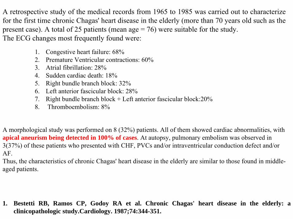
A retrospective study of the medical records from 1965 to 1985 was carried out to characterize for the first time chronic Chagas' heart disease in the elderly (more than 70 years old such as the present case). A total of 25 patients (mean age = 76) were suitable for the study. The ECG changes most frequently found were:
1. Congestive heart failure: 68%2. Premature Ventricular contractions: 60%3. Atrial fibrillation: 28%4. Sudden cardiac death: 18% 5. Right bundle branch block: 32%6. Left anterior fascicular block: 28%7. Right bundle branch block + Left anterior fascicular block:20%8. Thromboembolism: 8%
A morphological study was performed on 8 (32%) patients. All of them showed cardiac abnormalities, with apical aneurism being detected in 100% of cases. At autopsy, pulmonary embolism was observed in 3(37%) of these patients who presented with CHF, PVCs and/or intraventricular conduction defect and/or AF. Thus, the characteristics of chronic Chagas' heart disease in the elderly are similar to those found in middle-aged patients.
1. Bestetti RB, Ramos CP, Godoy RA et al. Chronic Chagas' heart disease in the elderly: a clinicopathologic study.Cardiology. 1987;74:344-351.