office practice in neonatology- costly misseswith respiratory infection was prescribed saline nasal...
TRANSCRIPT

Office practice in Neonatology-costly misses
Moderator-Dr Suresh Kumar Surapaneni
Panelists-Dr Alok BhandariDr Hemant JainDr Jaydeb ray Dr Asutosh Mohapatra

Dr Suresh Kumar Surapaneni
Head of Neonatology,Pragna Hospitals and KIMS
Kondapur,HYDERABAD
MD DNB DM(PGI)FRCPCH(UK)FNNF
VLBW,SGA, ventilation, nutrition

Professor of Pediatrics and HOD, Institute of Child Health, Kolkata
MD(Paed), DCH (Cal), DNB (Paed), MD (Com.Med), FIAMS(Paed), FNNF, FIAP
Neonatology----President(2016-18) NEONATOLOGY SOCIETY, National Executive (2011-12), NNF,INDIAGrowth and Development--National chairperson(2012-13), National secretary ,(2010-11) Growth, Development & Behavioral chapter of IAP,, Nutrition, Infectious diseases, More than 30 national and international Publication.
Prof. Jaydeb Ray

Dr. Asutosh MahapatraBhubaneswar, Odisha
Consultant in Pediatrics & Neonatology
MBBS, MD
Child ‘N’ Child, BhubaneswarNeonatology, Pediatric Infectious Disease, Medicolegal,
Adolescent PediatricsPresent Vice-President, NNF OdishaPast Secretary NNF, Odisha 2009-18
Governing Body Member – NNF India 2013-1418nos. Of publication indexed journal
Dr Hemant Jain
Prof and Head of Pediatrics,MGM Medical College, Indore
MD ,Fellowship in Neonatology USA ,FIAP
Neonatology and clinical research

Baby of S, born in a corporate hospital ,seen by a senior neonatal team
Had eye discharge on day 4 , treated and followed in opd
CATARACT DIAGNOSED at 4 months age

Eyes
Presence of bilateral red reflex
Structure
• Periorbital anatomy,Spacing and size,Eyelids,
lashes, eyebrows
• Nasolacrimal puncta nad duct
pathway,Palpebral fissures
• Conjunctiva,Sclera
• Anterior segment- Cornea, Irides,Lens
• Fundoscopic examination
Function:assessed throughout examination of
structure
• Visual acuity,Pupil reactivity,Extraocular
muscle activity

Baby A
Baby A, born of LSCS , weight 3.2 kg,
neonatal screening done but parents did not collect reports
Presented at 2 months age in OPD of another pediatrician with complaints of constipation
Neonatal screening report traced back at birth hospital- TSH high by 3 fold,
Repeat thyroid profile at age of 2 months -T4 and
T3 low and TSH high by 5 fold

❖with low T4
<10 μg/dL or
FT4 <1.17
ng/dL

Newborn screening for Congenital
Hypothyroidism
■ Imaging is recommended -- radionuclide scintigraphy and ultrasonography but treatment should not be delayed till scans are performed
■ Levothyroxine is commenced at 10 to 15 μg/kg in the neonatal period.
BIOCHEMICAL FOLLOW UP PLANE
2 wk --Serum T4/FT4
1 mo ---TSH and T4/FT4
2 monthly till 6 mo,
3 monthly from 6 mo-3 y
>3years -- every 3–6 mo
■ Babies with the possibility of transient congenital hypothyroidism should be re-evaluated at age 3 y, to assess the need for lifelong therapy

Baby of N , born of normal vaginal delivery, 2.65 kg born as breech ,
Developmental dysplasia of Hip DDH - diagnosed at 6 months

Baby P , Male 2.9 kg , had mild RD , kept NICU for 24 hours
Had Stridor in newborn , dismissed as laryngomalacia,
Persistent stridor and repeated episodes of excessive cry
ENT consultation for direct laryngoscopy and 2 D ECHO was done-diagnosed as Vascular ring

■ Baby of A,Male child 3rd in birth order
■ G1- FTND,boy died in 3rd monthof life with vomiting G2 –
FTND,boy,died at age of 5months with vomitings and
loosemotions,
■ G3 –PP –ANC uneventful FTNVD,APGAR 10,10 ,BW
3.2 kg
■ Baby presented on 15th day with vomitings 15 times in a
day
■ Weight 2.3 kg,severe dehydration,Fluid correction given
but iv fluid requirement over next 24 hours was in 350
ml/kg/day

■ S electrolytes Na 136 meq/l,K 6.5 meq/l
■ S cortisol 8 am - low and 17 hydroxy progesterone
markedly elevated
■ Diagnosis –Congenital adrenal hyperplasia
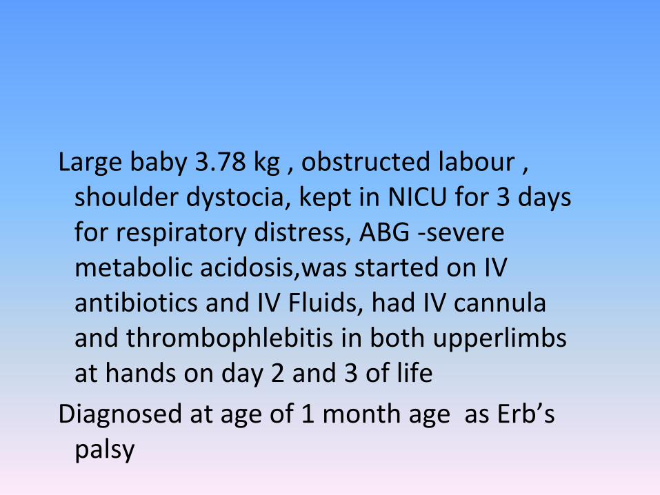
Large baby 3.78 kg , obstructed labour , shoulder dystocia, kept in NICU for 3 days for respiratory distress, ABG -severe metabolic acidosis,was started on IV antibiotics and IV Fluids, had IV cannula and thrombophlebitis in both upperlimbs at hands on day 2 and 3 of life
Diagnosed at age of 1 month age as Erb’s palsy

ERB’S PALSY

■ Baby of S,Born to G4 Lo mother
■ G1 – new born male,FTNVD,breast fed, had jaundice on day 4 onwards,died after 1 month
■ G2-girl baby, FTNVD,breast fed,had jaundice on day 3 died on 21st day of life
■ G3- boy ,full term,FTNVD,breast fed,had jaundice from day 2 of life,needed exchange transfusion twice, died at age of 2 months
■ G4 –PP- antenatal period uneventful
■ FTNVD,3 KG APGAR GOOD
■ Had jaundice on day 3 of life

■ Neonatal screening done after 2 days of breast feeding-positive for galactosemia
■ Galactosemia confirmed with urinary chromatography and gene mapping at CDFD
■ Child started on lactose free milk from day 4 of life and continued since then

Galactosemia1. Do newborn screening to all neonates
2. Do metabolic screening of all newborns bor of out of consanguineous marriage and bad obstetric
history.
3. Prenatal diagnosis & counseling in subsequent pregnancies
4. Early diagnosis and treatment is needed to prevent death and morbidity in newborns with IEM
5. A high index of of suspicion must be maintained in all neonates with history of previous siblings having
a neonatal demise.

⮚ 2 month old male child presented with
cough, distress, feeding difficulty and failure
to thrive
On examination had - SPO2
without oxygen of 86%,Tachypnea, distress,
wheeze++
No murmur, Liver – 3cm palpable
⮚ X Ray chest – Haziness in both the lung
fields
⮚ Received multiple antibiotics no response
Echo done showed RA/ RV hypertrophy, High
PA Pressure with ASD (OS) with Left to Right
shunt and normally related great vessels.

⮚ CT Chest - Bilateral
Consolidations
⮚ No response to Antibiotics
⮚ CT Angiography done showed
TAPVC draining into vertical vein
⮚ Patient operated, doing well on
follow up.

Baby R, born at 26 weeks , 810 gm , stayed for 11
weeks in NICU for RDS, sepsis , NNEC III B
Had first screening at 37 weeks , showing
advanced ROP
Preterm babies missing ROP screening and landing
in retinal detachment , visual deficit and poor
visual outcome later
Reasons told- late recovery , was on respiratory
support and oxygen longer, kept in Nicu longer,
did not get appointment, thought to go after 3
months, tired of visiting hospital, went to local
eye doctor, thought baby was too small to take
out to eye hospitals it was air-conditioned and we
don’t want to come out as it may cause infections
etc

■ ROP screening should be done at 4 weeks after birth
• However in babies born earlier than 30 weeks of GA or BW < 1200 grams, it should be done at 2-3 weeks after birth (Keep in mind that it should not be later than 3 weeks)
• The screening should be done by trained ophthalmologists using indirect ophthalmoscope in NICU/SCNU, if the baby is still admitted. If baby is discharged, then screening can be done in a defined area in SCNU on an outpatient basis
• The frequency of follow up depends upon the zone and stage of ROP
JAYDEB RAY
ROP screening

Baby M, 3.6kg , 21 day old Newborn
with respiratory infection was
prescribed saline nasal drops ad lib
After 48 hours child and still parents
complained blocked nose and Duty SR
prescribed nasal decongestant drops
After 24 hours child presented to OPD
with tachycardia and respiratory
distress
Chest- conducted sounds B/L, spo2 91
to 95 % all limbs, pulse rate 220/min

Neonate with mild respiratory distress and chest xray reported normal
Close repeat look revealed congenital lobar emphysema

Baby of N, birth weight 3.23kg , had flat occiput, SR saw at birth and advised follow up in opd
Came to OPD on busy day at 15 days age and Junior consultant was of same opinion
Repeat consult at 6 weeks with Senior consultant-flat occiput and head growth of only 0.5 cm
Suspected craniostenosis due to lamboid suture fusion got CT BRAIN ,which confirmed diagnosis


Why costly misses happen
• POOR TECHNIQUE
• OMISSION OF AREAS IN THE EXAMINATION
• FAILURE TO RECOGNISE SIGNS
• INCOMPLETE OR POOR RECORDING OF POSITIVE OR NEGATIVE FINDINGS
• IMPROPER INTERPRETATION OF FINDINGS
• BUSY CLINIC OR BUSY MIND

Suggested order
• General observation
• Head and Neck-Facies,nose,mouth,ears
• Trunk –cardiorespiratory system,abdomen,back,genitalia and rectum
• Extremities
• Neurologic examination
• Head circumference and length
• Eye examination

Take Home
Learn for others mistakes because
-You can’t make all mistakes yourself
its tension free
its cheaper

▶ B/O-S, a 26 day old male child brought with yellowishdiscoloration of skin since 5th day of life
▶ Child born to a primi mother at 33 wks of gestation byvaginal delivery , cried immediately at birth, birth wtof 2.4 kg.
▶ Developed respiratory distress soon after birth ,wasadmitted in NICU for 3 days during which o2 iv fluidsand antibiotics were given , child was weaned off o2and started on feeds by third day of life.
▶ Jaundice was first noticed on 5th dol, for whichphototherapy was given for two days

Baby discharged on 7th dol, with TSB of 11 mg /dl
Baby was readmitted again on 22 nd day of life with TSB of 15 mg/dl, child was discharged after 48 hrs with TSB of 12 mg/dl
NO h/o – discharge from umblical cord- ABO or Rh incompatibility- high coloured urine and clay coloured stools - hypotonia, coarse skin ,delayed passage of stools- cephalhematoma

Antenatal history
▶ Primi mother, on regular ANC check up▶ On iron and follic acid supplementation▶ h/o of fever with rash during 7 th month of
pregnancy, fever lasted for 2 days only anti pyretics were given ,
▶ No h/o of lymphadenopathy
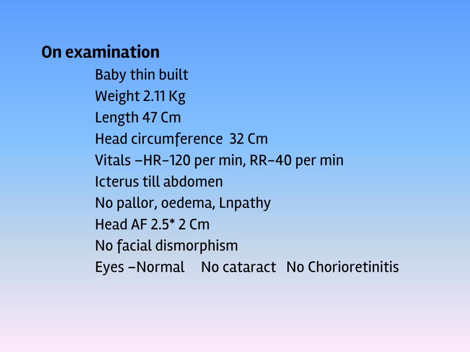
On examination Baby thin built Weight 2.11 KgLength 47 CmHead circumference 32 CmVitals –HR-120 per min, RR-40 per minIcterus till abdomenNo pallor, oedema, LnpathyHead AF 2.5* 2 CmNo facial dismorphismEyes –Normal No cataract No Chorioretinitis

Ear, Nose, Throat- NormalNeck-NormalSkull, Spine, Genitalia- NormalP/A- Soft, no organomegalyCVS-S1,S2-Normal, No murmurCNS-cry, tone, activity- Normal
Moro’s reflex present

TSB Values
Dol TSB D I
7th 11 0.7 10.3
22nd 15 1.9 13.0
24th 12 1.2 10.8
27th 16 1.5 14.5

InvestigationsLFT TSB 16.1 DCT – ve
D 1.5 I 14.6 Retic count - normalAST 85ALT 57 G6PD assay - normal ALKPo4 1164
Coagulation Profile PT 24/15 Peripheral smear- NAD APTT 46/35
TSH 3.9 IU/ml URINE M/E- NAD
CMV IgG+ve IgM -Ve
CT Brain s/o Bilateral parafalcine parietal lobe calcifications


41 days Female childBorn in non consanguineous marriage brought withChief complaints: Yellowish discoloration of skin and eyes- 20 days.White colored stool - 20 daysDark yellow colored urine - 20 daysAntenatal history: Uneventful
Birth history:Baby born by LSCS Full term Birth wt 4.0Kg

GENERAL EXAMINATION:HR 120 /minRR 34 /minCRT < 2 secIcterus noted in eyes & skin Mild pallor present.S/E:P/A Hepatomegaly 3 cm Spleen not palpable CVS S1 S2 normal R/S AEBECNS conscious with CTA goodReflexes normal.

INVESTIGATION
LIVER FUNCTION TEST : TSB 21.97 mg/dl
Direct 13.77 mg/dl Indirect 8.20
mg/dl
ALKPm855 u/lSGOTm125 u/l SGPT 80 u/l
Albumin 3.30 gms/dl Globulin 2.80 gms/dlA/G Ratio
1.17
CBP:
HB 15.8gm%Total WBC26,300Cells/cumm
Platelet count 1.80 Lakhs/cumm Retic count
0.9%
URINE EXAMINATION
Bile salt presentBile pigment present
COAGULATION PROFILE
PT 18sec APTT 58sec
HEPATO BILIARY SCINTIGRAPHY
Scan findings are consistent with features of extra hepatic biliary
obstruction (?Atresia)
USG ABDOMEN
Gallbladder – collapsed

D/DInspissated bile syndrome Biliary atresiaNeonatal hepatitisCholedochal cystBile duct stenosisPausity of intrahepatic bile ductAlagille syndrome

COURSE IN HOSPITAL:
➢Patient treated on IV fluids & FFP ➢Opinion of pediatric surgeon➢In view of extra hepatic biliary atresia –
plan for laparotomy.➢On laparotomy surgeon noted minimal
nodular liver with contracted gallbladder.➢Operative cholangiogram showed dye
entering in G B & C B D .➢Distally dye entered in duodenum.➢So, flushing done & liver biopsy taken.

INTRA – OPERATIVE CHOLANGIOGRAM
