of the mitral valve: clinical, cineangiographic study
TRANSCRIPT

July 1967 Post-concussional Sequela-Taylor BRITIS 71
and biochemical abnormalities. It is suggested that researchinto the large and important group of "non-serious" headinjuries has been neglected, to the detriment of their manage-ment.
Figs. 1, 2, and 3 are reproduced by kind permission of Dr.N. C. Nevin.
REFERENCES
Becker, R. F., Groat, R. A., and Windle, W. F. (1946). Fed. Proc., 5, 7.Biggart, J. H. (1936). Pathology of the Nervous System. Edinburgh.Breig, A. (1960). Biomechanics of the Central Nervous System. Stock-
holm.Brock, S. (1960). Injuries of the Brain and Spinal Cord and Their
Coverings, 4th ed. New York.Caveness, W. F. (1966). In Head Injury Conference Proceedings, edited
by W. F. Caveness and A. E. Walker, p. 209. Philadelphia.Denny-Brown, D., and Russell, W. R. (1941). Brain, 64, 93.Durand Wever, A. M. (1929). Munch. med. Wschr., 76, 1879.Erichsen, J. E. (1866). On Concussion of the Spine, Nervous Shock, and
Other Obscure Injuries of the Nervous System. New York.Friede, R. L. (1961). Arch. Neurol. (Chic.), 4, 449.Friedman, A. P., Brenner, C., and Denny-Brown, D. (1945). 7. Neuro-
surg. (Chic.), 2, 36.Goldstein, K. (1942). After Effects of Brain Injuries in War, p. 220.
London.Groat, R. A., and Simmons, J. Q. (1950). 7. Neuropath. exp. Neurol., 9,
150.
Haxhe, J. J. (1963). Blood C.S.F. barrier breakdown. Personal com-munication.
Hilton, J. (1863). Lectures on Rest and Pain. London.Houdart, R., Cathala, F., Pialoux, P., Raby, D., Fontelle, P., and Vollmer,
D. (1966). Presse mid., 74, 449.Ishii, S. (1966). In Head Injury Conference Proceedings, edited by W. F.
Caveness and A. E. Walker, p. 276. Philadelphia.Kurze, T., Tranquada, R. E., and Benedict, K. (1966). Ibid., p. 254.
Philadelphia.Lishman, W. A. (1966). Proc. roy. Soc. Med., 59, 261.Mayer, E. (1929). Munch. med. Wschr., 76, 2135.Miller, H. (1961). Brit. med. 7., 1, 919.
(1966). Proc. roy. Soc. Med., 59, 257.Nevin, N. C. (1964). " Pathology in Cerebral Concussion." Thesis, The
Queen's University of Belfast.Pott, P. (1808). The Chirurgical Works. London.Regler, J. (1879). Uber die Folgen der Verletzung auf Eisenbahnen.
Berlin.Russell, W. R. (1933-4). Trans. med.-chir. Soc. (Edinb.), 129.
and Smith, A. (1961). Arch. Neurol. (Chic.), 5, 4.Strauss, I., and Savitsky, N. (1934). Arch. Neurol. Psychiat. (Chic.), 31,
893.Strich, S. J. (1961). Lancet, 2, 443.Strimpell, A. (1888). Uber die traumatischen Neurosen. Berlin.Symonds, C. (1962). Lancet, 1, 1.- and Russell, W. R. (1943). Ibid., 1, 7.Taylor A R., and Bell, T. K. (1966). Ibid., 2, 178.Van iHarreveld, A. (1966). Brain Tissue Electrolytes. Washington, D.C.Wilmot, T. J. (1966). 7. Laryng., 80, 1156.Windle, W. F., Groat, R. A., and Fox, C. A. (1944). Surg. Gynec.
Obstet., 79, 561.Zangwill, 0. L. (1966). Proc. roy. Soc. Med., 59, 266.Zetterholm, S. (1947). Acta psychiat. scand., Suppl. No. 45.
Prolapse of the Posterior Leaflet of the Mitral Valve: A Clinical,Familial, and Cineangiographic Study
MARY STANNARD,* M.B., B.S.; J. G. SLOMAN,t M.B., B.SC., M.R.C.P., M.R.C.P.ED., Ml.R.A.C.P.
W. S. C. HARE,: M.D., M.R.A.C.P., F.F.R., F.C.R.A.; A. J. GOBLE,§ M.D., M.R.C.P., F.R.A.C.P.
Brit. med. 7., 1967, 3, 71-74
Mid and late systolic clicks and late systolic murmurs have inthe past been regarded as " innocent," and were thought to bedue to pericardial adhesions (Wells, 1957; McKusick, 1958;Ongley et al., 1960; Segal and Likoff, 1964). Recently therehas been evidence to show that these auscultatory findings aredue to an abnormality of the mitral valve and sometimes areassociated with a history of anterior chest pain and an abnormalelectrocardiogram (Barlow et al., 1963 ; Barlow, 1965; Tavelet al., 1965 ; Barlow and Bosman, 1966; Criley et al., 1966;Leon et al., 1966; Linhart and Taylor, 1966). The purposeof this paper is to present our findings in 13 patients with midor late systolic clicks and late systolic murmurs, with particularreference to the familial nature of this entity and the findingson left ventricular cineangiography and selective coronaryarteriography.
Methods and Materials
Of the 13 patients studied six were referred with anteriorchest pain, four because a murmur had been detected on routineexamination, and three were siblings of patients already knownto us. All were assessed clinically, and an electrocardiogramand a phonocardiogram were made. In six cases haemodynamicstudies and left ventricular cineangiography were carried out.
Selective coronary arteriography was performed in two patientswho presented with severe anterior chest pain.
*Research Assistant, Cardiac Department, Royal Melbourne Hospital.Grant in Aid No. G.428, National Heart Foundation of Australia.
t Director, Cardiac Laboratory, Royal Melbourne Hospital.* Professor of Radiology, University of Melbourne.5 Cardiologist, Royal Melbourne Hospital.
Findings
The clinical features are set out in Table I. There was adefinite female preponderance in the series, 11 patients beingfemale and two male. Their ages ranged from 28 to 67,with a mean of 46 years.
Chest pain was a significant symptom in six patients. Thepain occurred anteriorly in the chest, was stabbing in quality,and. was not related to exertion. In two cases it was severeenough to require analgesics by injection, but most patientshad been regarded as suffering from "cardiac neurosis." Inno case was there a past history of rheumatic fever or a historyof an illness which in retrospect could have been pericarditis.One patient had been involved in a motor-car accident shortlybefore the murmur was detected, but she could recall no chestinjury. Two patients had thyrotoxicosis ; one had symptoms ofthis at the time her murmur was detected and the otherdeveloped it subsequently. One patient had been thought tohave had a myocardial infarct some years previously, but therewas no evidence of this on her electrocardiogram.The family history was of significance in only one patient
whose father and four brothers were said to have coronaryartery disease. However, because a familial incidence of thisentity had been suggested, we examined the siblings of our
on 19 Novem
ber 2021 by guest. Protected by copyright.
http://ww
w.bm
j.com/
Br M
ed J: first published as 10.1136/bmj.3.5557.71 on 8 July 1967. D
ownloaded from

72 8 July 1967 Prolapse of Mitral Valve-Stannard et al.
TABLE 1.-Clinical Features, Electrocardiogram, and Left Ventricular Angiogram m 13 Patients with Mid or Late Systolic Clicks andLate Systolic Murmurs
15 years' anterior chestpain
Nil
Severe anterior chestpain
Anterior chest pain.Dyspnoea
Anterior chest pain.Dyspnoea
Dyspnoea on exertion
Chest pain. Thyro-toxicosis
Nil
Angina pectoris
Nil
V.E.S. = Ventricular extrasystoles.
Familial
+
_, +
_ +
2 Children normal
_ +Brother normal3 Children and half-
sister normal
Murmur aged10 years
? Infarct
- l4 Brothers66 coronaries "
Auscultation andPhonocardiogram Electrocardiogram
V.E.S. L.V.H. ST-Twave changes II, III,V5, V6
Normal
lILate click. Late systolicmurmur
Mid and late clicks. Latesystolic murmur
Mid-late click. Late sys-tolic murmur
Mid and late clicks. Latesystolic murmur
Late click. Soft, late sys-tolic murmur
Mid-late click. No sys-tolic murmur
Mid and late clicks. Latesystolic murmur
Mid click. Ejectionmurmur
Angiogram
Prolapse posterior cusp mitralvalve. Moderate mitral in-competence
Partial right bundle-branch block
L.V.H. Flat T waves Prolapse posterior cusp mitralwith inversion II, III, valve. Mild mitral incom-AVF,V4-V6. V.E.S. petence
Normal
Normal
Normal
V.E.S. Flat ST V4-V6.
Mid click. Late systolic L.V.H.murmur
Late click. Early systolicmurmur
Late click. Late systolicmurmur
Late click. Late systolicmurmur
Multiple clicks
L.V.H. Tall T wavesanterior chest leads
ST depression II andIII
ST depression II andIII
Flat T waves II, III,AVF, V5, V6
Prolapse posterior cusp mitralvalve. No mitral incom-petence. Normal coronaryarteries
Prolapse posterior cusp mitralvalve. Mitral incompetence.Normal coronary arteries
Prolapse posterior cusp mitralvalve. Mild mitral incom-petence
Prolapse posterior cusp mitralvalve. Moderate mitral in-competence
L.V.H. = Left ventricular hypertrophy.
patients as the opportunity arose. Three sisters in one family(Cases 1-3) had similar auscultatory findings (Fig. 1): haemo-dynamic studies were carried out in one of them. In thesecond family two sisters (Cases 4 and 5) had similar ausculta-tory findings (Fig. 2), though their brother and the children ofone of them were normal. The half-sister and three children ofanother patient (Case 6) were examined and found to be normal.The abnormal findings in all cases were limited to ausculta-
tion. In no case was there any clinical evidence of ventricularhypertrophy or cardiac failure, and all had normal bloodpressure. The first and second sounds were normal, but in allcases clicks were present. In seven cases the click was late-systolic, in two cases mid-systolic, and in four cases multiple.
FIG. 1.-Phonocardiograms of three sisters showing first and second heartsounds (1 and 2), the mid-systolic click, and the late systolic murmur.
(PA= pulmonary area; MA= mitral area; HF= high frequency.)
FIG. 2.-Phonocardiograms of two sisters showingthe systolic click and late systolic murmur.
In nine cases a late systolic murmur (Fig. 3) was heard, loudestat the apex, but in two cases isolated clicks were the onlyabnormal findings. In one case the murmur was very loud and" musical," varying with posture (Fig. 4). The remaining twopatients had systolic murmurs.
FIG. 3.-Case 9. Phonocardiogram showing thecharacteristic mid-systolic click (X) and late systolicmurmur (SM). (ii = electrocardiogram lead 11, Car.=carotid tracing, PA= pulmonary area, MA= mitral area,
HF= high frequency.)
FIG. 4.-Case 4. Phonocardiogram showing loud " musical " late systolicmurmur.
ByarrMEDWL JouRnce
2
3
4
5
6
7
8
9
10
11
12
13
F
F
F
F
F
F
F
M
F
F
F
M
55
57
32
40
36
53
44
28
59
48
67
40
_..~~~~~~~~~~~~~~~~~~~~~~~~~~~~~
on 19 Novem
ber 2021 by guest. Protected by copyright.
http://ww
w.bm
j.com/
Br M
ed J: first published as 10.1136/bmj.3.5557.71 on 8 July 1967. D
ownloaded from
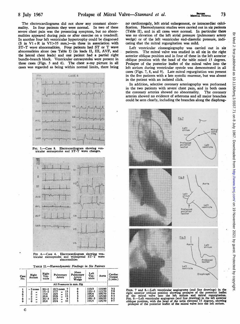
The electrocardiograms did not show any constant abnor-mality. In four patients they were normal. In two of thesesevere chest pain was the presenting symptom, but no abnor-malities appeared during pain or after exercise on a treadmill.In another four left ventricular hypertrophy could be diagnosed(S in VI +R in V5>35 mm.)-in three in association withST-T wave abnormalities. Four patients had ST or T waveabnormalities alone (see Table I) (in leads II, III, AVF, andthe lateral chest leads) and one patient had a partial rightbundle-branch block. Ventricular extrasystoles were present inthree cases (Figs. 5 and 6). The chest x-ray picture in allcases was regarded as being within normal limits, there being
FIG. 5.-Case 8. Electrocardiogram showing ven-tricular extrasystoles and ST-T wave changes.
BRmsUMEDICAL JOURNAL 73
nc cardiomegaly, left atrial enlargement, or intracardiac calci-fication. Haemodynamic studies were carried out in six patients(Table II), and in all cases were normal. In particular therwas no elevation of the left atrial pressure (pulmonary arterywedge) or of the left ventricular end-diastolic pressure, indi-cating that the mitral regurgitation was mild.
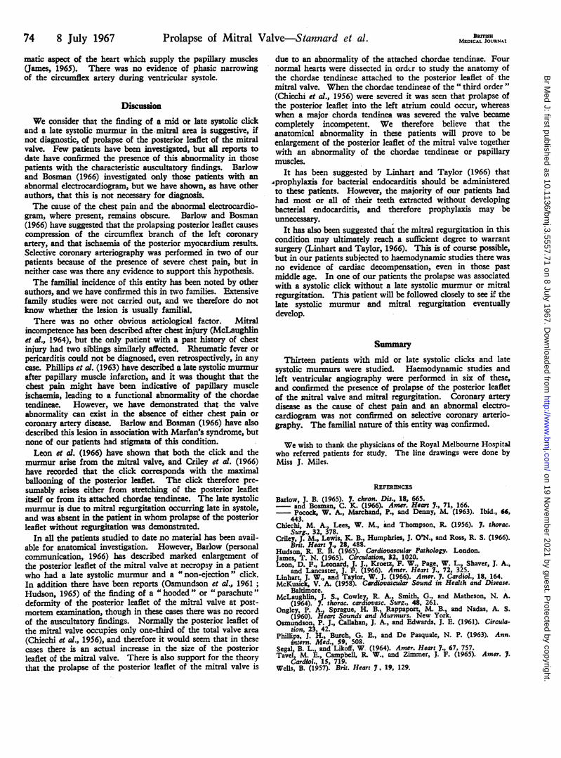
Left ventricular cineangiography was carried out in sixpatients. The mitral valve was studied in all six in the rightanterior oblique position and in four of these in the left anterioroblique position with the head of the table raised 15 degrees.Prolapse of the posterior leaflet of the mitral valve into theleft atrium during ventricular systole was demonstrated in allcases (Figs. 7, 8, and 9). Late mitral regurgitation was presentin the five patients with a late systolic murmur, but was absentin the patient with an isolated click.
In addition, selective coronary arteriography was performedin the two patients with severe chest pain, and in both casesthe coronary arteries showed no abnormality. The coronaryarteries showed no evidence of atheroma and all major branchescould be seen clearly, including the branches along the diaphrag-
atrium Mta
-'~ rin~gposterior ~~Left
le*flet ventricle
Fig.8
FIQ.9
Leftatrium
FIG 6.-Case 4. Electrocardiogram showing ven-tricular extrasystoles and widespread ST-T wave
abnormalities
TABLE II.-Haemodynamic Findings in Six Patients
Right PlMean LeftCase Right Ven- Pulmonary Pulmonary Ven- Aorta CardiacNo. Atrium tricle Artery Artery tricle IOutput~~~~~~~Wedge (1./min.)
All Pressures in mm. Hg-3 mean 20/-2 18/3 mean 7 0 |110/3 110/60 3-2
4 2 IN 17/-2 17/7 13 6 125/-1 115/70 5-86 0 " 17/0 18/5 " 11 5 105/4 105/70 5-67 2 ,, 25/0 25/8 ", 13 3 125/0 125/60 5-38 -2 20/-3 20/3 " 8 5 100/-3 100/55 6-2
9 0 , 25/0 23/4 ,, 10 3 155/5 125/70 5-7
C
FIGS. 7 and 8.-Left ventricular angiograms (and line drawings) in theright anterior oblique position showing prolapse of the posterior leafletof the mitral valve into the left atrium and mitral regurgitation.FIG. 9.-Left ventricular angiogram (and line drawing) in the left anterioroblique position, with the head of the table elevated 15 degrees, showingprolapse of the posterior leaflet of the mitral valve into the left atrium.
8 July 1967 Prolapse of Mitral Valve-Stannard et al.
Lef tven tricle
Diaphragm
_~1~-I1~
on 19 Novem
ber 2021 by guest. Protected by copyright.
http://ww
w.bm
j.com/
Br M
ed J: first published as 10.1136/bmj.3.5557.71 on 8 July 1967. D
ownloaded from
74 8 July 1967 Prolapse of Mitral Valve-Stannard et al. MEDICALJOURNAJ
matic aspect of the heart which supply the papillary muscles(James, 1965). There was no evidence of phasic narrowingof the circumflex artery during ventricular systole.
DiscussionWe consider that the finding of a mid or late systolic click
and a late systolic murmur in the mitral area is suggestive, ifnot diagnostic, of prolapse of the posterior leaflet of the mitralvalve. Few patients have been investigated, but all reports todate have confirmed the presence of this abnormality in thosepatients with the characteristic auscultatory findings. Barlowand Bosman (1966) investigated only those patients with anabnormal electrocardiogram, but we have shown, as have otherauthors, that this is not necessary for diagnosis.The cause of the chest pain and the abnormal electrocardio-
gram, where present, remains obscure. Barlow and Bosman(1966) have suggested that the prolapsing posterior leaflet causescompression of the circumflex branch of the left coronaryartery, and that ischaemia of the posterior myocardium results.Selective coronary arteriography was performed in two of ourpatients because of the presence of severe chest pain, but inneither case was there any evidence to support this hypothesis.The familial incidence of this entity has been noted by other
authors, and we have confirmed this in two families. Extensivefamily studies were not carried out, and we therefore do notknow whether the lesion is usually familial.
There was no other obvious aetiological factor. Mitralincompetence has been described after chest injury (McLaughlinet al., 1964), but the only patient with a past history of chestinjury had two siblings similarly affected. Rheumatic fever orpericarditis could not be diagnosed, even retrospectively, in anycase. Phillips et al. (1963) have described a late systolic murmurafter papillary muscle infarction, and it was thought that thechest pain might have been indicative of papillary muscleischaemia, leading to a functional abnormality of the chordaetendineae. However, we have demonstrated that the valveabnormality can exist in the absence of either chest pain orcoronary artery disease. Barlow and Bosman (1966) have alsodescribed this lesion in association with Marfan's syndrome, butnone of our patients had stigmata of this condition.Leon et al. (1966) have shown that both the click and the
murmur arise from the mitral valve, and Criley et al. (1966)have recorded that the click corresponds with the maximalballooning of the posterior leaflet. The click therefore pre-sumably arises either from stretching of the posterior leafletitself or from its attached chordae tendineae. The late systolicmurmur is due to mitral regurgitation occurring late in systole,and was absent in the patient in whom prolapse of the posteriorleaflet without regurgitation was demonstrated.
In all the patients studied to date no material has been avail-able for anatomical investigation. However, Barlow (personalcommunication, 1966) has described marked enlargement ofthe posterior leaflet of the mitral valve at necropsy in a patientwho had a late systolic murmur and a "non-ejection" click.In addition there have been reports (Osmundson et al., 1961;Hudson, 1965) of the finding of a " hooded " or " parachute"deformity of the posterior leaflet of the mitral valve at post-mortem examination, though in these cases there was no recordof the auscultatory findings. Normally the posterior leaflet ofthe mitral valve occupies only one-third of the total valve area(Chiechi et al., 1956), and therefore it would seem that in thesecases there is an actual increase in the size of the posteriorleaflet of the mitral valve. There is also support for the theorythat the prolapse of the posterior leaflet of the mitral valve is
due to an abnormality of the attached chordae tendinae. Fournormal hearts were dissected in order to study the anatomy ofthe chordae tendineae attached to the posterior leaflet of themitral valve. When the chordae tendineae of the " third order "(Chiechi et al., 1956) were severed it was seen that prolapse ofthe posterior leaflet into the left atrium could occur, whereaswhen a major chorda tendinea was severed the valve becamecompletely incompetent. We therefore believe that theanatomical abnormality in these patients will prove to beenlargement of the posterior leaflet of the mitral valve togetherwith an abnormality of the chordae tendineae or papillarymuscles.
It has been suggested by Linhart and Taylor (1966) that,prophylaxis for bacterial endocarditis should be administeredto these patients. However, the majority of our patients hadhad most or all of their teeth extracted without developingbacterial endocarditis, and therefore prophylaxis may beunnecessary.
It has also been suggested that the mitral regurgitation in thiscondition may ultimately reach a sufficient degree to warrantsurgery (Linhart and Taylor, 1966). This is of course possible,but in our patients subjected to haemodynamic studies there wasno evidence of cardiac decompensation, even in those pastmiddle age. In one of our patients the prolapse was associatedwith a systolic click without a late systolic murmur or mitralregurgitation. This patient will be followed closely to see if thelate systolic murmur and mitral regurgitation eventuallydevelop.
SummaryThirteen patients with mid or late systolic clicks and late
systolic murmurs were studied. Haemodynamic studies andleft ventricular angiography were performed in six of these,and confirmed the presence of prolapse of the posterior leafletof the mitral valve and mitral regurgitation. Coronary arterydisease as the cause of chest pain and an abnormal electro-cardiogram was not confirmed on selective coronary arterio-graphy. The familial nature of this entity was confirmed.
We wish to thank the physicians of the Royal Melbourne Hospitalwho referred patients for study. The line drawings were done byMiss J. Miles.
REFERENCES
Barlow, J. B. (1965). 7. chron. Dis., 18, 665.and Bosman, C. K. (1966). Amer. Heart 7., 71, 166.Pocock, W. A., Marchand, P., and Denny, M. (1963). Ibid., 66,
443.Chiechi, M. A., Lees, W. M., and Thompson, R. (1956). 7. thorac.
Surg., 32, 378.Criley, J. M., Lewis, K. B., Humphries, J. O'N., and Ross, R. S. (1966).
Brit. Heart 7., 28, 488.Hudson, R. E. B. (1965). Cardiovascular Pathology. London.James, T. N. (1965). Circulation, 32, 1020.Leon, D. F., Leonard, J. J., Kroetz F W., Page, W. L., Shaver, J. A.,
and Lancaster, J. F. (1966). Amer. Heart 7., 72, 325.Linhart, J. W., and Taylor W. J. (1966). Amer. 7. Cardiol., 18, 164.McKusick, V. A. (1958). Cardiovascular Sound in Health and Disease.
Baltimore.McLaughlin, J. S., Cowley, R. A., Smith, G., and Matheson, N. A.
(1964). 7. thorac. cardiovasc. Surg., 48, 261.Ongley, P. A., Sprague, H. B., Rappaport, M. B., and Nadas, A. S.
(1960). Heart Sounds and Murmurs. New York.Osmundson, P. J., Callahan, J. A., and Edwards, J. E. (1961). Circula-
tion, 23, 42.Phillips, J. H., Burch, G. E., and De Pasquale, N. P. (1963). Ann.
intern. Med., 59, 508.Segal, B. L., and Likoff, W. (1964). Amer. Heart 7., 67, 757.Tavel, M. E., Campbell, R. W., and Zimmer, J. F. (1965). Amer. 7.
Cardiol., 15, 719.Wells, B. (1957). Brit. Heart 7, 19, 129.
on 19 Novem
ber 2021 by guest. Protected by copyright.
http://ww
w.bm
j.com/
Br M
ed J: first published as 10.1136/bmj.3.5557.71 on 8 July 1967. D
ownloaded from