of escherichia coli - journal of bacteriology - american society for
TRANSCRIPT

Vol. 160, No. 1
Kinetic Analysis of the Synthesis and Assembly of Type 1 Fimbriaeof Escherichia coli
DOUGLAS C. DODD1 AND BARRY I. EISENSTEIN12*Departments of Microbiologyl and Medicine,2 The University of Texas Health Science Center at San Antonio, San
Antonio, Texas 78284
Received 11 June 1984/Accepted 17 July 1984
The adhesive organelles (type 1 fimbriae) of K-12 and other isolates of Escherichia coli are composed ofidentical 17,000-dalton subunits. We examined the assembly of these subunits into fimbrial organelles. Aftersynthesis, the nascent subunits were first processed and then assembled into the organelles; the assembly steptook almost 3 min in log-phase cultures at 37°C. Even during blockage of protein synthesis, the free subunitscontinued to assemble until the pool was depleted. This pool was small in comparison with the amount of totalfimbrial protein already assembled into surface organelles and was not sufficient to regenerate new detectableorganelles after the removal of preexistent ones by blending. Assembly appeared to slow when the metabolicrate of the bacterial cells slowed, since subunits took longer to appear in the organelles at lower than optimaltemperatures or as a culture entered the stationary phase. The synthetic rate of subunits slowed sooner thanthat of total cellular proteins as a culture approached the stationary phase and ceased completely as the cultureentered the stationary phase. The amount of fimbrial antigen expressed on the surface of the cells remainedrelatively constant during growth of a culture.
Type 1 fimbriae (or pili) are proteinaceous appendages 7nm in width projecting from the surface of many gram-neg-ative bacteria (4). They consist of aggregates of helicallyarranged identical 17,000-dalton polypeptides with 3.1 sub-units per turn of the helix (1). These structures are highlystable; they are resistant to disaggregation by sodium do-decyl sulfate (SDS) or urea (1, 2, 11, 13) but can be brokeninto individual subunits by saturated guanidine (1, 8) or hotacid (1, 2).
Studies have shown that for many bacterial diseases theadherence of bacteria to the mucosa is a prerequisite forsubsequent infection (for reviews see references 4 and 17).Type 1 fimbriae, which are the most commonly foundadhesin among Escherichia coli isolates (4), are termedmannose sensitive since the adherence mediated by theseorganelles can be inhibited or reversed by the addition ofmannose or closely related compounds (19). These organel-les also mediate pellicle formation (17), which occurs innonshaking rich media when fimbria-bearing cells form adense mat (or pellicle) at the surface of the broth. IndividualE. coli cells undergo phase variation, whereby fimbriaeexpression can oscillate between on-and-off at a rate of ca.l0-, per generation (1, 5). Since pellicle formation allowsfimbriate-phase bacteria to outgrow non-fimbriate-phase bac-teria in rich nonaerated broth (7, 17), bacteria in oldercultures are more fimbriate than those in younger cultures. Itis not known how the level of fimbriation varies in a culturenot undergoing a selection for one phase or the other duringbacterial growth.We have recently shown that fimbrial subunits are initially
synthesized as signal sequence-containing precursors (2a),which are rapidly processed into mature subunits beforeassembly into organelles. It has been suggested that there isa sufficient number of these nonassembled subunits to re-
generate fimbriae after their removal by blending (1). In thiscommunication we describe the kinetics of assembly ofthese mature subunits.
* Corresponding author.
MATERIALS AND METHODS
Strains and culture conditions. Strain CSH50 is a ColdSpring Harbor E. coli K-12-derived strain with the genotypeF- ara A(lac-pro) rpsL thi (14). VL386 is a nonfimbriatestrain derived from CSH50 after insertion of the bacterio-phage Mu d(Apr lac) into a gene necessary for the productionof fimbriae and subsequent replacement of Mu d by lambdap209 (9). For all experiments, cells grown overnight in min-imal medium A (14) supplemented with 0.5% glucose (or0.4% glycerol and 0.4% maltose in experiments in which themaltose-binding protein was immunoprecipitated) were in-oculated in dilutions between 1:150 and 1: 10 into the samebroth and grown aerated at 37°C. The cell number wasdetermined from a standard curve of viable counts versusoptical density at 550 nm measured in a Spectronic 21 spec-trophotometer (Bausch & Lomb, Inc., Rochester, N.Y.)SDS-PAGE. SDS-polyacrylamide gel electrophoresis
(PAGE) was performed in a 1.5-mm slab gel apparatus(Hoefer Scientific, San Francisco, Calif.) by the method ofLaemmli (12) as previously described (2).
Chemicals and buffers. All reagents were as previouslydescribed (2, 6) except that [3H]leucine (55 Ci/mmol) wasfrom Schwarz/Mann (Spring Valley, N.Y.), chloramphenicolwas from Sigma Chemical Co. (St. Louis, Mo.), and glyceroland formaldehyde were from Fisher Scientific (Pittsburgh,Pa.).
Antibodies. For the enzyme-linked immunosorbent assay(ELISA) inhibition assay, we used rabbit serum specific fortype 1 fimbriae as previously described (2). For immunopre-cipitations, we used this serum, a monoclonal antibody(MAb) previously described (6), and monospecific rabbitanti-maltose-binding protein serum, kindly provided by PhilBassford, Jr. (10). Rabbit antimouse serum was kindlyprovided by Judy Teale (6).
RIP. For radioimmunoprecipitation (RIP) we used themethod of Ito et al. (10) as follows. Bacteria in 1 ml ofminimal medium were labeled for 20 to 30 s with 15 RCi of[3H]leucine. A chase was performed by the addition of 200,ug of L-leucine per ml and 50 ,ug of L-isoleucine per ml.
227
JOURNAL OF BACTERIOLOGY, OCt. 1984, p. 227-2320021-9193/84/100227-06$02.00/0Copyright ©) 1984, American Society for Microbiology
Dow
nloa
ded
from
http
s://j
ourn
als.
asm
.org
/jour
nal/j
b on
25
Nov
embe
r 20
21 b
y 10
9.19
1.39
.89.
228 DODD AND EISENSTEIN
Reactions were stopped by the addition of an equal volumeof ice-cold 10% trichloracetic acid (TCA). We recovered theprecipitate by centrifuging the solution for 3 min at 4°C in aBeckman Microfuge model B (Fullerton, Calif.). After wash-ing the precipitate twice with ice-cold acetone, we solubil-ized it in 0.1 ml of a 1% SDS-50 mM Tris-hydrochloride (pH8)-i mM EDTA solution and performed the immunoprecipi-tation as previously described (2, 6). The TCA-acetonetreatment did not affect the efficiency of immunoprecipita-tion or cause any detectable disaggregation of organelles intosubunits.ELISA inhibition assay. We performed the ELISA for
quantitative determination of fimbrial antigen on the surfaceof E. coli as previously described (2, 6). To prevent anychange in the amount of fimbriae present in a culture untilcompletion of the assay, we fixed samples in 1% formalde-hyde at the time of removal from a culture. The formalde-hyde fixation did not affect the assay.Temperature studies. To determine the effect of various
temperatures on assembly, we placed samples of a culturegrowing at 37°C in shaking water baths at the appropriatetemperature 30 min before labeling. We checked the temper-ature of the samples by immersing a thermometer (Fisher)directly into the culture tube.
RESULTS
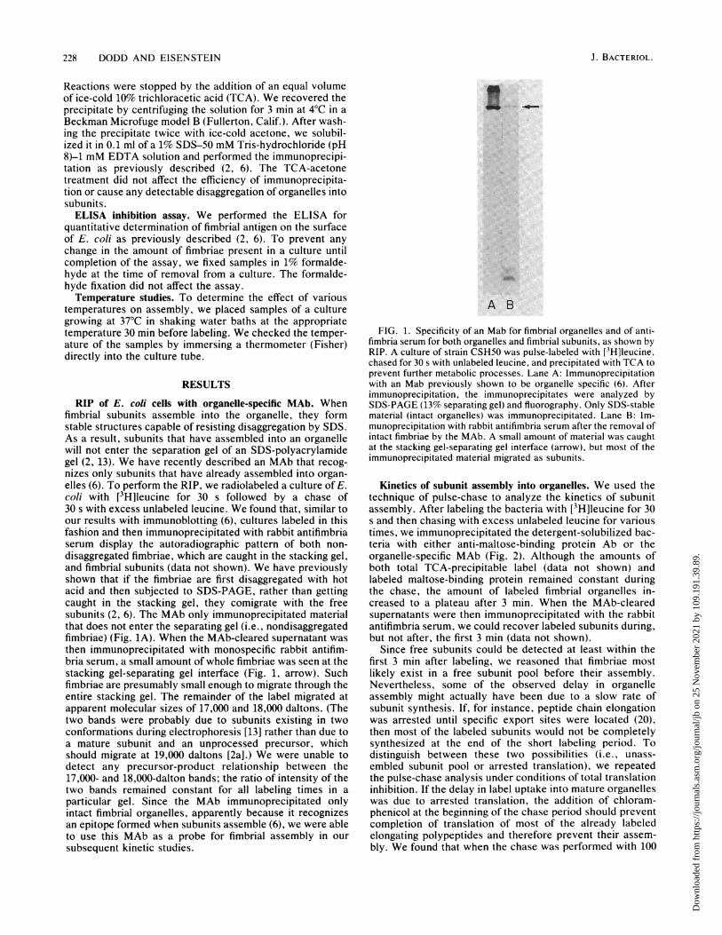
RIP of E. coli cells with organelle-specific MAb. Whenfimbrial subunits assemble into the organelle, they formstable structures capable of resisting disaggregation by SDS.As a result, subunits that have assembled into an organellewill not enter the separation gel of an SDS-polyacrylamidegel (2, 13). We have recently described an MAb that recog-nizes only subunits that have already assembled into organ-elles (6). To perform the RIP, we radiolabeled a culture of E.coli with [3H]leucine for 30 s followed by a chase of30 s with excess unlabeled leucine. We found that, similar toour results with immunoblotting (6), cultures labeled in thisfashion and then immunoprecipitated with rabbit antifimbriaserum display the autoradiographic pattern of both non-disaggregated fimbriae, which are caught in the stacking gel,and fimbrial subunits (data not shown). We have previouslyshown that if the fimbriae are first disaggregated with hotacid and then subjected to SDS-PAGE, rather than gettingcaught in the stacking gel, they comigrate with the freesubunits (2, 6). The MAb only immunoprecipitated materialthat does not enter the separating gel (i.e., nondisaggregatedfimbriae) (Fig. 1A). When the MAb-cleared supernatant wasthen immunoprecipitated with monospecific rabbit antifim-bria serum, a small amount of whole fimbriae was seen at thestacking gel-separating gel interface (Fig. 1, arrow). Suchfimbriae are presumably small enough to migrate through theentire stacking gel. The remainder of the label migrated atapparent molecular sizes of 17,000 and 18,000 daltons. (Thetwo bands were probably due to subunits existing in twoconformations during electrophoresis [13] rather than due toa mature subunit and an unprocessed precursor, whichshould migrate at 19,000 daltons [2a].) We were unable todetect any precursor-product relationship between the17,000- and 18,000-dalton bands; the ratio of intensity of thetwo bands remained constant for all labeling times in aparticular gel. Since the MAb immunoprecipitated onlyintact fimbrial organelles, apparently because it recognizesan epitope formed when subunits assemble (6), we were ableto use this MAb as a probe for fimbrial assembly in oursubsequent kinetic studies.
A B
FIG. 1. Specificity of an Mab for fimbrial organelles and of anti-fimbria serum for both organelles and fimbrial subunits, as shown byRIP. A culture of strain CSH50 was pulse-labeled with [VH]leucine,chased for 30 s with unlabeled leucine, and precipitated with TCA toprevent further metabolic processes. Lane A: Immunoprecipitationwith an Mab previously shown to be organelle specific (6). Afterimmunoprecipitation, the immunoprecipitates were analyzed bySDS-PAGE (13% separating gel) and fluorography. Only SDS-stablematerial (intact organelles) was immunoprecipitated. Lane B: Im-munoprecipitation with rabbit antifimbria serum after the removal ofintact fimbriae by the MAb. A small amount of material was caughtat the stacking gel-separating gel interface (arrow), but most of theimmunoprecipitated material migrated as subunits.
Kinetics of subunit assembly into organelles. We used thetechnique of pulse-chase to analyze the kinetics of subunitassembly. After labeling the bacteria with [3H]leucine for 30s and then chasing with excess unlabeled leucine for varioustimes, we immunoprecipitated the detergent-solubilized bac-teria with either anti-maltose-binding protein Ab or theorganelle-specific MAb (Fig. 2). Although the amounts ofboth total TCA-precipitable label (data not shown) andlabeled maltose-binding protein remained constant duringthe chase, the amount of labeled fimbrial organelles in-creased to a plateau after 3 min. When the MAb-clearedsupernatants were then immunoprecipitated with the rabbitantifimbria serum, we could recover labeled subunits during,but not after, the first 3 min (data not shown).
Since free subunits could be detected at least within thefirst 3 min after labeling, we reasoned that fimbriae mostlikely exist in a free subunit pool before their assembly.Nevertheless, some of the observed delay in organelleassembly might actually have been due to a slow rate ofsubunit synthesis. If, for instance, peptide chain elongationwas arrested until specific export sites were located (20),then most of the labeled subunits would not be completelysynthesized at the end of the short labeling period. Todistinguish between these two possibilities (i.e., unass-embled subunit pool or arrested translation), we repeatedthe pulse-chase analysis under conditions of total translationinhibition. If the delay in label uptake into mature organelleswas due to arrested translation, the addition of chloram-phenicol at the beginning of the chase period should preventcompletion of translation of most of the already labeledelongating polypeptides and therefore prevent their assem-bly. We found that when the chase was performed with 100
J. BACTERIOL.
Dow
nloa
ded
from
http
s://j
ourn
als.
asm
.org
/jour
nal/j
b on
25
Nov
embe
r 20
21 b
y 10
9.19
1.39
.89.

KINETICS OF E. COLI FIMBRIAL EXPRESSION 229
to 4-0x
E 3-C.)u° 2-.)_
I 0lr O
-0
P --0--------- o
v -- - _--
II I I1
0 2 4 6 8
Chase Period (minutes)
5 10 10 1 2sec minutes
3 5 10
NMaltose Binding Protein Fimbrial Organelles
FIG. 2. SDS-PAGE analysis of the time course of assembly ofsubunits into a form recognizable by the organelle-specific MAb. Aculture of strain CSH50 was pulse-labeled with [3H]leucine for 30 sfollowed by a chase with unlabeled leucine for the indicated times.The first six lanes result from immunoprecipitations of the maltose-binding protein done at the designated time points. Note that theintensity of the band remains constant over the chase period. Thelast six lanes result from immunoprecipitations done with theorganelle-specific MAb at the designated time points. Note that thetotal amount of immunoprecipitable material (fimbrial organelles)increases for the first few time points and that the amount of smallerfimbriae (those that migrate to the separating gel [arrow]) decreasesat the later times relative to the larger fimbriae (those that do notmigrate through the stacking gel).
,ug of chloramphenicol per ml (a concentration that inhibitsboth new TCA-precipitable label and new fimbrial subunitsby at least 99% within 10 s), the kinetics of fimbrial assemblywas virtually unaffected (Fig. 3). Moreover, the inhibition ofsubunit incorporation into organelles (39%, relative to thesample without chloramphenicol) was not much differentfrom that of the rapidly synthesized maltose-binding protein(40%) or from that of total TCA-precipitable material (31%).Taken together, these results suggest that the delay inlabeled subunits appearing in the organelles is almost en-tirely due to the time required for assembly and that assem-bly can occur in the absence of protein synthesis.
Effect of temperature on assembly. In contrast to manymannose-resistant fimbriae (3) and F-pili (16), type 1 fimbriaecan be expressed at low temperatures such as 18°C (3, 4). Itis not known, though, what the effect of temperature is onthe rates of synthesis and of assembly of type 1 fimbriae.Therefore, we next examined the kinetics of fimbrial expres-
sion of cultures grown at 18, 24, 30, and 37°C. The propor-tion of TCA-precipitable label that was finally incorporatedinto the organelles was not affected significantly by temper-ature (Fig. 4), although the rate at which the subunits were
incorporated into the organelles slowed somewhat withlower temperatures. Since the amount of TCA-precipitablelabel remained constant during the chase period for eachtemperature, it seems likely that this slowing of incorpora-tion of labeled subunits into fimbriae is due to a decrease inassembly rate. The culture growing at 18°C is growing muchslower than the 37°C culture, taking up less than 10% of the[3H]leucine into TCA-precipitable material compared withthe 370 culture during the 30-s labeling time.
FIG. 3. Effect of chloramphenicol on the synthesis and assemblyof fimbriae and on the synthesis of the maltose-binding protein.Immunoprecipitations were performed after different chase periodswith either anti-maltose-binding protein serum or antifimbriae MAb,described in the legend to Fig. 2, except that, in addition, the chasewas performed either in the presence or in the absence of 100 pug ofchloramphenicol per ml. Shown are total counts immunoprecipi-tated by the MAb in the absence (O) or presence (0) of chloram-phenicol or by anti-maltose-binding protein in the absence (U) or
presence (0) of chloramphenicol.
Production of fimbriae during growth. To determine theeffect of growth phase on fimbrial assembly, we examinedincorporation of subunits into organelles at various timesduring the growth of the culture. Figure 5 shows the points(A) at which samples were taken and examined for subunitassembly and fimbrial expression. Cultures were pulsed for30 s and then chased for either 90 s (early assembly phase) or20 min (total assembly). After the chase periods, the sampleswere analyzed to determine the fraction of the label that hadbeen incorporated into fimbrial organelles. Figure 6 shows a
fluorograph of the stacking gel of an SDS-polyacrylamide gelafter immunoprecipitation with the organelle-specific MAb.At all time points, free subunits could no longer be detectedat 20 min, and further chasing resulted in no additionalsubunit-to-organelle incorporation. During the log phase ofgrowth, the amount of subunits incorporated at 90 s was
2.0-
1.5
1205
0 2 4 6 8 lo0Chase Period (minutes)
FIG. 4. Kinetics of fimbrial assembly as a function of tempera-ture, as determined by immunoprecipitation with the organelle-specific MAb. Shown are the relative amounts of total TCA-precipitable counts that were specifically immunoprecipitated (inpercentages) after various chase periods. The temperatures exam-ined were 37°C (A), 300C (0), 220C (A), and 180C (0).
Length of 10 1 2 3chase sec minutes
10
VOL. 160, 1984
Dow
nloa
ded
from
http
s://j
ourn
als.
asm
.org
/jour
nal/j
b on
25
Nov
embe
r 20
21 b
y 10
9.19
1.39
.89.

230 DODD AND EISENSTEIN
-
I
5
0 1 2 3 4 5 6 7 8 9
Hours of Culture
FIG. 5. Growth curve of strain CSH50 in glucose-supplementedminimal medium at 37°C. Optical density readings (at 550 nm)(OD550) were taken at each point indicated (0). Samples were takenat points marked (A) for examination of fimbrial synthesis, assem-
bly, and expression.
roughly equal to that incorporated at 20 min, indicating that,during the log phase, most of the subunits assembled within90 s. In contrast, as the culture approached the stationaryphase of growth, subunit assembly slowed appreciably, as
shown by the fact that a lower percentage of incorporated[3H]leucine was incorporated into fimbriae relative to totalcellular synthesis (Table 1). (We used the ratio of countsincorporated after 90 s of chase to counts incorporated after20 min of chase as a measure of rapidly assembling sub-units.) We found that the percentage of rapidly assemblingsubunits decreased dramatically as the culture entered thestationary phase (Table 1), which could be due to slowing ofpolypeptide chain elongation, assembly, or both. In addi-tion, a sample of the culture at each time point was assayedfor total amount of fimbriae present on the surface of thebacteria by the ELISA inhibition assay. The amount offimbriae measured from the time of passage to 24 h of culturevaried between 4 and 5 ng/106 cells. After assaying samplesfrom several growth curves, we found that this small varia-tion is unrelated to the period of growth. Thus, in theabsence of selection for fimbriation (fimbrate bacteria out-grow nonfimbriate bacteria when grown without aeration inrich broth [7]), a culture remains relatively constant in itsdegree of fimbriation during the growth cycle.
Size of the free subunit pool. We have demonstrated thatfimbrial subunits are first synthesized and processed (2a) andthen, within 3 min of synthesis, assembled into quarternary-structured organelles (see above). Since these data indicate
SXE .
TABLE 1. Kinetics of fimbrial expressions during bacterialgrowth in broth
Start of labeling" Immunoprecipitable label infimbriae organelles
Time in % Relative to % of new subunitsgrowth curve OD550 total protein rdly assembled"
(min) synthesis" rapi
95 0.197 0.98 95155 0.415 0.67 120185 0.600 0.37 12240 0.680 0.25 10280 0.690 0.20 7.5
aSee Fig. 5 for growth curve. Samples (2 ml) were removed from culture atthe times indicated and labeled for 30 s. Half of each sample underwent a 90-schase before TCA precipitation; the other half was chased for 20 min. ODss,Optical density at 550 nm.bImmunoprecipitable counts after 20 min of chase/total TCA-precipitable
counts after 20 min of chase x 100.' Immunoprecipitable counts after 90 s of chase/immunoprecipitable counts
after 20 min of chase x 100.
that there is a preassembled pool of mature subunits withinthe cell, we next determined an upper bound of the size ofthis pool. We reasoned that if the pool was large enough,even total interruption of protein synthesis (by chloram-phenicol) should still allow measurable elongation of theorganelles given the observation that assembly is indepen-dent of protein synthesis (see above). In the absence of newsubunit synthesis, the degree of elongation of fimbriaeshould be proportional to the size of the preexistent subunitpool.We measured organelle elongation by two independent
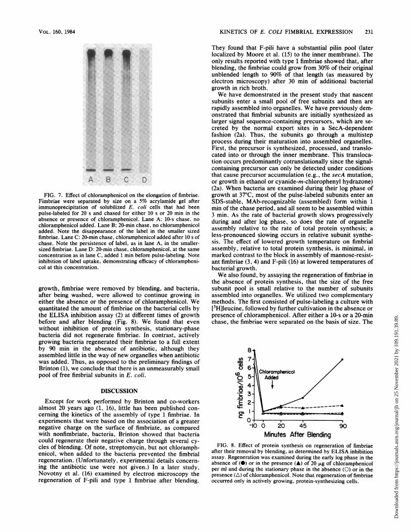
techniques. In the first, we performed a standard pulse-chase of growing bacteria and then determined the length ofthe labeled (new) organelles by RIP, SDS-PAGE in a longstacking gel, and fluorography (Fig. 7). When bacteria wereharvested after a very short chase (10 s), we found that thelabeled fimbriae were distributed in a broad size range (Fig.7A). In contrast, a long chase (20 min) allowed all of theshort, labeled fimbriae enough time to grow to large size(Fig. 7B). The addition of chloramphenicol at the beginningof the chase period had a pronounced effect on elongation;despite a chase of 20 min, there was no appreciable growthof labeled fimbriae (Fig. 7C). Thus, the size of the subunitpool must be small enough to prevent detectable furthergrowth of the organelles.To confirm these findings, we measured new organelle
formation after defimbriation of the bacteria. Cultures wereharvested in either the stationary or the mid-log phase of
Chose 90sec 20min 90sec 20min 90sec 20fmin 90sec 20min 90sec 20min
0D550 0.197 0.415 0.600 0.680 0.690
FIG. 6. Kinetics of new organelle expression at various times in the bacterial growth cycle. Bacteria at times indicated in Fig. 5 were
pulse-labeled for 30 s with [3H]leucine and chased for either 90 s or 20 min with unlabeled leucine before immunoprecipitation with the
organelle-specific MAb. The optical density at 550 nm (OD550) of the culture at the time of initial harvesting is indicated below each pair oflanes, which were prepared as described in the legend to Fig. 1. The arrow indicates the stacking gel-separating gel interface.
0Uc)I')0
0.70.E0.5OA0.30.20.1C
J. BACTERIOL.
Dow
nloa
ded
from
http
s://j
ourn
als.
asm
.org
/jour
nal/j
b on
25
Nov
embe
r 20
21 b
y 10
9.19
1.39
.89.
KINETICS OF E. COLI FIMBRIAL EXPRESSION 231
A B C D
FIG. 7. Effect of chloramphenicol on the elongation of fimbriae.Fimbriae were separated by size on a 5% acrylamide gel afterimmunoprecipitation of solubilized E. coli cells that had beenpulse-labeled for 20 s and chased for either 10 s or 20 min in theabsence or presence of chloramphenicol. Lane A: 10-s chase, no
chloramphenicol added. Lane B; 20-min chase, no chloramphenicoladded. Note the disappearance of the label in the smaller sizedfimbriae. Lane C: 20-min chase, chloramphenicol added after 10 s ofchase. Note the persistence of label, as in lane A, in the smaller-sized fimbriae. Lane D: 20-min chase, chloramphenicol, at the same
concentration as in lane C, added 1 min before pulse-labeling. Noteinhibition of label uptake, demonstrating efficacy of chlorampheni-col at this concentration.
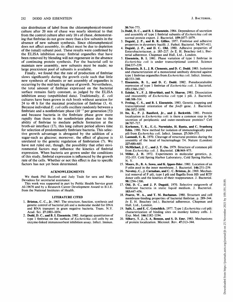
growth, fimbriae were removed by blending, and bacteria,after being washed, were allowed to continue growing ineither the absence or the presence of chloramphenicol. Wequantitated the amount of fimbriae on the bacterial cells bythe ELISA inhibition assay (2) at different times of growthbefore and after blending (Fig. 8). We found that evenwithout inhibition of protein synthesis, stationary-phasebacteria did not regenerate fimbriae. In contrast, activelygrowing bacteria regenerated their fimbriae to a full extentby 90 min in the absence of antibiotic, although theyassembled little in the way of new organelles when antibioticwas added. Thus, as opposed to the preliminary findings ofBrinton (1), we conclude that there is an unmeasurably smallpool of free fimbrial subunits in E. coli.
DISCUSSION
Except for work performed by Brinton and co-workersalmost 20 years ago (1, 16), little has been published con-cerning the kinetics of the assembly of type 1 fimbriae. Inexperiments that were based on the association of a greaternegative charge on the surface of fimbriate, as comparedwith nonfimbriate, bacteria, Brinton showed that bacteriacould regenerate their negative charge through several cy-
cles of blending. Of note, streptomycin, but not chloramph-enicol, when added to the bacteria prevented the fimbrialregeneration. (Unfortunately, experimental details concern-
ing the antibiotic use were not given.) In a later study,Novotny et al. (16) examined by electron microscopy theregeneration of F-pili and type 1 fimbriae after blending.
They found that F-pili have a substantial pilin pool (laterlocalized by Moore et al. (15) to the inner membrane). Theonly results reported with type 1 fimbriae showed that, afterblending, the fimbriae could grow from 30% of their originalunblended length to 90% of that length (as measured byelectron microscopy) after 30 min of additional bacterialgrowth in rich broth.We have demonstrated in the present study that nascent
subunits enter a small pool of free subunits and then arerapidly assembled into organelles. We have previously dem-onstrated that fimbrial subunits are initially synthesized aslarger signal sequence-containing precursors, which are se-creted by the normal export sites in a SecA-dependentfashion (2a). Thus, the subunits go through a multistepprocess during their maturation into assembled organelles.First, the precursor is synthesized, processed, and translo-cated into or through the inner membrane. This transloca-tion occurs predominantly cotranslationally since the signal-containing precursor can only be detected under conditionsthat cause precursor accumulation (e.g., the secA mutation,or growth in ethanol or cyanide-m-chlorophenyl hydrazone)(2a). When bacteria are examined during their log phase ofgrowth at 37°C, most of the pulse-labeled subunits enter anSDS-stable, MAb-recognizable (assembled) form within 1min of the chase period, and all seem to be assembled within3 min. As the rate of bacterial growth slows progressivelyduring and after log phase, so does the rate of organelleassembly relative to the rate of total protein synthesis; aless-pronounced slowing occurs in relative subunit synthe-sis. The effect of lowered growth temperature on fimbrialassembly, relative to total protein synthesis, is minimal, inmarked contrast to the block in assembly of mannose-resist-ant fimbriae (3, 4) and F-pili (16) at lowered temperatures ofbacterial growth.We also found, by assaying the regeneration of fimbriae in
the absence of protein synthesis, that the size of the freesubunit pool is small relative to the number of subunitsassembled into organelles. We utilized two complementarymethods. The first consisted of pulse-labeling a culture with[3H]leucine, followed by further cultivation in the absence orpresence of chloramphenicol. After either a 10-s or a 20-minchase, the fimbriae were separated on the basis of size. The
8
Chioramphenicol
003-
.0
-10 0 20 45 90Minutes After Blending
FIG. 8. Effect of protein synthesis on regeneration of fimbriaeafter their removal by blending, as determined by ELISA inhibitionassay. Regeneration was examined during the early log phase in theabsence of (0) or in the presence (A) of 20 jig of chloramphenicolper ml and during the stationary phase in the absence (0) or in thepresence (A) of chloramphenicol. Note that regeneration of fimbriaeoccurred only in actively growing, protein-synthesizing cells.
VOL. 160, 1984
Dow
nloa
ded
from
http
s://j
ourn
als.
asm
.org
/jour
nal/j
b on
25
Nov
embe
r 20
21 b
y 10
9.19
1.39
.89.
232 DODD AND EISENSTEIN
size distribution of label from the chloramphenicol-treatedculture after 20 min of chase was nearly identical to thatfrom the control culture after only 10 s of chase, demonstrat-ing that fimbriae do not grow more than a few subunits in theabsence of new subunit synthesis. Since chloramphenicoldoes not affect assembly, its affect must be due to depletionof the (small) subunit pool. These results were confirmed bythe ELISA inhibition assay: fimbrial organelles that havebeen removed by blending will not regenerate in the absenceof continuing protein synthesis. For the bacterial cell tomaintain new assembly, new subunits must be made; nolarge preexistent pool of subunits is available.
Finally, we found that the rate of production of fimbriaeslows significantly during the growth cycle such that littlenew synthesis of subunits or net assembly of organelles isoccurring by the mid-late log phase of growth. Nevertheless,the total amount of fimbriae expressed on the bacterialsurface remains fairly constant, as judged by the ELISAinhibition assay (unpublished data). Traditionally, E. colicultures have been grown in rich broth without aeration for24 to 48 h for the maximal production of fimbriae (3, 4).Because individual E. coli cells oscillate randomly between afimbriate and a nonfimbriate phase (10-3 per generation [5])and because bacteria in the fimbriate phase grow morerapidly than those in the nonfimbriate phase due to theability of fimbriae to mediate pellicle formation at thebroth-air interface (17), this long culture period allows timefor selection of predominantly fimbriate bacteria. This selec-tive growth advantage is abrogated by the addition of asugar-such as glucose; moreover, the effect of glucose isunrelated to the genetic regulation of fimbriation (7). Wehave not ruled out, though, the possibility that other envi-ronmental factors may influence the kinetics of fimbrialexpression. When bacteria are grown under the conditionsof this study, fimbrial expression is influenced by the growthrate of the cells. Whether or not this effect is due to specificfactors has not yet been determined.
ACKNOWLEDGMENTSWe thank Phil Bassford and Judy Teale for sera and Mary
Devadoss for secretarial assistance.This work was supported in part by Public Health Service grant
AI-19670 and by a Research Career Development Award to B.I.E.from the National Institutes of Health.
LITERATURE CITED1. Brinton, C. C., Jr. 1965. The structure, function, synthesis and
genetic control of bacterial pili and a molecular model for DNAand RNA transport in gram negative bacteria. Trans. N.Y.Acad. Sci. 27:1003-1054.
2. Dodd, D. C., and B. I. Eisenstein. 1982. Antigenic quantitation oftype 1 fimbriae on the surface of Escherichia coli cells by anenzyme-linked immunosorbent inhibition assay. Infect. Immun.
38:764-773.2a.Dodd, D. C., and B. I. Eisenstein. 1984. Dependence of secretion
and assembly of type 1 fimbrial subunits of Escherichia coli onnormal protein export. J. Bacteriol. 159:1077-1079.
3. Duguid, J. P., and R. R. Gillies. 1957. Fimbriae and adhesiveproperties in dysentery bacilli. J. Pathol. Bacteriol. 74:397-411.
4. Duguid, J. P., and D. C. Old. 1980. Adhesive properties ofEnterobacteriaceae, p. 185-217. In E. H. Beachey (ed.), Bac-terial adherence. Chapman and Hall, Ltd., London.
5. Eisenstein, B. I. 1981. Phase variation of type 1 fimbriae inEscherichia coli is under transcriptional control. Science214:337-339.
6. Eisenstein, B. I., J. R. Clements, and D. C. Dodd. 1983. Isolationand characterization of a monoclornal antibody directed againsttype 1 fimbriae organelles from Escherichia coli. Infect. Immun.42:333-340.
7. Eisenstein, B. I., and D. C. Dodd. 1982. Pseudocataboliterepression of type 1 fimbriae of Escherichia coli. J. Bacteriol.151:1560-1567.
8. Eshdat, Y., F. J. Silverblatt, and N. Sharon. 1981. Dissociationand reassembly of Escherichia coli type 1 pili. J. Bacteriol.148:308-314.
9. Freitag, C. S., and B. I. Eisenstein. 1983. Genetic mapping andtranscriptional orientation of the fimD gene. J. Bacteriol.156:1052-1058.
10. Ito, K., P. J. Bassford, Jr., and J. Beckwith. 1981. Proteinlocalization in Escherichia coli: is there a common step in thesecretion of periplasmic and outer-membrane proteins? Cell24:707-717.
11. Korhonen, T. K., E.-L. Nurmiaho, H. Ranta, and C. SvanborgEden. 1980. New method for isolation of immunologically purepili from Escherichia coli. Infect. Immun. 27:569-575.
12. Laemmll, U. K. 1970. Cleavage of structural proteins during theassembly of the head of bacteriophage T4. Nature (London)227:680-685.
13. McMichael, J. C., and J. T. Ou. 1979. Structure of common pilifrom Escherichia coli. J. Bacteriol. 138:969-975.
14. Miller, J. H. 1972. Experiments in molecular genetics, p.352-355. Cold Spring Harbor Laboratory, Cold Spring Harbor,N. Y.
15. Moore, D., B. A. Sowa, and K. Ippen-Ihler. 1981. Location of anF-pilin pool in the inner membrane. J. Bacteriol. 146:251-259.
16. Novotny, C., J. Carnahan, and C. C. Brinton, Jr. 1969. Mechan-ical removal of F pili, type I pili and flagella from Hfr and RTFdonor cells and the kinetics of their reappearance. J. Bacteriol.98:1294-1306.
17. Old, D. C., and J. P. Duguid. 1970. Selective outgrowth offimbriate bacteria in static liquid medium. J. Bacteriol.103:447-456.
18. Pearce, W. A., and T. M. Buchanan. 1980. Structure and cellmembrane-binding properties of bacterial fimbriae, p. 289-344.In E. H. Beachey (ed.), Bacterial adherence. Chapman andHall, Ltd., London.
19. Salit, I., and E. C. Gotschlich. 1977. Type 1 Escherichia coli pili:characterization of binding sites on monkey kidney cells. J.Exp. Med. 146:1182-1194.
20. Silhavy, T. J., S. A. Benson, and S. D. Emr. 1983. Mechanismsof protein localization. Microiol. Rev. 47:313-344.
J. BACTERIOL.
Dow
nloa
ded
from
http
s://j
ourn
als.
asm
.org
/jour
nal/j
b on
25
Nov
embe
r 20
21 b
y 10
9.19
1.39
.89.