docserv.uni-duesseldorf.dedocserv.uni-duesseldorf.de/servlets/derivateservlet/...table of contents 1...
TRANSCRIPT
Mimicry of a constitutively active pre-B cell
receptor in BCR-ABL1-transformed pre-B
leukemia cells
Inaugural-Dissertation
zur
Erlangung des Doktorgrades der
Mathematisch-Naturwissenschaftlichen Fakultät
der Heinrich-Heine-Universität Düsseldorf
vorgelegt von
Niklas Feldhahn
aus Bad Oldesloe
November 2005
Aus dem Labor für molekulare Stammzellbiologie der Heinrich-Heine-Universität Düsseldorf
Gedruckt mit der Genehmigung der
Mathematisch-Naturwissenschaftlichen Fakultät der
Heinrich-Heine-Universität Düsseldorf
Referent: Prof. Müschen
Koreferent: Prof. Riesner
Externer Referent: Prof. Reth (Freiburg)
Tag der mündlichen Prüfung: 02.05.2006

Table of contents
1 Introduction 1-28
1.1 Commitment to the B cell lineage 1
1.2 V(D)J recombination 3
1.3 Selection for the expression of a pre-B cell receptor 5
1.4 Pre-B cell receptor signaling 6
1.5 Bruton’s tyrosine kinase 10
1.6 X-linked agammaglobulinemia 13
1.7 Chromosomal translocations as a cause of B cell precursor leukemia 14
1.8 Signaling activity of the oncogenic BCR-ABL1 kinase 16
1.9 Aims of the thesis 17
2 Results and discussion 29-42 2.1 The BCR-ABL1 kinase bypasses the selection for the expression of 29
a pre-B cell receptor in transformed pre-B cells
2.2. BCR-ABL1 interferes with differentiation by the induction of a 31
dominant-negative isoform of the transcription factor IKAROS
2.3 Inhibition of BCR-ABL1 kinase activity relieves the differentiation 32
block of the transformed pre-B cells and indicates that the Igκ and
Igλ light chain loci rearrange sequentially during early B cell
development
2.4 Mimicry of a constitutively active pre-B cell receptor: Aberrant 35
splicing links Bruton’s tyrosine kinase to BCR-ABL1 in pre-B
lymphoblastic leukemia cells
2.5 Activation-induced cytidine deaminase acts as a mutator in pre-B 38
cell leukemia cells carrying a BCR-ABL1 gene rearrangement
2.6 Deficiency of Bruton’s tyrosine kinase in B cell precursor leukemia 39
cells without a BCR-ABL1 rearrangement

3 Concluding remarks 43-49
3.1 Splicing for survival−selection for aberrant splice variants 43
in BCR-ABL1-transformed pre-B cell leukemia cells
3.2 The pre-B cell receptor pathway is a target for malignant 44
transformation of pre-B cells by BCR-ABL1
3.3 Tumor suppressor or oncogenic function of the pre-B cell 46
receptor pathway
4 Summary 50-51
4.1 Summary 50
4.2 Zusammenfassung 51
5 Published Results 52-192
5.1 The BCR-ABL1 kinase bypasses selection for the expression 52
of a pre-B cell receptor in pre-B acute lymphoblastic leukemia
cells
5.2 BCR-ABL1 induces aberrant splicing of IKAROS and 80
lineage infidelity in pre-B lymphoblastic leukemia cells
5.3 Tracing the pre-B to immature B cell transition in human 92
leukemia cells reveals a coordinated sequence of primary and
secondary IGK gene rearrangement, IGK deletion and IGL
gene rearrangement
5.4 Mimicry of a constitutively active pre-B cell receptor: Aberrant 113
splicing links Bruton’s tyrosine kinase to BCR-ABL1 in
pre-B lymphoblastic leukemia
5.5 Activation-induced cytidine deaminase acts as a mutator in 148
BCR-ABL1-induced pre-B lymphoblastic leukemia and
lymphoid blast crisis of chronic myeloid leukemia

5.6 Deficiency of Bruton’s tyrosine kinase in B cell precursor 172
leukemia cells
6 Appendix 193-204
6.1 Supplementary information 193
6.2 Abbreviations 195
6.3 Curriculum vitae 197
6.4 List of publications 198
6.5 Specific contribution of Niklas Feldhahn to the 201-206
publications shown in chapter 5.1-5.2
6.6 Danksagung 205

Chapter 1
Introduction
Chapter 1 Introduction
1
1.1 Commitment to the B cell lineage
The development of B cells in the human originates from pluripotent hematopoietic stem cells
(HSCs) in the bone marrow or the fetal liver. HSCs exhibit extensive self-renewal capacity
and regenerate all hematopoietic lineages throughout life. Development from the HSC
towards the lymphoid lineages is accompanied with the loss of the self-renewal capacity of
HSCs (Adolfsson et al., 2001) and guided by several transcription factors that initiate or
maintain gene expression specific for lineage and stage of differentiation. Early B cell
development is characterized by the opening and rearrangement of the immunoglobulin heavy
chain (IGH) locus, which is mediated by the recombination activating genes 1 and 2
(RAG1/2). Somatic rearrangement of gene segments within the IGH locus encoding the
variable region of the immunoglobulin heavy chain occurs at the transition to the pro-B cell
and pre-B cell stages of development, respectively (Figure 1). Functional rearrangement of
IGH V (variable) region gene segments is a prerequisite for the expression of the pre-B cell
receptor and further differentiation to mature B cells (Kitamura et al., 1991).
Commitment to the B- or T-lymphoid lineages is dependent on the activity of two key
transcription factors, IKAROS and PU.1, thereby promoting the differentiation of HSCs into
common lymphoid progenitor (CLP) cells: Deficiency of IKAROS in mice prevents the
development of all lymphoid cells (Georgopoulos et al., 1994). Likewise, the level of PU.1
expression controls B versus myeloid differentiation as shown by retroviral reconstitution
experiments with PU.1 deficient progenitor cells (DeKoter and Singh; 2000). Notably,
IKAROS is an unusual transcription factor as it may either repress or activate target genes by
closing or opening their loci through the induction of chromatin remodeling (Kim et al.,
1999).
The differentiation from the CLP to the pro-B cell stage, which represents the
commitment to the B cell lineage, critically depends on the three transcription factors E2A,
EBF and PAX5. Deficiency of any of these transcription factors results in a B cell
differentiation arrest at earliest stages and in the absence of IGH V region gene
rearrangements (Bain et al., 1994; Lin and Grosschedl, 1995; Urbanek et al., 1994). In
agreement with the phenotype of E2A- and EBF-deficient mice, it has been shown that E2A
and EBF act in concert to regulate the expression of RAG1 and RAG2 and germline
transcription of the IGH locus (Romanow et al., 2000), which are both essential for
recombination processes within the IGH locus. E2A and EBF also promote the transition from
the pro-B cell to the pre-B cell stage as they induce the transcription of several crucial
components of the pre-B cell receptor complex, like the elements of the surrogate light chain

Chapter 1 Introduction
2
VpreB and λ5 (Sigvardsson et al., 1997; Kee and Murre, 1998). Deficiency of these
components leads to a developmental arrest at the pro-B cell stage (Papavasiliou et al., 1996).
Despite already advanced differentiation, B cell lineage commitment is still reversible at this
stage of development and critically depends on the sustained expression of PAX5.
Conditional deletion of PAX5 in mice is sufficient to reactivate in early B cells a broad
developmental potential (Mikkola et al., 2002). Even in mature B cells, the conditional
inactivation of PAX5 leads to the loss of B cell identity (Horcher et al., 2001), demonstrating
the crucial role of continuous PAX5 expression for the maintenance of the B cell identity.
Figure 1: Early B cell development is guided by distinct transcription factors
Schematic diagram of early B cell development. Differentiation of a hematopoietic stem cell (HSC) into the lymphoid lineage (CLP, common lymphoid progenitor) is guided by the transcription factors IKAROS and PU.1. Early B cell development is defined by a sequence of somatic rearrangements within the immunoglobulin heavy chain locus (IGH): The rearrangement of a DH to JH gene segment occurs during the transition from the CLP to the pro-B cell stage followed by a rearrangement of a VH gene segment to the DJH joint during the transition from the pro-B to the pre-B cell stage. Early B cell development is guided by the transcription factors E2A, EBF and PAX5. Pre-B cells express a the µ-heavy chain (black) in combination with the surrogate light chain (red) on the surface. Selected pre-B cells downregulate their pre-B cell receptor, initiate the rearrangement of the immunoglobulin light chain loci and further differentiate into immature and mature B cells, which express a B cell receptor on their surface.
Peripheral blood
HSC CLP pro-B pre-B immature B mature B
Early B cell development
Bone marrow
DJ
DH to JH VH to DJH
VDJGLGL VDJ VDJ
IKAROSPU.1
E2A EBFPAX5
Peripheral blood
HSC CLP pro-B pre-B immature B mature B
Early B cell development
Bone marrow
DJ
DH to JH VH to DJH
VDJGLGL VDJ VDJ
IKAROSPU.1
E2A EBFPAX5

Chapter 1 Introduction
3
1.2 V(D)J recombination
Early B cell differentiation is characterized by a sequence of somatic rearrangements within
the immunoglobulin heavy chain (IGH) locus. If the rearrangement was functional, pre-B
cells are able to express the gene product of the rearrangement, the µ-heavy chain, as part of
the pre-B cell receptor on their surface and are selected to undergo further differentiation
(Kitamura et al., 1991; Rajewsky, 1996).
The IGH locus contains clusters of V (variable), D (diversity) and J (joining) gene
segments for the generation of a variable region and a cluster of C (constant) genes encoding
the different constant regions of an immunoglobulin heavy chain (Ravetch et al., 1981;
Matsuda et al., 1988; Matsuda et al., 1998; Figure 2a).
Figure 2: V(D)J recombination is a prerequisite for the expression of a µ-heavy chain
(A) The rearrangement of a DH segment to a JH segment followed by the rearrangement of a VH segment to the DJH-joint during early B cell development is a prerequisite for the expression of the µ-heavy chain within the pre-B cell receptor. The VHDJH-joint encodes the variable region and is expressed with a gene segment encoding the constant region µ. The newly formed heavy chain expressed in combination with the surrogate light chain (stripes) forms the pre-B cell receptor. (B) V(D)J-recombination is initiated by RAG1/2 proteins, which recognize RSS motifs and induce DNA double-strand breaks (DSBs). The coding ends are sealed by hairpin formation and the signal ends are sealed resulting in an excision circle. The activity of ARTEMIS, TdT and components of the NHEJ pathway mediate coding end processing and end joining to generate a unique junction.
VH DH JH CH
VHDJH-joint
DJH-joint
5‘ Poly(A) 3‘
IGH rearrangement
transcription/pre-mRNAsplicing
translation
A B---AGTCG ---TCAGC
CTTGA---GAACT---
VH DJHRAG1/2 RAG1/2
---AGTCG ---TCAGC
CTTGA---GAACT---
VH DJH
RAG1/2 cleavage
hairpin openingARTEMIS
ARTEMIS
VH--- AGT CGCG ATGCGGCAAT--- TCA
TGA---AGCTTGACGGA TCGA ACT---
TdT
coding end processingand joining
VH--- AGT CGCG ATGCGGCAATTGAAATTTCGAACTGCCT AGCT --- TCA
TGA---GCGC TACGCCGTTAACTTTAAAGCTTGACGGA TCGA ACT---
DJH
DJH
p-nucleotides n-nucleotides p-nucleotides
excisioncircle
RSSRSS
TdT
VH DH JH CH
VHDJH-joint
DJH-joint
5‘ Poly(A) 3‘
IGH rearrangement
transcription/pre-mRNAsplicing
translation
A B---AGTCG ---TCAGC
CTTGA---GAACT---CTTGA---GAACT---
VH DJHRAG1/2 RAG1/2
---AGTCG ---TCAGC
CTTGA---GAACT---
VH DJH
RAG1/2 cleavage
hairpin openingARTEMIS
ARTEMIS
VH--- AGT CGCG ATGCGGCAAT--- TCA
TGA---AGCTTGACGGA TCGA ACT---
TdTTdT
coding end processingand joining
VH--- AGT CGCG ATGCGGCAATTGAAATTTCGAACTGCCT AGCT --- TCA
TGA---GCGC TACGCCGTTAACTTTAAAGCTTGACGGA TCGA ACT---
DJH
DJH
p-nucleotides n-nucleotides p-nucleotides
excisioncircle
RSSRSS
TdTTdT

Chapter 1 Introduction
4
Prior to expression of a functional µ-chain, one V, D and J gene segment has to be
recombined by a process termed V(D)J recombination, thereby generating the variable region
(Ravetch et al., 1981). The somatic recombination process occurs in two steps: a
rearrangement of a DH to a JH gene segment (DJH-joint) at the transition to the pro-B cell
stage of development is followed by the rearrangement of a VH segment to the DJH-joint
(VHDJH-joint) at the transition to the pre-B cell stage of development (Figure1 and 2a). At the
transcriptional level, the variable region is linked to the constant region by pre-mRNA
splicing. Each VH, DH and JH gene segment carries recombination signal sequences (RSS),
immediately flanking VH and JH segments at their 3’ and 5’ border, respectively, or on both
sides for DH segments (Sakano et al., 1981). RSS sequences are the target sequences for the
V(D)J recombinase encoded by the recombination-activating genes 1 and 2 (RAG1/2). The
RSS motif consists of a conserved heptamer and nonamer sequence separated by a spacer of a
conserved length of either 12 or 23 bp. Efficient V(D)J recombination requires participitation
of two RSS motifs, one with a spacer of 12 bp and one with a spacer of 23 bp in length (12/23
rule; Akamatsu et al., 1994). Before the onset of recombination, the IGH locus has to be
rendered accessible for germline transcription. The introduction of DNA double-strand breaks
(DSBs) between the RSS site and the flanking gene segment results in two asymmetric DNA
ends: the coding end is covalently sealed into a hairpin while the signal end is blunt ended, 5’
phosphorylated (Schlissel et al., 1993) and subsequently ligated to form an episomal excision
circle (Figure 2b). The hairpins represent the substrate for ARTEMIS, which opens the
hairpins by single-strand cleavage at random position (Ma et al., 2002; see Figure 2b).
Subsequently, unfolding of the hairpin results in a palindromic sequence (P-nucleotides),
which can be further extended by the addition of non-templated nucleotides (N-nucleotides)
through enzymatic activity of the terminal deoxynucleotidyl transferase (TdT; Alt and
Baltimore, 1982; see Figure 2b). Joining of the ends of the two coding joints involves the
components of the non-homologous end joining (NHEJ) pathway (Grawunder et al., 1998).
Statistically, two thirds of all V(D)J recombination events result in an out-of-frame
rearrangement lacking coding capacity for a µ-heavy chain. As ARTEMIS and TdT activity is
randomly, the frequency of functional rearrangements is further decreased by the possible
generation of stop-codons within the junction. If V(D)J recombination resulted in a
productively rearranged IGH V region gene, the pre-B cell is capable of expressing a µ heavy
chain, which in combination with the surrogate light chain forms the pre-B cell receptor. If
the initial rearrangement was non-functional, the pre-B cell can initiate V(D)J recombination
on the second allele. Furthermore, the already rearranged VHDJH-joint can undergo another

Chapter 1 Introduction
5
recombination process termed VH replacement (Figure 3). In this case, the VH gene segment
of the VHDJH-joint is replaced by an upstream located VH gene segment. Within the
previously rearranged VH segment, RAG1/2 proteins recognize a cryptic RSS motif (cRSS;
see Figure 3). Such an cRSS motif is present within the extreme 3’ region of 40 out of 44
functional VH gene segments. However, VH replacement is relatively rare as only 5 % of
normal immature B cells show traces of VH replacement (Zhang et al., 2003).
Figure 3: VH-DJH-joints can undergo a further rearrangement by VH-replacement
1.3 Selection for the expression of a pre-B cell receptor
The pre-B cell receptor is assembled by the µ-heavy chain, the two signaling chains Igα and
Igβ, and the surrogate light chain (SL), which consists of the two proteins VpreB and λ5
(Tsubata et al., 1990; Karasuyama et al., 1990; Lassoued et al., 1996). A functional
rearrangement of the µ-chain by V(D)J recombination is a prerequisite for the expression of a
pre-B cell receptor. Remarkably, only the half of all newly rearranged functional µ-chains are
capable to pair with the SL to form a pre-B cell receptor in mice (ten Boekel et al., 1997).
However, if the configuration of the µ-heavy chain allows its expression within the pre-B cell
receptor on the surface, pre-B cell receptor signaling is initiated. Whether this signaling is
ligand-dependent or autonomous, remains controversial: Galectin-1 (GAL1), anchored to
glycosylated counterreceptors on stromal cells, was proposed to represent a ligand for the pre-
B cell receptor selection in the human, as GAL1 can induce pre-B cell receptor signaling
through the interaction with the non-Ig portion of the λ5 protein (Gauthier et al., 2002).
An already rearranged VHDJH-joint can undergo further rearrangements by VH-replacement. While the original RSS site is lost during the initial V(D)J-recombination, rearrangement is initiated at the cRSS motifs present in the 3’ end of the coding region of 40 out of 44 functional VH gene segments. Replacement of a VH gene segment usually leaves a footprint of the previously used VH within the newly formed junction which represent traces of the VH-replacement process.
upstream VH CHVH-DJH JH
cRSScRSS RSS RSS
traces of previously joined VH gene segments
upstream VH
upstream VH
CHJH
CHJH
VH-DJH
VH-DJH
upstream VH CHVH-DJH JH
cRSScRSS RSS RSS
traces of previously joined VH gene segments
upstream VH
upstream VH
CHJH
CHJH
VH-DJH
VH-DJH

Chapter 1 Introduction
6
However, it was shown that GAL1 does not exhibit this function in mice, instead heparan
sulfate was proposed to act as a pre-B cell receptor ligand in mice (Bradl et al., 2003). In
striking contrast with this notion, ex vivo isolated µ-chain expressing pre-B cells undergo
further cell divisions and differentiation in the absence stromal cells (Rolink et al., 2000).
Autonomous pre-B cell receptor signaling can be explained by crosslinking of pre-B cell
receptor molecules through the non-Ig portion of λ5 (Ohnishi and Melchers, 2003).
The expression of a pre-B cell receptor on the cell surface is necessary for cell cycle
progression of pre-B cells (Hess et al., 2001; Flemming et al., 2003). To limit proliferation
and induce further differentiation, pre-B cell receptor signals initiate a negative feedback
signal: The expression of the pre-B cell receptor downregulates the expression of VpreB and
λ5 (Lu et al., 2003; Parker et al., 2005) and the pre-B cell receptor molecules present on the
cell surface are presumably downregulated by proliferation-mediated dilution. The pre-B cell
receptor-dependent change in gene expression is mediated by transcriptional activation of
IRF4 and IRF8 (Lu et al., 2003). These transcription factors do not only terminate pre-B cell
receptor signaling but also promote further differentiation by the initiation of the
rearrangement of Igκ and Igλ light chain genes leading to the expression of a B cell receptor
(Lu et al., 2003). Therefore, the expression of a pre-B cell receptor on the surface of pre-B
cells and subsequent pre-B cell receptor signaling represents a positive selection mechanism,
which controls expansion and further differentiation of pre-B cells.
1.4 Pre-B cell receptor signaling
Signal transduction of the pre-B cell receptor is essential for the survival, proliferation and
differentiation of pre-B cells (Hess et al., 2001; Tarlinton et al; 1997). Transmission of pre-B
cell receptor signals is mediated by three types of protein tyrosine kinases (PTKs): the SRC
(cellular homolog of Rous sarcoma virus) family kinases (Aoki et al., 1994; Saijo et al.,
2003), the SYK (spleen tyrosine kinase) family kinases (Schweighoffer et al., 2003) and the
TEC family kinases (Ellmeier et al., 2000). The correct assembly of the pre-B cell receptor
signalosome also requires the presence of adapter molecules, which stabilize the interaction of
signaling molecules (Su et al., 1999; Flemming et al., 2003; Ishiai et al., 1999; Figure 4).
Signaling activity of these molecules is regulated through autoinhibition (Shiue et al., 1995;
Rolli et al., 2002; Tolar et al., 2005), regulation of membrane-localisation (Bolland et al.,
1998) and the activity of inhibitory protein tyrosine phosphatases (PTPs; Ravetch and Lanier,
2000). The need for this tight regulation is underscored by the finding that molecules
Chapter 1 Introduction
7
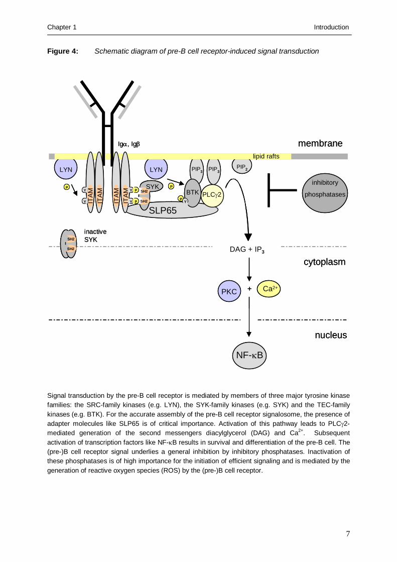
Figure 4: Schematic diagram of pre-B cell receptor-induced signal transduction
Signal transduction by the pre-B cell receptor is mediated by members of three major tyrosine kinase families: the SRC-family kinases (e.g. LYN), the SYK-family kinases (e.g. SYK) and the TEC-family kinases (e.g. BTK). For the accurate assembly of the pre-B cell receptor signalosome, the presence of adapter molecules like SLP65 is of critical importance. Activation of this pathway leads to PLCγ2-mediated generation of the second messengers diacylglycerol (DAG) and Ca2+. Subsequent activation of transcription factors like NF-κB results in survival and differentiation of the pre-B cell. The (pre-)B cell receptor signal underlies a general inhibition by inhibitory phosphatases. Inactivation of these phosphatases is of high importance for the initiation of efficient signaling and is mediated by the generation of reactive oxygen species (ROS) by the (pre-)B cell receptor.
SLP65
SH2
SH2
PKC Ca2+
NF-κB
ITAM
ITAM
ITAM
ITAM
Igα, Igβ
Y
Y
P
P
Y
Y
SYK
SH2
SH2
inactive SYK
lipid rafts
LYN
P P
BTK PLCγ2
LYN PIP3 PIP3PIP2
DAG + IP3
inhibitory
phosphatases
+
membrane
cytoplasm
nucleus
P Y
SLP65
SH2
SH2
SH2
SH2
PKC Ca2+
NF-κB
ITAM
ITAM
ITAM
ITAM
Igα, Igβ
YY
YY
PP
PP
YY
YY
SYK
SH2
SH2
SH2
SH2
inactive SYK
lipid rafts
LYN
PP PP
BTK PLCγ2
LYN PIP3 PIP3PIP2
DAG + IP3
inhibitory
phosphatases
+
membrane
cytoplasm
nucleus
PP Y

Chapter 1 Introduction
8
implicated in pre-B cell receptor signaling have initially been identified as oncogenes
(Rodrigues and Park, 1994).
Crosslinking of the pre-B cell receptor leads to the accumulation of signaling
molecules within cell surface microdomains of high activation, the glycolipid-enriched
membrane domains (GEMs) or so-called lipid rafts (Guo et al., 2000). The assembly of this
microdomains is induced by pre-B cell receptor activation and essential for effective signaling
(Saeki eta al., 2003). Recruitment of the pre-B cell receptor to the lipid raft fraction allows the
phosphorylation of the pre-B cell receptor associated signal chains Igα and Igβ by the SRC
kinase LYN, which is constitutively present in these microdomains (Cheng et al., 1999).
Interestingly, localisation within the lipid raft fraction itself maintains LYN in an active state
(Young et al., 2003). In parallel to its function in pre-B cell activation, LYN also initiates the
termination of pre-B cell receptor signaling by the activation of inhibitory phosphatases
through the phosphorylation of inhibitory receptors (Smith et al., 1998). In agreement with
this, B cells from LYN-deficient mice exhibit enhanced signaling in response to B cell
receptor stimulation (Chan et al., 1997). LYN positively transmits the initial signal by
phosphorylation of one tyrosine within an immunoreceptor tyrosine-based activation motif
(ITAM; Reth, 1989) of Igα and Igβ (Flaswinkel et al., 1994; Rolli et al., 2002; see Figure 4).
Phosphorylated ITAMs allow binding of the tyrosine kinase SYK, which is thereby released
from autoinhibition (Rolli et al., 2002). The autoinhibition of SYK is mediated through
binding one of the two N-terminal SH2 domains to the own kinase domain (Wossning and
Reth, 2004) and relieved through binding of the SH2 domains to the tyrosine-phosphorylated
ITAMs (Rolli et al., 2002). In addition to the activation of downstream targets, SYK initiates
a positive feedback loop by tyrosine-phosphorylation of ITAMs of further Igα and
Igβ molecules as well as by tyrosine-phosphorylation of further SYK molecules (Rolli et al.,
2002; Keshvara et al., 1998). As a consequence, the amount of initially activated SYK
molecules is largely amplified. SYK activity is highly regulated by dephosphorylation of its
binding partners Igα and Igβ by the inhibitory phosphatase SHP1 (Adachi et al., 2001). In
addition, attenuation of SYK activity is mediated by ubiquitin ligase CBL, which targets SYK
for degradation (Sohn et al., 2003). The important role of SYK family kinases is demonstrated
by the complete block at the pro-B cell stage in SYK family kinases deficient mice
(Schweighoffer et al., 2003). Activated SYK phosphorylates SLP65, an adapter molecule that
stabilizes the pre-B cell receptor signalosome through binding to phosphorylated Igα and
downstream targets including GRB2, VAV, NCK, BTK and PLCγ2 (Fu et al., 1998; see
Figure 4). SYK also induces the recruitment of the TEC family kinase BTK (Bruton’s

Chapter 1 Introduction
9
tyrosine kinase) and PLCγ2 to the pre-B cell receptor signalosome at the plasma membrane by
the activation of phosphatidylinositol-3-kinase (PI3K; Beitz et al., 1999). PI3K activity
generates the membrane bound phospho-lipid phosphatidylinositol-3,4,5-triphosphat (PIP3),
which serves as a membrane-anchor for the PH-domains of BTK and PLCγ2 (Saito et al.,
2003). The activity of both enzymes is negatively regulated by their membrane-delocalisation
through PIP3-degradation mediated by the inhibitory phosphatidyl-inositol phosphatases SHIP
(Bolland et al., 1998) and PTEN (Satterthwaite et al., 2000). Binding of membrane-localized
BTK to the phosphorylated adapter molecule SLP65 brings BTK in close proximity to LYN,
which in turn activates BTK through phosphorylation (Rawlings et al., 1996). BTK and
SLP65 are essential for further transmission of pre-B cell receptor-induced differentiation
signals, as either BTK- or SLP65-deficiency results in a severe B cell differentiation arrest in
the human (Noordzij et al., 2002; Minegishi et al., 1999). However, proliferation signals can
also be transduced in a SLP65- and BTK-independent pathway in mice, documented by the
rapid expansion of pre-B cells in tumors in the absence of SLP65 and BTK (Kersseboom et
al., 2003). Binding of BTK to SLP65 brings BTK also in close proximity to his target PLCγ2.
Activated PLCγ2 generates diacylglycerol (DAG) and inositol-3-phosphate (IP3) through the
hydrolysis of phosphatidylinositol-4,5-bisphosphate (PIP2), which in turn results in the release
of Ca2+ from intracellular stores by triggering IP3 receptors on the endoplasmic reticulum
(ER). The second messengers DAG and Ca2+ promote differentiation and survival by
induction of the NF-κB pathway.
The pre-B cell receptor-mediated activation of a network of tyrosine kinases (PTKs)
allows rapid amplification of the initial stimulus. While the tight regulation of their activity by
autoinhibition and delocalisation is necessary for the specificity of the pre-B cell receptor
signal, their inhibition by inhibitory PTPs is superior to the activating signal: PTPs have a 10-
to 100-fold higher turnover rate than PTKs (Honjo T, Molecular Biology of B cells, 2004,
S.162). Efficient pre-B cell receptor signaling therefore requires both, kinase activation and
phosphatase inhibition. Recently, the oxidation of a cystein within the catalytic center of PTPs
has been identified as a mechanism for the reversible inactivation of PTPs (Meng et al.,
2002). The oxidation is mediated by H2O2, which is generated by the membrane-bound
NADPH-oxidase complex in response to (pre-) B cell receptor stimulation (Singh et al.,
2005). The stimulation of the pre-B cell receptor therefore induces a highly specific signal in
a limited time frame of activation resulting in survival, proliferation and differentiation of pre-
B cells.

Chapter 1 Introduction
10
1.5 Bruton’s tyrosine kinase
In 1993, mutations of a gene encoding a novel cytoplasmic tyrosine kinase, termed Bruton’s
tyrosine kinase (BTK), were decribed as the cause of the human immunodeficiency X-linked
agammaglobulinemia (XLA; Vetrie et al., 1993; Tsukada et al., 1993). Defective expression
of this tyrosine kinase leads to a severe differentiation arrest at the pre-B cell stage in the bone
marrow and the absence of mature B cells in the periphery of XLA patients (de Weers et al.,
1993; Noordzij et al., 2002). Expression of BTK in healthy humans appears from the early
pro-B cell till the mature memory B cell stage, but is downregulated in plasma cells (de
Weers et al., 1993). While BTK has a crucial function in normal B cells, BTK is also
expressed in myeloid cells but does not play an essential role, since myeloid differentiation in
XLA patients does not seem to be affected (de Weers et al., 1993).
BTK belongs to the SRC-related family of TEC protein tyrosine kinases (Desiderio
and Siliciano, 1994; Ohta et al., 1994), which share the following structural domains: The
amino-terminal Pleckstrin-homology (PH) domain, the TEC-homology (TH) domain, the
SRC homology 3 (SH3) domain, the SH2 domain and the carboxy-terminal kinase domain
(Figure 5). With the exception of one member, termed TXK (Ohta et al., 1996), all TEC
family kinases carry a PH domain, which confers membrane-localisation of BTK depending
on PIP3 and brings BTK into close proximity with its interaction partners within the (pre-) B
cell receptor signalosome. Each domain of BTK is required for a specific interaction with
specific molecules of the (pre-)B cell receptor, thereby also acting as a linker in addition to its
kinase function (Figure 5): The PH and kinase domain of BTK together allow binding to the
cytoplasmic domain of CD95 (Vassilev et al., 1999). The TH domain of BTK contains a
proline-rich region which binds the SH3 domain of other signaling molecules (Hansson et al.,
2001). Those intermolecular interactions can be formed between BTK and the SRC kinases
LYN, FYN and HCK (Cheng et al., 1994; Yang et al., 1995), the G protein Gα (Ma et al.,
1998) as well as in self-association of BTK molecules (Laederach et al., 2002). The SH3 and
SH2 domains also confer intermolecular interactions by binding to the proto-oncogene CBL
(Patel et al., 1997), the spleen tyrosine kinase (SYK; Morrogh et al., 1999), the WAS protein
(WASP; Morrogh et al., 1999), the guanine exchange factor VAV, the RNA-binding protein
SAM68 (Guinamard et al., 1997) and the adapter molecule SLP65 (Su et al., 1999). In
agreement with this, recent work demonstrated that kinase-inactive BTK, acting as a linker,
can partially restore the differentiation arrest in BTK-deficient mice (Middendorp et al.,
2003).
Chapter 1 Introduction
11
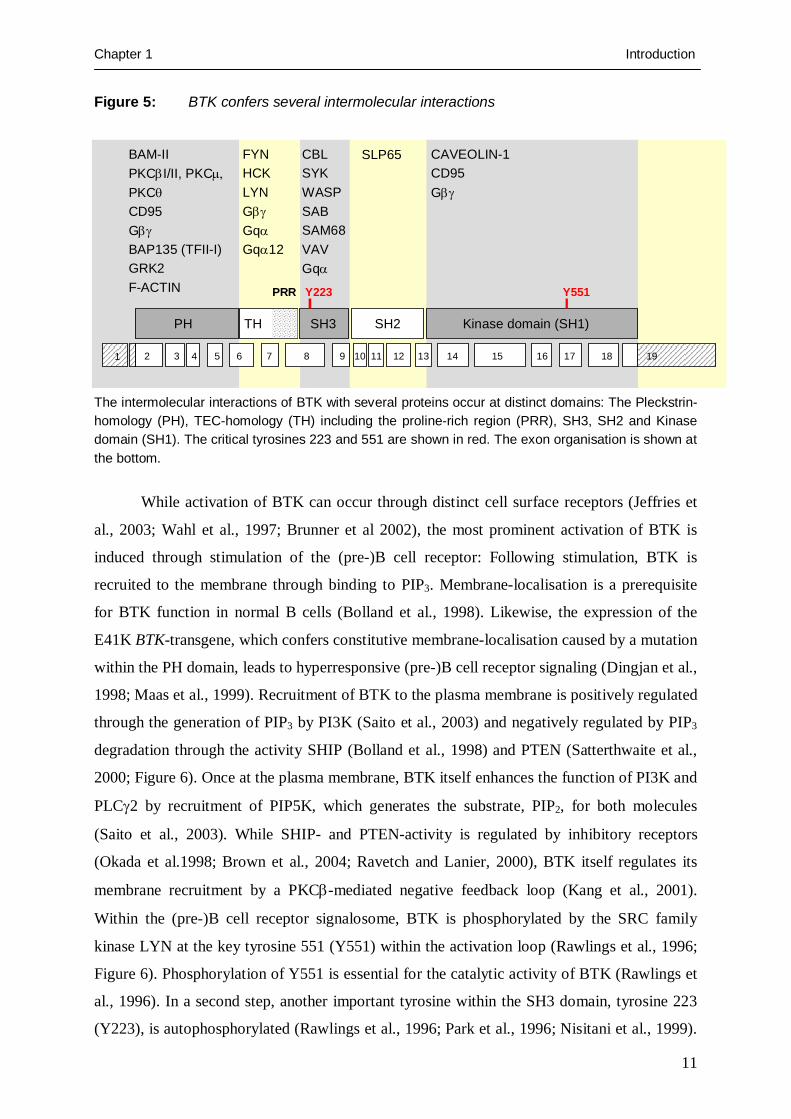
Figure 5: BTK confers several intermolecular interactions
The intermolecular interactions of BTK with several proteins occur at distinct domains: The Pleckstrin-homology (PH), TEC-homology (TH) including the proline-rich region (PRR), SH3, SH2 and Kinase domain (SH1). The critical tyrosines 223 and 551 are shown in red. The exon organisation is shown at the bottom.
While activation of BTK can occur through distinct cell surface receptors (Jeffries et
al., 2003; Wahl et al., 1997; Brunner et al 2002), the most prominent activation of BTK is
induced through stimulation of the (pre-)B cell receptor: Following stimulation, BTK is
recruited to the membrane through binding to PIP3. Membrane-localisation is a prerequisite
for BTK function in normal B cells (Bolland et al., 1998). Likewise, the expression of the
E41K BTK-transgene, which confers constitutive membrane-localisation caused by a mutation
within the PH domain, leads to hyperresponsive (pre-)B cell receptor signaling (Dingjan et al.,
1998; Maas et al., 1999). Recruitment of BTK to the plasma membrane is positively regulated
through the generation of PIP3 by PI3K (Saito et al., 2003) and negatively regulated by PIP3
degradation through the activity SHIP (Bolland et al., 1998) and PTEN (Satterthwaite et al.,
2000; Figure 6). Once at the plasma membrane, BTK itself enhances the function of PI3K and
PLCγ2 by recruitment of PIP5K, which generates the substrate, PIP2, for both molecules
(Saito et al., 2003). While SHIP- and PTEN-activity is regulated by inhibitory receptors
(Okada et al.1998; Brown et al., 2004; Ravetch and Lanier, 2000), BTK itself regulates its
membrane recruitment by a PKCβ-mediated negative feedback loop (Kang et al., 2001).
Within the (pre-)B cell receptor signalosome, BTK is phosphorylated by the SRC family
kinase LYN at the key tyrosine 551 (Y551) within the activation loop (Rawlings et al., 1996;
Figure 6). Phosphorylation of Y551 is essential for the catalytic activity of BTK (Rawlings et
al., 1996). In a second step, another important tyrosine within the SH3 domain, tyrosine 223
(Y223), is autophosphorylated (Rawlings et al., 1996; Park et al., 1996; Nisitani et al., 1999).
PH TH SH2SH3 Kinase domain (SH1)
1 2 3 4 5 6 7 8 9 10 11 12 13 14 15 16 17 18 19
BAM-IIPKCβI/II, PKCμ, PKCθCD95GβγBAP135 (TFII-I)GRK2F-ACTIN
FYNHCKLYNGβγGqαGqα12
CBLSYKWASPSABSAM68VAVGqα
SLP65 CAVEOLIN-1CD95Gβγ
Y551Y223PRR
PH TH SH2SH3 Kinase domain (SH1)
1 2 3 4 5 6 7 8 9 10 11 12 13 14 15 16 17 18 19
BAM-IIPKCβI/II, PKCμ, PKCθCD95GβγBAP135 (TFII-I)GRK2F-ACTIN
FYNHCKLYNGβγGqαGqα12
CBLSYKWASPSABSAM68VAVGqα
SLP65 CAVEOLIN-1CD95Gβγ
Y551Y223PRR

Chapter 1 Introduction
12
The relevance of Y223 phosphorylation for BTK function remains controversial:
Phosphorylation of Y223 enhances binding properties of signaling molecules like SYK to the
SH3 domain of BTK (Morrogh et al., 1999), suggesting a positive role of Y223. In contrast,
an inhibitory function of Y223 was proposed, as it was shown that mutation of Y223
potentiates the capacity of a gain-of-function mutant to transform fibroblasts (Park et al.,
1996). Moreover, it was shown that the expression of a BTK-transgene with an Y233F
mutation could reconstitute B cell development in BTK-deficient mice (Middendorp et al.,
2003).
Activated BTK is essential for cell cycle progression and survival in response to (pre-)
B cell receptor stimulation (Petro et al., 2000). Following activation, BTK phosphorylates
PLCγ2 facilitated by the interaction with the adapter molecules CBL-B and SLP65 (Yasuda et
al., 2002). PLCγ2-mediated release of the second messengers diacylglycerol (DAG) and Ca2+
into the cytoplasm induces the activation of PKCβ (Saijo et al., 2002). The subsequent
activation of NF-κB and CYCLIN D2 allows entry into the S phase of the cell cycle and cell
growth (Petro et al., 2001; Glassford et al., 2003). However, while BTK-dependent activation
of PLCγ2 and the subsequent Ca2+ release is mediated by tyrosine-phosphorylation (Watanabe
et al., 2001), it has recently been shown, that also a BTK-transgene lacking kinase activity
could partially restore B cell development in BTK-deficient mice (Middendorp et al., 2003).
In parallel to cell cycle progression, BTK provides survival signals through NF-κB
associated expression of the anti-apoptotic protein BCLXL (Suzuki et al., 2003). In addition,
BTK activity leads to the activation of STAT5 (Mahajan et al., 2001), which also results in
the upregulation of BCLXL (Nosaka et al., 1999). Contrary to the function in survival
signaling by prevention of CD95-mediated apoptosis (Vassilev et al., 1999) and upregulation
of BCLXL, BTK can also act as a mediator of apoptosis: BTK kinase activity is responsible
for radiation-induced apoptosis in the chicken lymphoma cell line DT40 (Uckun et al., 1996)
and forced expression induces apoptosis in the human epithelial carcinoma cell line HELA
(Islam et al., 2000; Islam and Smith, 2000).
There may be additional functions of BTK in regulating gene expression. BTK binds
to the two transcription factors BAP135/TFII-I (Yang and Desiderio, 1997) and BRIGHT
(Rajaiya et al., 2005) and can translocate to the nucleus, which may be critical during B cell
development and differentiation (Mohamed et al., 2000).
In summary, it has been shown that Bruton’s tyrosine kinase (BTK) represents a
crucial component of the (pre-) B cell receptor signal transduction cascade (Fluckiger et al.,
1998). Deficiency of BTK leads to a differentiation arrest at the pre-B cell stage (Noordzij et

Chapter 1 Introduction
13
al., 2002) and reduced survival and proliferation, as in the bone marrow of XLA patients the
amount of B cells is decreased compared to bone marrow healthy humans (Genevier and
Callard, 1997). Surprisingly, in cooperation with the linker molecule SLP65, BTK was
recently described as a tumor suppressor in mice (Kersseboom et al., 2003). This underlines
the important role of BTK in different signaling pathways leading to survival, proliferation or
differentiation of pre-B cells.
1.6 X-linked agammaglobulinemia
The severe human immunodeficiency X-linked agammaglobulinemia (XLA) was firstly
characterized as human agammaglobulinemia in 1952 by Ogden C. Bruton, who reported the
complete absence of the immunoglobulin γ-fraction in the serum of an eight year old boy
(Bruton, 1952). The immune defect was further characterized by the X-linked pattern of
inheritance (Janeway et al., 1953) and renamed as XLA. The disease is, at a prevalence of 1
out of 200.000 individuals (Sideras and Smith, 1995), rare and characterized by an increased
susceptibility to extracellular bacterial and enteroviral infections (Lederman and Winkelstein,
1985). While in XLA patients the myeloid cell and T cell populations are unaffected, the B
cell population exhibits a differentiation arrest between the pro-B and pre-B cell stage
resulting in a near complete loss of mature B cells (Campana et al., 1990; Noordzij et al.,
2002). The gene defective in XLA was mapped to the Xq21.3-22 region (Guioli et al., 1989)
and afterwards described as the gene encoding a cytoplasmic tyrosine termed ATK
(agammablobilinemia tyrosine kinase; Vetrie et al., 1993) or BPK (B cell progenitor kinase;
Tsukada et al., 1993), later renamed as Bruton’s tyrosine kinase (BTK). BTK is currrently
characterized as a crucial component of the (pre-) B cell receptor signalosome (Fluckiger et
al., 1998) promoting the developmental progression of pre-B cells (Middendorp et al., 2003).
Mutation of the mouse homolog causes a less severe phenotype termed X-linked
immunodeficiency (Xid; Rawlings et al., 1993; Thomas et al., 1993), which is owing to the
presence of BTK-redundant TEC family kinases that may serve as a backup in the absence of
BTK (Ellmeier et al., 2000). In contrast to XLA, Xid mice still have about 50 % of the normal
number of B cells and are able to generate an antibody response against T cell dependent
antigens (Klaus et al., 1997; Wicker and Scher, 1986). In the human, about 85 % of defects in
early B cell development are caused by a mutation in BTK, while 5-10 % are caused by
defects in other components of the (pre-) B cell receptor signalosome (Conley et al., 2005).
The so far described mutations within the BTK gene are collected in the BTK database
BTKbase (http://bioinf.uta.fi/BTKbase/), which represents a public XLA-mutation registry

Chapter 1 Introduction
14
established in 1994 (Vihinen et al., 1996). Currently, about 561 unique mutations within the
BTK gene from 839 families are known, of which 185 are missense mutations. While
frameshift and nonsense mutations are distributed randomly over the whole gene, missense
mutations are predominantly present within the carboxyterminal kinase domain and the
aminoterminal PH domain of BTK (Lindvall et al., 2005). Notably, only the SH3 domain of
BTK is completely devoid of missense mutations in XLA patients. From in vitro kinase
assays of BTK mutants derived from BTK-deficient X-linked agammaglobulinemia (XLA)
patients, it is known that in all cases of XLA, BTK kinase activity is abolished or severely
reduced (http://bioinf.uta.fi/BTKbase/BTKbasebrowser.html). Even replacement mutations in
the distal portion of the kinase domain often result in a complete loss of BTK kinase activity
(Holinski-Feder et al., 1998).
1.7 Chromosomal translocations as a cause of B cell precursor leukemia
Acute lymphoblastic leukemia (ALL) with a pre-B cell immunophenotype is characterized by
deregulated cell growth, differentiation and survival signaling. In many cases, leukemogenic
deregulation is owing to translocation events. In 52% of cases of all ALLs in children and
68% of cases of all ALLs in adults, chromosomal aberrations were detected in the leukemia
cells (Pui et al., 2004). As a general phenomenon of cancer, translocations typically result in
transcriptional deregulation of the target gene or lead to the generation of a fusion gene if the
breakpoint involves coding exons on both chromosomes. In this regard it is noteworthy that
recurrent chromosomal translocations in mature B cell lymphoma cells, in almost all
instances, result in transcriptional deregulation. In contrast, most if not all recurrent
chromosomal rearrangements in the leukemia cells result in the expression of chimeric
proteins (Look , 1997).
The most frequent genetic aberration in children (22% of all ALLs) involves a
rearrangement of the oligomerisation domain of the TEL gene on chromosome 12 to the entire
coding region of the AML1 gene on chromosome 21 (Pui et al., 2004). While TEL belongs to
the ETS family of transcriptional repressors and AML1 encodes the RUNT-domain of the
transcription factor termed core-binding factor (CBF), the TEL-AML1 gene product
oligomerizes and represses genes normally activated by AML1 (Hiebert et al., 1996). In
contrast to the normal AML1 proteins, which recruit other co-activators like histone-acetyl-
transferases (HATs) to the target gene, the oncogenic TEL-AML1 protein reverses the initial
function by recruiting histone-deacetylases (HDACs) that close the chromatin structure by

Chapter 1 Introduction
15
methylation and inhibit transcription (Hiebert et al., 1996). In addition to the reversal of
AML1 function, the translocation event also prevents normal function of TEL.
Another example for an oncogenic trancription factor chimera generated by a
translocation event in B cell precursor leukemia represents the fusion protein E2A-PBX1.
Fusion of the transcriptional activation domains of the E2A gene on chromosome 19 to the
DNA-binding domain of the PBX1 gene on chromosome 1 results in a new transcription
factor that deregulates PBX1-target genes. This deregulation alone is sufficient to promote
proliferation and survival of leukemia cells as transgenic expression of E2A-PBX1 leads to
leukemia in mice (Dedera et al., 1993).
The most frequent type of lymphoid leukemia in adults (25%) is the pre-B cell
leukemia carrying a translocation of the BCR gene on chromosome 22 to the ABL1 gene on
chromosome 9, the so-called Philadelphia (Ph) chromosome (Piu et al., 2004; Rowley, 1973).
With identification of the BCR-ABL1 translocation, it was for the first time that a
chromosomal aberration could be linked to a specific cancer (Nowell and Hungerford, 1960).
The BCR-ABL1 rearrangement is present in B cell progenitor leukemia, but also drives
malignant transformation in >95% if not all cases of chronic myeloid leukemia (CML;
Shepherd et al., 1995). While in CML, leukemia cells express predominantly a BCR-ABL1
onco-protein of 210 kd (BCR-ABL1p210), pre-B cell leukemias with a BCR-ABL1 gene
rearrangement mainly express the form of 190 kd size (BCR-ABL1p190; Clark et al., 1988).
Notably, BCR-ABL1–positive CML can be cured in many cases with the BCR-ABL1 kinase
inhibitor STI571 (also termed Imatinib or Gleevec), but the same treatment leads to a relapse
in all patients with BCR-ABL1–positive pre-B cell leukemia within a median of 4 months
(Druker et al., 2001). However, the reasons for this striking difference have yet remained
unclear. The translocation of the BCR to the ABL1 gene deletes the autoinhibition domain of
the ABL1 tyrosine kinase resulting in a constitutively active BCR-ABL1 tyrosine kinase. The
expression of BCR-ABL1 alone is sufficient for malignant transformation of pre-B cells
(Klucher et al., 1998). The transformation capacity is dependent on the kinase activity of
BCR-ABL1 as kinase-inhibitors against BCR-ABL1 induce apoptosis in the leukemia cells
(Druker et al., 1996). BCR-ABL1 kinase activity leads to the deregulation of cellular
signaling pathways and is of critically importance for the promotion of survival of the
leukemia cells (Huettner et al., 2000).

Chapter 1 Introduction
16
1.8 Signaling activity of the oncogenic BCR-ABL1 kinase
The activity of the normal c-ABL1 tyrosine kinase is regulated by an autoinhibition
mechanism (Pluk et al., 2002). This autoinhibition is mediated through binding of the N-
terminal myristoyl modification of c-ABL1 to the kinase domain, thereby stabilizing the
inactive conformation of c-ABL1 (Nagar et al., 2003). The translocation resulting in the BCR-
ABL1 fusion gene deletes the N-terminal part of c-ABL1 required for autoinhibition. To the
contrary, the Abelson murine leukemia virus gene product v-Abl, which also exhibits
constitutive ABL1 kinase activity, still contains the complete N-terminal domain but carries a
deletion within the C-terminus also resulting in the relief of autoinhibition (Shore et al.,
1990). The rearrangement of BCR and ABL1 results in fusion proteins of different sizes
depending on the breakpoint within the BCR gene. Comparison of the kinase activity of the
two major forms of BCR-ABL1, p190 and p210, demonstrated that the p190 isoform exhibits
increased kinase activity compared to p210 (Lugo et al., 1990). BCR-ABL1p190 also induces
lymphoid leukemia with a shorter latency than p210 in mice (Li et al., 1999). However, both
forms are present in BCR-ABL1-positive pre-B cell leukemia and a significant difference in
the response to treatment has not been observed (Gleissner et al., 2002).
For the activation of c-ABL1, the phosphorylation of tyrosine 412 is of critical
importance (Hantschel et al., 2003). Relief of the autoinhibition of c-ABL1 results in the
recruitment to specific membrane domains like lipid rafts, where c-ABL1 is activated by
phosphorylation. In contrast, BCR-ABL1 is not membrane-associated but requires
oligomerization to homotetramers for full activation (Maru et al., 1996). Oligomerization is
dependent on a coiled-coiled domain within the BCR gene which is present in all BCR-ABL1
forms (Tauchi et al., 1997). The oligomerisation status of BCR-ABL1 proteins within the cell
directly determines their autophosphorylation and transformation capacity (Maru et al., 1996).
The activated BCR-ABL1 kinase is sufficient to modulate distinct cellular signaling
pathways promoting cell growth and survival: Several molecules that promote cell-cycle
progression and growth like CYCLIN D2 (Parada et al., 2001), MYC (Skorski et al., 1997)
and the RAS pathway (Cortez et al., 1997) have been identified to be regulated by BCR-
ABL1 in leukemia cells. In addition, BCR-ABL1 prevents apoptosis by upregulation of the
anti-apoptotic protein BCLXL through phosphorylation of STAT5 (Gesbert and Griffin, 2000)
and activation of the anti-apoptoic protein BCL2 through the PI3K/AKT pathway (Skorski et
al., 1997). Also molecules involved in cytoskeletal modulation (including PAXILLIN and
CBL), DNA repair (including RAD51 and DNA-PK) and signal transduction (including RAS
and JNK) have been described to be also regulated by BCR-ABL1 (Wong and Witte, 2004).

Chapter 1 Introduction
17
As several of BCR-ABL1-regulated molecules are also implicated in the signal
transduction of the pre-B cell receptor (e.g. PI3K, AKT, SHP1, CBL, GRB2 and VAV),
potential interference of BCR-ABL1-induced oncogenic signaling with pre-B cell receptor
signaling in the pre-B leukemia cells was systematically investigated in this thesis.
1.9 Aims of the thesis
During early B cell development, B cell precursor cells undergo a sequence of somatic
rearrangements within the IGH locus, which are required for the expression of a µ-heavy
chain within the pre-B cell receptor complex on the surface of pre-B cells. Expression of a
functional pre-B cell receptor and subsequent signal transduction serves as an early
checkpoint of B cell development, thereby initiating the expansion, survival and further
differentiation of pre-B cells into immature B cells (Hess et al., 2001; Tarlinton et al., 1997).
B cell lineage leukemia cells expressing the oncogenic BCR-ABL1 tyrosine kinase
typically exhibit a differentiation block at the pre-B cell stage (Young and Witte, 1988).
Oncogenic signaling initiated by the BCR-ABL1 kinase is sufficient for malignant
transformation of pre-B cells (Huettner et al., 2000).
Based on these findings, the following questions should be addressed within this
thesis:
i) Is the differentiation block of BCR-ABL1-transformed pre-B cells induced by
the oncogenic BCR-ABL1 kinase and how is this arrest maintained in the
leukemia cells?
ii) BCR-ABL1-transformed pre-B leukemia cells exhibit enhanced proliferation,
which could also be induced by pre-B cell receptor signaling. Does the pre-B
cell receptor function as an oncogene within the leukemia cells?
iii) Do the oncogenic BCR-ABL1 signaling pathway and the pre-B cell receptor
signal transduction pathway coincide within the leukemia cell and if so, do
they interfere with each other?

Chapter 1 Introduction
18
References
Adachi T, Wienands J, Wakabayashi C, Yakura H, Reth M, Tsubata T. SHP-1 requires inhibitory co-receptors to down-modulate B cell antigen receptor-mediated phosphorylation of cellular substrates. J Biol Chem. 2001; 276: 26648-55 Adolfsson J, Borge OJ, Bryder D, Theilgaard-Monch K, Astrand-Grundstrom I, Sitnicka E, Sasaki Y, Jacobsen SE. Upregulation of Flt3 expression within the bone marrow Lin(-)Sca1(+)c-kit(+) stem cell compartment is accompanied by loss of self-renewal capacity. Immunity. 2001; 15: 659-69 Akamatsu Y, Tsurushita N, Nagawa F, Matsuoka M, Okazaki K, Imai M, Sakano H. Essential residues in V(D)J recombination signals. J Immunol. 1994; 153: 4520-9 Alt FW, Baltimore D. Joining of immunoglobulin heavy chain gene segments: implications from a chromosome with evidence of three D-JH fusions. Proc Natl Acad Sci U S A. 1982; 79: 4118-22 Aoki Y, Isselbacher KJ, Cherayil BJ, Pillai S. Tyrosine phosphorylation of Blk and Fyn Src homology 2 domain-binding proteins occurs in response to antigen-receptor ligation in B cells and constitutively in pre-B cells. Proc Natl Acad Sci U S A. 1994; 91: 4204-8 Bain G, Maandag EC, Izon DJ, Amsen D, Kruisbeek AM, Weintraub BC, Krop I, Schlissel MS, Feeney AJ, van Roon M, et al. E2A proteins are required for proper B cell development and initiation of immunoglobulin gene rearrangements. Cell. 1994; 79: 885-92 Beitz LO, Fruman DA, Kurosaki T, Cantley LC, Scharenberg AM. SYK is upstream of phosphoinositide 3-kinase in B cell receptor signaling. J Biol Chem. 1999; 274: 32662-6 Bolland S, Pearse RN, Kurosaki T, Ravetch JV. SHIP modulates immune receptor responses by regulating membrane association of Btk. Immunity. 1998; 8: 509-16 Bradl H, Wittmann J, Milius D, Vettermann C, Jäck HM. Interaction of murine precursor B cell receptor with stroma cells is controlled by the unique tail of lambda 5 and stroma cell-associated heparan sulfate. J Immunol. 2003; 171: 2338-48 Brown KS, Blair D, Reid SD, Nicholson EK, Harnett MM. FcgammaRIIb-mediated negative regulation of BCR signalling is associated with the recruitment of the MAPkinase-phosphatase, Pac-1, and the 3'-inositol phosphatase, PTEN. Cell Signal. 2004 ; 16: 71-80 Brunner C, Avots A, Kreth HW, Serfling E, Schuster V. Bruton's tyrosine kinase is activated upon CD40 stimulation in human B lymphocytes. Immunobiology. 2002; 206: 432-40 Bruton OC. Agammaglobulinemia. Pediatrics. 1952; 9: 722-728 Campana D, Farrant J, Inamdar N, Webster AD, Janossy G. Phenotypic features and proliferative activity of B cell progenitors in X-linked agammaglobulinemia. J Immunol. 1990; 145: 1675-80 Chan VW, Meng F, Soriano P, DeFranco AL, Lowell CA. Characterization of the B lymphocyte populations in Lyn-deficient mice and the role of Lyn in signal initiation and down-regulation. Immunity. 1997; 7: 69-81 Chen J, Trounstine M, Alt FW, Young F, Kurahara C, Loring JF, Huszar D. Immunoglobulin gene rearrangement in B cell deficient mice generated by targeted deletion of the JH locus. Int Immunol. 1993; 5: 647-56 Cheng G, Ye ZS, Baltimore D. Binding of Bruton's tyrosine kinase to Fyn, Lyn, or Hck through a Src homology 3 domain-mediated interaction. Proc Natl Acad Sci U S A. 1994; 91: 8152-5

Chapter 1 Introduction
19
Cheng PC, Dykstra ML, Mitchell RN, Pierce SK. A role for lipid rafts in B cell antigen receptor signaling and antigen targeting. J Exp Med. 1999; 190: 1549-60 Clark SS, McLaughlin J, Timmons M, Pendergast AM, Ben-Neriah Y, Dow LW, Crist W, Rovera G, Smith SD, Witte ON. Expression of a distinctive BCR-ABL oncogene in Ph1-positive acute lymphocytic leukemia (ALL). Science. 1988; 239: 775-7 Conley ME, Broides A, Hernandez-Trujillo V, Howard V, Kanegane H, Miyawaki T, Shurtleff SA. Genetic analysis of patients with defects in early B-cell development. Immunol Rev. 2005; 203: 216-34 Cortez D, Reuther G, Pendergast AM. The Bcr-Abl tyrosine kinase activates mitogenic signaling pathways and stimulates G1-to-S phase transition in hematopoietic cells. Oncogene. 1997; 15: 2333-42 DeKoter RP, Singh H. Regulation of B lymphocyte and macrophage development by graded expression of PU.1. Science. 2000; 288: 1439-41 Dedera DA, Waller EK, LeBrun DP, Sen-Majumdar A, Stevens ME, Barsh GS, Cleary ML. Chimeric homeobox gene E2A-PBX1 induces proliferation, apoptosis, and malignant lymphomas in transgenic mice. Cell. 1993; 74: 833-43 de Weers M, Verschuren MC, Kraakman ME, Mensink RG, Schuurman RK, van Dongen JJ, Hendriks RW. The Bruton's tyrosine kinase gene is expressed throughout B cell differentiation, from early precursor B cell stages preceding immunoglobulin gene rearrangement up to mature B cell stages. Eur J Immunol. 1993; 23: 3109-14 Dingjan GM, Maas A, Nawijn MC, Smit L, Voerman JS, Grosveld F, Hendriks RW. Severe B cell deficiency and disrupted splenic architecture in transgenic mice expressing the E41K mutated form of Bruton's tyrosine kinase. EMBO J. 1998; 17: 5309-20 Desiderio S, Siliciano JD. The Itk/Btk/Tec family of protein-tyrosine kinases. Chem Immunol. 1994; 59: 191-210 Druker BJ, Tamura S, Buchdunger E, Ohno S, Segal GM, Fanning S, Zimmermann J, Lydon NB. Effects of a selective inhibitor of the Abl tyrosine kinase on the growth of Bcr-Abl positive cells. Nat Med. 1996; 2: 561-6 Druker BJ, Sawyers CL, Kantarjian H, Resta DJ, Reese SF, Ford JM, Capdeville R, Talpaz M. Activity of a specific inhibitor of the BCR-ABL tyrosine kinase in the blast crisis of chronic myeloid leukemia and acute lymphoblastic leukemia with the Philadelphia chromosome. N Engl J Med. 2001; 344: 1038-42 Ellmeier W, Jung S, Sunshine MJ, Hatam F, Xu Y, Baltimore D, Mano H, Littman DR. Severe B cell deficiency in mice lacking the tec kinase family members Tec and Btk. J Exp Med. 2000; 192: 1611-24 Flaswinkel H, Reth M. Dual role of the tyrosine activation motif of the Ig-alpha protein during signal transduction via the B cell antigen receptor. EMBO J. 1994 ; 13: 83-9 Flemming A, Brummer T, Reth M, Jumaa H. The adaptor protein SLP-65 acts as a tumor suppressor that limits pre-B cell expansion. Nat Immunol. 2003; 4: 38-43

Chapter 1 Introduction
20
Fluckiger AC, Li Z, Kato RM, Wahl MI, Ochs HD, Longnecker R, Kinet JP, Witte ON, Scharenberg AM, Rawlings DJ. Btk/Tec kinases regulate sustained increases in intracellular Ca2+ following B-cell receptor activation. EMBO J. 1998; 17: 1973-85 Fu C, Turck CW, Kurosaki T, Chan AC. BLNK: a central linker protein in B cell activation. Immunity. 1998; 9: 93-103 Gauthier L, Rossi B, Roux F, Termine E, Schiff C. Galectin-1 is a stromal cell ligand of the pre-B cell receptor (BCR) implicated in synapse formation between pre-B and stromal cells and in pre-BCR triggering. Proc Natl Acad Sci U S A. 2002; 99: 13014-9 Genevier HC, Callard RE. Impaired Ca2+ mobilization by X-linked agammaglobulinaemia (XLA) B cells in response to ligation of the B cell receptor (BCR). Clin Exp Immunol. 1997; 110: 386-91 Georgopoulos K, Bigby M, Wang JH, Molnar A, Wu P, Winandy S, Sharpe A. The Ikaros gene is required for the development of all lymphoid lineages. Cell. 1994; 79: 143-56 Gesbert F, Griffin JD. Bcr/Abl activates transcription of the Bcl-X gene through STAT5. Blood. 2000; 96: 2269-76 Glassford J, Soeiro I, Skarell SM, Banerji L, Holman M, Klaus GG, Kadowaki T, Koyasu S, Lam EW. BCR targets cyclin D2 via Btk and the p85alpha subunit of PI3-K to induce cell cycle progression in primary mouse B cells. Oncogene. 2003; 22: 2248-59 Gleissner B, Gokbuget N, Bartram CR, Janssen B, Rieder H, Janssen JW, Fonatsch C, Heyll A, Voliotis D, Beck J, Lipp T, Munzert G, Maurer J, Hoelzer D, Thiel E; German Multicenter Trials of Adult Acute Lymphoblastic Leukemia Study Group. Leading prognostic relevance of the BCR-ABL translocation in adult acute B-lineage lymphoblastic leukemia: a prospective study of the German Multicenter Trial Group and confirmed polymerase chain reaction analysis. Blood. 2002; 99: 1536-43 Grawunder U, Zimmer D, Fugmann S, Schwarz K, Lieber MR. DNA ligase IV is essential for V(D)J recombination and DNA double-strand break repair in human precursor lymphocytes. Mol Cell. 1998; 2: 477-84 Guinamard R, Fougereau M, Seckinger P. The SH3 domain of Bruton's tyrosine kinase interacts with Vav, Sam68 and EWS. Scand J Immunol. 1997; 45: 587-95 Guioli S, Arveiler B, Bardoni B, Notarangelo LD, Panina P, Duse M, Ugazio A, Oberle I, de Saint Basile G, Mandel JL, et al.Close linkage of probe p212 (DXS178) to X-linked agammaglobulinemia. Hum Genet. 1989; 84: 19-21 Guo B, Kato RM, Garcia-Lloret M, Wahl MI, Rawlings DJ. Engagement of the human pre-B cell receptor generates a lipid raft-dependent calcium signaling complex. Immunity. 2000; 13: 243-53 Hantschel O, Nagar B, Guettler S, Kretzschmar J, Dorey K, Kuriyan J, Superti-Furga G. A myristoyl/phosphotyrosine switch regulates c-Abl. Cell. 2003; 112: 845-57 Hansson H, Okoh MP, Smith CI, Vihinen M, Hard T. Intermolecular interactions between the SH3 domain and the proline-rich TH region of Bruton's tyrosine kinase. FEBS Lett. 2001; 489: 67-70 Hashimoto S, Tsukada S, Matsushita M, Miyawaki T, Niida Y, Yachie A, Kobayashi S, Iwata T, Hayakawa H, Matsuoka H, Tsuge I, Yamadori T, Kunikata T, Arai S, Yoshizaki K, Taniguchi N, Kishimoto T. Identification of Bruton's tyrosine kinase (Btk) gene mutations and characterization of the derived proteins in 35 X-linked agammaglobulinemia families: a nationwide study of Btk deficiency in Japan. Blood. 1996; 88: 561-73

Chapter 1 Introduction
21
Hess J, Werner A, Wirth T, Melchers F, Jack HM, Winkler TH. Induction of pre-B cell proliferation after de novo synthesis of the pre-B cell receptor. Proc Natl Acad Sci U S A. 2001; 98: 1745-50 Hiebert SW, Sun W, Davis JN, Golub T, Shurtleff S, Buijs A, Downing JR, Grosveld G, Roussell MF, Gilliland DG, Lenny N, Meyers S. The t(12;21) translocation converts AML-1B from an activator to a repressor of transcription. Mol Cell Biol. 1996; 16: 1349-55 Holinski-Feder E, Weiss M, Brandau O, Jedele KB, Nore B, Backesjo CM, Vihinen M, Hubbard SR, Belohradsky BH, Smith CI, Meindl A. Mutation screening of the BTK gene in 56 families with X-linked agammaglobulinemia (XLA): 47 unique mutations without correlation to clinical course. Pediatrics. 1998; 101: 276-84 Horcher M, Souabni A, Busslinger M. Pax5/BSAP maintains the identity of B cells in late B lymphopoiesis. Immunity. 2001; 14: 779-90 Huettner C.S., P. Zhang, R.A. Van Etten, D.G. Tenen. Reversibility of acute B-cell leukaemia induced by BCR-ABL1. Nat Genet. 2000; 24: 57-60 Ishiai M, Kurosaki M, Pappu R, Okawa K, Ronko I, Fu C, Shibata M, Iwamatsu A, Chan AC, Kurosaki T. BLNK required for coupling Syk to PLC gamma 2 and Rac1-JNK in B cells. Immunity. 1999; 10: 117-25 Islam TC, Branden LJ, Kohn DB, Islam KB, Smith CI. BTK mediated apoptosis, a possible mechanism for failure to generate high titer retroviral producer clones. J Gene Med. 2000; 2: 204-9 Islam TC, Smith CI. The cellular phenotype conditions Btk for cell survival or apoptosis signaling. Immunol Rev. 2000; 178: 49-63 Janeway CA, Apt L, Gitlin D. Agammaglobulinemia. Trans Assoc Am Physicians. 1953; 66: 200-2 Jefferies CA, Doyle S, Brunner C, Dunne A, Brint E, Wietek C, Walch E, Wirth T, O'Neill LA. Bruton's tyrosine kinase is a Toll/interleukin-1 receptor domain-binding protein that participates in nuclear factor kappaB activation by Toll-like receptor 4. J Biol Chem. 2003; 278: 26258-64 Kang SW, Wahl MI, Chu J, Kitaura J, Kawakami Y, Kato RM, Tabuchi R, Tarakhovsky A, Kawakami T, Turck CW, Witte ON, Rawlings DJ. PKCbeta modulates antigen receptor signaling via regulation of Btk membrane localization. EMBO J. 2001; 20: 5692-702 Karasuyama H, Kudo A, Melchers F. The proteins encoded by the VpreB and lambda 5 pre-B cell-specific genes can associate with each other and with mu heavy chain. J Exp Med. 1990; 172: 969-72 Kato I., T. Takai, A. Kudo The pre-B cell receptor signaling for apoptosis is negatively regulated by Fc gamma RIIB. J Immunol. 2002; 168: 534-629 Kee BL, Murre C. Induction of early B cell factor (EBF) and multiple B lineage genes by the basic helix-loop-helix transcription factor E12. J Exp Med. 1998; 188: 699-713 Kersseboom R, Middendorp S, Dingjan GM, Dahlenborg K, Reth M, Jumaa H, Hendriks RW. Bruton's tyrosine kinase cooperates with the B cell linker protein SLP-65 as a tumor suppressor in Pre-B cells. J Exp Med. 2003; 198: 91-8 Keshvara LM, Isaacson CC, Yankee TM, Sarac R, Harrison ML, Geahlen RL. Syk- and Lyn-dependent phosphorylation of Syk on multiple tyrosines following B cell activation includes a site that negatively regulates signaling. J Immunol. 1998; 161: 5276-83
Chapter 1 Introduction
22
Kim J, Sif S, Jones B, Jackson A, Koipally J, Heller E, Winandy S, Viel A, Sawyer A, Ikeda T, Kingston R, Georgopoulos K. Ikaros DNA-binding proteins direct formation of chromatin remodeling complexes in lymphocytes. Immunity. 1999; 10: 345-55 Kim YJ, Sekiya F, Poulin B, Bae YS, Rhee SG. Mechanism of B-cell receptor-induced phosphorylation and activation of phospholipase C-gamma2. Mol Cell Biol. 2004; 24: 9986-99 Kitamura D, Roes J, Kuhn R, Rajewsky K. A B cell-deficient mouse by targeted disruption of the membrane exon of the immunoglobulin mu chain gene. Nature. 1991; 350: 423-6 Klaus GG, Holman M, Johnson-Leger C, Elgueta-Karstegl C, Atkins C. A re-evaluation of the effects of X-linked immunodeficiency (xid) mutation on B cell differentiation and function in the mouse. Eur J Immunol. 1997; 27: 2749-56 Klucher KM, Lopez DV, Daley GQ. Secondary mutation maintains the transformed state in BaF3 cells with inducible BCR/ABL expression. Blood. 1998; 91: 3927-34 Laederach A, Cradic KW, Brazin KN, Zamoon J, Fulton, BD, Huang XY, Andreotti AH. Competing modes of self-association in the regulatory doamins of Bruton’s tyrosine kinase: Intramolecular contact versus asymmetric homodimerization. Prot Science 2002; 11: 36-45 Lam KP, Kuhn R, Rajewsky K. In vivo ablation of surface immunoglobulin on mature B cells by inducible gene targeting results in rapid cell death. Cell. 1997; 90: 1073-83 Lassoued K, Illges H, Benlagha K, Cooper MD. Fate of surrogate light chains in B lineage cells. J Exp Med. 1996; 183: 421-9 Lederman HM, Winkelstein JA. X-linked agammaglobulinemia: an analysis of 96 patients. Medicine (Baltimore). 1985; 64: 145-56 Li S, Ilaria RL Jr, Million RP, Daley GQ, Van Etten RA. The P190, P210, and P230 forms of the BCR/ABL oncogene induce a similar chronic myeloid leukemia-like syndrome in mice but have different lymphoid leukemogenic activity. J Exp Med. 1999; 189: 1399-412 Lin H, Grosschedl R. Failure of B-cell differentiation in mice lacking the transcription factor EBF. Nature. 1995; 376: 263-7 Lindvall JM, Blomberg KE, Wennborg A, Smith CI. Differential expression and molecular characterisation of Lmo7, Myo1e, Sash1, and Mcoln2 genes in Btk-defective B-cells. Cell Immunol. 2005; 235: 46-55 Lindvall JM, Blomberg KE, Valiaho J, Vargas L, Heinonen JE, Berglof A, Mohamed AJ, Nore BF, Vihinen M, Smith CI. Bruton's tyrosine kinase: cell biology, sequence conservation, mutation spectrum, siRNA modifications, and expression profiling. Immunol Rev. 2005; 203: 200-15 Liu W, Quinto I, Chen X, Palmieri C, Rabin RL, Schwartz OM, Nelson DL, Scala G. Direct inhibition of Bruton's tyrosine kinase by IBtk, a Btk-binding protein. Nat Immunol. 2001; 2: 939-46 Look A.T. 1997. Oncogenic transcription factors in the human acute leukemias. Science 278: 1059-1064 Lu R, Medina KL, Lancki DW, Singh H. IRF-4,8 orchestrate the pre-B-to-B transition in lymphocyte development. Genes Dev. 2003; 17: 1703-8 Lugo TG, Pendergast AM, Muller AJ, Witte ON. Tyrosine kinase activity and transformation potency of bcr-abl oncogene products. Science. 1990; 247: 1079-82

Chapter 1 Introduction
23
Ma YC, Huang XY. Identification of the binding site for Gqalpha on its effector Bruton's tyrosine kinase. Proc Natl Acad Sci U S A. 1998; 95: 12197-201 Ma Y, Pannicke U, Schwarz K, Lieber MR. Hairpin opening and overhang processing by an Artemis/DNA-dependent protein kinase complex in nonhomologous end joining and V(D)J recombination. Cell. 2002; 108: 781-94 Maas A, Dingjan GM, Grosveld F, Hendriks RW. Early arrest in B cell development in transgenic mice that express the E41K Bruton's tyrosine kinase mutant under the control of the CD19 promoter region. J Immunol. 1999; 162: 6526-33 Mahajan S, Vassilev A, Sun N, Ozer Z, Mao C, Uckun FM. Transcription factor STAT5A is a substrate of Bruton's tyrosine kinase in B cells. J Biol Chem. 2001; 276: 31216-28 Matsuda F, Ishii K, Bourvagnet P, Kuma K, Hayashida H, Miyata T, Honjo T. The complete nucleotide sequence of the human immunoglobulin heavy chain variable region locus. J Exp Med. 1998; 188: 2151-62 Matsuda F, Lee KH, Nakai S, Sato T, Kodaira M, Zong SQ, Ohno H, Fukuhara S, Honjo T. Dispersed localization of D segments in the human immunoglobulin heavy-chain locus. EMBO J. 1988; 7: 1047-51 Meng TC, Fukada T, Tonks NK. Reversible oxidation and inactivation of protein tyrosine phosphatases in vivo. Mol Cell. 2002; 9: 387-99 Middendorp S, Dingjan GM, Maas A, Dahlenborg K, Hendriks RW. Function of Bruton's tyrosine kinase during B cell development is partially independent of its catalytic activity. J Immunol. 2003; 171: 5988-96 Minegishi Y, Rohrer J, Coustan-Smith E, Lederman HM, Pappu R, Campana D, Chan AC, Conley ME. An essential role for BLNK in human B cell development. Science. 1999; 286: 1954-7 Mikkola I, Heavey B, Horcher M, Busslinger M. Reversion of B cell commitment upon loss of Pax5 expression. Science. 2002; 297: 110-3 Mohamed AJ, Vargas L, Nore BF, Backesjo CM, Christensson B, Smith CI. Nucleocytoplasmic shuttling of Bruton's tyrosine kinase. J Biol Chem. 2000; 275: 40614-9 Morrogh LM, Hinshelwood S, Costello P, Cory GO, Kinnon C. The SH3 domain of Bruton's tyrosine kinase displays altered ligand binding properties when auto-phosphorylated in vitro. Eur J Immunol. 1999; 29: 2269-79 Nagar B, Hantschel O, Young MA, Scheffzek K, Veach D, Bornmann W, Clarkson B, Superti-Furga G, Kuriyan J. Structural basis for the autoinhibition of c-Abl tyrosine kinase. Cell. 2003; 112: 859-71 Nisitani S, Kato RM, Rawlings DJ, Witte ON, Wahl MI.In situ detection of activated Bruton's tyrosine kinase in the Ig signaling complex by phosphopeptide-specific monoclonal antibodies. Proc Natl Acad Sci U S A. 1999; 96: 2221-6 Noordzij JG, de Bruin-Versteeg S, Comans-Bitter WM, Hartwig NG, Hendriks RW, de Groot R, van Dongen JJ. Composition of precursor B-cell compartment in bone marrow from patients with X-linked agammaglobulinemia compared with healthy children. Pediatr Res. 2002; 51: 159-68 Nosaka T, Kawashima T, Misawa K, Ikuta K, Mui AL, Kitamura T. STAT5 as a molecular regulator of proliferation, differentiation and apoptosis in hematopoietic cells. EMBO J. 1999; 18: 4754-65

Chapter 1 Introduction
24
Nowell PC, Hungerford DA: A minute chromosome in human chronic granulocytic leukemia. Science 1960; 132: 1497 Ohnishi K, Melchers F. The nonimmunoglobulin portion of lambda5 mediates cell-autonomous pre-B cell receptor signaling. Nat Immunol. 2003; 4: 849-56 Ohta Y, Haire RN, Litm an RT, Fu SM, Nelson RP, Kratz J, Kornfeld SJ, de la Morena M, Good RA, Litman GW. Genomic organization and structure of Bruton agammaglobulinemia tyrosine kinase: localization of mutations associated with varied clinical presentations and course in X chromosome-linked agammaglobulinemia. Proc Natl Acad Sci U S A. 1994; 91: 9062-6 Ohta Y, Haire RN, Amemiya CT, Litman RT, Trager T, Riess O, Litman GW. Human Txk: genomic organization, structure and contiguous physical linkage with the Tec gene. Oncogene. 1996; 12: 937-42 Okada H, Bolland S, Hashimoto A, Kurosaki M, Kabuyama Y, Iino M, Ravetch JV, Kurosaki T. Role of the inositol phosphatase SHIP in B cell receptor-induced Ca2+ oscillatory response. J Immunol. 1998 161: 5129-32 Papavasiliou F, Jankovic M, Nussenzweig MC. Surrogate or conventional light chains are required for membrane immunoglobulin mu to activate the precursor B cell transition. J Exp Med. 1996; 184: 2025-30 Patel HV, Tzeng SR, Liao CY, Chen SH, Cheng JW. SH3 domain of Bruton's tyrosine kinase can bind to proline-rich peptides of TH domain of the kinase and p120cbl. Proteins. 1997; 29: 545-52 Parada Y, Banerji L, Glassford J, Lea NC, Collado M, Rivas C, Lewis JL, Gordon MY, Thomas NS, Lam EW. BCR-ABL and interleukin 3 promote haematopoietic cell proliferation and survival through modulation of cyclin D2 and p27Kip1 expression. J Biol Chem. 2000; 276: 23572-80 Park H, Wahl MI, Afar DE, Turck CW, Rawlings DJ, Tam C, Scharenberg AM, Kinet JP, Witte ON. Regulation of Btk function by a major autophosphorylation site within the SH3 domain. Immunity. 1996; 4: 515-25 Parker MJ, Licence S, Erlandsson L, Galler GR, Chakalova L, Osborne CS, Morgan G, Fraser P, Jumaa H, Winkler TH, Skok J, Martensson IL. The pre-B-cell receptor induces silencing of VpreB and lambda5 transcription. EMBO J. 2005 Petro JB, Rahman SM, Ballard DW, Khan WN. Bruton's tyrosine kinase is required for activation of IkappaB kinase and nuclear factor kappaB in response to B cell receptor engagement. J Exp Med. 2000; 191: 1745-54 Petro JB, Khan WN.Phospholipase C-gamma 2 couples Bruton's tyrosine kinase to the NF-kappaB signaling pathway in B lymphocytes. J Biol Chem. 2001; 276: 1715-9 Pluk H, Dorey K, Superti-Furga G. Autoinhibition of c-Abl. Cell. 2002; 108: 247-59 Pui CH, Relling MV, Downing JR. Acute lymphoblastic leukemia. N Engl J Med. 2004; 350: 1535-48 Rajaiya J, Hatfield M, Nixon JC, Rawlings DJ, Webb CF. Bruton's tyrosine kinase regulates immunoglobulin promoter activation in association with the transcription factor Bright. Mol Cell Biol. 2005; 25: 2073-84 Rajewsky K. Clonal selection and learning in the antibody system. Nature. 1996; 381: 751-758

Chapter 1 Introduction
25
Ravetch JV, Siebenlist U, Korsmeyer S, Waldmann T, Leder P. Structure of the human immunoglobulin mu locus: characterization of embryonic and rearranged J and D genes. Cell. 1981; 27: 583-91 Ravetch JV, Lanier LL. Immune inhibitory receptors. Science. 2000; 290: 84-9 Rawlings DJ, Saffran DC, Tsukada S, Largaespada DA, Grimaldi JC, Cohen L, Mohr RN, Bazan JF, Howard M, Copeland NG, Mutation of unique region of Bruton's tyrosine kinase in immunodeficient XID mice. Science. 1993; 261: 358-61 Rawlings DJ, Scharenberg AM, Park H, Wahl MI, Lin S, Kato RM, Fluckiger AC, Witte ON, Kinet JP. Activation of BTK by a phosphorylation mechanism initiated by SRC family kinases. Science. 1996; 271: 822-5 Reth M. Antigen receptor tail clue. Nature. 1989; 338: 383-4 Reth M. Hydrogen peroxide as second messenger in lymphocyte activation. Nat Immunol. 2002; 3: 1129-34 Rodrigues GA, Park M. Oncogenic activation of tyrosine kinases. Curr Opin Genet Dev. 1994; 4: 15-24 Rolink AG, Winkler T, Melchers F, Andersson J. Precursor B cell receptor-dependent B cell proliferation and differentiation does not require the bone marrow or fetal liver environment. J Exp Med. 2000; 191: 23-32 Rolli V, Gallwitz M, Wossning T, Flemming A, Schamel WW, Zurn C, Reth M. Amplification of B cell antigen receptor signaling by a Syk/ITAM positive feedback loop. Mol Cell. 2002; 10: 1057-69 Romanow WJ, Langerak AW, Goebel P, Wolvers-Tettero IL, van Dongen JJ, Feeney AJ, Murre C. E2A and EBF act in synergy with the V(D)J recombinase to generate a diverse immunoglobulin repertoire in nonlymphoid cells. Mol Cell. 2000; 5: 343-53 Rowley JD. A new consistent chromosomal abnormality in chronic myelogenous leukaemia identified by quinacrine fluorescence and Giemsa staining. Nature. 1973; 243: 290-3 Saeki K, Miura Y, Aki D, Kurosaki T, Yoshimura A. The B cell-specific major raft protein, Raftlin, is necessary for the integrity of lipid raft and BCR signal transduction. EMBO J. 2003; 22: 3015-26 Saijo K, Mecklenbrauker I, Santana A, Leitger M, Schmedt C, Tarakhovsky A. Protein kinase C beta controls nuclear factor kappaB activation in B cells through selective regulation of the IkappaB kinase alpha. J Exp Med. 2002; 195: 1647-52 Saijo K, Schmedt C, Su IH, Karasuyama H, Lowell CA, Reth M, Adachi T, Patke A, Santana A, Tarakhovsky A. Essential role of Src-family protein tyrosine kinases in NF-kappaB activation during B cell development. Nat Immunol. 2003; 4: 274-9 Saito K, Tolias KF, Saci A, Koon HB, Humphries LA, Scharenberg A, Rawlings DJ, Kinet JP, Carpenter CL. BTK regulates PtdIns-4,5-P2 synthesis: importance for calcium signaling and PI3K activity. Immunity. 2003; 19: 669-78 Sakano H, Kurosawa Y, Weigert M, Tonegawa S. Identification and nucleotide sequence of a diversity DNA segment (D) of immunoglobulin heavy-chain genes. Nature. 1981; 290: 562-5 Satterthwaite AB, Willis F, Kanchanastit P, Fruman D, Cantley LC, Helgason CD, Humphries RK, Lowell CA, Simon M, Leitges M, Tarakhovsky A, Tedder TF, Lesche R, Wu H, Witte ON. A

Chapter 1 Introduction
26
sensitized genetic system for the analysis of murine B lymphocyte signal transduction pathways dependent on Bruton's tyrosine kinase. Proc Natl Acad Sci U S A. 2000; 97: 6687-92 Satterthwaite A.B., O.N. Witte The role of Bruton's tyrosine kinase in B-cell development and function: a genetic perspective. Immunol Rev. 2000; 175:120-127 Schweighoffer E, Vanes L, Mathiot A, Nakamura T, Tybulewicz VL. Unexpected requirement for ZAP-70 in pre-B cell development and allelic exclusion. Immunity. 2003; 18: 523-33 Shepherd P, Suffolk R, Halsey J, Allan N. Analysis of molecular breakpoint and m-RNA transcripts in a prospective randomized trial of interferon in chronic myeloid leukaemia: no correlation with clinical features, cytogenetic response, duration of chronic phase, or survival. Br J Haematol. 1995; 89: 546-54 Shiue L, Zoller MJ, Brugge JS. Syk is activated by phosphotyrosine-containing peptides representing the tyrosine-based activation motifs of the high affinity receptor for IgE. J Biol Chem. 1995; 270: 10498-502 Shore SK, Bogart SL, Reddy EP. Activation of murine c-abl protooncogene: effect of a point mutation on oncogenic activation. Proc Natl Acad Sci U S A. 1990; 87: 6502-6 Sigvardsson M, O'Riordan M, Grosschedl R. EBF and E47 collaborate to induce expression of the endogenous immunoglobulin surrogate light chain genes. Immunity. 1997; 7: 25-36 Skorski T, Bellacosa A, Nieborowska-Skorska M, Majewski M, Martinez R, Choi JK, Trotta R, Wlodarski P, Perrotti D, Chan TO, Wasik MA, Tsichlis PN, Calabretta B. Transformation of hematopoietic cells by BCR/ABL requires activation of a PI-3k/Akt-dependent pathway. EMBO J. 1997; 16: 6151-61 Singh DK, Kumar D, Siddiqui Z, Basu SK, Kumar V, Rao KV. The strength of receptor signaling is centrally controlled through a cooperative loop between Ca2+ and an oxidant signal. Cell. 2005; 121: 281-93 Smith KG, Tarlinton DM, Doody GM, Hibbs ML, Fearon DT. Inhibition of the B cell by CD22: a requirement for Lyn. J Exp Med. 1998; 187: 807-11 Smith KM, Yacobi R, Van Etten RA. Autoinhibition of Bcr-Abl through its SH3 domain. Mol Cell. 2003; 12: 27-37 Sideras P, Smith CI. Molecular and cellular aspects of X-linked agammaglobulinemia. Adv Immunol. 1995; 59: 135-223 Sohn HW, Gu H, Pierce SK. Cbl-b negatively regulates B cell antigen receptor signaling in mature B cells through ubiquitination of the tyrosine kinase Syk. J Exp Med. 2003; 197: 1511-24 Su YW, Zhang Y, Schweikert J, Koretzky GA, Reth M, Wienands J. Interaction of SLP adaptors with the SH2 domain of Tec family kinases. Eur J Immunol. 1999; 29: 3702-11 Suzuki H, Matsuda S, Terauchi Y, Fujiwara M, Ohteki T, Asano T, Behrens TW, Kouro T, Takatsu K, Kadowaki T, Koyasu S. PI3K and Btk differentially regulate B cell antigen receptor-mediated signal transduction. Nat Immunol. 2003; 4: 280-6 Tarlinton DM, Corcoran LM, Strasser A. Continued differentiation during B lymphopoiesis requires signals in addition to cell survival. Int Immunol. 1997; 9: 1481-94

Chapter 1 Introduction
27
Tauchi T, Miyazawa K, Feng GS, Broxmeyer HE, Toyama K. A coiled-coil tetramerization domain of BCR-ABL is essential for the interactions of SH2-containing signal transduction molecules. J Biol Chem. 1997; 272: 1389-94 ten Boekel E, Melchers F, Rolink AG. Changes in the V(H) gene repertoire of developing precursor B lymphocytes in mouse bone marrow mediated by the pre-B cell receptor. Immunity. 1997; 7: 357-68 Thomas JD, Sideras P, Smith CI, Vorechovsky I, Chapman V, Paul WE. Colocalization of X-linked agammaglobulinemia and X-linked immunodeficiency genes. Science. 1993; 261: 355-8 Tolar P, Sohn HW, Pierce SK. The initiation of antigen-induced B cell antigen receptor signaling viewed in living cells by fluorescence resonance energy transfer. Nat Immunol. 2005; 6: 1168-76 Tsubata T, Reth M. The products of pre-B cell-specific genes (lambda 5 and VpreB) and the immunoglobulin mu chain form a complex that is transported onto the cell surface. J Exp Med. 1990; 172: 973-6 Tsukada S, Saffran DC, Rawlings DJ, Parolini O, Allen RC, Klisak I, Sparkes RS, Kubagawa H, Mohandas T, Quan S, Belmont JW, Cooper MD, Conley ME, Witte OW. Deficient expression of a B cell cytoplasmic tyrosine kinase in human X-linked agammaglobulinemia. Cell. 1993; 72: 279-90 Uckun FM, Waddick KG, Mahajan S, Jun X, Takata M, Bolen J, Kurosaki T. BTK as a mediator of radiation-induced apoptosis in DT-40 lymphoma B cells. Science. 1996; 273: 1096-100 Urbanek P, Wang ZQ, Fetka I, Wagner EF, Busslinger M. Complete block of early B cell differentiation and altered patterning of the posterior midbrain in mice lacking Pax5/BSAP. Cell. 1994; 79: 901-12 Vassilev A, Ozer Z, Navara C, Mahajan S, Uckun FM.Bruton's tyrosine kinase as an inhibitor of the Fas/CD95 death-inducing signaling complex. J Biol Chem. 1999; 274: 1646-56 Vetrie D, Vorechovsky I, Sideras P, Holland J, Davies A, Flinter F, Hammarstrom L, Kinnon C, Levinsky R, Bobrow M, Smith CI, Bentley DR. The gene involved in X-linked agammaglobulinaemia is a member of the src family of protein-tyrosine kinases. Nature. 1993; 361: 226-33 Vihinen M, Iwata T, Kinnon C, Kwan SP, Ochs HD, Vorechovsky I, Smith CI. BTKbase, mutation database for X-linked agammaglobulinemia (XLA). Nucleic Acids Res. 1996; 24: 160-5 Wahl MI, Fluckiger AC, Kato RM, Park H, Witte ON, Rawlings DJ. Phosphorylation of two regulatory tyrosine residues in the activation of Bruton's tyrosine kinase via alternative receptors. Proc Natl Acad Sci U S A. 1997; 94: 11526-33 Watanabe D, Hashimoto S, Ishiai M, Matsushita M, Baba Y, Kishimoto T, Kurosaki T, Tsukada S. Four tyrosine residues in phospholipase C-gamma 2, identified as Btk-dependent phosphorylation sites, are required for B cell antigen receptor-coupled calcium signaling. J Biol Chem. 2001; 276: 38595-601 Wicker LS, Scher I. X-linked immune deficiency (xid) of CBA/N mice. Curr Top Microbiol Immunol. 1986; 124:87-101 Wong S, Witte ON. The BCR-ABL story: bench to bedside and back. Annu Rev Immunol. 2004; 22: 247-306 Wossning T, Reth M. B cell antigen receptor assembly and Syk activation in the S2 cell reconstitution system. Immunol Lett. 2004; 92: 67-73

Chapter 1 Introduction
28
Yang W, Malek SN, Desiderio S. An SH3-binding site conserved in Bruton's tyrosine kinase and related tyrosine kinases mediates specific protein interactions in vitro and in vivo. J Biol Chem. 1995; 270: 20832-40 Yang W, Desiderio S. BAP-135, a target for Bruton's tyrosine kinase in response to B cell receptor engagement. Proc Natl Acad Sci U S A. 1997; 94: 604-9 Yasuda T, Tezuka T, Maeda A, Inazu T, Yamanashi Y, Gu H, Kurosaki T, Yamamoto T. Cbl-b positively regulates Btk-mediated activation of phospholipase C-gamma2 in B cells. J Exp Med. 2002; 196: 51-63 Young JC, Witte ON. Selective transformation of primitive lymphoid cells by the BCR/ABL oncogene expressed in long-term lymphoid or myeloid cultures. Mol Cell Biol. 1988; 8: 4079-87 Young RM, Holowka D, Baird B. A lipid raft environment enhances Lyn kinase activity by protecting the active site tyrosine from dephosphorylation. J Biol Chem. 2003; 278: 20746-52 Zhang Z, Zemlin M, Wang YH, Munfus D, Huye LE, Findley HW, Bridges SL, Roth DB, Burrows PD, Cooper MD. Contribution of Vh gene replacement to the primary B cell repertoire. Immunity. 2003; 19: 21-31

Chapter 2
Results and discussion
Chapter 2 Results and discussion
29
2.1 The BCR-ABL1 kinase bypasses the selection for the expression of
a pre-B cell receptor in transformed pre-B cells
Published in the Journal of Experimental Medicine (5.1 in chapter 5).
The expression of a pre-B cell receptor on the cell surface and its signaling capacity
represents a critical checkpoint during early B cell development and confers survival and
proliferation (Rajewsky, 1996; Hess et al., 2001).
In order to investigate if BCR-ABL1-positive leukemia cells are selected for the
expression of a pre-B cell receptor, the configuration of the IGH locus of twelve leukemia
cases was analyzed for the capacity to encode a functional µ-heavy chain. Surprisingly, only
three out of twelve cases harbored a functionally rearranged IGH allele, which was
compatible with the expression of a pre-B cell receptor. Within the non-functional IGH gene
rearrangements, traces of secondary VH rearrangements were found, which may have
rendered an initially productive rearrangement non-functional (see 1.2). Notably, the usuage
of this mechanism in normal B cells is rare as only 5 % of normal immature B cells show
traces of secondary VH rearrangements (Zhang et al., 2003).
Comparing genome-wide gene expression profiles of pre-B leukemia cells harboring a
BCR-ABL1 rearrangement and human pre-B cells by the serial analysis of gene expression
(SAGE) method (Velculescu et al., 1995), the leukemia cells exhibit specific silencing of
many genes involved in pre-B cell receptor signaling. The presence of non-functional IGH V
region gene rearrangements in the majority of cases and the severely reduced expression of
pre-B cell receptor signaling molecules indicates the absence of selection pressure for the
expression of a functional pre-B cell receptor in the leukemia cells.
Secondary VH rearrangement may result from an ongoing recombination process in
the leukemia cells. To investigate possible reasons for ongoing recombination, the expression
of the adapter molecule SLP65, which stabilizes the correct assembly of the pre-B cell
receptor signaling cascade, was examined. SLP65 functions as a sensor for the expression of
a productive IGH V region gene rearrangement within the pre-B cell receptor and halts the
recombination machinery to prevent further, potentially inactivating, rearrangement at the
IGH locus (Schebesta et al., 2002). Indeed, BCR-ABL1 kinase activity was linked to the
expression of a truncated non-functional isoform of SLP65 (SLP65ΔC). In addition to
ongoing rearrangement within the IGH locus resulting from defective SLP65 expression
(Sprangers et al., 2005), truncated SLP65 splice variants may also interfere with normal

Chapter 2 Results and discussion
30
SLP65 function in pre-B cell receptor signal transduction by competition for upstream
binding partners like the SYK kinase. Inhibition of BCR-ABL1 kinase activity resulted in
apoptosis in the majority of leukemia cells, but leads to the outgrowth of a small subset of
leukemia cells, which survived the inhibition of BCR-ABL1. In the absence of BCR-ABL1
kinase activity, the surviving cells expressed a pre-B cell receptor on their surface and
showed normal expression of SLP65 and other molecules related to pre-B cell receptor
signaling. Specific outgrowth of pre-B cell receptor-responsive leukemia cells surviving
inhibition of BCR-ABL1 indicates that selection for the capacity to initiate pre-B cell receptor
signaling is reconstituted upon inhibition of BCR-ABL1 (Figure 1).
As three out of twelve leukemia cases tested are able to express a pre-B cell receptor
on their surface, the signaling capacity of the pre-B cell receptor was analyzed in these cells.
Stimulation of the pre-B cell receptor did not lead to a Ca2+ response in any of the three cases
tested. Instead, the leukemia cells exhibit an autonomous oscillatory Ca2+ signaling, which
was dependent on BCR-ABL1 kinase activity.
In summary, these results demonstrate that the BCR-ABL1 kinase bypasses the
selection for the expression of a pre-B cell receptor. In addition, BCR-ABL1 also actively
interferes with pre-B cell receptor signal transduction in the leukemia cells by the expression
of defective SLP65 and the induction of an autonomous Ca2+ signal.
Figure 1: BCR-ABL1 bypasses the selection for the expression of a pre-B cell receptor
BCR−ABL1 kinase
Selection for survival signals through the pre−B cell receptor
Functional IGH gene rearrangement
Expression of functional SLP65
Secondary VH−
rearrangement
BCR−ABL1 kinase
Selection for survival signals through the pre−B cell receptor
Functional IGH gene rearrangement
Expression of functional SLP65
Secondary VH−
rearrangement

Chapter 2 Results and discussion
31
2.2. BCR-ABL1 interferes with differentiation by the induction of a
dominant-negative isoform of the transcription factor IKAROS
Published in Oncogene (5.2 in chapter 5).
Early B cell development in the human is guided by several transcription factors that initiate
differentiation and maintain the identity of B cell precursors (Busslinger et al., 2004). B
lymphoid cells transformed by the BCR-ABL1 kinase typically exhibit an a differentiation
block at the pre-B cell stage (Young and Witte, 1988).
Comparison of genome-wide gene expression profiles of leukemia cells carrying a
BCR-ABL1 rearrangement and normal human pre-B cells by SAGE, revealed that multiple
components of the pre-B cell receptor signaling cascade and molecules maintaining B cell
identity were transcriptionally silenced in the leukemia cells (see 2.1). In addition, myeloid
lineage-specific molecules were upregulated, thereby promoting a mixed-lineage phenotype
of the transformed pre-B cells. The usage of a BCR-ABL1-specific kinase inhibitor, termed
STI571, and a murine cell line carrying an inducible BCR-ABL1 transgene showed that this
mixed lineage phenotype was dependent on BCR-ABL1 kinase activity. Furthermore,
induced expression of BCR-ABL1 resulted in aberrant splicing of IKAROS, a transcription
factor essential for the commitment to the lymphoid lineages (Georgopoulos et al., 1994).
Previous studies have shown that deficiency of IKAROS prevents early B cell development
(Georgopoulos et al., 1994) and even prevents normal function of mature B cells (Kirstetter et
al., 2002). In transformed pre-B cells, BCR-ABL1 induces the expression of a dominant-
negative IKAROS isoform, termed IK6, which has the capacity to block B lymphoid
differentiation of HSCs (Tonnelle et al., 2001). Unlike full-length IKAROS, alternatively-
spliced IK6 is lacking the IKAROS DNA-binding domain (Sun et al., 1996; Sun et al., 1999).
As a consequence, IK6 is localized to the cytoplasm and also prevents nuclear localisation of
full-length IKAROS molecules through formation of heterodimers (Sun et al., 1996). To
investigate the specific contribution of IK6 expression to the mixed-lineage phenotype of
BCR-ABL1-positive B cell precursor leukemia, IK6 transcripts were specifically silenced in
the leukemia cells using RNA interference. Upon inhibition of IK6 expression, the leukemia
cells partially restored their B lineage identity while the expression of myeloid lineage-
specific molecules was downregulated.
In summary, it has been shown that BCR-ABL1 kinase activity promotes a loss of B
cell identity by the relief of selection pressure for the expression of a pre-B cell receptor (see

Chapter 2 Results and discussion
32
2.1). In addition, the BCR-ABL1 kinase directly interferes with differentiation by the
induction of the expression of a dominant-negative isoform of IKAROS, IK6, which results in
the downregulation of B cell-specific genes and upregulation of myeloid-specific genes
(Figure 2).
Figure 2: The BCR-ABL1 kinase interferes with IKAROS-dependent B lymphoid
differentiation by the induction of the dominant-negative isoform IK6
2.3 Inhibition of BCR-ABL1 kinase activity relieves the differentiation block
of transformed pre-B cells and indicates that the Igκ and Igλ light chain
loci rearrange sequentially during early B cell development
Published in the Journal of Immunology (5.3 in chapter 5).
BCR-ABL1-transformed B cell progenitor cells exhibit a differentiation block at the pre-B cell
stage of development. While normal pre-B cells are selected for the expression of a pre-B cell
receptor and further differentiate into immature B cells, this selection process is impaired in
the leukemia cells (see 2.1). However, inhibition of BCR-ABL1 kinase activity by STI571,
induces apoptosis in the majority of the leukemia cells, but also partially relieves the
differentiation block mediated by BCR-ABL1: A small subset of pre-B cell receptor
expressing leukemia cells selectively survives, by their capacity to generate survival signals
BCR−ABL1 kinase
B cell lineage-specific gene expression
myeloid lineage-specific gene expression
IKAROS IK6
early B cell development
BCR−ABL1 kinase
B cell lineage-specific gene expression
myeloid lineage-specific gene expression
IKAROS IK6
early B cell development
BCR-ABL1 kinase

Chapter 2 Results and discussion
33
through the pre-B cell receptor. These cells further differentiate to the immature B cell stage
demonstrated by the expression of λ-light chains within the B cell receptor complex on their
surface. Likewise, treatment of patients suffering from BCR-ABL1-positive pre-B cell
leukemia with STI571 induces the expression of the transcription factors IRF4 and SPIB in
the surviving leukemia cells, both of which are neccessary for efficient immunoglobulin light
chain (IGL) rearrangement (Lu et al., 2003; Muljo and Schlissel, 2003). To investigate the
cause for the selective expression of λ-light chains in the surviving leukemia cells, the
specific targeting of the recombination machinery within the IGL loci was monitored by the
amplification of DNA double-strand break (DSB) intermediates, reflecting an active
recombination process: Surprisingly, DSBs within the Igκ light chain locus were already
detectable after one hour of STI571 treatment. After six hours, initially rearranged VκJκ
joints were deleted by the rearrangement of the κ-deleting element (KDE, see Figure 3A)
within the Igκ light chain locus followed by rearrangment of the Igλ light chain locus after 12
hours.
Figure 3A: Initially rearranged Vκ-Jκ joints can be deleted by the rearrangement of the κ-
deleting element (KDE)
A preformed Vκ-Jκ-joint (top) can be inactivated by two types of rearrangement of the κ-deleting element (KDE). Using an upstream Vκ-RSS of an unrearranged Vκ-gene segment, rearrangement of the KDE results in a large deletion within the κ locus including downstream unrearranged Vκ-gene segments, the pre-existing Vκ-Jκ-joint, unrearranged Jκ-gene segments and the Cκ-gene (left). In alternative, the KDE can rearrange to an intronic RSS between the cluster of Jκ-gene segments and the Cκ gene. In this case, KDE-rearrangement results in a deletion of the Cκ-gene and both κ enhancers (right).
Jκ Intron-KDEVκ-KDE
Vκ-Jκ Jκ Intron IE Cκ 3‘E KDE germlineVκ
Jκ Intron-KDEVκ-KDE
Vκ-Jκ Jκ Intron IE Cκ 3‘E KDE germlineVκ

Chapter 2 Results and discussion
34
Therefore, the selective expression of Igλ-light chains by the surviving leukemia cells is
explained as a result of continuous activity of the recombination machinery within the Igκ
and Igλ light chain loci. This likely reflects the absence of a negative feedback regulation of
the recombinase in the differentiating leukemia cells.
In summary, inhibition of BCR-ABL1 kinase activity demonstrates that BCR-ABL1
continuously maintains the differentiation block in the leukemia cells. Furthermore, inhibition
of BCR-ABL1 kinase activity directly shows that the Igκ light chain locus, KDE and the Igλ
light chain locus are rearranged in a sequential order (Figure 3B).
Figure 3B: Inhibition of BCR-ABL1 kinase activity relieves the differentiation block and
induces the pre-B to immature B cell transition
BCR−ABL1 kinase pre-B cell receptor selection
Inhibition of BCR-ABL1 by STI571 IGH rearrangement
IGκ rearrangement
IGλ rearrangement
IGκ deletion by KDE
BCR−ABL1 kinase pre-B cell receptor selection
Inhibition of BCR-ABL1 by STI571 IGH rearrangement
IGκ rearrangement
IGλ rearrangement
IGκ deletion by KDE
Igκ light chain gene rearrangement
Igλ light chain gene rearrangement
Igκ deletion by rearrangement of KDE

Chapter 2 Results and discussion
35
2.4 Mimicry of a constitutively active pre-B cell receptor: Aberrant splicing
links Bruton’s tyrosine kinase to BCR-ABL1 in pre-B lymphoblastic
leukemia cells
Published in the Journal of Experimental Medicine (5.4 in chapter 5).
BCR-ABL1-transformed pre-B cells have lost their capacity to express a functional pre-B cell
receptor, which is consistent with lack of selection pressure for the expression of an active
pre-B cell receptor (see 2.1). While pre-B cell receptor signals are critical for the survival and
proliferation of normal human pre-B cells, BCR-ABL1 induces an autonomous oscillatory
Ca2+ signal that interferes with signals initiated by the pre-B cell receptor (see 2.1).
In order to identify the mechanism by which BCR-ABL1 kinase activity interferes
with pre-B cell receptor signaling, the activation of the proximal signaling cascade in
response to stimulation of the pre-B cell receptor was analyzed. As expected, the key
molecules SYK, SLP65 and Bruton’s tyrosine kinase (BTK) could not be activated in
response to pre-B cell receptor engagement. Instead, BTK was constitutively phosphorylated
in the leukemia cells. The analysis of BTK in B cell precursor leukemia cells harboring other
chromosomal aberrations demonstrated, that constitutively activated BTK is a specific feature
of BCR-ABL1-positive leukemia cells. Usage of the BCR-ABL1-specific kinase inhibitor
STI571 and a murine pro-B cell line carrying an inducible BCR-ABL1-transgene revealed that
BTK is specifically tyrosine-phosphorylated by BCR-ABL1.
Further investigations showed that BTK is “captured” by the oncogenic BCR-ABL1
kinase: BTK was identified as a crucial component of the BCR-ABL1 signalosome
responsible for the initiation of survival signals through STAT5-mediated upregulation of the
anti-apoptotic protein BCLXL. Inhibition of BTK kinase activity induced apoptosis in the
leukemia cells at comparable levels as inhibition of the BCR-ABL1 kinase activity itself. The
initially observed autonomous oscillatory Ca2+ signal initiated by BCR-ABL1 (see 2.1) was
also dependent on BTK kinase activity and mediated by activation of PLCγ1. Notably, even
inhibition of PLCγ1 expression decreased survival of the leukemia cells significantly.
However, the interaction between BCR-ABL1 and BTK is difficult to explain,
because previous studies have shown that BCR-ABL1 cannot directly bind to BTK. Instead,
BCR-ABL1 kinase activity induces the expression of a splice variant of BTK that has the
capacity to bind to BCR-ABL1 as well as to full-length BTK molecules. This BTK isoform,
termed BTKp52 (referring to its molecular weight of 52 kD), is C-terminally truncated and

Chapter 2 Results and discussion
36
lacks kinase activity. Notably, BTKp52 can act as a linker between the two kinases thereby
facilitating the activation of BTK by BCR-ABL1. Indeed, specific inhibition of BTKp52
expression by RNA interference abolished the oncogenic activation of full-length BTK and
downstream targets like STAT5 and PLCγ1 and subsequently induced apoptosis in the
leukemia cells. In a co-transfection assay, the SH3 domain of BTKp52 was sufficient to induce
BCR-ABL1- and BTK-dependent phosphorylation of STAT5 and PLCγ1.
BCR-ABL1-induced aberrant splicing of BTK interferes with normal membrane-
associated pre-B cell receptor signaling, because the kinase-inactive BTK isoforms are
exluded from BTK-dependent phophorylation events and may also compete with full-length
BTK molecules for binding to upstream interaction partners. In contrast, truncated BTK
isoforms are essential for the transmission of the oncogenic signal initiated by BCR-ABL1
providing survival of the leukemia cells.
In conclusion, BTK function in the leukemia cells exemplifies that components of the
pre-B cell receptor signal cascade are essential for the autonomous survival signal in BCR-
ABL1-positive B cell precursor leukemia cells. Instead of being dependent on pre-B cell
receptor signals, which can also initiate apoptosis, the BCR-ABL1 kinase mimics an aberrant
pre-B cell receptor signal by activation of BTK, which leads to proliferation and survival of
the leukemia cells (Figure 4).
Legend to Figure 4: Mimicry of a constitutively active pre-B cell receptor in pre-B
cell leukemia cells by the oncogenic BCR-ABL1 kinase
In normal pre-B cells (left), signals initiated by the pre-B cell receptor result in the activation of the tyrosine kinases SYK and BTK and the subsequent initiation of PLCγ2-mediated Ca2+ release. The adapter molecule SLP65 stabilizes the accurate assembly of the pre-B cell receptor signalosome. As a result, the trancription factor NF-κB translocates into the nucleus and initiates gene expression leading to survival and differentiation. In BCR-ABL1-transformed pre-B cells (right), the pre-B cell receptor is not active. Instead, the oncogenic BCR-ABL1 kinase activates BTK and induces an autonomous survival signal mediated by PLCγ1 and STAT5. For the interaction between the two kinases, BCR-ABL1 and BTK, the presence of a BCR-ABL1-induced isoform of BTK, BTKp52, is essential, which lacks kinase activity. Acting as a linker, BTKp52 facilitates BCR-ABL1-mediated activation of full-length BTK and downstream signaling molecules.

Chapter 2 Results and discussion
37
DA
G +
IP 3
PIP2
SYK
SLP
65P
LCγ2
BTK
PIP3
PIP3
SHIP
PLC
γ1P
LCγ1
STAT
5
PLC
γ1
BTK
p52 B
TK
BC
R-
AB
L1
PK
CC
a2+
NF-
κB
PIP2
BC
LXL
pre-
B c
ellr
ecep
tors
igna
ling
onco
geni
cB
CR
-AB
L1 s
igna
ling
hydr
olys
isof
PIP
2
SYK
SLP
65P
LCγ2
BTK
PIP3
PIP
3
DAG
+ I
P3
PK
CC
a2+
NF-
κB
DA
G +
IP 3
PIP2
SYK
SLP
65P
LCγ2
BTK
PIP3
PIP3
SHIP
PLC
γ1P
LCγ1
STAT
5
PLC
γ1
BTK
p52 B
TK
BC
R-
AB
L1
PK
CC
a2+
NF-
κB
PIP2
BC
LXL
pre-
B c
ellr
ecep
tors
igna
ling
onco
geni
cB
CR
-AB
L1 s
igna
ling
hydr
olys
isof
PIP
2
SYK
SLP
65P
LCγ2
BTK
PIP3
PIP
3
DAG
+ I
P3
PK
CC
a2+
NF-
κB
Figu
re 4
:M
imic
ry o
f a c
onst
itutiv
ely
activ
e pr
e-B
cell
rece
ptor
inpr
e-B
leuk
emia
cel
ls b
y th
e on
coge
nic
BCR
-ABL
1 ki
nase

Chapter 2 Results and discussion
38
2.5 Activation-induced cytidine deaminase acts as a mutator in pre-B cell
leukemia cells carrying a BCR-ABL1 gene rearrangement
Submitted to the Journal of Experimental Medicine (5.5 in chapter 5).
Pre-B cell leukemia carrying a BCR-ABL1 fusion gene defines a subset of acute
lymphoblastic leukemia (ALL) with a particular unfavorable prognosis (Druker et al., 2001).
Since malignant transformation by the oncogenic BCR-ABL1 tyrosine kinase has been
extensively studied, a specific inhibitor of BCR-ABL1 kinase activity was identified, termed
STI571, which is used in the therapy of BCR-ABL1-positive leukemia (Wong and Witte,
2004). However, while BCR-ABL1-positive chronic myeloid leukemia (CML) can be treated
efficiently by STI571, BCR-ABL1-positive pre-B cell leukemia cells usually become resistant
to STI571 in most if not all cases after a median of four months (Druker et al., 2001). A
frequent cause leading to resistance is the pre-existence of point mutations within the kinase
domain of BCR-ABL1, which prevent binding of STI571 (von Bubnoff et al., 2002). The
origin of these mutations is to date not known.
Within the germinal center reaction, normal mature B cells modify the affinity and
effector function of their B cell receptor by somatic hypermutation (SHM) and class switch
recombination (CSR), respectively. Both events require the activity of the activation-induced
cytidine deaminase (AID), which introduces DNA-single strand breaks (SSB) into the
immunoglobulin (Ig) variable (V) region or constant (C) region genes, thereby resulting in
SHM or the initiation of CSR. At low frequency, AID can also target non-Ig genes in
germinal center B cells (Pasqualucci et al., 1998; Shen et al., 1998; Müschen et al., 2000) and
may even act as a genome-wide mutator (Wang et al., 2004).
To examine the potential contribution of AID to leukemia development, B cell
precursor leukemia cells were analyzed for AID expression. Neither normal pre-B cells nor
pre-B cell leukemia cells express AID. Surprisingly, AID expression was specifically found in
leukemia cells with a BCR-ABL1 gene rearrangement. AID expression was directly dependent
on BCR-ABL1 kinase activity in vitro and in vivo, demonstrated by retroviral delivery of
BCR-ABL1 expression in a human BCR-ABL1-negative cell line and the usage of matched
patient samples, respectively. Consistent with aberrant AID expression, immunoglobulin V
region genes were mutated in 28 out of 32 cases of BCR-ABL1-positive pre-B cell leukemia
analyzed, but unmutated in 44 out of 48 cases among BCR-ABL1-negative pre-B cell
leukemia. In 5 out of 21 cases of BCR-ABL1-positive leukemia, also class-switched Ig
Chapter 2 Results and discussion
39
transcripts could be detected. To further investigate the capacity of AID to act as a mutator in
the leukemia cells, Ig and non-Ig genes were analyzed for the presence of DNA-SSBs. In
both, imunoglobulin V region genes and the non-Ig gene CDKN2B, DNA-SSBs could be
detected in BCR-ABL1-positive cells. These DNA-SSBs were dependent on AID activity, as
demonstrated by specific silencing of AID expression by RNA interference.
In summary, AID could be identified as a mutator of Ig and and non-Ig genes in pre-B
cell leukemia cells harboring a BCR-ABL1 gene rearrangement. AID activity induced by the
BCR-ABL1 kinase might lead to an accumulation of somatic mutations that favor the
development of resistance against chemotherapy and treatment with STI571.
2.6 Deficiency of Bruton’s tyrosine kinase in B cell precursor leukemia cells
without a BCR-ABL1 rearrangement
Published in Proceedings of the National Academy of Science USA
(5.6 in chapter 5).
Pre-B cell receptor-induced signal transduction is essential for the initiation of survival,
proliferation and differentiation of human pre-B cells. In normal B cells, defective expression
of components of the pre-B cell receptor complex or the pre-B cell receptor signaling cascade
usually results in a developmental block at the pre-B cell stage. Likewise, inherited deficiency
of BTK results in a differentiation block at the pre-B cell stage leading to the severe
immunodeficiency XLA in the human and in the milder form XID in mice (Campana et al.,
1990; Rawlings et al., 1993).
As shown in chapter 2.4 and 5.4 of this thesis, oncogenic activation of BTK is
essential for BCR-ABL1-induced transformation. Since BCR-ABL1-induced oncogenic
activation of BTK provides survival and proliferation-related signaling pathways but does not
promote BTK-induced differentiation, the leukemia cells typically exhibit a pre-B cell
differentiation block. A pre-B cell differentiation block is also a common feature of many
other types of B cell lineage leukemia besides BCR-ABL1-induced leukemogenesis (Schiff et
al., 1991; LeBien, 2000). To investigate a potential contribution of BTK to the differentiation
block of BCR-ABL1-negative B cell precursor leukemia cells, BTK expression was analyzed
in leukemia cells harboring an E2A-PBX1, MLL-AF4, TEL-AML1 or TEL-PDGFRB

Chapter 2 Results and discussion
40
rearrangement. In contrast to BCR-ABL1-positive pre-B cell leukemia cells, where the BTK
kinase has a unique role within the transmission of oncogenic signaling, in none of the BCR-
ABL1-negative leukemias constitutive activation of BTK by tyrosine-phosphorylation could
be detected. However, all leukemia types tested, expressed aberrant splice variants of BTK,
which had in common that they lack kinase activity. Unlike BCR-ABL1-positive pre-B cell
leukemia (see 2.4), the expression of a specific splice variant was not linked to a specific
leukemia type. While in four out of eight cases of MLL-AF4-positive B cell progenitor
leukemia analyzed, expression of full-length BTK was missing, the majority of leukemia
cases typically coexpressed BTK splice variants with full-length BTK.
To investigate if the expression of kinase-inactive BTK splice variants could interfere
with pre-B cell receptor-mediated differentiation and therefore could contribute to the
differentiation block of the leukemia cells, we transfected a pre-B cell line, which exhibited a
normal pre-B cell receptor signal and only expression of wildtype BTK, with a retroviral
expression vector encoding kinase-inactive BTK. While in the presence of only wildtype
BTK, the engagement of the pre-B cell receptor resulted in a typical Ca2+ response, this signal
was completely blocked upon coexpression of kinase-inactive BTK. In another assay, in
which the differentiation from the pre-B to immature B cell stage could be induced, the
presence of kinase-deficient BTK splice variants prevented this differentiation process.
Therefore, kinase-deficient BTK isoforms, frequently expressed in leukemia cells, can act as
dominant-negative BTK in human pre-B cells regarding pre-B cell receptor-induced
differentiation.
Surprisingly, reconstitution of B cell precursor leukemia cells expressing kinase-
deficient BTK splice variants with full-length BTK rendered the cells sensitive to apoptosis,
In order to examine if the expression of dominant-negative BTK splice variants contributes to
prevention of BTK mediated apoptosis, B cell precursor leukemia cells were γ-irradiated and
BTK isoform expression was monitored in the surviving and in pre-apoptotic cells. Consistent
with a role of wildtype BTK as apoptosis-sentisizer, kinase-deficient BTK splice variants in
the leukemia cells were predominantly expressed by the non-apoptotic leukemia cells.
The specific contribution of the expression of dominant-negative BTK isoforms to the
differentiation arrest and apoptosis prevention in BCR-ABL1-negative pre-B cell leukemia
cells remains unclear as full-length BTK is still expressed in the leukemia cells. Though,
these findings show that the expression of dominant-negative splice variants of BTK i) can
inhibit differentiation beyond the pre-B cell stage and ii) can interfere with BTK-mediated
apoptosis signals initiated by the pre-B cell receptor.

Chapter 2 Results and discussion
41
References Bräuninger A, Goossens T, Rajewsky K, Kuppers R. Regulation of immunoglobulin light chain gene rearrangements during early B cell development in the human. Eur J Immunol. 2001; 31: 3631-7 Busslinger M. Transcriptional control of early B cell development. Annu Rev Immunol. 2004; 22: 55-79 Campana D, Farrant J, Inamdar N, Webster AD, Janossy G. Phenotypic features and proliferative activity of B cell progenitors in X-linked agammaglobulinemia. J Immunol. 1990; 145: 1675-80 Druker BJ, Sawyers CL, Kantarjian H, Resta DJ, Reese SF, Ford JM, Capdeville R, Talpaz M. Activity of a specific inhibitor of the BCR-ABL tyrosine kinase in the blast crisis of chronic myeloid leukemia and acute lymphoblastic leukemia with the Philadelphia chromosome. N Engl J Med. 2001; 344: 1038-42 Georgopoulos K, Bigby M, Wang JH, Molnar A, Wu P, Winandy S, Sharpe A. The Ikaros gene is required for the development of all lymphoid lineages. Cell. 1994; 79: 143-56 Hess J, Werner A, Wirth T, Melchers F, Jack HM, Winkler TH. Induction of pre-B cell proliferation after de novo synthesis of the pre-B cell receptor. Proc Natl Acad Sci U S A. 2001; 98: 1745-50 Hu Y, Liu Y, Pelletier S, Buchdunger E, Warmuth M, Fabbro D, Hallek M, Van Etten RA, Li S. Requirement of Src kinases Lyn, Hck and Fgr for BCR-ABL1-induced B-lymphoblastic leukemia but not chronic myeloid leukemia. Nat Genet. 2004; 36: 453-61 Kirstetter P, Thomas M, Dierich A, Kastner P, Chan S. Ikaros is critical for B cell differentiation and function. Eur J Immunol. 2002; 32: 720-30 Korsmeyer SJ, Hieter PA, Sharrow SO, Goldman CK, Leder P, Waldmann TA. Normal human B cells display ordered light chain gene rearrangements and deletions. J Exp Med. 1982; 156: 975-85 LeBien TW. Fates of human B-cell precursors. Blood. 2000; 96: 9-23 Look A.T. Oncogenic transcription factors in the human acute leukemias. Science 1997; 278: 1059-1064 Lu R, Medina KL, Lancki DW, Singh H. IRF-4,8 orchestrate the pre-B-to-B transition in lymphocyte development. Genes Dev. 2003; 17: 1703-8 Müschen M, Re D, Jungnickel B, Diehl V, Rajewsky K, Küppers R. Somatic mutation of the CD95 gene in human B cells as a side-effect of the germinal center reaction. J Exp Med. 2000; 192: 1833-40 Muljo SA, Schlissel MS. A small molecule Abl kinase inhibitor induces differentiation of Abelson virus-transformed pre-B cell lines. Nat Immunol. 2003; 4: 31-7 Nadel B, Cazenave PA, Sanchez P. Murine lambda gene rearrangements: the stochastic model prevails over the ordered model. EMBO J. 1990; 9: 435-40 Pasqualucci L, Migliazza A, Fracchiolla N, William C, Neri A, Baldini L, Chaganti RS, Klein U, Küppers R, Rajewsky K, Dalla-Favera R. BCL-6 mutations in normal germinal center B cells: evidence of somatic hypermutation acting outside Ig loci. Proc Natl Acad Sci U S A. 1998; 95: 11816-21

Chapter 2 Results and discussion
42
Pasqualucci L, Neumeister P, Goossens T, Nanjangud G, Chaganti RS, Küppers R, Dalla-Favera R. Hypermutation of multiple proto-oncogenes in B-cell diffuse large-cell lymphomas. Nature. 2001; 412: 341-346 Rajewsky K. Clonal selection and learning in the antibody system. Nature. 1996; 381: 751-758 Rawlings DJ, Saffran DC, Tsukada S, Largaespada DA, Grimaldi JC, Cohen L, Mohr RN, Bazan JF, Howard M, Copeland NG, Mutation of unique region of Bruton's tyrosine kinase in immunodeficient XID mice. Science. 1993; 261: 358-61 Schebesta, M., P.L. Pfeffer, and M. Busslinger. 2002. Control of pre-BCR signaling by Pax5-dependent activation of the BLNK gene. Immunity 17: 473-485. Schiff C, Milili M, Bossy D, Tabilio A, Falzetti F, Gabert J, Mannoni P, Fougereau M. Lambda-like and V pre-B genes expression: an early B-lineage marker of human leukemias. Blood. 1991; 78:1 516-25 Shen HM, Peters A, Baron B, Zhu X, Storb U. Mutation of BCL-6 gene in normal B cells by the process of somatic hypermutation of Ig genes. Science. 1998; 280: 1750-2 Sprangers M, Feldhahn N, Liedtke S, Jumaa H, Siebert S, Müschen M. SLP65-deficiency results in perpetual V(D)J-recombinase activity in pre-B lymphoblastic leukemia and B cell lymphoma cells. Oncogene. 2005; in press Sun L., Liu A., Georgopoulos K. Zinc finger-mediated protein interactions modulate Ikaros activity, a molecular control of lymphocyte development. EMBO J. 1996, 15: 5358-5369 Sun L, Goodman PA, Wood CM, Crotty ML, Sensel M, Sather H, Navara C, Nachman J, Steinherz PG, Gaynon PS, Seibel N, Vassilev A, Juran BD, Reaman GH, Uckun FM. Expression of aberrantly spliced oncogenic ikaros isoforms in childhood acute lymphoblastic leukemia. J Clin Oncol. 1999; 17: 3753-66 Tonnelle C, Bardin F, Maroc C, Imbert AM, Campa F, Dalloul A, Schmitt C, Chabannon C. Forced expression of the Ikaros 6 isoform in human placental blood CD34(+) cells impairs their ability to differentiate toward the B-lymphoid lineage. Blood. 2001; 98: 2673-80 Velculescu VE, Zhang L, Vogelstein B, Kinzler KW. Serial analysis of gene expression. Science. 1995; 270: 484-7 von Bubnoff N, Schneller F, Peschel C, Duyster J. BCR-ABL gene mutations in relation to clinical resistance of Philadelphia-chromosome-positive leukaemia to STI571: a prospective study. Lancet. 2002; 359: 487-91 Wang CL, Harper RA, Wabl M. Genome-wide somatic hypermutation. Proc Natl Acad Sci U S A. 2004; 101: 7352-7356 Young JC, Witte ON. Selective transformation of primitive lymphoid cells by the BCR/ABL oncogene expressed in long-term lymphoid or myeloid cultures. Mol Cell Biol. 1988; 8: 4079-87 Zhang Z, Zemlin M, Wang YH, Munfus D, Huye LE, Findley HW, Bridges SL, Roth DB, Burrows PD, Cooper MD. Contribution of Vh gene replacement to the primary B cell repertoire. Immunity. 2003; 19: 21-31 Zou YR, Takeda S, Rajewsky K. Gene targeting in the Ig kappa locus: efficient generation of lambda chain expressing B cells, independent of gene rearrangements in Ig kappa. EMBO J. 1993; 12: 811-20

Chapter 3
Concluding remarks

Chapter 3 Concluding remarks
43
3.1 Splicing for survival − selection for aberrant splice variants in BCR-ABL1-
transformed pre-B cell leukemia cells
Accurate splice site targeting is mandatory for efficient protein synthesis. Previous studies
(Jumaa et al., 2003) and the results described here (see 2.1, 2.2, 2.4 and 2.6) demonstrate that
inaccurate splice site targeting is a frequent event in B cell progenitor leukemia cells.
Aberrant splice events in leukemia cells may reflect advantageous changes in protein
function, which were positively selected during leukemogenesis. As shown for BTK,
IKAROS and SLP65, deranged function as a result of aberrant splicing promotes tumor
progression and survival of the leukemia cells (see 2.1, 2.2, 2.4 and 2.6). While many types of
B cell progenitor leukemia exhibit aberrant splicing of B cell-specific proteins (Jumaa et al.,
2003 and chapter 2.6), a direct relation between aberrant splicing and a specific leukemia type
could only be established for pre-B cell leukemia cells harboring a BCR-ABL1 gene
rearrangement.
As shown here, aberrant splicing of BTK, IKAROS and SLP65 is induced by BCR-
ABL1 kinase activity. Previous studies indicate that these processes are enabled by the
increased expression of splice factors including hnRNPA2/B1, TLS, RNA-helicase II and
SRPK1 in BCR-ABL1-positive cells (Salesse et al., 2004; Perrotti et al., 1998). Increased
expression of these proteins is directly dependent on BCR-ABL1 kinase activity (Salesse et
al., 2004) and in the case of hnRNPA1 and TLS mediated by increased protein stability owing
to reduced ubiquitin/proteasome-dependent degradation (Iervolino et al., 2002; Perrotti et al.,
2000). Interference with one of these splice factors resulted in the reduction of the
tumorigenic potential of the leukemia cells in either case (Perrotti et al., 1998; Iervolino et al.,
2002).
While it remains unclear whether the expression of aberrant splice variants of SLP65,
IKAROS and BTK in BCR-ABL1-positive pre-B cell leukemia is a result of loss of stringency
in splice site selection or represents specific targeting of particular pre-mRNAs, it is evident
that these aberrant splice events contribute to survival signaling by BCR-ABL1: SLP65-
deficiency caused by aberrant splicing in the leukemia cells results in ongoing recombination
within the IGH locus (Sprangers et al., 2005), thereby perturbing pre-B cell receptor
expression at the plasma membrane. SLP65-mediated absence of pre-B cell receptor
expression favors the maintenance of BCR-ABL1-induced leukemia in the human (described
in 2.1). SLP65-deficiency was also described to promote uncontrolled pre-B cell expansion in
mice (Flemming et al., 2003). Deranged expression of IKAROS contibutes to the

Chapter 3 Concluding remarks
44
differentiation block in BCR-ABL1-positive pre-B cell leukemia (see 2.2). Interference with
differentiation maintains the leukemia cell in an autonomous state independent from
regulation by a specific developmental program. Moreover, deranged expression of IKAROS
may also contribute to the malignant transformation itself as reduced expression of IKAROS
in IKAROS +/− mice leads to lymphoproliferation and rapid development of T cell leukemia
(Winandy et al., 1995) and is associated with disease progression of CML (Nakayama et al.,
1999). The most striking example of aberrant splicing in BCR-ABL1-induced
leukemogenesis involves BTK: The survival of the leukemia cells strictly relies on the
presence of an aberrant BTK splice variant, the isoform BTKp52. Acting as a linker, BTKp52
facilitates the activation of full-length BTK by BCR-ABL1 and subsequently the activation of
downstream targets of BTK (see 2.4). Constitutive activation of BTK mimics a constitutively
active pre-B cell receptor thereby providing survival signals for the leukemia cells.
3.2 The pre-B cell receptor pathway is a target of malignant
transformation of pre-B cells by BCR-ABL1
In normal pre-B cells, signal transduction through the pre-B cell receptor initiates the
transition from the pre-B to immature B cell stage by a combination of signals leading to
survival, proliferation and differentiation (Hess et al., 2001). In contrast, BCR-ABL1-positive
leukemia cells typically exhibit an enhanced capacity to proliferate and to maintain survival in
the absence of developmental progression.
The findings presented in this thesis identify the pre-B cell receptor pathway as a
target for malignant transformation of BCR-ABL1-positive pre-B cell leukemia cells:
Interference with the normal pre-B cell receptor signal and oncogenic activation of essential
components of the pre-B cell receptor signal transduction cascade results in independence
from regulation by the pre-B cell receptor. BCR-ABL1 interferes with pre-B cell receptor
signal transduction at nearly all levels of the signaling cascade (Figure 1): At the cell surface,
the expression of the pre-B cell receptor itself is prevented, as ongoing recombination activity
within the IGH locus renders previously productive VH-DJH-joints non-functional. The relief
of selection pressure for the expression of a functional pre-B cell receptor (see 2.1) enables
the leukemia cells to survive even in the absence of a functional µ-heavy chain. Within the
proximal signaling cascade, uncoupling of BTK from the pre-B cell receptor complex by
BCR-ABL1 interferes with pre-B cell receptor signaling by the disruption of the signaling

Chapter 3 Concluding remarks
45
cascade (see 2.4). Interestingly, also the SRC-kinases LYN, HCK and FGR were collectively
identified as a target of BCR-ABL1 (Hu et al., 2004). BCR-ABL1-induced expression of non-
functional isoforms of the adapter molecule SLP65 also interferes with an efficient assembly
of the pre-B cell receptor signalosome (see 2.1). Aberrant expression of SLP65 also leads to
ongoing IGH V region gene rearrangement (Sprangers et al., 2005), which may render a
previously productively rearranged VHDJH-joint non-functional. Within the nucleus, BCR-
ABL1-induces deranged expression of the transcription factor IKAROS (see 2.2), which is
consistent with global silencing of genes important for the maintenance of B cell identity. As
a result, the transformed pre-B cells exhibit a developmental arrest and become independent
from regulation through the pre-B cell receptor. Oncogenic activation of BTK independently
from the pre-B cell receptor generates a signal which is comparable to constitutive pre-B cell
receptor signaling but results selectively in autonomous proliferation and survival of the
leukemia cells.
In summary, aberrant splicing in BCR-ABL1-induced leukemia may interfere with
membrane-associated pre-B cell receptor signaling and allow BCR-ABL1-induced oncogenic
activation of components of the pre-B cell receptor signal transduction cascade (Figure 1).
Figure 1: Targeting at various levels of the pre-B cell receptor pathway by the BCR-
ABL1 kinase
cytoplasm
nucleus
SYK
SLP65PLCγ2
BTK
PIP3PIP3
Ca2+ signaling
differentiation / B cell identity / gene expression
membrane
BCR-ABL1 bypasses the selection for the expression of a pre-B cell receptor
ongoing IGH gene rearrangement in BCR-ABL1-positive pre-B leukemia cells prevents continuous expression of a functional pre-B cell receptor
BCR-ABL1 captures BTK and thereby terminates pre-B cell receptor responsiveness
BCR-ABL1 interferes with the accurate assembly of the pre-B cell receptor signalosome by the induction of aberrant SLP65 isoforms
BCR-ABL1 interferes with pre-B cell receptor-induced Ca2+
signaling by the induction of an autonomous oncogenic Ca2+ signal
BCR-ABL1 interferes with B cell specific gene expression important for differentiation, the maintenance of B cell identity and the efficient expression of components of the pre-B cell receptor pathway
Transcription factors BCR-ABL1 induces the expression of a dominant-negative splice variant (IK6) of the transcription factor IKAROS

Chapter 3 Concluding remarks
46
3.3 Tumor suppressor or oncogenic function of the pre-B cell receptor
pathway
Recently, an important component of the pre-B cell receptor cascade, SLP65, was described
to function as a tumor suppressor in mice to limit proliferation of murine pre-B cells and
suppress leukemogenesis (Flemming et al., 2003). Remarkably, SLP65-/-BTK-/- double-
deficient mice exhibit a dramatic increase of leukemia-development: While about 6% of
SLP65-/- single-deficient mice develop leukemia, this is the case in about 75% of SLP65-/-
BTK-/- double-deficient mice (Kersseboom et al., 2003). In both mice strains, the
proliferating cells exhibited a differentiation arrest at the pre-B cell receptor-positive stage.
The proliferation capacity of the tumor cells was dependent on the expression of a pre-B cell
receptor and linked to deranged assembly of the pre-B cell receptor signalosome. Of note,
both molecules, SLP65 and BTK, exhibit linker function and, as demonstrated for BTK, loss
of linker function is the pivotal cause for the development of tumors in SLP65/BTK-double
deficient mice (Middendorp et al., 2005). However, as the majority of SLP65-deficient mice
did not develop tumors and even in SLP65/BTK-double deficient mice, leukemia did not arise
in all instances (75% of mice developed tumors; Kersseboom et al., 2003), other linker
molecules likely have residual capacity to prevent tumor development. These could be adapter
molecules like LAT and SLP76, which, indeed in the case of LAT in SLP65/LAT double-
deficient mice, also exhibit the capacity to limit uncontrolled pre-B cell expansion (Su et al.,
2004). Therefore, it seems that uncontrolled pre-B cell receptor signaling caused by
deficiency of adapter proteins, may lead to enhanced pre-B cell expansion and the
development of leukemia. Likewise, oncogenic activation of BTK by BCR-ABL1 represents
an equivalent scenario of uncontrolled constitutive pre-B cell receptor signaling independent
of the activity of adapter molecules. Moreover, constitutively active variants of BLK and
SYK, which result in an deregulated pre-B cell receptor signal, enhance proliferation of early
B cells which overrides developmental regulation: Expression of active BLK in mice lacking
a functional pre-BCR induced proliferation and relieved the selection for productive Ig heavy
chain rearrangement (Tretter et al., 2003). In addition, a constitutively active TEL-SYK
fusion gene has the capacity transform the IL-3-dependent murine pro-B cell line BAF3 to be
growth factor independent (Kanie et al., 2004).
However, a comprehensive analysis of the configuration of the IGH locus in 317 cases
of B cell precursor leukemia revealed that in only 21% of cases the leukemia cells carried a
productive IGH V region gene rearrangement compatible with the expression of a pre-B cell

Chapter 3 Concluding remarks
47
receptor (Li et al., 2004). This is even below the statistical probability of 33% to generate a
functional rearrangement in the complete absence of selection. Loss of pre-B cell receptor
expression on the majority of leukemia cells suggests that the expression of a pre-B cell
receptor as such, despite its oncogenic potential, does not confer a survival advantage in
leukemia cells. Furthermore, loss of the pre-B cell receptor may provide independence of
regulation by the pre-B cell receptor pathway, which may also induce apoptosis or
differentiation rather than proliferation and apoptosis-resistance. Indeed, the normal pre-B cell
receptor signal can also act as a tumor suppressor, as reconstitution of the deranged pre-B cell
receptor signal in SLP65-single or SLP65/BTK-double deficient mice can limit uncontrolled
proliferation of pre-B cells (Flemming et al., 2003; Middendorp et al., 2005) and in the
majority of B lineage leukemia cells pre-B cell receptor expression and signal transduction is
impeded. However, abrogation of pre-B cell receptor signaling, for example by deletion of the
µ-heavy chain, does not lead to enhanced proliferation or tumor development (Hess et al.,
2001).
The pre-B cell receptor pathway therefore appears ambiguous with respect to
leukemogenesis: The pre-B cell receptor limits uncontrolled pre-B cell expansion thereby
acting as a tumor suppressor. In contrast, the pre-B cell receptor also induces pre-B cell
proliferation thereby acting as an oncogene.
Chapter 3 Concluding remarks
48
References Flemming A, Brummer T, Reth M, Jumaa H. The adaptor protein SLP-65 acts as a tumor suppressor that limits pre-B cell expansion. Nat Immunol. 2003; 4: 38-43 Goodman PA, Wood CM, Vassilev A, Mao C, Uckun FM. Spleen tyrosine kinase (Syk) deficiency in childhood pro-B cell acute lymphoblastic leukemia. Oncogene. 2001; 20: 3969-78 Hess J, Werner A, Wirth T, Melchers F, Jack HM, Winkler TH. Induction of pre-B cell proliferation after de novo synthesis of the pre-B cell receptor. Proc Natl Acad Sci U S A. 2001; 98: 1745-50 Hu Y, Liu Y, Pelletier S, Buchdunger E, Warmuth M, Fabbro D, Hallek M, Van Etten RA, Li S. Requirement of Src kinases Lyn, Hck and Fgr for BCR-ABL1-induced B-lymphoblastic leukemia but not chronic myeloid leukemia. Nat Genet. 2004; 36: 453-61 Iervolino A, Santilli G, Trotta R, Guerzoni C, Cesi V, Bergamaschi A, Gambacorti-Passerini C, Calabretta B, Perrotti D. hnRNP A1 nucleocytoplasmic shuttling activity is required for normal myelopoiesis and BCR/ABL leukemogenesis. Mol Cell Biol. 2002; 22: 2255-66 Jumaa H, Bossaller L, Portugal K, Storch B, Lotz M, Flemming A, Schrappe M, Postila V, Riikonen P, Pelkonen J, Niemeyer CM, Reth M. Deficiency of the adaptor SLP-65 in pre-B-cell acute lymphoblastic leukaemia. Nature. 2003; 423: 452-6 Jumaa H, Hendriks RW, Reth M. B cell signaling and tumorigenesis. Annu Rev Immunol. 2005; 23: 415-45 Kanie T, Abe A, Matsuda T, Kuno Y, Towatari M, Yamamoto T, Saito H, Emi N, Naoe T. TEL-Syk fusion constitutively activates PI3-K/Akt, MAPK and JAK2-independent STAT5 signal pathways. Leukemia. 2004; 18: 548-55 Kersseboom R, Middendorp S, Dingjan GM, Dahlenborg K, Reth M, Jumaa H, Hendriks RW. Bruton's tyrosine kinase cooperates with the B cell linker protein SLP-65 as a tumor suppressor in Pre-B cells. J Exp Med. 2003; 198: 91-8 Li A, Rue M, Zhou J, Wang H, Goldwasser MA, Neuberg D, Dalton V, Zuckerman D, Lyons C, Silverman LB, Sallan SE, Gribben JG; Dana-Farber Cancer Institute ALL Consortium. Utilization of Ig heavy chain variable, diversity, and joining gene segments in children with B-lineage acute lymphoblastic leukemia: implications for the mechanisms of VDJ recombination and for pathogenesis. Blood. 2004; 103: 4602-9 Middendorp S, Zijlstra AJ, Kersseboom R, Dingjan GM, Jumaa H, Hendriks RW. Tumor suppressor function of Bruton tyrosine kinase is independent of its catalytic activity. Blood. 2005; 105: 259-65 Nakayama H, Ishimaru F, Avitahl N, Sezaki N, Fujii N, Nakase K, Ninomiya Y, Harashima A, Minowada J, Tsuchiyama J, Imajoh K, Tsubota T, Fukuda S, Sezaki T, Kojima K, Hara M, Takimoto H, Yorimitsu S, Takahashi I, Miyata A, Taniguchi S, Tokunaga Y, Gondo H, Niho Y, Harada M, et al. Decreases in Ikaros activity correlate with blast crisis in patients with chronic myelogenous leukemia. Cancer Res. 1999; 59: 3931-4 Perrotti D, Bonatti S, Trotta R, Martinez R, Skorski T, Salomoni P, Grassilli E, Lozzo RV, Cooper DR, Calabretta B. TLS/FUS, a pro-oncogene involved in multiple chromosomal translocations, is a novel regulator of BCR/ABL-mediated leukemogenesis. EMBO J. 1998; 17: 4442-55 Perrotti D, Iervolino A, Cesi V, Cirinna M, Lombardini S, Grassilli E, Bonatti S, Claudio PP, Calabretta B. BCR-ABL prevents c-jun-mediated and proteasome-dependent FUS (TLS) proteolysis through a protein kinase CbetaII-dependent pathway. Mol Cell Biol. 2000; 20: 6159-69

Chapter 3 Concluding remarks
49
Salesse S, Dylla SJ, Verfaillie CM. p210BCR/ABL-induced alteration of pre-mRNA splicing in primary human CD34+ hematopoietic progenitor cells. Leukemia. 2004; 18: 727-33 Sprangers M, Feldhahn N, Liedtke S, Jumaa H, Siebert S, Müschen M. SLP65-deficiency results in perpetual V(D)J-recombinase activity in pre-B lymphoblastic leukemia and B cell lymphoma cells. Oncogene. 2005; in press
Su YW, Herzog S, Lotz M, Feldhahn N, Muschen M, Jumaa H. The molecular requirements for LAT-mediated differentiation and the role of LAT in limiting pre-B cell expansion. Eur J Immunol. 2004; 34: 3614-22 Tretter T, Ross AE, Dordai DI, Desiderio S. Mimicry of pre-B cell receptor signaling by activation of the tyrosine kinase Blk. J Exp Med. 2003; 198: 1863-73 Winandy S, Wu P, Georgopoulos K. A dominant mutation in the Ikaros gene leads to rapid development of leukemia and lymphoma. Cell. 1995; 83: 289-99

Chapter 4
Summary

Chapter 4 Summary
50
4.1 Summary
The expression of a pre-B cell receptor on the surface of pre-B cells represents a critical
checkpoint during early B cell development. While pre-B cells expressing a functional pre-B
cell receptor are selected to proliferate and initiate further differentiation, they undergo
apoptosis in the absence of pre-B cell receptor-mediated survival signals. The regulation by
the pre-B cell receptor is of critical importance to selectively induce the proliferation of
further differentiating cells but also to prevent uncontrolled pre-B cell expansion. Bruton’s
tyrosine kinase (BTK) is an essential component of the proximal signal transduction cascade
of the pre-B cell receptor.
BCR-ABL1 represents an oncogenic tyrosine kinase sufficient to transform pre-B cells
by the induction of an oncogenic survival and proliferation signal. In order to investigate if
BCR-ABL1-transformed pre-B cell leukemia cells are still selected for the expression of a pre-
B cell receptor, the capacity of the leukemia cells to express a functional pre-B cell receptor
was examined. Surprisingly, in the vast majority of cases, the leukemia cells were unable to
express a pre-B cell receptor, which would otherwise be a requirement for survival of normal
pre-B cells. Studying how the BCR-ABL1-mediated oncogenic signal helps the pre-B
leukemia cells to escape apoptosis, BCR-ABL1-dependent signal transduction was
investigated. The leukemia cells were identified to capture and constitutively activate BTK by
the oncogenic BCR-ABL1 kinase. Uncoupling of BTK from the pre-B cell receptor thereby
prevents the capacity of the pre-B cell receptor to limit pre-B cell expansion, but induces an
aberrant and oncogenic mimicry of the pre-B cell receptor signal leading to uncontrolled
proliferation and survival of the leukemia cells. In addition, the oncogenic signaling by the
BCR-ABL1 kinase interferes with differentiation of the leukemia cells, which are typically
arrested at the pre-B cell stage of development. Interference with differentiation further
maintains the autonomous proliferation capacity of the leukemia cells.
The oncogenic pre-B cell receptor signal in the leukemia cells also induces the
expression of a B cell-specific mutator enzyme, termed AID, which targets Ig and non-Ig
genes in the leukemia cells. The accumulation of mutations might contribute to the frequent
development of therapy-resistance, which notoriously develops in BCR-ABL1-induced pre-B
cell leukemia.
In summary, the BCR-ABL1 kinase induces an oncogenic mimicry of a constitutively
active pre-B cell receptor, which drives uncontrolled pre-B cell expansion and maintains the
leukemia cells in an undifferentiated state. Furthermore, the induction of a mutator phenotype
in the leukemia cells may lead to the development of therapy-resistance.

Chapter 4 Summary
51
4.2 Zusammenfassung
Die Expression des Prä-B Zell Rezeptors auf der Oberfläche von Prä-B Zellen stellt einen
zentralen Kontrollpunkt während der frühen B Zell Entwicklung im Menschen dar. Während
die Expression des Prä-B Zell Rezeptors in Prä-B Zellen Proliferation und Differenzierung
auslöst, erleiden Prä-B Zellen bei Abwesenheit von Prä-B Zell Rezeptor-abhängigen
Überlebenssignalen Apoptose. Die Regulation über den Prä-B Zell Rezeptor garantiert daher
die gezielte Expansion von differenzierenden Prä-B Zellen und verhindert unkontrollierte
Proliferation. Bruton’s Tyrosin Kinase (BTK) ist ein essentieller Bestandteil der proximalen
Signaltransduktion des Prä-B Zell Rezeptors.
BCR-ABL1 ist eine onkogene Tyrosin-Kinase, die für die Entstehung eines
autonomen Überlebens- und Proliferationssignals in Prä-B Leukämiezellen notwendig und
ausreichend ist. Um zu klären, ob BCR-ABL1-transformierte Prä-B Zellen auf die Expression
eines Prä-B Zell Rezeptors angewiesen sind, wurde untersucht, ob die Leukämiezellen die
Fähigkeit besitzen, einen Prä-B Zell Rezeptor zu exprimieren. Überraschender Weise war die
Mehrheit der getesteten Leukämien nicht in der Lage, einen Prä-B Zell Rezeptor zu
exprimieren, obwohl dieser für das Überleben von normalen Prä-B Zellen notwendig ist. Um
zu klären, wie BCR-ABL1-transformierte Prä-B Zellen Apoptose bei Abwesenheit von Prä-B
Zell Rezeptor-vermittelten Überlebenssignalen verhindern, wurden die von BCR-ABL1
ausgehenden onkogenen Signale untersucht. Es konnte festgestellt werden, daß in den
Leukämiezellen BTK gezielt von BCR-ABL1 aktiviert wird. Die Vereinnahmung von BTK
durch BCR-ABL1 verhindert die Kontrolle der Prä-B Zell Expansion durch den Prä-B Zell
Rezeptor und induziert zudem ein Mimikry des Prä-B Zell Rezeptor Signals, welches die
Proliferation und das Überleben der Leukämiezellen ermöglicht. Dieses von BCR-ABL1-
ausgehende onkogene Signal verhindert die weitere Differenzierung der transformierten Prä-B
Zellen und sichert damit die autonome Proliferation der Leukämiezellen.
Über das onkogene Prä-B Zell Rezeptor-ähnliche Signal wird außerdem die
Expression des B Zell-spezifischen Mutator-Enzyms AID induziert. AID Aktivität innerhalb
und außerhalb von Immunglobulingenen führt zu einer Ansammlung von Mutationen, die
vermutlich zur Entwicklung von Therapieresistenz beitragen, die typischerweise bei der
Behandlung von BCR-ABL1-induzierten Prä-B Zell Leukämien auftritt.
Es konnte gezeigt werden, daß die BCR-ABL1 Kinase das onkogene Mimikry eines
konstitutiv-aktiven Prä-B Zell Rezeptors induziert. Dieses Signal führt zur unkontrollierten
Vermehrung der Prä-B Leukämiezellen und sichert deren Überleben und Unabhängigkeit von
Differenzierungsvorgängen. Die Expression von AID verursacht die Ansammlung von
Mutationen und unterstützt somit vermutlich die Entwicklung von Therapieresistenz.

Chapter 5
Published results
Chapter 5 Published results
52
5.1 The BCR-ABL1 kinase bypasses selection for the expression of a pre-B
cell receptor in pre-B acute lymphoblastic leukemia cells
published in the Journal of Experimental Medicine; 2004; 199: 673-685
By Florian Klein1, Niklas Feldhahn1, Lana Harder2, Hui Wang1, Maria
Wartenberg3, Wolf-Karsten Hofmann4, Peter Wernet1, Reiner Siebert2 &
Markus Müschen1
1Laboratory for Molecular Stem Cell Biology, Institute for Transplantation Diagnostics and Cell Therapeutics, Heinrich-Heine-Universität Düsseldorf, Düsseldorf, Germany; 2Institute for Human Genetics, University Hospital Schleswig-Holstein, Campus Kiel, Kiel, Germany; 3Institute for Neurophysiology Universität zu Köln, Cologne, Germany; 4Department of Hematology, University Hospital, Frankfurt/Main, Germany
Summary
The BCR-ABL1 kinase expressed in acute lymphoblastic leukemia (ALL)
drives malignant transformation of human pre-B cells. Comparing genome-
wide gene expression profiles of BCR-ABL1+ pre-B ALL and normal bone
marrow pre-B cells by SAGE, many genes involved in pre-B cell receptor
signaling are silenced in the leukemia cells. While normal pre-B cells are
selected for the expression of a functional pre-B cell receptor, BCR-ABL1+
ALL cells mostly do not harbor a productively rearranged IGH allele. In these
cases, we identified traces of secondary VH gene rearrangements, which may
have rendered an initially productive VH region gene nonfunctional. Even
BCR-ABL1+ ALL cells harboring a functional VH region gene are unresponsive
to pre-B cell receptor engagement and exhibit autonomous oscillatory Ca2+
signaling activity. Conversely, leukemia subclones surviving inhibition of
BCR-ABL1 by STI571 restore responsiveness to antigen receptor engagement
and differentiate into immature B cells expressing Ig light chains. BCR-ABL1
kinase activity is linked to defective pre-B cell receptor signaling and the
expression of a truncated isoform of the pre-B cell receptor-associated linker
molecule SLP65. Also in primary leukemia cells, truncated SLP65 is expressed
before but not after treatment of the patients with STI571. We conclude that
inhibition of BCR-ABL1 reconstitutes selection for leukemia cells expressing
a functional (pre-) B cell receptor.

Chapter 5 Published results
53
Introduction
A number of transcription factors, including PAX5, E2A and EBF, guide commitment of
hematopoietic stem cells to the B cell lineage (1). Committed B cell precursors then undergo a
sequence of immunoglobulin (Ig) gene rearrangements defining distinct stages of early B cell
development (2). During their early development within the bone marrow, B cell precursors
have to pass checkpoints, at which only cells carrying functional Ig gene rearrangements are
selected for further development along the B cell lineage (3). For instance, the presence of a
productive IGH gene rearrangement is a prerequisite for the expression of the Ig μ heavy
chain as a component of the pre-B cell receptor on the surface of a pre-B cell (4). If these cells
initially fail to express a functional pre-B cell receptor on their surface because of a
nonproductive IGH gene rearrangement on one allele, they can continue to rearrange Ig V
region genes on the second IGH allele or undergo secondary VH gene recombination on the
nonproductively rearranged allele (5). Pre-B cells are destined to die by apoptosis unless they
are rescued by survival signals through their pre-B cell receptor. The pre-B cell receptor
complex including the Ig μ heavy chain, the surrogate light chain composed of λ5 and VpreB,
CD19 and the Igα and Igβ signal chains does not only convey survival signals but also
terminates the rearrangement process within the IGH locus. A critical component in the pre-B
cell receptor signaling cascade is the adapter molecule SLP65, which links SYK to
downstream effector pathways including PLCγ2, BTK and VAV (6). While somatic SLP65-
deficiency is a frequent aberration in pre-B ALL in humans (7), SLP65-/- mutant mice exhibit
a differentiation block at the pre-B cell stage (8), autonomous proliferation, ongoing
rearrangement of IGH V region genes and development of leukemia (9).
In most cases of acute lymphoblastic leukemia (ALL), pre-B cells represent the
normal counterpart of the malignant clone, which in many cases carries specific oncogenic
gene rearrangements defining both biological and clinical subentities (10). Among these
translocation events, the t(9;22) (q34;q11) results in a fusion of the BCR and ABL1 genes
(11), which codes for a potent tyrosine kinase and represents the most frequent recurrent
genetic aberration leading to ALL in adults (12). Comparing pre-B ALL carrying a BCR-
ABL1 gene rearrangement to pre-B ALL harboring other translocations, recent studies using
the cDNA microarray technique identified differentially expressed genes predicting a BCR-
ABL1 fusion. Aiming at molecular classification of leukemia, these studies searched for
differentially expressed genes discriminating between various subentities of ALL (13, 14). In
an alternative approach using the SAGE technique, we compared genome-wide gene
expression profiles of normal hematopoietic bone marrow populations including pre-B cells,

Chapter 5 Published results
54
hematopoietic progenitor cells, myeloid progenitor cells, T lymphoid precursors and BCR-
ABL1+ pre-B ALL, which are thought to represent the malignant outgrowth of pre-B cells
(12). The leukemia cells were also compared to mature B cell populations including naïve B
cells, germinal center B cells, memory B cells and plasma cells. These comparisons were
meant i.) to identify novel target genes of BCR-ABL1-mediated transformation and ii.) to
elucidate how the oncogenic BCR-ABL1 kinase may interfere with normal pre-B cell receptor
signaling.
Results and Discussion
Pre-B cell receptor-associated signaling molecules are silenced in BCR-ABL1+ pre-B ALL
cells
To identify target genes of the oncogenic BCR-ABL1 kinase, we compared genome-wide
gene expression profiles of two cases of BCR-ABL1+ pre-B ALL and normal hematopoietic
populations using the serial analysis of gene expression (SAGE) technique. For the SAGE-
analysis of normal hematopoietic populations, a total of 592,000 SAGE-tags have been
collected, including CD10+ CD19+ human bone marrow pre-B cells as the normal precursors
of pre-B ALL, CD7+ CD10+ T lymphoid progenitor cells (TLP), CD15+ common myeloid
progenitor cells (CMP), CD34+ CD38low hematopoietic progenitor cells (HSC), CD19+ CD27-
naïve (NBC), CD20+ CD77+ germinal center (GCB) and CD19+ CD27+ memory B cells
(MBC) and CD19+ CD138+ plasma cells (PC; Figure 1). Comparing the gene expression
pattern in BCR-ABL1+ pre-B ALL and normal human pre-B cells, many genes conferring B
cell lineage commitment and signal transduction through the pre-B cell receptor were
transcriptionally silenced in the leukemic cells (Figures 1 and 2). Loss of pre-B cell receptor-
related molecules in the pre-B ALL cells involves nuclear transcription factors (OBF1, PAX5,
E2A, OCT2, EBF and IRF4; see Figure 2), cytoplasmic kinases and linker molecules (LYN,
BLK, BTK, BRAG, SLP65, SYK, BAP37, IgαBP1, BRDG1, PLCγ2, VAV1-3, HPK1, LCK,
FYN, BAM32, AKT, SHC1, SAP, p62DOK, CIN85, NIK and IKK; Figure 1) and membrane
associated receptor molecules (CD19, IGHμ, VpreB, Igβ, Igα; Figure 1). Conversely,
transcription factors related to primitive hematopoiesis including AML1 and GATA1 are
upregulated in the leukemia cells as compared to their normal counterpart (see Figure 2).
Also, signaling molecules related to NF-κB, JAK-STAT, GAB2 and GRB2 pathways are
expressed in the leukemia cells at similar or higher levels than in pre-B cells (see bottom part
of Figure 1).

Chapter 5 Published results
55
Figu
re 1
:
S
ilenc
ing
of p
re-B
cel
l rec
epto
r-re
late
d m
olec
ules
in B
CR
-AB
L1+ p
re-B
ALL
H
SC
CM
P TL
P AL
L II
ALL
IX
pre-
B N
BC
GC
B
MBC
P
C
Nam
e Pr
opos
ed fu
nctio
n
Ref
eren
ce
1
1 30
4 40
8 32
99
184
497
628
32
4
C
D19
co
-liga
tion
enha
nces
BC
R s
igna
ls
Car
ter &
Fea
ron,
199
2 7
10
8 48
14
4 86
5 14
4 33
4 45
1
425
IGH
Cμ
IGH
μ c
hain
R
aff e
t al.,
197
6
1
4 12
27
8
Vp
reB
su
rrog
ate
light
cha
in
Kudo
& M
elch
ers,
198
7 1
19
4
12
5
R
AG2
V(D
)J re
com
bina
tion
Sc
hatz
et a
l., 1
989
4 22
23
4
8 99
51
21
13
41
LY
N
atte
ntua
tes
BCR
sig
nalin
g
Yam
anas
hi e
t al.,
199
1 1
1
101
17
13
16
BL
K
recr
uits
GR
B2
Aoki
et a
l., 1
994
3 17
10
9 14
31
12
5 8
27
17
12
NFA
T ac
tivat
ed b
y Va
v2 u
pon
BCR
cro
sslin
king
D
oody
et a
l., 2
002
8
4 62
8
73
58
68
Igα
IT
AM
Fl
asw
inke
l et a
l., 1
995
5
16
4 1
83
RAG
1 V(
D)J
reco
mbi
natio
n
M
enet
ski &
Gel
lert,
199
0 1
38
49
44
15
5 4
3
Td
T ge
nera
tion
of ju
nctio
nal d
iver
sity
Kung
et a
l., 1
975
3 1
1 4
71
4
17
27
8
BT
K
PLC
γ ac
tivat
ion
upon
BC
R c
ross
linki
ng
Che
ng e
t al.,
199
4 1
8
77
11
13
4
BRAG
en
hanc
es s
urvi
val s
igna
ls th
roug
h BC
R
Verk
oczy
et a
l., 2
000
1 1
4
8 73
4
17
33
8
SL
P65
links
Syk
to P
LCγ2
, GR
B2, V
av a
nd N
CK
Fu
et a
l., 1
998
3 22
13
4
8 65
8
41
44
4
SY
K
prom
otes
BC
R s
igna
ling
Ku
rosa
ki e
t al.,
199
4 2
74
51
8 8
45
8 11
5 76
36
C
D45
an
tago
nize
s SH
P1,
CS
K
Pa
ni e
t al.,
199
7 3
1
118
81
175
46
166
121
81
Igβ
ITAM
Cas
sard
et a
l., 1
996
21
2 4
16
16
76
11
21
16
BA
P37
Ig
M s
peci
fic tr
ansm
embr
ane
sign
alin
g Te
rash
ima
et a
l., 1
994
2 15
42
31
12
79
4
11
21
32
RH
O p
115
actin
pol
ymer
isat
ion
upon
BC
R li
gatio
n M
oore
s et
al.,
200
0 1
8 1
19
8 61
11
37
4
Ig
αBP1
si
gnal
tran
sduc
tion
thro
ugh
Igα
Gru
pp e
t al.,
199
5 1
4
38
7
7
BRD
G1
BCR
-dep
ende
nt a
ctiv
atio
n of
CR
EBP
O
hya
et a
l., 1
999
3 7
12
33
4 41
23
12
PL
Cγ2
pa
rt of
dow
nstre
am B
CR
sig
nalin
g co
mpl
ex
Oka
zaki
et a
l., 2
001
3
15
12
12
21
44
VA
V2
BCR
sig
nals
syn
ergi
stic
with
CD
19
Doo
dy e
t al.,
200
0 2
4 16
4
15
12
34
53
8
HPK
1 au
gmen
ts B
CR
sig
nalin
g th
roug
h BL
NK
Ts
uji e
t al.,
200
1
2 2
16
7
17
7
PI
3K p
110
activ
ates
BTK
O
kken
haug
et a
l., 2
002
1 2
72
13
21
11
10
LC
K
activ
ated
upo
n BC
R li
gatio
n
Gol
d et
al.,
199
4 1
7
8
22
11
11
4
FYN
re
duct
ion
of B
CR
sig
nalin
g in
FY
N-/-
mic
e Ap
pleb
y et
al.,
199
5 1
5
4 4
12
4 34
21
16
W
IP
cyto
scel
eton
-ass
ocia
ted
activ
atio
n si
gnal
s An
ton
et a
l., 2
000
1 2
9
9
11
17
12
BA
M32
hi
gher
Ca2+
upo
n BC
R c
ross
linki
ng
Niir
o et
al.,
200
2 1
12
4 7
7 4
AKT
prom
otes
PI3
K si
gnal
s up
on B
CR
liga
tion
Asto
ul e
t al.,
199
9 2
1
11
5
9 4
SHC
1 R
as-a
ctiv
atio
n up
on B
CR
-cro
sslin
king
Sa
xton
et a
l., 1
994
7 15
18
1 16
4 10
4 23
1 8
3 37
16
N
F-κB
2 re
duct
ion
of B
CR
-sig
nal t
hres
hold
C
laud
io e
t al.,
200
2
1
6
7 13
8
SAP
re
crui
tmen
t of S
HIP
or S
HP2
to S
LAM
Sh
lapa
tska
et a
l., 2
001
7 3
6
4
16
4
p6
2DO
K
med
iate
s IT
IM-d
epen
dent
BC
R-in
hibi
tion
Tam
ir et
al.,
200
0 2
4
17
13
4
C
IN85
In
hibi
tion
of C
BL
Beck
with
et a
l., 1
996
6
4
6
3
7 4
VAV
1 B
cell
activ
atio
n th
roug
h BC
R
M
oore
s et
al.,
200
0 13
74
25
19
12
11
31
24
24
56
Iκ
Bα
inhi
bits
NF-
κB a
ctiv
atio
n up
on B
CR
liga
tion
Petro
et a
l., 2
000
1 1
1
11
11
SHP2
lin
ks B
CR
to G
RB2
, act
ivat
es R
AS
Nak
amur
a et
al.,
199
8 6
1 1
18
CBL
re
crui
ts C
BL
Wak
ioka
et a
l., 1
999
25
32
16
SI
RPα
1 SH
P1 s
ubst
rate
C
ambi
er, 1
997
1 1
1
13
SI
T in
hibi
tion
of B
CR
by
ITIM
-dep
ende
nt s
igna
ls
Mar
ie-C
ardi
ne e
t al.,
199
9 1
1 19
4
4 6
7 8
SLP7
6 Ig
α-d
owns
tream
sig
nalin
g in
ear
ly B
cel
ls
Nag
ata
et a
l., 1
997
1 1
1
3
4
NIK
ac
tivat
es N
F-κB
by
IκBα
deg
rada
tion
Cla
udio
et a
l., 2
002
4
4 4
3
7 8
VAV
3 ac
tivat
ion
of P
I3K,
incr
ease
d C
a2+ fl
ux
Inab
e et
al.,
200
2
13
61
34
16
33
17
17
30
IK
K
degr
adat
es Iκ
Bα u
pon
activ
atio
n by
BTK
Pe
tro e
t al.,
200
0 2
4
4 4
7 13
GAB
1 am
plifi
es B
CR
sig
nalin
g vi
a PI
3 ki
nase
In
gham
et a
l., 2
001
12
12
12
4
7 21
8
REL
pr
otec
ts fr
om B
CR
-med
iate
d ap
opto
sis
Che
n et
al.,
200
1 10
23
26
27
16
19
58
7
7 24
SH
IP
ITIM
-dep
ende
nt in
hibi
tion
of B
CR
-sig
nals
O
no e
t al.,
199
7 15
8
4
19
3
AP
S
recr
uits
CBL
W
akio
ka e
t al.,
199
9 1
27
45
31
44
24
47
33
32
G
RB2
in
tera
cts
with
SH
C, a
ctiv
ates
Ras
Sm
it et
al.,
199
4 9
9 11
22
40
7
70
3 9
52
CSK
in
hibi
ts B
CR
-sig
nals
toge
ther
with
p62
DO
K
Vuic
a et
al.,
199
7 8
31
15
42
16
7 36
11
7
25
SHP1
an
tago
nize
s C
D45
Pa
ni e
t al.,
199
7 6
6
33
51
23
4 28
44
4
NF-
κB
BCR
-indu
ced
prol
ifera
tion
Petro
et a
l., 2
000
3 2
32
48
23
3 13
GAB
2 su
bstra
te o
f SH
P2
Nis
hida
et a
l., 1
999
1
38
61
18
3 17
JAK3
re
quire
d fo
r pre
-BC
R s
igna
l tra
nsdu
ctio
n Th
omis
et a
l., 1
995
HSC
CMP
TLP
ALLII
ALLIX
pre-B
NBC
GCB
MBC
PC

Chapter 5 Published results
56
Legend to Figure 1:
Using the SAGE method, ten genome-wide gene expression profiles have been generated for CD34+ CD38low hematopoietic progenitor cells (HSC), CD15+ common myeloid progenitor cells (CMP), CD7+ CD10+ T cell lineage precursors (TLP), CD10+ CD19+ Igμ+ pre-B cells (pre-B), CD19+ CD27- naïve B cells (NBC), CD20+ CD77+ germinal center B cells (GCB), CD19+ CD27+ memory B cells (MBC), CD19+ CD138+ plasma cells (PC) and compared with two cases of pre-B ALL carrying a BCR-ABL1 gene rearrangement (ALL, case II and IX). SAGE-tag counts are given as numbers and depicted in colors, with red indicating high and green low levels or no expression. SAGE data were sorted according to the ratio of SAGE-tag counts for pre-B cells and SAGE-tag counts in BCR-ABL1+ ALL cases. In addition to gene names, annotations of proposed gene functions (including a reference) are given.
However, NF-κB activation (23) and GAB2/ GRB2 phosphorylation (24, 25) together with
expression of JAK and STAT proteins (26) reflect oncogenic BCR-ABL1 kinase activity or
requirements for transformation by BCR-ABL1 rather than pre-B cell receptor signaling in
the leukemia cells.
BCR-ABL1+ pre-B ALL cells carry nonproductive VH gene rearrangements in most cases
These findings suggest that BCR-ABL1+ pre-B ALL cells as opposed to normal bone marrow
pre-B cells do not express an active pre-B cell receptor. Therefore, we investigated whether
the genomic configuration of the IGH loci in BCR-ABL1+ pre-B ALL cells is compatible with
the expression of a functional pre-B cell receptor. Only three out of 12 BCR-ABL1+ pre-B
ALL cases harbored a potentially functional IGH gene rearrangement (Table II). In the
remaining nine cases, the coding capacity of IGH alleles was compromised by loss of the
reading frame or generation of stop codons within the junction between V, D and J segments
during the rearrangement process. To formally exclude that in these cases a productive VH-
DHJH rearrangement on the second allele might have been missed during the PCR
amplification, we also amplified the second IGH allele using primers specific for IGH
germline configuration or DH-JH rearrangements (Table II). In one case (case IV, Table II),
two IGH VDJ rearrangements were amplified, both of which were nonfunctional.
Hence, the majority of pre-B ALL clones carrying the oncogenic BCR-ABL1 gene
rearrangement can survive and even clonally expand in the absence of pre-B cell receptor
survival signals. We conclude that BCR-ABL1+ leukemia cells can bypass selection for the
expression of a pre-B cell receptor.
However, since clonal VH gene rearrangements were amplified in a single round of
PCR amplification at a relatively low sensitivity (detection limit of about 104 clonal cells in 5
x 106 total cells), we cannot exclude that small subclones carrying a productive IGH allele
were among the leukemia cells. Of note, the two leukemia cases analyzed by SAGE also

Chapter 5 Published results
57
Pre-B BCR-ABL1+ pre-B ALL SAGE-tag counts (per 105 total tags)
? ? + + STI571
CD10+
CD19+ ? μ? μ+ MACS pre-B ALL II ALL IX UniGene
AML1 6 57 40 129914
GATA1 1 8 11 765
E2A 164 12 12 101047
EBF 178 185708
OBF1 118 8 4 2407
OCT2 48 1101
IRF4 18 4 82132
PAX5 66 4 4 22030
GAPDH 311 503 541 169476
Pre-B BCR-ABL1+ pre-B ALL SAGE-tag counts (per 105 total tags)
? ? + + STI571
CD10+
CD19+ ? μ? μ+ MACS pre-B ALL II ALL IX UniGene
AML1 6 57 40 129914
GATA1 1 8 11 765
E2A 164 12 12 101047
EBF 178 185708
OBF1 118 8 4 2407
OCT2 48 1101
IRF4 18 4 82132
PAX5 66 4 4 22030
GAPDH 311 503 541 169476
differred with respect to the coding capacity of their IGH alleles. However, irrespective of
whether the leukemia cells harbored a potentially productive IGH allele or not, mRNA
expression of pre-B cell receptor-related signaling molecules was invariably downregulated or
missing in both cases (Figure 1).
Figure 2: Inhibition of BCR-ABL1 in pre-B ALL cells can restore expression of
B cell-specific transcription factors
SAGE-tag counts of hematopoietic stem cell-related transcription factors AML1 and GATA1 and B cell-specific transcription factors E2A, EBF, OBF1, OCT2, IRF4 and PAX5 are compared between normal pre-B cells and two cases of BCR-ABL1+ pre-B ALL (cases II and IX). Differential expression of these transcription factors in pre-B as compared to leukemia cells was verified by semiquantitative RT-PCR using cDNAs from normal pre-B cells and BCR-ABL1+ SUP-B15 cells. Inhibition of BCR-ABL1 by STI571 could restore a pre-B cell-like expression pattern of these transcription factors in Igµ+ but not in Igµ− leukemia cells. Igµ+ and Igµ− leukemia cells were separated by MACS.
Inhibition of BCR-ABL1 kinase activity reconstitutes selection of pre-B ALL cells expressing
Ig μ-chains at high levels
To determine whether the BCR-ABL1 kinase has an effect on the expression of the pre-B cell
receptor, we measured expression levels of surface Ig μ heavy chain on three BCR-ABL1+
pre-B ALL cell lines (SUP-B15, BV173 and NALM1) in the presence and absence of STI571,
a specific inhibitor of BCR-ABL1 kinase activity. CD19+ bone marrow B lineage cells exhibit
surface Ig μ heavy chain expression at variable levels (Figure 3A). In contrast, Ig μ heavy
− μ− μ+
− − + +

Chapter 5 Published results
58
chain protein was detectable only at very low levels on the surface of SUP-B15 cells (Figure
3B) and NALM1 cells (not shown). On BV173 cells, Ig μ heavy chains were detected neither
on the cell membrane nor in the cytoplasm (not shown), which is consistent with the absence
of a productive IGH allele in these cells (case XI, Table II). However, after 19 hours of
STI571-treatment, a small subset of about 3% of the leukemia cells expressed surface Ig μ
heavy chain at high levels (Figure 3C). Only few ALL cells survived continued STI571-
treatment, which further increased the fraction of Ig μhigh cells to 23% after four days (Figure
3F) and 48% after six days (Figure 3D). In STI571-treated leukemia cells, expression of Ig μ
heavy chain surface correlated with viability of the cells (i.e. lack of both annexin V
membrane expression and uptake of propidium iodide; Figure 3F). Conversely, almost no Ig μ
heavy chain protein could be detected on the surface of cells undergoing apoptosis induced by
STI571 (Figure 3F). During six days of STI571-treatment more than 95% of all leukemia
cells died by apoptosis. Thus, the relative gain of Ig μhigh cells can be fully attributed to
selective deletion of Ig μ− leukemia cells. We conclude that ablation of BCR-ABL1 kinase
activity by STI571 induces selection of leukemia subclones expressing high levels of μ-chains
on their surface.
Table I: Clinical data on BCR-ABL1+ pre-B ALL cases studied Case Age Sex BCR-ABL1 Clinical course Fusion
I 34 m p190 Complete remission after BMT II 32 m p190 Died after BMT III 76 m p190 Chemotherapy, died three weeks after onset of therapy IV 30 m p190 Complete remission after BMT, currently GVHD V 66 f p190 Complete remission after chemotherapy VI 10 f p190 Died after BMT VII 56 m p190 Chemotherapy, currently in remission VIII 10 f p190 Died after BMT, GVHD IX 16 m p210 Complete remission after BMT X 9 m p190 Died after relapse (SUP-B15 cell line) XI 45 m p210 Died after relapse (BV173 cell line) XII 3 f p210 Died after relapse (NALM1 cell line) XIII 39 m p210 Relapse after STI571 treatment XIV 54 f p190 Relapse after STI571 treatment XV 61 f p190 Relapse after STI571 treatment XVI 51 m p190 Relapse after STI571 treatment XVII N.N. N.N. p190 Relapse after STI571 treatment XVIII 65 m p190 Relapse after STI571 treatment XIX 47 f p190 Relapse after STI571 treatment
Notes: m, male; f, female; BMT, bone marrow transplantation; N.N., not known; GVHD, graft versus host disease; Sequencing analysis for IGH alleles: cases I to XII; SAGE analysis: cases II and IX; Comparison of SLP65 isoform expression before and after STI571 treatment: cases XIII to XIX

Chapter 5 Published results
59
In STI571-treated patients, expression of μ-chains on few leukemia subclones may confer a
survival advantage and, hence, represent an escape mechanism, through which some leukemic
cells can temporarily evade STI571-induced apoptosis.
In the absence of BCR-ABL1 kinase activity, the malignant cells would undergo
apoptosis unless they successfully reactivate other survival signals by default, e.g. through the
(pre-) B cell receptor. After extended STI571-treatment, Ig μhigh cells were not only seen in
SUP-B15 cells and NALM1 cells, which both carry a functional IGH allele (cases X and XII,
Table II). Unexpectedly, also BV173 cells, from which we amplified two non-productive IGH
alleles, gave rise to Ig μhigh cells after prolonged exposure to STI571. This could be explained
by selective outgrowth of few Ig μhigh subclones of the BV173 cell line that were already
existing before STI571-treatment, e.g. as a result of spontaneous secondary IGH gene
rearrangement. Alternatively, secondary IGH gene rearrangements might be induced by
STI571-mediated inhibition of BCR-ABL1. Remedy of compromised coding capacity for a
functional (pre-) B cell receptor by BCR-ABL1-inhibition in the case of BV173 cells
therefore exemplifies that survival signals through the (pre-) B cell receptor would indeed
allow leukemia cells to temporarily evade STI571-mediated apoptosis.
BCR-ABL1 kinase activity overrides (pre-) B cell receptor signaling in leukemia cells
To elucidate whether the BCR-ABL1 kinase activity in leukemic pre-B cells can not only
bypass selection for but also interfere with (pre-) B cell receptor signaling, we compared
antigen receptor responsiveness in human bone marrow pre-B cells (VpreB+) and immature B
cells (Igκ+ or Igλ+) and three BCR-ABL1+ leukemia cell lines. Two of the three cell lines
(SUP-B15 and NALM1, cases X and XII, Table II) indeed carry a productive IGH allele and
express surface Ig μ-heavy chain at low levels together with VpreB (Figure 4). To this end,
we measured Ca2+ release in response to antigen receptor engagement by anti-VpreB, anti-κ,
anti-λ and anti-μ antibodies in normal pre-B and immature B cells and BCR-ABL1+ leukemia
cells at the single-cell level. Pre-B cells and immature B cells showed the typical increase of
cytoplasmic Ca2+ reaching a plateau about one minute after pre-B cell receptor crosslinking
(Figure 5A). Comparing engagement of pre-B cell receptors by anti-VpreB and crosslinking
of B cell receptors by anti-κ or anti-λ, the Ca2+ signal initiated by the pre-B cell receptor had a
lower amplitude and was slightly delayed (Figure 5A). In contrast, BCR-ABL1+ SUP-B15
leukemia cells did not respond to anti-Ig μ trigger at all even if 5-fold higher antibody
concentrations were used. Instead, the leukemia cells displayed a peculiar autonomous
oscillatory Ca2+ signal activity (Figure 5B).

Chapter 5 Published results
60
Tabl
e II:
S
eque
nce
anal
ysis
of I
GH
alle
les
in B
CR
-AB
L1+ p
re-B
ALL
cas
es
Cas
e
Alle
le
Con
figur
atio
n of
IGH
loci
Cod
ing
capa
city
In
dica
tion
for s
econ
dary
VH-g
ene
rear
rang
emen
t I
#1
VH4.
34-D
H6.
13-J
H5
-
out-o
f-fra
me
+
(pre
viou
sly
rear
rang
ed: V
H1.
8)
#2
DH5-
J H
II
#1
VH4.
61-D
H2.
2-J H
4
-
out-o
f-fra
me,
2 s
top
codo
ns in
junc
tion
+
(pre
viou
sly
rear
rang
ed: V
H1.
58)
#2
D
H2.
4-J H
4
III
#1
VH3.
33-D
H3.
22-J
H4
- ou
t-of-f
ram
e
- #2
D
H7.
27-J
H; g
erm
line
IV
#1
V
H5.
51-D
H3.
22-J
H4
-
in fr
ame;
sto
p co
don
in ju
nctio
n
-
#2
VH5.
51-D
H6.
19-J
H3
- ou
t-of-f
ram
e
-
V
#1
VH5.
51-D
H1.
26-J
H1
-
in
fram
e; s
top
codo
n in
junc
tion
+
(pre
viou
sly
rear
rang
ed: V
H2.
5)
#2
DH7.
27-J
H; g
erm
line
VI
#1
VH3.
30-D
H3.
22-J
H3
- ou
t-of-f
ram
e
+ (p
revi
ousl
y re
arra
nged
: VH1.
2)
#2
DH5-
J H
VI
I #1
V
H1.
46-D
H6.
13-J
H4
- ou
t-of-f
ram
e
+ (p
revi
ousl
y re
arra
nged
: VH2.
5)
#2
DH5-
J H
VI
II #1
V
H2.
5-D
H3.
22-J
H4
- in
fram
e; 2
sto
p co
dons
in ju
nctio
n
+ (p
revi
ousl
y re
arra
nged
: VH2.
70)
#2
DH7.
27-J
H; g
erm
line
IX
#1
V
H1.
46-D
H3.
10-J
H5
+ in
fram
e
+ (p
revi
ousl
y re
arra
nged
: VH1.
24)
#2
DH3.
9-J H
6
Xa
#1
VH3.
53-D
H2.
8-J H
6
+
in fr
ame
+
(pre
viou
sly
rear
rang
ed: V
H3.
38)
#2
n.d.
X
Ia #1
V
H3.
48-D
H2.
15-J
H3
- ou
t-of-f
ram
e, s
top
codo
n in
junc
tion
+
(pre
viou
sly
rear
rang
ed: V
H3.
38)
#2
D
H2-
J H
XIIa
#1
VH3.
9-D
H2.
21-J
H3
+ in
fram
e
-
#2
D
H4-
J H
Not
e:
a: c
ases
X, X
I and
XII
corr
espo
nd to
BC
R-A
BL1+ p
re-B
ALL
cel
l lin
es S
UP-
B15,
BV1
73 a
nd N
ALM
1, re
spec
tivel
y.

Chapter 5 Published results
61
Autonomous Ca2+ oscillations were not only seen in SUP-B15 cells (Figure 5B) but also in
BCR-ABL1+ BV173 and NALM1 leukemia cells (not shown). To test whether autonomous
undulatory Ca2+ signals are causally linked to BCR-ABL1 kinase activity, we treated BCR-
ABL1+ SUP-B15, BV173 and NALM1 cells with STI571. In the presence of STI571, the
leukemia cells did not exhibit any autonomous Ca2+ signal activity. Instead, antigen receptor
responsiveness was partially restored in STI571-treated leukemia cells. Upon pre-B or B cell
receptor engagement by anti-μ chain antibodies, some of the STI571-treated leukemia cells
responded by release of Ca2+ from cytoplasmic stores while many other leukemia cells did not
respond to antigen receptor crosslinking (not shown).
In order to determine whether regained responsiveness of (pre-) B cell receptors was
restricted to differentiating Ig μhigh cells among the STI571-treated leukemia cells, Ig μhigh and
Ig μ− cells were separated by MACS after prolonged STI571-treatment. While 48 of 56 Ig
μhigh STI571-treated cells responded by an oscillatory Ca2+ signal, all 45 Ig μ− STI571-treated
cells remained silent upon (pre-) B cell receptor engagement (Figure 5C).
We conclude that the BCR-ABL1 kinase establishes an unusual autonomous signaling
activity, which overrides signal transduction initiated by the (pre-) B cell receptor. While the
kinetics of the Ca2+ signal in STI571-treated leukemia cells still clearly differs from the
signaling pattern in normal pre-B and immature B cells, inhibition of BCR-ABL1 kinase
activity restores responsiveness of the (pre-) B cell receptor in these leukemia cells.
BCR-ABL1+ leukemia cells surviving STI571-treatment differentiate into immature B cells
Assuming a pre-B cell origin of the three BCR-ABL1+ leukemia cell lines analyzed, Ig μ
heavy chains on the surface of leukemia cells surviving extended STI571-treatment were
expressed at surprisingly high levels (Figure 3D, E), which are characteristic for μ-chain
expression in the context of a B cell receptor rather than a pre-B cell receptor (12). We
therefore tested whether cells surviving prolonged STI571-treatment were in fact
differentiating into immature B cells, which express high levels of surface μ-chain as part of a
B cell receptor. As shown in Figure 4, Ig μhigh leukemia cells downregulated VpreB as
compared to Ig μlow cells (top). Indeed, surrogate light chains on Ig μhigh leukemia cells were
replaced with conventional light chains (Figure 4, middle and bottom). We conclude that the
three BCR-ABL1+ leukemia cell lines treated with STI571 exhibit preferential outgrowth of
subclones differentiating into immature B cells. These findings are in agreement with a recent
study on the effect of STI571 on v-abl-mediates differentiation arrest in murine pre-B cells
(27).

Chapter 5 Published results
62
Figure 3: Inhibition of BCR-ABL1 induces selection for μ−chain+ leukemia cells
Normal bone marrow mononuclear cells (MNCs) include B lineage cells expressing Ig μ heavy chains at variable levels, depending on their differentiation stage as pro-B cells (Igµ−) pre-B cells (Igµlow) or immature B cells (Igµhigh; A). BCR-ABL1+ SUP-B15 (B-F), BV173 (not shown) and NALM1 (not shown) pre-B ALL cells were incubated in the absence (B, E) or presence of 10 µmol/l STI571 (C, D, F) for 19 hours (B, C), two (E, F) or six days (D) and measured for surface expression of CD19 (A-D), Annexin V (E, F) and Ig μ chains (A-F). Comparing BCR-ABL1+ SUP-B15 pre-B ALL cells to BV173 and NALM1 cells, similar results were obtained (not shown).
μ ch
ain
Bon
e m
arro
w M
NC
s A B C D
CD
19
μ chain
E
F
Annexin V
μ ch
ain
Unt
reat
ed p
re-B
ALL
cel
ls
STI
571-
treat
ed p
re-B
ALL
cel
ls
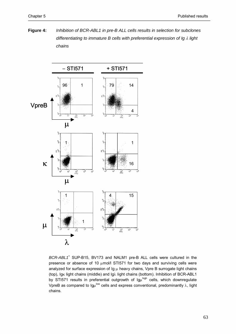
Chapter 5 Published results
63
μ
μ
μ
λ
κ
VpreB
96 1 79 14
4
1
16
1
1
1
4 15
− STI571 + STI571
μ
μ
μ
λ
κ
VpreB
96 1 79 14
4
1
16
1
1
1
4 15
− STI571 + STI571
Figure 4: Inhibition of BCR-ABL1 in pre-B ALL cells results in selection for subclones
differentiating to immature B cells with preferential expression of Ig λ light
chains
BCR-ABL1+ SUP-B15, BV173 and NALM1 pre-B ALL cells were cultured in the presence or absence of 10 μmol/l STI571 for two days and surviving cells were analyzed for surface expression of Ig μ heavy chains, Vpre B surrogate light chains (top), Igκ light chains (middle) and Igλ light chains (bottom). Inhibition of BCR-ABL1 by STI571 results in preferential outgrowth of Igμhigh cells, which downregulate VpreB as compared to Igμlow cells and express conventional, predominantly λ, light chains.

Chapter 5 Published results
64
However, in the three BCR-ABL1+ leukemia cell lines tested, the pattern of light chain
expression was heavily biased in favor of λ-light chains (Figure 4, bottom) and only few κ-
expressing cells could be detected (Figure 4, middle) while Igκ+ cells in control stainings can
easily be identified by the antibody we used (not shown).
Extensive κ-deleting element rearrangement results in preferential λ-light chain expression
on STI571-surviving leukemia cells
Searching for a potential explanation for predominant λ-light chain expression on
differentiating leukemia cells surviving STI571-treatment, we investigated whether
rearrangement of the so-called κ deleting element (KDE) may contribute to reduced κ light
chain usage in STI571-treated leukemia cells. KDE rearrangement may lead to inactivation of
productively recombined Vκ-Jκ joints and hence represents another level at which Ig light
chain expression can be regulated (28).
We amplified specific DNA fragments for KDE-germline configuration, KDE
rearrangement to an RSS within the Jκ intron and KDE rearrangement to RSS-sites flanking
Vκ gene segments from three BCR-ABL1+ leukemia cells in the absence or presence of
STI571. While KDE was found in germline configuration in untreated leukemia cells,
STI571-mediated inhibition of BCR-ABL1 was associated with rearrangement of KDE to
Jκ intron RSS sites and –to lesser extent- to Vκ RSS sites (not shown). Thus, predominant
expression of λ light chains is consistent with extensive KDE rearrangement observed here. In
the absence of BCR-ABL1 kinase activity, differentiating leukemia cells seem to undergo
multiple rounds of rearrangement targeting both κ and λ light chain loci, which ultimately
leads to preferential λ light chain expression expression due to KDE-mediated deletion of
potentially productive Vκ-Jκ joints.
Ig μhigh leukemia cells surviving STI571-treatment restore expression of B cell-specific
transcription factors
Propensity of STI571-treated leukemia cells to differentiate into light chain-expressing
immature B cells suggests that these cells exhibit a more mature phenotype as compared to
untreated leukemia cells. Therefore, we tested whether STI571-treated differentiating
leukemia cells can restore expression of B cell-specific transcription factors, which were
downregulated in the untreated leukemia cells as compared to normal pre-B cells (SAGE
data; Figure 2). BCR-ABL1+ SUP-B15 pre-B ALL cells were cultured either in the presence

Chapter 5 Published results
65
Figure 5: Inhibition of BCR-ABL1 restores (pre-) B cell receptor responsiveness in pre-B
ALL cells
0 50 100 150 200 250 300
0
40
80
120
160
200
0
40
80
120
160
200
0 50 100 150 200 250 300
0
0 50 100 150 200 250 300
0
40
80
120
160
200
antibody
anti-μ
anti-μ
anti-κ, anti-λ
anti-VpreB
Bone marrow CD19+ B lymphoid cells
BCR-ABL1+ pre-B ALL cells, untreated
BCR-ABL1+ pre-B ALL cells, + 10 μM STI571
MACS: μ+
MACS: μ-
Ca2+
i [nm
ol/l]
Ca2+
i [nm
ol/l]
Ca2+
i [nm
ol/l]
A
B
C
Time [sec]
Time [sec]
Time [sec]
0 50 100 150 200 250 300
0
40
80
120
160
200
0
40
80
120
160
200
0 50 100 150 200 250 300
0
0 50 100 150 200 250 300
0
40
80
120
160
200
antibody
anti-μanti-μ
anti-μanti-μ
anti-κ, anti-λ
anti-VpreB
Bone marrow CD19+ B lymphoid cells
BCR-ABL1+ pre-B ALL cells, untreated
BCR-ABL1+ pre-B ALL cells, + 10 μM STI571
MACS: μ+
MACS: μ-
Ca2+
i [nm
ol/l]
Ca2+
i [nm
ol/l]
Ca2+
i [nm
ol/l]
A
B
C
Time [sec]
Time [sec]
Time [sec]
CD19+ B lineage cells were purified from bone marrow mononuclear cells using immunomagnetic beads. Release of Ca2+ from cytoplasmic stores in these cells in response to pre-B or B cell receptor engagement by anti-VpreB or a mixture of anti-κand anti-λ chain antibodies (arrows) was measured using a laser scanning microscope (A). Untreated BCR-ABL1+ SUP-B15 (B), BV173 and NALM1 (not shown) pre-B ALL cells and BCR-ABL1+ pre-B ALL cells incubated with 10μmol/l STI571 for 24 hours (C) were stimulated using an anti-μ chain antibody, which can crosslink both pre-B and B cell receptors. Among STI571-treated leukemia cells, Igμ+ cells were separated from Igµ− cells and analyzed independently (C). In (B), 5-fold higher concentrations of the anti-μ antibody than in (A) and (C) were used. The data shown here refer to SUP-B15 cells. Analyzing BV173 and NALM1 cells, similar results were obtained (not shown).

Chapter 5 Published results
66
or absence of STI571. Among STI571-treated leukemia cells, Ig μhigh and Ig μ− cells were
separated by MACS using immunmagnetic beads. From normal pre-B cells, untreated ALL
cells, STI571-treated Ig μhigh and STI571-treated Ig μ− cells, cDNAs were synthesized and
subjected to semiquantitative RT-PCR analysis for mRNA levels of B cell-specific
transcription factors (E2A, EBF, OBF1, OCT2, IRF4 and PAX5) and the stem cell-related
transcription factors AML1 and GATA1 (Figure 2). The expression patterns of these
transcription factors in STI571-treated differentiating Ig μhigh and normal pre-B cells were
similar indicating that STI571-treated Ig μhigh leukemia cells indeed reversed the de-
differentiated stem cell-like phenotype seen in untreated leukemia cells and STI571-treated Ig
μ− leukemia cells (Figure 2).
Ablation of BCR-ABL1 kinase activity corrects expression of truncated SLP65 in pre-B ALL
cells from STI571-treated patients
An earlier study demonstrated that in the human, functional SLP65 is required for the
expression of a pre-B cell receptor: B cell precursors in a patient carrying a deleterious
mutation of the SLP65 gene are arrested at the pro-B cell stage of development (29). In mice,
SLP65-deficiency does not prevent expression of a pre-B cell receptor but results in a partial
differentiation block at the pre-B cell stage (7). Of note, in about 50 percent of childhood pre-
B ALL cases, SLP65 expression is defective, which results in compromised pre-B cell
receptor signaling (8). In these cases, either no SLP65 was expressed or an aberrant splice
variant including one or two additional exons inserted between SLP65 exons III and IV (8).
Inclusion of additional exons, termed IIIa and IIIb introduced a premature translation stop
resulting in loss of the C-terminus of SLP65 (SLP65 ΔC). The C-terminus of SLP65
encompasses tyrosine 96, whose phosphorylation is critical for SLP65 activity in mice (8).
Therefore, we tested whether SLP65 expression is deranged in BCR-ABL1+ pre-B ALL cells
and whether SLP65 deficiency is linked to the BCR-ABL1 kinase activity. To this end, we
analyzed SLP65 isoform expression in primary pre-B cells from the bone marrow of four
healthy donors and STI571-treated and untreated BCR-ABL1+ pre-B ALL cells (Figure 6A).
While normal pre-B cells expressed high levels of the canonical SLP65 isoform and only very
little alternatively spliced SLP65 ΔC, this pattern was almost inverted in BCR-ABL1+ pre-B
ALL cells.

Chapter 5 Published results
67
Figure 6: Expression of SLP65 isoforms in BCR-ABL1+ pre-B ALL cells
SLP65 SLP65 ΔC GAPDH
Pre-B BCR-ABL1+ pre-B ALL ─ ─ + + STI571
CD10+ ─ μ─ μ+ MACS/ FACS CD19+ μ+
SLP65 SLP65 ΔC GAPDH
XIII XIV XV XVI XVII XVIII XIX ALL cases
─ + ─ + ─ + ─ + ─ + ─ + ─ + STI571
A
B
SLP65
SLP65 ΔC GAPDH
Pro-B Pre-B Memory B CD19+ CD10+ CD19+ μ- VpreB- CD19+ μ+ CD27+
C
55 48 91 42 n.d. 72 90 54 64 n.d. 60 64 CD10a 97 2 98 8 n.d. 73 97 46 82 n.d. 90 81 CD34 a
Note: a. Percentage of CD19+ leukemia cells coexpressing CD10 or CD34

Chapter 5 Published results
68
Legend to Figure 6:
SLP65 cDNA fragments were amplified from normal human CD19+ CD34+ Igµ− pro-B, CD10+ CD19+ Igμlow pre-B and CD10− CD19+ CD27+ Igμhigh memory B cells (C) and BCR-ABL1+ ALL cells, including a BCR-ABL1+ cell line in the presence or absence of STI571 (A) and enriched ALL cells from seven leukemia patients (cases XIII to XIX, Table I) before and after treatment with STI571 (B). cDNA amounts were normalized using GAPDH as a standard (A-C). The primers used amplified full-length SLP65 and an isoform encoding truncated SLP65 (SLP65 ΔC). For leukemia cases XIII, XIV, XVI, XVII and XIX described in (B), FACS data on coexpression of CD10 and/ or CD34 on leukemic CD19+ cells was available before and after STI571-treatment (B, lower panel).
Also, when BCR-ABL1+ pre-B ALL cells were incubated in the presence of STI571 for 48
hours, the leukemia cells expressed the SLP65 ΔC transcript, although at lower levels. Given
that short-term treatment with STI571 partially restored (pre-) B cell receptor responsiveness
(Figure 5C) and induced selection for differentiating leukemia cells expressing high levels of
Ig μ heavy chains (Figure 3), we sorted Ig μ+ leukemia cells. STI571-treated leukemia cells,
which have recovered (pre-) B cell receptor expression also regained normal SLP65
expression (Figure 6A) in parallel with restored pre-B cell receptor responsiveness (Figure
5C). In humans, deleterious mutation of the SLP65 gene prevents expression and function of
the pre-B cell receptor on mutant B cell precursors (29). However, inactivating mutations
within pre-B cell receptor-related genes (including IGHCμ, Igα, Igβ, VpreB and λ5) do not
interfere with SLP65 expression (29). Therefore, deranged SLP65 expression is likely the
cause rather than the consequence of compromised pre-B cell receptor signaling in BCR-
ABL1+ pre-B ALL cells.
To determine whether deranged SLP65 expression is linked to BCR-ABL1 kinase
activity also in primary BCR-ABL1+ pre-B ALL leukemia cells, we analyzed the pattern of
SLP65 isoform expression in enriched leukemia cells from seven patients before and after
treatment with STI571. Comparing matched pairs of leukemia samples, we identified three
patients, in which the leukemia cells expressed SLP65 only at low levels, if at all (cases XVI,
XVII and XIX; Figure 6B). In these cases, treatment by STI571 had no effect on SLP65
isoform expression. Given that the leukemia samples had a purity of about 80 percent, weak
SLP65 expression in these cases can be attributed to few contaminating normal bystander B
cells. In four cases, expression of the canonical SLP65 isoform encoding the full-length
protein was accompanied by the expression of SLP65 ΔC. Treatment with STI571 not only
increased mRNA levels of full-length SLP65 but also abolished expression of the truncated
splice variant (Figure 6B).
Given that STI571-treatment of the BCR-ABL1+ ALL cell lines BV173, NALM1 and
SUP-B15 resulted in the preferential outgrowth of more differentiated subclones (Figures 2

Chapter 5 Published results
69
and 3), which further differentiate into Ig μhigh light chain-expressing immature B cells
(Figure 4), we wondered whether also in patients the BCR-ABL1+ leukemia cells surviving
STI571-treatment tend to exhibit a more mature phenotype. Therefore, we analyzed primary
B lymphoid bone marrow cells from seven leukemia patients before and after STI571-
treatment by flow cytometry and calculated the percentage of CD19+ cells, which coexpress
CD34 and/ or CD10. In the human, CD19+ CD34+ cells are considered as pro-B cells, CD19+
CD10+ CD34− cells include pre-B cells and immature B cells, whereas mature B cell subsets
in human bone marrow and the peripheral blood typically express CD19 but neither CD10 nor
CD34 (12). Based on this rough classification, between 46 and 98 percent of BCR-ABL1+
leukemia cells coexpress CD34 and thus exhibit a pro-B cell-like phenotype (Figure 6B).
After STI571-treatment, the percentage of cells displaying a pro-B cell-like phenotype
remains high in three cases (XVI, XVII and XIX). In two other cases, however, the fraction of
pro-B cell-like leukemia cells drops to 2 and 8 percent (cases XIII and XIV; Figure 6B, lower
panel). The remaining cells show a more mature phenotype resembling pre-B or immature B
cells. Of note, STI571-induced conversion of primary leukemia cases from a pro-B cell-like
to a pre-B cell-/ immature B cell-like phenotype correlates with therapy-related correction of
SLP65 isoform expression in these cases (Figure 6B). Three cases, in which STI571-treatment
had no effect on SLP65 mRNA expression, also did not convert to a more differentiated
phenotype in response to STI571-therapy (cases XVI, XVII and XIX; Figure 6B). From two
cases, no flow cytometry data was available. SLP65 ΔC is nonfunctional and by competing
with full-length SLP65 for binding to upstream kinases (e.g. SYK), it may have an inhibitory
effect on signal transduction initiated from the pre-B cell receptor in pre-B ALL cells. While
the negative regulatory effect of truncated SLP65 is not fully elucidated, our results indicate
that its expression is linked to BCR-ABL1 kinase activity.
A potential link between SLP65 deficiency, secondary VH gene rearrangement and loss of
coding capacity for a μ heavy chain in BCR-ABL1+ pre-B ALL cells
SLP65 also functions as a sensor for the expression of a productive VH region gene within a
pre-B cell receptor complex and then halts the recombination machinery to prevent further,
potentially inactivating, rearrangement of IGH gene segments (1). In the absence of functional
SLP65, the recombination machinery remains active and generates secondary VH region gene
rearrangements even if V, D and J segments have been productively rearranged (9). Assuming
that SLP65 deficiency is a common feature of BCR-ABL1+ ALL, one would predict that in
most if not all cases the leukemia clone has undergone one or more rounds of secondary VH
gene rearrangement. Such secondary rearrangements can occur by replacement of an already

Chapter 5 Published results
70
rearranged VH gene segment by another (in most cases upstream) VH gene segment (22). VH
gene replacement typically uses a cryptic recombination signal sequence (cRSS), which is
present in the 3’ part of 40 out of 44 functional VH gene segments (22). A hallmark for VH
gene replacement are short sequences (5 to 7 bp) within the VH-DH junction that are foreign to
the actually rearranged VH gene segment but are matching exactly the 3’ end adjacent to the
cRSS of another previously rearranged VH gene. It is very unlikely that 5 to 7 bp stretches
matching the extreme 3’ part of another VH gene within the VH-DH joint can be randomly
generated as a byproduct of junctional diversification (p=0.009; reference 22). Therefore,
these short sequences are considered as vestiges of a previously rearranged VH gene segment.
Analysing the IGH VDJ-rearrangements for footprints of secondary rearrangements, we
found indication of VH-gene replacement in 9 of 12 leukemia cases (Table III). In eight of
nine cases, the potential secondary rearrangement uses a donor VH segment located upstream
from the initially rearranged (recipient) VH-gene segment (Table III). One potential VH-
replacement might reflect an inversion or transrecombination event as described earlier (22).
A comprehensive analysis of IGH VDJ-rearrangements amplified from normal human
immature B cells identified traces of VH-gene replacement in 16 out of 343 sequences (5%;
reference 22). Hence, the frequency of VH-gene replacement in BCR-ABL1+ ALL cases is, at
about 75%, surprisingly high.
Therefore, defective SLP65 expression (Figure 6) is associated with and potentially the
reason for secondary, mostly nonproductive, VH gene rearrangements in 9 of the 12 leukemia
cases we analyzed (Table II). In agreement with a pre-B cell origin of BCR-ABL1+ ALL one
might envision that the precursor of the leukemia clone underwent malignant transformation
after having passed the pre-B cell receptor checkpoint but subsequently lost coding capacity
of an initially productive Ig VH region gene by a deleterious secondary VH gene
rearrangement.
Assuming that BCR-ABL1 can relieve the selection pressure for survival signals
through the pre-B cell receptor, we next tested whether truncated SLP65 is also expressed in
normal pro-B cells, in which selection for the expression of a functional pre-B cell receptor is
not operative. While CD19+ Ig μ chain− VpreB− pro-B cells express full-length SLP65 at
similar levels as in CD19+ Ig μ chain+ Vpre-B+ pre-B cells and CD19+ CD27+ memory B
cells, SLP65 ΔC is only expressed in pro-B cells (Figure 6C). As SLP65 isoform expression is
similar in BCR-ABL1+ pre-B ALL and normal human CD19+ Igμ¯ VpreB¯ pro-B cells, we
conclude that SLP65 ΔC is not a marker for B lineage leukemia cells but rather for B
lymphoid cells that are not selected for the expression of an active (pre-) B cell receptor.

Chapter 5 Published results
71
Tabl
e III
: In
dica
tion
of p
oten
tial V
H-g
ene
repl
acem
ent i
n V
H re
gion
gen
es o
f BC
R-A
BL1
+ pre
-B A
LL c
lone
s
Cas
e
3’ p
art o
f rec
ipie
nt V
H
3’
par
t of d
onor
VH
V H
-DH
DH
I
VH1.
8 CGTGTAT
TA
CT
GT
GCGAGAGG
V
H4.
34
GTGTAT
TA
CT
GT
GCG
AGAGGGGGGTA
GGGAATAGCA
D
H6.
13-J
H5
II V
H1.
58
CGTGTAT
TA
CT
GT
GCGGCAGA
V
H4.
61
GTGTAT
TA
CT
GT
GCGAGAGA
CACGGCAGATG
TATTGTAGTAGTACCCCC
DH2.
2-J H
4
V
VH2.
5 CACATAT
TA
CT
GT
GCACACAGACC
V
H5.
51
ATGTAT
TA
CT
GT
GCG
AGACACACAGT
GAGAGAAACCAGCC
DH1.
26-J
H1
VI
VH1.
2a CGTGTAT
TA
CT
GT
GCGAGAGA
V
H3.
30
GTGTAT
TA
CT
GT
GCG
CGAGAGATCCCCCTTC
TATTACTATGA
D
H3.
22-J
H3
VII
VH2.
5 CACATAT
TA
CT
GT
GCACACAGACC
V
H1.
46
GTGTAT
TA
CT
GT
GCGAG
AGACCGCT
GGGTATAGCAGCAGCTG
DH6.
13-J
H4
VIII
VH2.
70b
CACATAT
TA
CT
GT
GCACACAGAC
V
H2.
5 ACATAT
TA
CT
GT
GCA
CACAGCCCCGGGATCCC
CGGGATAGTAGTG
DH3.
22-J
H4
IX
VH1.
24
CGTGTAT
TA
CT
GT
GCAACAGA
V
H1.
46
GTGTAT
TA
CT
GT
GCGAGAGA
CCAACAG
ACCGTGGTTCGGGGA
DH3.
10-J
H5
X
VH3.
38
CGTGTAT
TA
CT
GT
GCCAGATATA
c
VH3.
53
GTGTAT
TA
CT
GT
GCGAGA
GTTGCCAGGGGG
TGGTGTATGCTATACC
DH2.
8-J H
6
XI
VH3.
38
CGTGTAT
TA
CT
GT
GCCAGATATA
V
H3.
48
GTGTAT
TA
CT
GT
GGCGA
GCCAGATATTGT
AGTGGTGGTAGCT
DH2.
15-J
H3
Not
es:
a: T
he fo
otpr
int o
f thi
s po
tent
ial V
H-r
epla
cem
ent m
ay b
e al
so d
eriv
ed fr
om V
H1.
3, V
H1.
18, V
H3.
7, V
H3.
11, V
H3.
20, V
H3.
21 a
nd V
H4.
4 ge
ne s
egm
ents
b:
Lik
ely
gene
rate
d by
inve
rsio
n or
tran
srec
ombi
natio
n ev
ents
c:
The
foot
prin
t of V
H-r
epla
cem
ent i
n th
is c
ase
also
invo
lves
the
last
two
nucl
eotid
es o
f the
cR
SS m
otif
bo
ld: c
RSS
mot
ifs; u
nder
lined
: foo
tprin
ts o
f rec
ipie
nt V
H-g
ene
segm
ents

Chapter 5 Published results
72
Perspective
A broad range of pre-B cell receptor-related signaling molecules and transcription factors is
transcriptionally silenced in BCR-ABL1+ pre-B ALL as compared to human pre-B cells. Loss
of (pre-) B cell receptor-related molecules as observed by SAGE (Figures 1 and 2) may reflect
that the BCR-ABL1 kinase may have relieved the selection pressure for the expression of
these molecules. In the presence of autonomous BCR-ABL1 kinase activity, pre-B ALL cells
become nonresponsive to (pre-) B cell receptor engagement (Figure 5). As a consequence,
molecules needed for transduction of survival signals through the pre-B cell receptor,
disappear from the gene expression program of the leukemic cells (SAGE, Figure 1).
Conversely, inhibition of BCR-ABL1 reconstitutes dependence on survival signals initiated
from surface receptors including the (pre-) B cell receptor (Figure 3). It is even conceivable
that default signaling through the (pre-) B cell receptor represents a mechanism to acquire
drug resistance that is available to BCR-ABL1+ pre-B ALL cells but not to other malignant
cells carrying a BCR-ABL1 gene rearrangement, e.g. chronic myeloid leukemia (CML) cells.
In this regard, it is interesting to note that treatment failure of STI571 is frequent in pre-B
ALL but not in CML harboring a BCR-ABL1 fusion gene (30). Moreover, STI571-resistant as
opposed to STI571-sensitive BCR-ABL1+ pre-B ALL cells exhibit high expression levels of
Bruton’s tyrosine kinase (BTK), which represents a critical component of (pre-) B cell
receptor signal transduction cascade (15). Therefore, resumption of (pre-) B cell receptor
signaling may represent a mechanism to escape immediate apoptosis induced by STI571 and
open a time frame, during which secondary transforming events that confer permanent
STI571-resistance (e.g. mutations within the ATP-binding site of the ABL1 kinase domain)
can occur.
Acknowledgements: We would like to thank Klaus Rajewsky, Janet D. Rowley, Matthias Fischer, Hassan Jumaa and Michael Reth for critical discussions, Ines Schwering and Ralf Küppers for sharing SAGE-data, Janet D. Rowley and Martin Krönke for continuous support and discussions and Jana L. Mooster for critically reading the manuscript and Stefanie Jauch, Jana L. Mooster and Peter Wurst for excellent technical assistance. F.K. is supported by scholarships of the Studienstiftung des deutschen Volkes and the Köln Fortune program of the Faculty of Medicine of the University of Cologne. H.W. is supported by the DAAD Bioscience program. M.M. is supported by the Deutsche Forschungsgemeinschaft through the Emmy-Noether-Programm. This work was supported by the Deutsche Forschungsgemeinschaft through grants MU1616/2-1 and MU1616/3-1 (to M.M.), the German José-Carreras-Leukemia-Foundation (grant to M.M.), the Deutsche Krebshilfe 10-1643-Si1 (grant to R.S.) and the Ministry of Science and Research for North Rhine-Westphalia through the Stem Cell Network NRW (to M.M.).

Chapter 5 Published results
73
Materials and Methods
Patient samples and cell purification
In total, 19 cases of BCR-ABL1+ pre-B ALL were studied. For cases I to IX (Table I), BCR-
ABL1+ pre-B ALL cells obtained from bone marrow samples of untreated patients had a
purity of >95% as assessed by bone marrow morphology. Cases X, XI and XII (Table I)
correspond to BCR-ABL1+ pre-B ALL cell lines, termed SUP-B15, BV173 and NALM1
(DSMZ, Braunschweig, Germany), respectively. Purification of ALL cells from cases XIII to
XIX (Table I) was described earlier (15). In these cases, SLP65 isoform expression before and
after treatment with the BCR-ABL1 inhibitor STI571 and expression of CD10, CD19 and
CD34 was studied (Figure 4; cases XIII to XIX; Table I). For nine primary cases (including
two cases analyzed by SAGE, cases II and IX) and three cell lines, the configuration of IGH
loci was examined (cases I to XII in Table II). For all cases, breakpoints within the major or
minor breakpoint cluster region of the BCR gene, leading to p210 and p190 fusion molecules,
respectively, were detected by PCR using primers and PCR conditions as previously
described (16). Clinical data for all 19 cases are given in Table I. Normal hematopoietic cell
populations including CD34+ CD38low CD133+ Lin− hematopoietic progenitor cells (HSC),
CD15+ myeloid progenitor cells (CMP), CD7+ CD10+ T lymphoid precursors (TLP), CD19+
VpreB− Igμ chain− pro-B cells (no SAGE-profile available) and CD10+ CD19+ Igμ chain+
pre-B cells (pre-B) were enriched from human bone marrow by depletion of unwanted cells
and enrichment of the populations of interest using immunomagnetic beads and cell sorting.
Mature B cell populations including CD19+ CD27− naïve B cells (NBC), CD19+ CD27+
memory B cells (MBC) and CD19+ CD138+ plasma cells (PC) were from pooled peripheral
blood samples of 12 healthy donors. CD20+ CD77+ germinal center B cells (GCB) were
isolated from human tonsillectomy resectates. HSC, CMP, TLP, pre-B cells, naïve B cells,
memory B cells and plasma cells were subjected to SAGE-analysis as described earlier (17-
19). SAGE data on germinal center B cells were kindly provided by Ines Schwering and Ralf
Küppers (University of Cologne, Cologne, Germany and University of Essen, Essen,
Germany).
SAGE analysis
cDNA-synthesis, SAGE analysis, cloning and sequencing of SAGE concatemers was carried
out as previously described (17-19). The UniGene reference data base (March 2001) was
obtained at http://www.sagenet.org/SAGEDatabases/unigene.htm. A total of 592,000 SAGE
tags were collected for 10 SAGE profiles. 106,000 tags were analyzed from the HSC library,
99,000 for common myeloid progenitor cells, 110,000 for pre-B cells, 96,500 for T lymphoid
Chapter 5 Published results
74
precursors and each about 30,000 tags for two cases of bone marrow-derived pre-B ALL
carrying a BCR-ABL1 gene rearrangement, naïve B cells, germinal center B cells, memory B
cells and plasma cells. All SAGE libraries were normalized to 100,000 tags. SAGE data were
graphically visualized using the Cluster and Treeview software (http://rana.lbl.gov/) and
sorted according to the ratio between SAGE-tag counts in pre-B cells and in BCR-ABL1+ ALL
cases. In a comprehensive search for pre-B cell receptor components and pre-B cell receptor-
related signaling molecules in PubMed, UniGene (http://www.ncbi.nlm.nih.gov/UniGene/)
and OMIM (http://www.ncbi.nlm.nih.gov/entrez/query.fcgi?db=OMIM), we identified 54
genes in at least one of the SAGE-libraries analyzed, for which a role in the assembly or
regulation of the pre-B cell receptor was shown.
Semiquantitative RT-PCR analysis and verification of quantitative accuracy of SAGE data
In order to corroborate quantitative differences in gene expression as determined by SAGE,
semiquantitative RT-PCR analysis was performed for a set of 45 selected genes differentially
expressed between HSC and pre-B cells and 41 genes differentially expressed between naïve
and memory B cells as previously described (17, 18).
To verify differential gene expression observed by SAGE-analysis of pre-B cells and the
BCR-ABL1+ pre-B ALL cell line SUP-B15, we performed semiquantitative RT-PCR using 5’-
TTAGCACCCCTGGCCAAG-3’ and 5’-CTTACTCCTTGGAGGCCATG-3’ for GAPDH
(for standardization), 5’-AATGATGAAAACTACTCGGCT-3’ and 5’-
TTGGTCTGATCATCTAGTTTC-3’ for AML1, 5’-AGTCTTTCAGGTGTACCCAT-3’ and
5’-AAAGAAGGTACTGGAAAAGTC-3’ for GATA1, 5’-
AAGGAAAAAGAAGCCAACAG-3’ and 5’-GGACAAACATGTTATCAGGA-3’ for EBF,
5’-CTGAAAGCAAGCAACAAAAC-3’ and 5’-CACGGGGTCTTTTTAATACA-3’ for E2A,
5’-CCATGTAAATACCTTCTTGC-3’ and 5’-ATTCTGCTTCGGAAAAGTAG-3’ for
PAX5, 5’-CAAAATAAGACCTCCCCATT-3’ and 5’-TGAGGTAGCTGGAATAGATT-3’
for OCT2, 5’-GGCTTCAAAGAGAAAAGGCA-3’ and 5’-TCTGTCGTGACATTGGTGAT-
3’ for OBF1 and 5’-CAAGAGCAATGACTTTGAGG-3’ and 5’-
TGGGACATTGGTACGGGAT-3’ for IRF4. The same primer sets were also used for RT-
PCR analysis of STI571-treated SUP-B15 ALL cells after MACS-enrichment of μ+ and μ−
subclones. In addition, quantitative differences between the SAGE-libraries for pre-B cells
and BCR-ABL1+ pre-B ALL cases were verified at the protein level for ten surface molecules
by flow cytometry using antibodies against CD19, IL7Rα, CD10, CD38, CD40, CD150/
SLAM, CD72, VpreB, Igα and CD34 (BD Biosciences, Heidelberg, Germany; data not
shown).

Chapter 5 Published results
75
Sequence analysis of IGH loci
In order to characterize the configuration of the IGH loci in nine primary BCR-ABL1+ pre-B
ALL cases (cases I to IX, Table II) and three cell lines (SUP-B15, BV173 and NALM1; cases
X to XII, Table II), three primer sets were used to amplify germline configuration, D-J and V-
DJ gene rearrangements from both alleles of the IGH locus. Clonal VH gene rearrangements
were amplified in a single round of PCR amplification using 35 PCR cycles at a detection
limit of about 104 clonal cells in 5 x 10⁶ total cells. PCR primers and conditions were used as
previously described (20) and amplification products were directly sequenced and aligned
according to Matsuda et al. (21) using the IMGT software at
http://www.dnaplot.de/input/human_v.html. To identify footprints from formerly rearranged
VH gene segments within potential secondary VDJ-rearrangements, a list of VH and DH
germline gene segments was obtained at http://imgt.cines.fr and cryptic recombination signal
sequences (cRSS) were identified as previously described (22).
Measurement of pre-B cell receptor responsiveness
Primary human CD19+ bone marrow mononuclear cells were enriched from bone marrow
samples of four healthy donors using immunomagnetic MACS beads as previously described
(17). SUP-B15, BV173 and NALM1 pre-B ALL cells (DMSZ, Braunschweig, Germany)
carrying a BCR-ABL1 gene rearrangement (Table II, cases X, XI and XII) were cultured with
20% fetal calf serum in RPMI 1640 medium in the presence or absence of 10 μM STI571
(Novartis, Basel, Switzerland) for the times indicated. Among STI571-treated leukemia cells,
Igμ+ and Igμ− cells were separated using a non-stimulating, biotin-conjugated anti-µ antibody
(BD Biosciences) together with streptavidin-microbeads (Miltenyi Biotech). After MACS-
enrichment or pre-incubation, cells were washed and stained with Fluo-3 dye (Calbiochem,
Bad Soden, Germany) for 30 minutes. Changes of cytosolic Ca2+ were measured by laser
scans using confocal microscopy (18, 19). After 10 to 30 seconds of measurement, antibodies
against human μ chains (Jackson ImmunoResearch), VpreB (BD Biosciences) or Igκ-
together with Igλ-light chains (BD Biosciences) were added to CD19+ bone marrow
mononuclear cells or preincubated pre-B ALL cells (in the presence or absence of STI571;
cases X to XII). Cytosolic Ca2+ concentrations were determined as previously described (18).
As a negative control, cells were also treated with an antibody against CD3 (BD Bioscience),
which induces Ca2+ mobilization in T but not B lineage cells.
Flow cytometry
Surface expression of VpreB surrogate- or Igκ- or Igλ-light chains, Ig μ-chains and CD19 on
BV173, NALM1, and SUP-B15 pre-B ALL cells in the presence or absence of 10 μM STI571

Chapter 5 Published results
76
was monitored using antibodies against VpreB, Igκ- or Igλ-light chains, the Ig μ heavy chain
and CD19 (BD Biosciences, Heidelberg, Germany) after the incubation times indicated.
Apoptotic or dead cells were identified by staining with anti-Annexin V antibodies and
propidium iodide (BD Biosciences).
RT-PCR analysis of SLP65 isoform expression
cDNA amounts were normalized by OD measurements and amplification of a specific
fragment of the GAPDH gene using 5’-TTAGCACCCCTGGCCAAGG-3’ as forward and 5’-
CTTACTCCTTGGAGGCCATG-3’ as reverse primers. For amplification of SLP65 isoforms,
forward primers specific for exon I (5’-TGGACAGTTATTCGTGTCTCTT-3’), and
additional exon IIIb (5’-AGAGTGTGTTGACCTTGGTG-3’) were used together with a
reverse primer specific for additional exon IIIb (5’-TTGGCTTAGAGGGTTTTGG-3’) and
exon VII (5’-GTGAACTGCTTTCTGTGGGA-3’) as described earlier (8). For amplification
of another additional exon IIIa, the forward primer 5’-TTTAATCTCTCCTGGAATGCAG-3’
was used. However, a splice variant including exon IIIa (8) could not be amplified with this
primer (not shown). cDNAs derived from human pre-B cells, BCR-ABL1+ pre-B ALL cells in
the presence or absence of STI571 and MACS-sorted μ-chain+ and μ-chain- BCR-ABL1+ pre-
B ALL cells after treatment with 10 μM STI571, were used as RT-PCR templates.

Chapter 5 Published results
77
References
1. Schebesta, M., P.L. Pfeffer, and M. Busslinger. 2002. Control of pre-BCR signaling by Pax5-
dependent activation of the BLNK gene. Immunity 17: 473-485.
2. Hardy, R.R., C.E. Carmack, S.A. Shinton, J.D. Kemp, and K. Hayakawa. 1991. Resolution
and characterization of pro-B and pre-pro-B cell stages in normal mouse bone marrow. J. Exp.
Med. 173: 1213-1225.
3. Rajewsky, K. 1996. Clonal selection and learning in the antibody system. Nature 381: 751-
758.
4. Thevenin, C., S.L. Nutt, and M. Busslinger. 1998. Early function of PAX5 (BSAP) before the
pre-B cell receptor stage of B lymphopoiesis. J. Exp. Med. 188: 735-744.
5. Nussenzweig, M.C. 1998. Immune receptor editing: revise and select. Cell 95: 875-878.
6. Saijo, K., C. Schmedt, I.H. Su, H. Karasuyama, C.A. Lowell, M. Reth, T. Adachi, A. Patke, A.
Santana, and A. Tarakhovsky. 2003. Essential role of Src-family protein tyrosine kinases in
NF-κB activation during B cell development. Nat. Immunol. 4: 274-279.
7. Jumaa, H., L. Bossaller, K. Portugal, B. Storch, M. Lotz, A. Flemming, V. Postila, P.
Riikonen, J. Pelkonen, C.M. Niemeyer, and M. Reth. 2003. Deficiency of the adaptor SLP65
in pre-B-cell acute lymphoblastic leukaemia. Nature 423: 452-456.
8. Jumaa, H., M. Mitterer, M. Reth, and P.J. Nielsen. 2001. The absence of SLP65 and BTK
blocks B cell development at the preB cell receptor-positive stage. Eur. J. Immunol. 31: 2164-
2169.
9. Flemming, A., T. Brummer, M. Reth, and H. Jumaa. 2003. The adaptor protein SLP65 acts as
a tumor suppressor that limits pre-B cell expansion. Nat. Immunol. 4: 38-43
10. Look, A.T. 1997. Oncogenic transcription factors in the human acute leukemias. Science 278:
1059-1064.
11. Rowley, J.D. 1973. A new consistent chromosomal abnormality in chronic myelogenous
leukaemia identified by quinacrine fluorescence and Giemsa staining. Nature 243: 290-293.
12. LeBien, T.W. 2000. Fates of human B-cell precursors. Blood 96: 9-23.
13. Yeoh, E.J., M.E. Ross, S.A. Shurtleff, W.K. Williams, D. Patel, R. Mahfouz, F.G. Behm, S.C.
Raimondi, M.V. Relling, A. Patel, C. Cheng, D. Campana, D. Wilkins, X. Zhou, J. Li, H. Liu ,
C.H. Pui, W.E. Evans, C. Naeve, L. Wong, and J.R. Downing. 2002. Classification, subtype
discovery, and prediction of outcome in pediatric acute lymphoblastic leukemia by gene
expression profiling. Cancer Cell 1: 133-143.
14. Armstrong, S.A., J.E. Staunton, L.B. Silverman, R. Pieters, M.L. den Boer, M.D. Minden,
S.E. Sallan, E.S. Lander, T.R. Golub, and S.J. Korsmeyer. 2002. MLL translocations specify a
distinct gene expression profile that distinguishes a unique leukemia. Nat. Genet. 30: 41-47.
15. Hofmann, W.K., S. de Vos, D. Elashoff, H. Gschaidmeier, D. Hoelzer, H.P. Koeffler, and
O.G. Ottmann. 2002. Relation between resistance of Philadelphia-chromosome-positive acute

Chapter 5 Published results
78
lymphoblastic leukaemia to the tyrosine kinase inhibitor STI571 and gene-expression profiles:
a gene-expression study. Lancet 359: 481-486.
16. Radich, J., G. Gehly, A. Lee, R. Avery, E. Bryant, S. Edmands, T. Gooley, P. Kessler, J. Kirk,
P. Ladne, E.D. Thomas, and F.R. Appelbaum. 1997. Detection of BCR-ABL transcripts in
Philadelphia chromosome-positive acute lymphoblastic leukemia after marrow
transplantation. Blood 89: 2602-2609.
17. Müschen, M., S. Lee, G. Zhou, N. Feldhahn, V.S. Barath, J. Chen, C. Moers, M. Krönke, J.D.
Rowley, and S.M. Wang. 2002. Molecular portraits of B cell lineage commitment. Proc. Natl.
Acad. Sci. U. S. A. 99: 10014-10019
18. Feldhahn, N., I. Schwering, S. Lee, M. Wartenberg, F. Klein, H. Wang, G. Zhou, S.M. Wang,
J.D. Rowley, J. Hescheler, M. Krönke, K. Rajewsky, R. Küppers R, and M. Müschen. 2002.
Silencing of B cell receptor signals in human naive B cells. J. Exp. Med. 196: 1291-1305.
19. Klein, F., N. Feldhahn, S. Lee, H. Wang, F. Ciuffi, M. von Elstermann, M.L. Toribio, H.
Sauer, M. Wartenberg, V.S. Barath, M. Krönke, P. Wernet, J.D. Rowley, M. Müschen. 2003.
T lymphoid differentiation in human bone marrow. Proc. Natl. Acad. Sci. U. S. A. 100: 6747-
6752.
20. Müschen, M., K. Rajewsky, A. Bräuninger, A.S. Baur, J.J. Oudejans, A. Roers, M.L.
Hansmann, and R. Küppers. 2000. Rare occurrence of classical Hodgkin's disease as a T cell
lymphoma. J. Exp. Med. 191: 387-394.
21. Matsuda, F., K. Ishii, P. Bourvagnet, K. Kuma, H. Hayashida, T. Miyata, and T. Honjo. 1998.
The complete nucleotide sequence of the human immunoglobulin heavy chain variable region
locus. J. Exp. Med. 188: 2151-2162.
22. Zhang, Z., M. Zemlin, Y.H. Wang, D. Munfus, L.E. Huye, H.W. Findley, S.L. Bridges, D.B.
Roth, P.D. Burrows, and M.D. Cooper. 2003. Contribution of VH gene replacement to the
primary B cell repertoire. Immunity 19: 21-31.
23. Reuther, J.Y., G.W. Reuther, D. Cortez, A.M. Pendergast, and A.S. Baldwin Jr. 1998. A
requirement for NF-κB activation in Bcr-Abl-mediated transformation. Genes Dev. 12: 968-
981.
24. Sattler, M., M.G. Mohi, Y.B. Pride, L.R. Quinnan, N.A. Malouf, K. Podar, F. Gesbert, H.
Iwasaki, S. Li, R.A. Van Etten, H. Gu, J.D. Griffin, and B.G. Neel. 2002. Critical role for
GAB2 in transformation by BCR/ABL. Cancer Cell 1: 479-492.
25. Li, S., A.D. Couvillon, B.B. Brasher, and R.A. Van Etten. 2001. Tyrosine phosphorylation of
GRB2 by Bcr/Abl and epidermal growth factor receptor: a novel regulatory mechanism for
tyrosine kinase signaling. E.M.B.O. J. 20: 6793-6804.
26. Danial, N.N., and P. Rothman. 2000. JAK-STAT signaling activated by Abl oncogenes.
Oncogene 19: 2523-2531.

Chapter 5 Published results
79
27. Muljo, S.A., and M.S. Schlissel. 2003. A small molecule Abl kinase inhibitor induces
differentiation of Abelson virus-transformed pre-B cell lines. Nat. Immunol. 4: 31-37.
28. Siminovitch, K.A., A. Bakhshi, P. Goldman, and S.J. Korsmeyer. 1985. A uniform deleting
element mediates the loss of kappa genes in human B cells. Nature 316: 260-262.
29. Minegishi, Y., J. Rohrer, E. Coustan-Smith, H.M. Lederman, R. Pappu, D. Campana, A.C.
Chan, and M.E. Conley. 1999. An essential role for BLNK in human B cell development.
Science 286: 1954-1957.
30. Druker, B.J., C.L. Sawyers, H. Kantarjian, D.J. Resta, S.F. Reese, J.M. Ford, R. Capdeville,
and M. Talpaz. 2001. Activity of a specific inhibitor of the BCR-ABL tyrosine kinase in the
blast crisis of chronic myeloid leukemia and acute lymphoblastic leukemia with the
Philadelphia chromosome. N. Engl. J. Med. 344: 1038-1042.

Chaper 5 Published results
80
5.2 BCR-ABL1 induces aberrant splicing of IKAROS and lineage infidelity
in pre-B lymphoblastic leukemia cells
published in Oncogene; 2005; 24: 1-7
By Florian Klein1, Niklas Feldhahn1, Sebastian Herzog2, Mieke Sprangers1,
Jana L. Mooster1, Hassan Jumaa2 and Markus Müschen1
1Laboratory for Molecular Stem Cell Biology, Heinrich-Heine-Universität Düsseldorf, Düsseldorf, Germany; 2Max-Planck-Institute for Immunobiology, Freiburg, Germany
Summary
Pre-B lymphoblastic leukemia cells carrying a BCR-ABL1 gene rearrangement
exhibit an undifferentiated phenotype. Comparing genome-wide gene
expression profiles of normal B cell subsets and BCR-ABL1+ pre-B
lymphoblastic leukemia cells by SAGE, the leukemia cells show loss of B
lymphoid identity and aberrant expression of myeloid lineage-specific
molecules. Consistent with this, BCR-ABL1+ pre-B lymphoblastic leukemia
cells exhibit defective expression of IKAROS, a transcription factor needed for
early lymphoid lineage commitment. As shown by inducible expression of
BCR-ABL1 in human and murine B cell precursor cell lines, BCR-ABL1
induces the expression of a dominant-negative IKAROS splice variant, termed
IK6. Comparing matched leukemia sample pairs from patients before and
during therapy with the BCR-ABL1 kinase inhibitor STI571 (Imatinib),
inhibition of BCR-ABL1 partially corrected aberrant expression of IK6 and
lineage-infidelity of the leukemia cells. To elucidate the contribution of IK6 to
lineage infidelity in BCR-ABL1+ cell lines, IK6-expression was silenced by
RNA interference. Upon inhibition of IK6, BCR-ABL1+ leukemia cells
partially restored B lymphoid lineage commitment. Therefore, we propose that
BCR-ABL1 induces aberrant splicing of IKAROS, which interferes with
lineage identity and differentiation of pre-B lymphoblastic leukemia cells.

Chaper 5 Published results
81
Introduction
The BCR-ABL1 gene rearrangement resulting from the t(9;22)(q34;q11) translocation
represents the most frequent recurrent genetic aberration in B lymphoid leukemia in adults
(Look, 1997). BCR-ABL1 fusion genes encode constitutively active tyrosine kinase molecules
mostly of a molecular weight of either 190 or 210 kD (p190 or p210, Laurent et al. 2001).
BCR-ABL1 kinase activity is required and sufficient to drive malignant transformation of B
cell precursors in mice (Huettner et al., 1998). Pre-B lymphoblastic leukemia cells carrying a
BCR-ABL1 gene rearrangement typically exhibit a differentiation block at the pre-B cell stage
of development (Klein et al., 2004; Klein et al., 2005).
Early B cell development is guided by a number of transcription factors including
PAX5, E2A, EBF and IKAROS (Busslinger, 2004). In order to search for downstream targets
of BCR-ABL1 that contribute to a differentiation block in BCR-ABL1+ pre-B lymphoblastic
leukemia cells, genome-wide gene expression profiles of this leukemia type were compared
with normal hematopoietic progenitor populations by serial analysis of gene expression
(SAGE; Velculescu et al., 1995).
Results and Diskussion
BCR-ABL1 interferes with B lymphoid differentiation in BCR-ABL1+ pre-B lymphoblastic
leukemia cells
Comparing genome-wide gene expression profiles of leukemia cells from two leukemia cases
of BCR-ABL1+ pre-B lymphoblastic leukemia with normal pre-B cells by SAGE, a number of
myeloid lineage-specific genes including transcription factors (MLF2, MZF1, AML1,
GATA1), surface receptors (CSF3R, CSF1R, CD14, CD11A) and signaling molecules (IRAK1,
MYD88) were upregulated in the pre-B lymphoblastic leukemia cells as in normal myeloid
progenitor cells (Figure 1). Hence, the pattern of gene expression in BCR-ABL1+ pre-B
lymphoblastic leukemia cells as compared to their normal pre-B cell counterpart is skewed to
the myeloid lineage. Consistent with this, transcription factors and signaling molecules
involved in early lymphoid differentiation (IKAROS, E2A, IL7Rα, RAG1, RAG2, TdT) and B
lymphoid lineage commitment (EBF, PAX5) were silenced in BCR-ABL1+ pre-B
lymphoblastic leukemia cells (Figure 1). These findings lead to the hypothesis that lymphoid
lineage commitment is impaired in BCR-ABL1+ pre-B lymphoblastic leukemia cells.
We next studied whether derangement of lymphoid lineage commitment and B cell
differentiation in the BCR-ABL1+ pre-B lymphoblastic leukemia cells are indeed owing to
BCR-ABL1 kinase activity. To this end, we induced BCR-ABL1 expression in a murine B
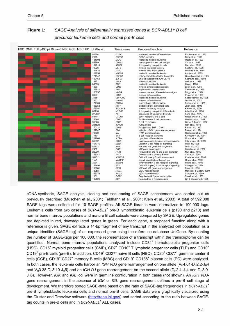
Chaper 5 Published results
82
Figure 1: SAGE-Analysis of differentially expressed genes in BCR-ABL1+ B cell
precursor leukemia cells and normal pre-B cells
81994 GYPC erythroid/ myeloid differentiation Robinson et al., 1981 2175 CSF3R GCSF-receptor Dong et al., 1996 181002 MSF1 related to myeloid leukemia Osaka et al., 199985289 CD133 hematopoietic stem cell antigen Yin et al., 1997 182018 IRAK1 IL1 signaling in myeloid cells Cao et al., 1996 79026 MLF2 myeloid leukemia factor 2 Kuefer et al., 1996 169832 MZF1 myeloid zinc finger gene 1 Hromas et al., 1991 112255 NUP98 related to myeloid leukemia Ahuja et al., 1999 174142 CSF1R colony-stimulating factor 1 receptor Gisselbrecht et al., 1987 460463 IL3Rα Shared subunit with GM-CSFR Kitamura et al., 19911817 MPO myeloperoxidase Weil et al., 198889633 PML related to myeloid leukemia Cleary, 19961239 CD13 myeloid differentiation antigen Look et al., 1986129914 AML1 implicated in myelopoiesis Tanaka et al., 1995153837 MNDA myeloid nuclear differentiation antigen Briggs et al., 199483731 CD33 myeloid differentiation Peiper et al., 198831551 CBFA2T2 related to myeloid leukemia Kitabayashi et al., 1998765 GATA1 myeloid differentiation Qian et al., 2002174103 CD11A macrophage differentiation Springer et al., 1985196352 NCF4 oxidative burst in myeloid cells Zhan et al., 1996279751 SIGLEC8 myeloid inhibitory receptor Kikly et al., 200082116 MYD88 IL1 signaling in myeloid differentiation Adachi et al., 1998272537 TdT generation of junctional diversity Kung et al., 197589414 CXCR4 SDF1 receptor, pre-B cells Nagasawa et al., 199625648 CD40 Proliferation of B cell precursors Hasbold et al., 199496023 CD19 B cell co-receptor Carter & Fearon, 1992285823 IGHCM IGH μ chain Raff et al., 1976170121 CD45 Antagonizes SHP1, CSK Pani et al., 1997101047 E2A Initiation of IGH gene rearrangement Bain et al., 199479630 Igα ITAM signaling chain Flaswinkel et al., 199574101 SYK B cell receptor signaling Kurosaki et al., 1994237868 IL7Rα Lymphoid differentiation Uckun et al., 199166052 CD38 Ligation causes tyrosine phosphorylation Kitanaka et al., 1999167746 BLNK Linker in B cell receptor signaling Fu et al., 199882132 IRF4 IGK and IGL gene rearrangement Lu et al., 20032407 OBF1 IGK gene transcription Casellas et al., 200222030 PAX5 Required for pro- to pre-B cell transition Nutt et al., 1999158341 TACI Growth control of early B cells Yan et al., 200154452 IKAROS Critical for early B cell development Kirstetter et al., 20021521 IgMBP2 Signal transduction through Igα Grupp et al., 19953631 IgαBP1 Involvement in B cell receptor signaling Fukita et al., 1993159494 BTK Critical for (pre-) B cell receptor signaling Cheng et al., 1994192861 SPIB IGK and IGL gene rearrangement Su et al., 199673958 RAG1 V(D)J recombination Menetski & Gellert, 1990159376 RAG2 V(D)J recombination Schatz et al., 19891101 OCT2 Regulates IGH gene transcription Staudt et al., 1988192824 EBF Required for B lymphopoiesis Lin & Grosschedl, 1995
HSC CMP TLP p190 p210 pre-B NBC GCB MBC PC UniGene Gene name Proposed function Reference
cDNA-synthesis, SAGE analysis, cloning and sequencing of SAGE concatemers was carried out as previously described (Müschen et al., 2001; Feldhahn et al., 2001; Klein et al., 2003). A total of 592,000 SAGE tags were collected for 10 SAGE profiles. All SAGE libraries were normalized to 100,000 tags. Leukemia cells from two cases of BCR-ABL1+ pre-B lymphoblastic leukemia cells (p190 and p210) and normal bone marrow populations and mature B cell subsets were compared by SAGE. Upregulated genes are depicted in red, downregulated genes in green. For each gene, a proposed function along with a reference is given. SAGE extracts a 14-bp fragment of any transcript in the analyzed cell population as a unique identifier (SAGE-tag) of an expressed gene using the reference database UniGene. By counting the number of SAGE-tags per 100,000, the representation of a transcript within the transcriptome can be quantified. Normal bone marrow populations analyzed include CD34+ hematopoietic progenitor cells (HSC), CD15+ myeloid progenitor cells (CMP), CD7+ CD10+ T lymphoid progenitor cells (TLP) and CD10+ CD19+ pre-B cells (pre-B). In addition, CD19+ CD27− naïve B cells (NBC), CD20+ CD77+ germinal center B cells (GCB), CD19+ CD27+ memory B cells (MBC) and CD19+ CD138+ plasma cells (PC) were analysed. In both cases, the leukemia cells harbor an IGH VDJ gene rearrangement on one allele (VH4.61-DH2.2-JH4 and VH3.38-DH3.10-JH5) and an IGH DJ gene rearrangement on the second allele (DH2.4-JH4 and DH3.9-JH6). However, IGK and IGL loci were in germline configuration in both cases (not shown). An IGH VDJ-gene rearrangement in the absence of IGK or IGL gene rearrangement defines a pre-B cell stage of development. We therefore sorted SAGE-data based on the ratio of SAGE-tag frequencies in BCR-ABL1+ pre-B lymphoblastic leukemia cells and normal pre-B cells. SAGE data were graphically visualized using the Cluster and Treeview software (http://rana.lbl.gov/) and sorted according to the ratio between SAGE-tag counts in pre-B cells and in BCR-ABL1+ ALL cases.

Chaper 5 Published results
83
lymphoid cell line carrying an inducible BCR-ABL1-transgene (Klucher et al., 1998, Figure
2A, B). BCR-ABL1 expression was induced by addition of doxycycline as monitored by
measurement of BCR-ABL1 mRNA expression and Western blot analysis (Figure 2B).
Induced expression of BCR-ABL1 resulted in upregulation of mRNA levels for the myeloid
lineage-specific genes Csfr1 and Gata1 and downregulation of mRNA levels for Il7rα, which
is critical for early lymphoid development. Of note, inducible BCR-ABL1 expression also
resulted in aberrant splicing of IKAROS, leading to the expression of a dominant-negative
form of Ikaros (IK6; Figure 2A). These findings indicate that BCR-ABL1 can promote
lineage infidelity in the murine B cell precursor line. However, mRNA levels for Oct2, Obf1
and Pax5 remained unchanged.
Attenuation of lymphoid lineage commitment was also linked to BCR-ABL1 kinase
activity in BCR-ABL1+ pre-B lymphoblastic leukemia cells directly isolated from leukemia
patients (Figure 2C, D). From three leukemia patients, matched sample pairs of leukemia cells
were available before and during treatment with the BCR-ABL1 kinase-inhibitor STI571
(Imatinib). Comparing leukemia cell samples before and during STI571-therapy by flow
cytometry, inhibition of BCR-ABL1 kinase activity resulted in downregulation of the myeloid
antigens GM-CSFRα, IL3Rα and CD13 (Figure 2C). Conversely, STI571-treated leukemia
cells upregulated IL7Rα surface expression. In agreement with this, we found that inhibition
of BCR-ABL1 kinase activity during STI571-therapy results in increased mRNA levels of
IL7Rα while mRNA levels of myeloid transcription factors AML1, GATA1 and CEBPα were
downregulated (Figure 2D).
BCR-ABL1 induces defective expression of IKAROS
Previous studies showed that loss of functional IKAROS prevents normal B cell development
(Kirstetter et al. 2002, Georgopoulos et al. 1994). Since the expression of a dominant negative
splice variant IK6 can inhibit B cell differentiation (Tonnelle et al. 2001), lineage infidelity in
human pre-B lymphoblastic leukemia cells may result from BCR-ABL1-induced
derangement of IKAROS pre-mRNA splicing leading to aberrant expression of IK6.
Furthermore, the expression of dominant negative IKAROS splice variants lacking the DNA-
binding domain was recently reported in both childhood and adult B cell lineage acute
lymphoblastic leukemia (Sun et al., 1999; Nakase et al., 2000). Studying IKAROS isoform
expression in BCR-ABL1+ pre-B lymphoblastic leukemia, expression of dominant-negative
IK6 was detected in 6 of 7 primary cases (Cases XIII to XIX in Klein et al., 2004) and two of
three cell lines (in BV173 and SUP-B15 but not in Nalm1 cells).

Chaper 5 Published results
84
Figure 2: BCR-ABL1 kinase activity results in lineage infidelity and differentially
regulates the expression of lymphoid versus myeloid-specific genes
(A, B) A lymphoid derivative of the murine cell line TONB210 (Klucher et al., 1998) carrying an inducible BCR-ABL1 transgene under the control of a doxycycline-dependent promoter was used for inducible expression of a BCR-ABL1 transgene. The cells were cultured in the presence or absence of 1 µg/ml doxycycline (DOX). In the absence of BCR-ABL1 expression the cells remain viable in the presence of 2 ng/ml IL-3. BCR-ABL1 expression was induced by addition of 1 µg/ml doxycycline and verified by Western blot analysis (B). EIF4e was used as a loading control. mRNA levels for human BCR-ABL1 and murine Csfr1, Gata1, Pax5, Ikaros splice variants, Il7rα, Oct2, Obf1 and Hprt were monitored (A). From three leukemia patients, matched leukemia sample pairs (Patients I to III correspond to cases XIII,
WB:
BCR-ABL1
EIF4e
BCR-ABL1
Csfr1
Gata1
Pax5
Ikaros
Ik6
Il7rαOct2
Obf1
Hprt
28 32 36 28 32 36 PCR Cycles
- DOX + DOX
28 32 36 28 32 36 28 32 36 28 32 36 PCR Cycles
Patient I: STI571-TherapyBefore During
IKAROS
IK6
PAX5IL7Rα
AML1
GATA1
CEBPα
BCR-ABL1
GAPDH
Patient II: STI571-TherapyBefore During
Patient III: STI571-TherapyBefore During
28 32 36 28 32 36
GM-CSFRα IL7Rα
D
IL3Rα CD13
Before STI571-therapy
During STI571-therapy
A
C
- DOX + DOXB

Chaper 5 Published results
85
Legend to Figure 2 (continued):
XIV and XVIII in Klein et al., 2004) before (white histograms) and during (grey histograms) therapy with the BCR-ABL1 kinase inhibitor STI571 were analyzed by flow cytometry for surface expression of GM-CSFRα, IL3Rα,CD13 and IL7Rα (C). One representative case out of three is shown. These patient samples were also subjected to RT-PCR analysis for mRNA expression of IKAROS splice variants, PAX5, IL7Rα and the myeloid transcription factors AML1, GATA1, CEBPα and normalized for cDNA amounts and leukemia cell content by amplification of BCR-ABL1 and GAPDH cDNA fragments (D). Specificity of the inhibitory effect of STI571 on BCR-ABL1 with respect to B lymphoid versus myeloid lineage markers was tested using three MLL-AF4+ leukemia cell lines (BEL1, RS4;11 and SEM) that did not carry a BCR-ABL1 gene rearrangement yet exhibited a mixed lineage (lymphoid/ myeloid) phenotype. These three cell lines were cultured in the presence or absence of 10 µmol/l STI571 for 48 hours. STI571 had no effect on expression levels (carried out by FACS analysis) of myeloid-lineage related surface molecules GM-CSFRα, IL3Rα and CD13 and the lymphoid lineage marker IL7Rα (not shown) . Primers used for semiquantitative RT-PCR analysis are listed in supplementary Table 1.
As shown by us and others, BCR-ABL1 can induce aberrant splicing of various genes
(Perrotti and Calabretta, 2001) including SLP65 (Jumaa et al., 2003; Klein et al., 2004), PYK2
(Salesse et al., 2004) and BTK (Feldhahn et al., 2005). Consistent with BCR-ABL1-induced
derangement of IKAROS pre-mRNA splicing, patient-derived leukemia cells express
dominant-negative IKAROS (IK6), before but not during extended therapy with the BCR-
ABL1 kinase inhibitor STI571 (Figure 2D).
To directly analyze the effect of BCR-ABL1 expression on IKAROS pre-mRNA
splicing in human pre-B lymphoblastic leukemia cells, 697 cells that carry an E2A-PBX1 but
not a BCR-ABL1 gene rearrangement and only exhibit expression of full-length IKAROS,
were transiently transfected with expression vectors encoding either GFP only or BCR-ABL1
and GFP (Figure 3A). For both transfections, GFP+ and GFP− cells were sorted and separately
analyzed for the expression of IKAROS splice variants. Expression of the BCR-ABL1 kinase
in transfected cells was verified by Western blot analysis (Figure 3B). As shown in Figure
3A, induced expression of BCR-ABL1 results in aberrant splicing of IKAROS leading to the
expression of IK6.
Inhibition of BCR-ABL1 kinase corrects splicing and nuclear localization of IKAROS in pre-B
lymphoblastic leukemia cells
Treatment of the leukemia cells with STI571 for four days induced selective outgrowth of
differentiating subclones that downregulate IL3Rα expression and exhibit surface IgM
expression (not shown), which indicates the pre-B to immature B cell transition (Klein et al.,
2005). We studied mRNA expression of IKAROS in undifferentiated leukemia cells and in
IgM+ MACS-enriched differentiating subclones. Whereas undifferentiated leukemia cells

Chaper 5 Published results
86
Figure 3: BCR-ABL1 induces aberrant splicing of IKAROS in human pre-B lymphoblastic
leukemia cells
MIG_GFP
GFP+ GFP−
28 32 36 45 28 32 36 45 28 32 36 45 28 32 36 45
IKAROS
BCR-ABL p210
GAPDH
MIG_BCR-ABL-GFP
GFP+ GFP−
MIG_GFP
GFP+ GFP−
28 32 36 45 28 32 36 45 28 32 36 45 28 32 36 45
IKAROS
BCR-ABL p210
GAPDH
MIG_BCR-ABL-GFP
GFP+ GFP−
28 32 36 45PCR-cycles
BCR-ABL1
IKAROS
IK6
no STI571no MACS
+ STI571MACS: IgM enrichment
Condition
28 32 36 4528 32 36 45PCR-cycles
BCR-ABL1
IKAROS
IK6
no STI571no MACS
+ STI571MACS: IgM enrichment
Condition
28 32 36 45
pMIG-tansfected 697 cells
BCR-ABL1 − +
WB:
BCR-ABL1
EIF4e
pMIG-tansfected 697 cells
BCR-ABL1 − +
WB:
BCR-ABL1
EIF4e
p190BCR-ABL1
no STI571no MACS
+ STI571MACS: IgM enrichment
Breakpoint
p210BCR-ABL1
red: propidium iodide; green: IKAROS
A
B C
D

Chaper 5 Published results
87
Legend to Figure 3:
(A) 697 pre-B lymphoblastic leukemia cells carrying an E2A-PBX1 gene rearrangement were transiently transfected through electroporation with an expression vector encoding only GFP (pMIG_GFP) or BCR-ABL1 and GFP (pMIG_BCR-ABL1/GFP). Expression of the BCR-ABL1 kinase was identified by Western blot analysis (B). EIF4e was used as loading control. 24 hours after electroporation, for each transfection, GFP+ and GFP- cells were sorted, subjected to RNA isolation, cDNA synthesis and analyzed for the expression of IKAROS splice variants (A). cDNA amounts were normalized for COX6B mRNA levels.
BCR-ABL1+ pre-B lymphoblastic leukemia cells (BV173) were cultured in the presence or absence of 10 µmol/l STI571 for four days. Differentiating IgM+ subclones were enriched by MACS as previously described (Klein et al., 2004). Undifferentiated BCR-ABL1+ pre-B lymphoblastic leukemia cells (IgM−) and differentiating subclones (IgM+) were subjected to RT-PCR analysis for IKAROS isoform expression (C). IK6 expression (IKAROS splice variant lacking exons 3 to 6) was identified by sequence analysis. (Sequence data is available from EMBL/GenBank under accession number, AM085310). cDNA amounts were normalized by amplification of BCR-ABL1 fusion transcripts. Localization of IKAROS (D) was studied by confocal laser microscopy in BCR-ABL1+ pre-B lymphoblastic leukemia cells (BV173 and SUP-B15) expressing either a p210 or p190 BCR-ABL1 fusion molecule, respectively. For localization of IKAROS, undifferentiated and MACS-enriched IgM+ differentiating subclones were stained with propidium iodide (nucleus) and with anti-IKAROS antibodies. Nuclear or cytoplasmic localization of IKAROS was analyzed using primary antibodies from Santa Cruz Biotechnology (Santa Cruz, CA) together with anti-rabbit IgG-Cy2 (Jackson Immunoresearch). Cells were fixed with 0.4% para-formaldehyde and incubated for 10 minutes in 90% methanol on ice and subjected to confocal laser-scanning microscopy as previously described (Klein et al., 2003).
predominantly express dominant negative IK6, expression of functional IKAROS transcripts
was largely restored in IgM+ differentiating subclones (Figure 3C).
While nuclear localization of IKAROS is critical for its function as a transcription
factor, in two untreated leukemia cell lines expressing either p190 or p210 BCR-ABL1
proteins, IKAROS protein is localized in the cytoplasm, which is consistent with the
expression of non-DNA binding IK6 (Figure 3D). However, differentiating subclones that
were MACS-enriched for IgM-expression, exhibit a nuclear pattern of staining for IKAROS,
which is consistent with expression of DNA-binding forms of IKAROS (Figure 3D).
Silencing of dominant negative IK6 partially restores B lymphoid lineage commitment
Expression of dominant-negative IK6 can effectively block B cell differentiation of
hematopoietic progenitor cells from umbilical cord blood (Tonnelle et al., 2001). In the
absence of functional IKAROS, hematopoietic stem cells cannot give rise to lymphoid cells
and are exclusively diverted into the myeloid lineages (Georgopoulos et al., 1994). In order to
provide evidence for a possible link between the expression of IK6 and lineage infidelity in
pre-B lymphoblatic leukaemia cells, we silenced IK6 expression in two BCR-ABL1+ pre-B

Chaper 5 Published results
88
Figure 4: Specific silencing of IK6 partially restores lineage determination in BCR-ABL1+
pre-B leukemia cells.
BCR-ABL1+ pre-B lymphoblastic leukemia cell lines (SUP-B15 and BV173) were transfected either with a pool of siRNA duplicies targeting IK6 or a non-targeting siRNA duplex serving as a negative control. Three different IK6 siRNAs were designed and synthesized (MWG Biotech, Ebersberg, Germany) to target the junction of exon2 and exon7, which defines the IKAROS splice variant IK6 (sequence data available from EMBL/GenBank under accession number, pending). The control non-targeting siRNA duplex did not match a known mRNA sequence. The IK6-specific siRNA duplexes were designed according to the guidelines described by Dr. Thomas Tuschl at http://www.rockefeller.edu/labheads/tuschl/sirna.html. All siRNA duplices were labeled with Cy2 using an siRNA labeling kit (Ambion, Austin, TX) according to the manufacturer’s protocol. siRNAs were applied to the cells with a final concentration of 100 nmol/l for each siRNA, using Oligofectamine (Invitrogen) in Opti-MEM1 medium (Invitrogen) according to the manufacturer’s instructions. After 24 hours, leukemia cells were re-transfected with labeled siRNAs and subsequently incubated for further 24 hours. The silencing effect of siRNAs for IKAROS isoform IK6 was controlled by RT-PCR analysis of IK6 in Cy2+ sorted cells. siRNA transfected cells were subjected to RT-PCR analysis as previously described and investigated for the expression of IKAROS, IK6, PAX5, RAG1, �5, GATA1 and GAPDH. Oligonucleotides used for semiquantitative RT-PCR analysis and siRNA oligonucleotides are listed in supplementary Table 1.
lymphoblastic leukemia cell lines (SUP-B15 and BV173). Three different siRNA duplices
against IK6 and one non-targeting siRNA duplex were used in transfection experiments. All
siRNA duplices were labeled with a Cy2 fluorochrome to identify transfected cells. Cy2+ cells
carrying siRNAs were sorted by FACS and subjected to RT-PCR analysis. In both cell lines,
IK6 expression was significantly diminished (Figure 4). Using cells transfected with a non-
targeting siRNA duplex as a reference, mRNA levels for the V(D)J-recombinase molecule
non-targeting siRNA IK6
SUP-B15
IKAROS
IK6
PAX5
RAG1
λ5
GATA1
GAPDH
PCR cycles32 36 45 32 36 45 32 36 45 32 36 45
BV-173
non-targeting siRNA IK6non-targeting siRNA IK6
SUP-B15
IKAROS
IK6
PAX5
RAG1
λ5
GATA1
GAPDH
PCR cycles32 36 45 32 36 45 32 36 45 32 36 45
BV-173
non-targeting siRNA IK6

Chaper 5 Published results
89
RAG1 and the B cell specific component of the surrogate light chain λ5 where increased,
while mRNA levels of the myeloid transcription factor GATA1 were reduced in parallel with
IK6. However, no differences were detected for the mRNA expression levels of PAX5 (Figure
4). Specificity of the IK6-targeting siRNA duplices were controlled by co-amplification of
GAPDH, mRNA levels of which remained stable (Figure 4). We conclude that BCR-ABL1-
induced expression of IK6 contributes to lineage infidelity observed in BCR-ABL1+ pre-B
lymphoblastic leukemia cells. However, we cannot exclude that also other molecules than IK6
also contribute BCR-ABL1-induced loss of lineage determination.
Taken together, these findings show that BCR-ABL1 kinase activity i.) induces
lineage infidelity in pre-B lymphoblastic leukemia cells ii.) interferes with IKAROS pre-
mRNA splicing and iii.) that aberrant expression of IK6 contributes to lineage infidelity.
Therefore, we propose that defective lineage commitment and B cell differentiation in BCR-
ABL1+ pre-B lymphoblastic leukemia cells can occur as a consequence of BCR-ABL1-
induced derangement of IKAROS-expression.
Acknowledgements:
We would like to thank Martin Krönke (Köln), Janet D. Rowley (Chicago) and Klaus Rajewsky (Boston) for
continuous support, Wolf-Karsten Hofmann (Berlin) for provision of leukemia samples and Stefanie Jauch and
Peter Wurst for excellent technical assistance. F.K. is supported by the Studienstiftung des deutschen Volkes and
the Köln Fortune program of the Faculty of Medicine of the University of Cologne. N.F. is supported by a
fellowship of the José-Carreras-Leukemia Foundation. M.M. is supported by the Deutsche
Forschungsgemeinschaft through the Emmy-Noether-Programm, the German José-Carreras-Leukemia-
Foundation (grant to M.M.), the Deutsche Krebshilfe through joint project grant ‘Mechanisms of Malignant
Lymphoma’ (to M.M.) and the Ministry of Science and Research for North Rhine-Westphalia through the Stem
Cell Network NRW (to M.M.).

Chaper 5 Published results
90
References
Busslinger M. Transcriptional control of early B cell development. Annu Rev Immunol. 2004; 22: 55-
79.
Feldhahn N, Klein F, Mooster JL, Hadweh P, Sprangers M, Wartenberg M, Bekhite MM, Hofmann
WK, Herzog S, Jumaa H, Rowley JD, Müschen M. Mimicry of a constitutively active pre–B
cell receptor in acute lymphoblastic leukemia cells. J Exp Med. 2005; 201: 1837-1852.
Feldhahn N, Schwering I, Lee S, Wartenberg M, Klein F, Wang H, Zhou G, Wang SM, Rowley JD,
Hescheler J, Krönke M, Rajewsky K, Küppers R, Müschen M. Silencing of B cell receptor
signals in human naive B cells. J Exp Med. 2002; 196:1291-305.
Georgopoulos K, Bigby M, Wang JH, Molnar A, Wu P, Winandy S, Sharpe A. The Ikaros gene is
required for the development of all lymphoid lineages. Cell. 1994; 79: 143-56
Huettner CS, Zhang P, Van Etten RA, Tenen DG. Reversibility of acute B-cell leukaemia induced by
BCR-ABL1. Nat Genet. 2000; 24: 57-60.
Jumaa H, Bossaller L, Portugal K, Storch B, Lotz M, Flemming A, Postila V, Riikonen P, Pelkonen J,
Niemeyer CM, Reth M. Deficiency of the adaptor SLP-65 in pre-B-cell acute lymphoblastic
leukaemia. Nature. 2003; 423: 452-456.
Klein F, Feldhahn N, Lee S, Wang H, Ciuffi F, von Elstermann M, Toribio ML, Sauer H, Wartenberg
M, Barath VS, Krönke M, Wernet P, Rowley JD, Müschen M. T lymphoid differentiation in
human bone marrow. Proc Natl Acad Sci U S A. 2003; 100: 6747-52.
Klein F, Feldhahn N, Harder L, Wang H, Wartenberg M, Hofmann WK, Wernet P, Siebert R,
Müschen M. The BCR-ABL1 kinase bypasses selection for the expression of a Pre-B cell
receptor in Pre-B acute lymphoblastic leukemia cells. J Exp Med. 2004; 199: 673-85.
Klein F, Feldhahn N, Mooster JL, Sprangers M, Hofmann WK, Wernet P, Wartenberg M, Müschen
M. Tracing the pre-B to immature B cell transition in human leukemia cells reveals a
coordinated sequence of primary and secondary IGK gene rearrangement, IGK deletion, and
IGL gene rearrangement. J Immunol. 2005; 174: 367-375.
Klucher KM, Lopez DV, Daley GQ. Secondary mutation maintains the transformed state in BaF3 cells
with inducible BCR/ABL expression. Blood. 1998; 91: 3927-34.
Laurent E, Talpaz M, Kantarjian H, Kurzrock R. The BCR gene and philadelphia chromosome-
positive leukemogenesis. Cancer Res. 2001;61(6): 2343-55.
Look AT. Oncogenic transcription factors in the human acute leukemias. Science. 1997; 278: 1059-
1064.

Chaper 5 Published results
91
Müschen M, Lee S, Zhou G, Feldhahn N, Barath VS, Chen J, Moers C, Krönke M, Rowley JD, Wang
SM. Molecular portraits of B cell lineage commitment. Proc Natl Acad Sci U S A. 2002; 99:
10014-9.
Nakase K, Ishimaru F, Avitahl N, Dansako H, Matsuo K, Fujii K, Sezaki N, Nakayama H, Yano T,
Fukuda S, Imajoh K, Takeuchi M, Miyata A, Hara M, Yasukawa M, Takahashi I, Taguchi H,
Matsue K, Nakao S, Niho Y, Takenaka K, Shinagawa K, Ikeda K, Niiya K, Harada M.
Dominant negative isoform of the Ikaros gene in patients with adult B-cell acute
lymphoblastic leukemia. Cancer Res. 2000; 60: 4062-5.
Perrotti D, Calabretta B. Post-transcriptional mechanisms in BCR/ABL leukemogenesis: role of
shuttling RNA-binding proteins. Oncogene. 2002; 21: 8577-83.
Salesse S, Dylla SJ, Verfaillie CM. p210 BCR/ABL-induced alteration of pre-mRNA splicing in
primary human CD34+ hematopoietic progenitor cells. Leukemia. 2004; 18: 727-733.
Sun L, Heerema N, Crotty L, Wu X, Navara C, Vassilev A, Sensel M, Reaman GH, Uckun FM.
Expression of dominant-negative and mutant isoforms of the antileukemic transcription factor
Ikaros in infant acute lymphoblastic leukemia. Proc Natl Acad Sci U S A. 1999; 96: 680-5.
Tonnelle C, Bardin F, Maroc C, Imbert AM, Campa F, Dalloul A, Schmitt C, Chabannon C. Forced
expression of the Ikaros 6 isoform in human placental blood CD34+ cells impairs their ability
to differentiate toward the B-lymphoid lineage. Blood. 2001; 98:2673-80.
Velculescu VE, Zhang L, Vogelstein B, Kinzler KW. Serial analysis of gene expression. Science.
1995; 270: 484-487.

Chapter 5 Published Results
92
5.3 Tracing the pre-B to immature B cell transition in human leukemia cells
reveals a coordinated sequence of primary and secondary IGK gene
rearrangement, IGK deletion and IGL gene rearrangement
published in the Journal of Immunology 2005; 174: 367-375
By Florian Klein*, Niklas Feldhahn*, Jana L. Mooster*, Mieke Sprangers*,
Wolf-Karsten Hofmann‡, Peter Wernet*, Maria Wartenberg† and Markus
Müschen*
*Laboratory for Molecular Stem Cell Biology, Institute for Transplantation Diagnostics and Cell Therapeutics, Heinrich-Heine-Universität Düsseldorf, Düsseldorf, Germany; †Institute for Neurophysiology, University of Cologne, Cologne, Germany; ‡Department of Hematology and Oncology, University Hospital "Benjamin Franklin”, 12200 Berlin, Germany
Summary
The BCR-ABL1 kinase expressed in acute lymphoblastic leukemia (ALL)
drives malignant transformation of pre-B cells and prevents further
development. We studied whether inhibition of BCR-ABL1 kinase activity
using STI571 can relieve this differentiation block. STI571 treatment of
leukemia patients induced expression of the Ig light chain-associated
transcription factors IRF4 and SPIB, upregulation of RAG1 and RAG2, Cκ and
Cλ germline transcription and rearrangement of IGK and IGL genes. However,
STI571-treated pre-B ALL cells expressed λ- but almost no κ-light chains.
This could be explained by STI571-induced rearrangement of the κ-deleting
element (KDE), which can delete productively rearranged Vκ-Jκ joints.
Amplifying double-strand breaks at recombination signal sequences within the
IGK, KDE and IGL loci revealed a coordinated sequence of rearrangement
events induced by STI571: Recombination of IGK gene segments is already
initiated within one hour after STI571-treatment, followed by KDE-mediated
deletion of Vκ-Jκ-joints six hours later and, ultimately, IGL gene
rearrangement after 12 hours. Consistently, upregulation of Cκ and Cλ
germline transcripts, indicating opening of IGK and IGL loci, was detected
after one and six hours for IGK and IGL, respectively. Continued activity of
the recombination machinery induced secondary IGK gene rearrangements,
which shifted preferential usage of upstream located Jκ- to downstream Jκ-

Chapter 5 Published Results
93
gene segments. Thus, inhibition of BCR-ABL1 in pre-B ALL cells i.)
recapitulates early B cell development, ii.) directly shows that IGK, KDE and
IGL genes are rearranged in sequential order and iii.) provides a model for Ig
light chain gene regulation in the human.
Introduction
B cell precursors within the bone marrow undergo a sequence of immunoglobulin (Ig) gene
rearrangements defining distinct stages of early B cell differentiation (1). During their early
development, B cell precursors have to pass checkpoints at which only cells carrying
functional Ig gene rearrangements are selected for further development along the B cell
lineage (2). For instance, the presence of a productive IGH gene rearrangement is a
prerequisite for the expression of the Ig μ heavy chain as a component of the pre-B cell
receptor on the surface of a pre-B cell. As soon as a B cell precursor successfully rearranges
VH, DH and JH gene segments on one allele, the recombination machinery is halted,
preventing further rearrangement of the second allele, which constitutes allelic exclusion at
the IGH locus (3). Termination of the rearrangement process at the IGH locus is followed by
the initiation of Ig light chain gene recombination at the IGK and IGL loci, which defines the
pre-B to immature B cell transition. Human B cells express more frequently κ- than λ-light
chains, at a κ/λ ratio of about 1.4 (4). To explain the relative overrepresentation of IGK gene
rearrangements, which is even more striking in mice, two models have been proposed: While
the ‘stochastic model’ (5, 6) postulates that IGK and IGL loci are independently accessible to
recombinase activity and that cis-acting regulatory elements (e.g. the 3’ κ-enhancer;
references 7, 8) determine the ratio of κ- and λ-light chain expressing cells. This model was
supported by the finding that λ-expressing B cells could arise from mice carrying inactivated
IGK loci on both alleles (9, 10). On the contrary, the ‘ordered model’ proposes that IGK
genes, as a rule, rearrange prior to IGL gene recombination (11, 12). Consistent with an
ordered model of IGK and IGL gene rearrangement, recent work showed that single human λ-
expressing B cells (13) or B cell precursor leukemia clones (14) harbored inactivated Vκ-Jκ
gene rearrangements almost in all instances in addition to productive Vλ-Jλ joints but not vice
versa: only few κ-expressing B cells or leukemic B cell clones also carried inactive Vλ-Jλ
joints in addition to productive Vκ-Jκ gene rearrangements. These findings argue for
sequential rearrangement of IGK and IGL genes. However, these studies do not formally
demonstrate that rearrangement of IGK genes in λ-expressing B cells do necessarily precede
successful Vλ-Jλ gene recombination. In λ-expressing B cells, for instance, IGL genes may

Chapter 5 Published Results
94
well be rearranged first, followed by needless Vκ-Jκ gene rearrangements, which then would
subsequently be inactivated by rearrangement of the κ-deleting element (KDE). The KDE
may rearrange to recombination signal sequences (RSS) flanking germline Vκ gene segments
or an RSS within the intron between the Jκ cluster and the Cκ gene, which leads to
inactivation of a preexisting Vκ-Jκ joint in either case (15). Also the existence of B cells
expressing κ- together with λ-light chains (16) suggests that recombination events in the IGK
and IGL loci are not mutually exclusive in all instances. Instead of κ- or λ-light chains, human
pre-B cells express surrogate light chains composed of VpreB and λ5.
In the vast majority of acute lymphoblastic leukemia (ALL), the malignant clone
represents the outgrowth of a transformed pre-B cell. In many cases, pre-B ALL clones carry
specific oncogenic gene rearrangements defining both biological and clinical subentities (17).
Among these translocation events, the t(9;22)(q34;q11) results in a fusion of the BCR and
ABL1 genes, which codes for a constitutive active variant of the ABL1 tyrosine kinase (18)
and represents the most frequent recurrent aberration leading to ALL in adults (19). ABL1
represents the human homologue of the transforming gene of the Abelson murine leukemia
virus, v-abl. While v-abl has been used for many years to transform murine pre-B cells, recent
work showed that v-abl also confers a differentiation block at the pre-B cell stage of
development and, hence, prevents rearrangement of IGK or IGL light chain genes (20, 21).
Recently, a specifically designed inhibitor of BCR-ABL1, termed STI571, has become
available and is now widely used as anti-leukemia drug for BCR-ABL1+ leukemias (22). We,
therefore, investigated whether and in which way inhibition of BCR-ABL1 kinase activity by
STI571 might induce differentiation including IGK and IGL gene rearrangement in pre-B
ALL cells.
Results and Discussion
Regulation of genes implicated in Ig-light chain expression by BCR-ABL1
Comparing genome-wide gene expression profiles from normal bone marrow pre-B cells with
two cases of BCR-ABL1+ pre-B ALL generated by the serial analysis of gene expression
(SAGE) technique, we identified 16,786 individual transcripts (unique tags) in pre-B cells
(24) and each about 9,200 in the pre-B ALL cases respectively. In order to elucidate
transcriptional regulation during the pre-B to immature B cell transition, we identified 182
genes, for which a specific role in early B cell development is known (24, 25, 30). Among
these genes, transcription factors, such as PAX5, E2A and EBF and genes coding for (pre-) B

Chapter 5 Published Results
95
(A) Focusing on thirty genes implicated in the regulation of Ig light chain expression, genome-wide SAGE-profiles of two BCR-ABL1+ pre-B ALL cases (ALL1 and ALL2) were compared to SAGE-profiles of normal CD34+ CD38low hematopoietic progenitor cells (HSC), CD15+ common myeloid progenitor cells (CMP), CD7+ CD10+ T lymphoid progenitor cells (TLP), CD10+ CD19+ pre-B cells (PBC), CD19+ CD27 naïve B cells (NBC), CD20+ CD77+ germinal center B cells (GCB), CD19+ CD27+ memory B cells (MBC) and CD19low CD138+ plasma cells (PC). SAGE-tag counts are visualized by colors, red indicating high and green depicting low levels or no expression, respectively. SAGE data were sorted according to the ratio of SAGE-tag counts in normal B cell subsets to leukemia samples. (B) From three patients with BCR-ABL1+ pre-B
IRF4 required for IGK and IGL gene rearrangement; Eisenbeis et al., 1995SPIB activates IGK and IGL transcription; Su et al., 1996RAG2 recombination activating gene; Schatz et al., 1989OBF1 required for recombination of some IGK genes; Casellas et al., 2002BLNK increases IGK light chain production; Xu and Lam, 2002RAG1 recombination activating gene; Schatz et al., 1989BTK increases IGL gene rearrangement; Dingjan et al., 2001IRF8 required for IGK and IGL gene rearrangement; Lu et al., 2003Igα opens IGK and IGL loci for rearrangement; Maki et al., 2000E2A activates IGK and IGL gene transcription; Kee and Murre, 1998RASA1 induces IGK and IGL gene rearrangement; Shaw et al., 1999AP4 activates IGK gene transcription; Aranburu et al., 2001KRAS2 induces IGK and IGL gene rearrangement; Shaw et al., 1999PU.1 activates IGK and IGL gene transcription; Schwarzenbach et al., 1995MEF2B activates IGK and IGL gene transcription; Satyaraj and Storb, 1998CREM activates IGK and IGL gene transcription; Pongubala and Atchison, 1995HRAS induces IGK and IGL gene rearrangement; Shaw et al., 1999MEF2A activates IGK and IGL gene transcription; Satyaraj and Storb, 1998ATF1 activates IGK and IGL gene transcription; Pongubala and Atchison, 1995JUN activates IGK and IGL gene transcription; Schanke et al.,1994TdT N-nucleotide addition Landau et al., 1987NRAS induces IGK and IGL gene rearrangement; Shaw et al., 1999Igβ opens IGK and IGL loci for rearrangement; Maki et al., 2000NF-κB2 activates IGK (and IGL) gene transcription; Sen and Baltimore, 1986REL-B activates IGK (and IGL) gene transcription; Sen and Baltimore, 1986FOS activates IGK and IGL gene transcription; Schanke et al.,1994REL-A activates IGK (and IGL) gene transcription; Sen and Baltimore, 1986POLμ required for IGK and IGL gene rearrangement; Satyaraj and Storb, 1998NF-κB1 activates IGK (and IGL) gene transcription; Sen and Baltimore, 1986YY1 represses IGK gene transcription; Park and Atchinson, 1991
HSC CMP TLP ALL I ALL II PBC NBC GCB MBC PC Gene Proposed function in light chain regulation Reference
Figure 1: mRNA levels of Ig light chain gene-associated genes in BCR-ABL1+ pre-B ALL cells
A
B Before after STI571 treatment
IRF4SPIBGAPDH
IRF4SPIBGAPDH
IRF4SPIBGAPDH
Case III
Case IV
Case V
36 32 28 36 32 28 PCR cycles
Before after STI571 treatment
IRF4SPIBGAPDH
IRF4SPIBGAPDH
IRF4SPIBGAPDH
Case III
Case IV
Case V
36 32 28 36 32 28 PCR cycles

Chapter 5 Published Results
96
Legend to Figure 1 (continued):
ALL, leukemia samples were available before and after treatment with the BCR-ABL1 kinase inhibitor STI571. In these matched leukemia sample pairs, mRNA levels of IRF4 and SPIB were compared by semiquantitative RT-PCR. cDNA amounts were normalized by OD measurements and amplification of a specific fragement of the housekeeping gene GAPDH.
cell receptor signaling molecules (e.g. LYN, BTK, SYK, FYN) are silenced in the leukemia
cells (30). Furthermore, we focused on a subset of 30 genes, which are specifically implicated
in Ig light chain regulation (Figure 1). Here, transcription factors that positively regulate Ig
light chain gene transcription (IRF4, SPIB, OBF1, IRF8, E2A, AP4, PU.1, CREM, MEF2,
ATF1, JUN) are downregulated in the leukemia cells compared to normal pre-B cells, which
argues for a differentiation block at the pre-B cell to immature B cell transition. However,
besides genes implicated in Ig light chain gene rearrangement (pre-B to immature B cell
transition), also genes coding for components of the pre-B cell receptor (μ-chain: 565 tags in
pre-B cells and 48 and 148 tags in leukemia cells (two cases in Figure 1); VpreB: 278 tags in
pre-B cells and 4 and 12 tags in leukemia cells; λ5: 1,932 tags in pre-B cells and 12 and 20
tags in leukemia cells) at the pro-B to pre-B cell transition are downregulated in the leukemia
cells. This may suggest that in the leukemia cells, expression and function of the pre-B cell
receptor is impaired in general, including its specific function at the pre-B cell to immature B
cell checkpoint (2). We next investigated whether downregulation of the Ig light chain-
associated transcription factors IRF4 and SPIB is indeed linked to BCR-ABL1 kinase activity.
To this end, we analysed primary BCR-ABL1+ leukemia samples, which were derived from
three patients before and after treatment with the BCR-ABL1-inhibitor STI571 (cases III, IV,
V, reference 30, Figure 1B). Comparing matched pairs of patient-derived leukemia cells,
IRF4 and SPIB mRNA levels were increased when BCR-ABL1 kinase activity was blocked
by STI571.
Before rearrangement of Ig light chain genes can be initiated, IGK and IGL loci have to be
opened to become accessible to the recombination machinery. Therefore, Cκ and Cλ germline
transcription indicating transcriptional activation of Ig light chain loci was analyzed by RT-
PCR in BCR-ABL1+ cell lines treated with or without STI571 (Figure 2A). Upon inhibition of
BCR-ABL1, germline transcription of Cκ was upregulated already after one hour, followed
by Cλ germline transcription after six hours. However, low germline transcription activity at
both loci could also be detected in the absence of STI571. These data suggest that IGK and
IGL light chain loci become in sequential order accessible to Ig-specific transcription factors
and the recombination machinery.

Chapter 5 Published Results
97
Figure 2: Inhibition of BCR-ABL1 by STI571 results in C κ and C λ germline transcription
after 1 and 6 - 9 hours respectively (A) and rapid upregulation of RAG1/2 (A,B)
0 1 3 6 9
27 30 33 36 27 30 33 36 27 30 33 36 27 30 33 36 27 30 33 36
Cκ (Α)
Cλ (Β)
BV173
NALM1
SUPB15
BV173
NALM1
SUPB15
RAG1 (C)
RAG2 (D)
BV173
NALM1
SUPB15
BV173
NALM1
SUPB15
Incubation time with STI571 (hours):
BV173
NALM1
SUPB15
GAPDH
A
Three BCR-ABL1+ pre-B ALL cell lines were incubated in the presence or absence of 10 µmol/L STI571 for the times indicated and analyzed for the expression of Cκ and Cλ germline transcription, RAG1, RAG2 and GAPDH (control) by RT-PCR (A). Nuclear RAG expression in individual leukemia cells was measured by staining for CD19 (green) and intracellular RAG1 expression (red; B).
27 30 33 36 27 30 33 36 27 30 33 36 27 30 33 36 27 30 33 36
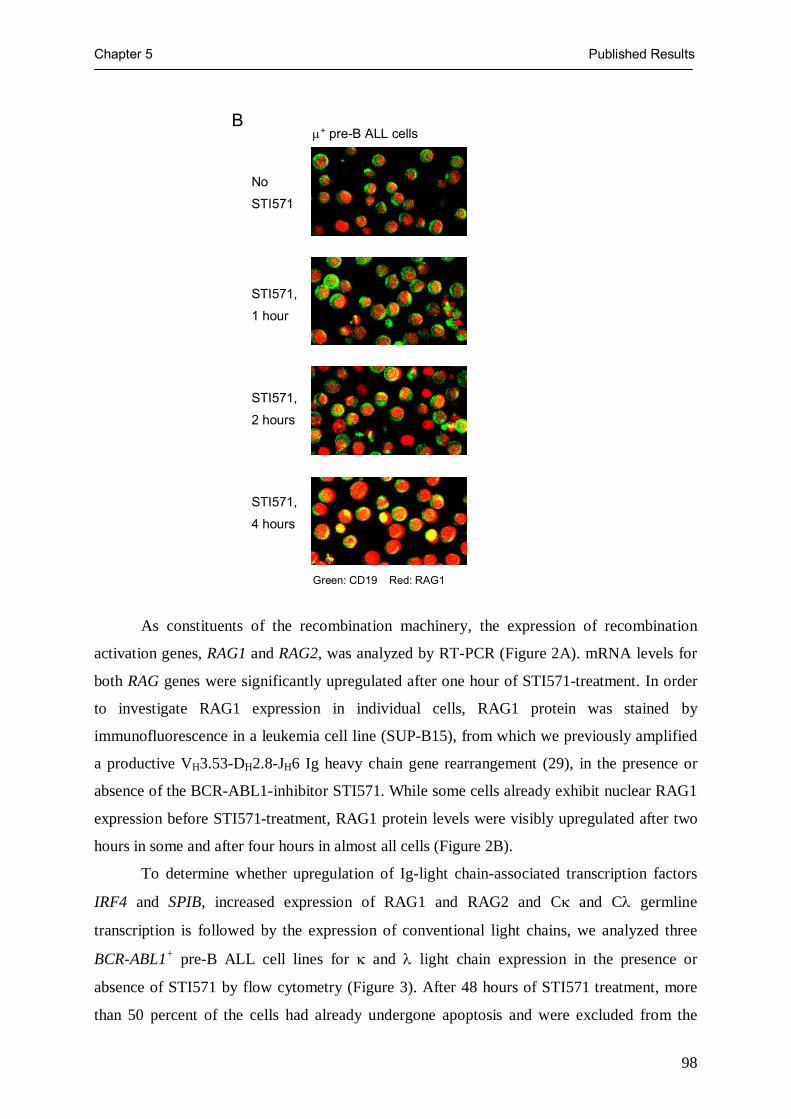
Chapter 5 Published Results
98
As constituents of the recombination machinery, the expression of recombination
activation genes, RAG1 and RAG2, was analyzed by RT-PCR (Figure 2A). mRNA levels for
both RAG genes were significantly upregulated after one hour of STI571-treatment. In order
to investigate RAG1 expression in individual cells, RAG1 protein was stained by
immunofluorescence in a leukemia cell line (SUP-B15), from which we previously amplified
a productive VH3.53-DH2.8-JH6 Ig heavy chain gene rearrangement (29), in the presence or
absence of the BCR-ABL1-inhibitor STI571. While some cells already exhibit nuclear RAG1
expression before STI571-treatment, RAG1 protein levels were visibly upregulated after two
hours in some and after four hours in almost all cells (Figure 2B).
To determine whether upregulation of Ig-light chain-associated transcription factors
IRF4 and SPIB, increased expression of RAG1 and RAG2 and Cκ and Cλ germline
transcription is followed by the expression of conventional light chains, we analyzed three
BCR-ABL1+ pre-B ALL cell lines for κ and λ light chain expression in the presence or
absence of STI571 by flow cytometry (Figure 3). After 48 hours of STI571 treatment, more
than 50 percent of the cells had already undergone apoptosis and were excluded from the
μ+ pre-B ALL cells
No
STI571
STI571,
1 hour
STI571,
2 hours
STI571,
4 hours
Green: CD19 Red: RAG1
B

Chapter 5 Published Results
99
analysis. Apoptotic or dead cells were identified by annexin V membrane expression and
uptake of propidium iodide. Among the surviving cells, about 10 percent exhibit de novo Ig
light chain expression (Figure 3). Unexpectedly, BCR-ABL1-inhibition resulted in a pattern
of light chain expression heavily biased for λ-light chains (Figure 3). Only a few κ-light
chain-producing leukemia cells were detectable, while the anti-κ antibody used clearly
identified normal κ-expressing B cells (Figure 3). For control stainings, B cells from
umbilical cord blood were used that were enriched by MACS for CD19 expression (Figure 3).
Extensive κ-deleting element rearrangement results in preferential λ-light chain expression
on STI571-surviving leukemia cells
Using three different PCR strategies (Figure 4), we investigated at which level the expression
of κ light chains may have been impaired. By ligation-mediated PCR (LM-PCR; Figure 4A),
we first showed that inhibition of BCR-ABL1 kinase activity induces DNA double-strand
breaks at recombination signal sequences (RSS) within the IGK locus after eight hours
(Figure 5A). For validation of the LM-PCR assay, PCR products were cloned and five
randomly chosen clones were sequenced. Sequence analysis confirmed Jκ1 RSS-linker
ligation products (not shown). Such RSS-specific DNA strand breaks represent an initial step
within the recombination process of IGK genes and demonstrate that the IGK locus is indeed
targeted by the recombination machinery upon treatment with STI571. Since Jκ-RSS-specific
DNA strand breaks represent a pre-condition for Vκ-Jκ gene rearrangements, we next
amplified Vκ-Jκ rearrangements from genomic DNA of B cell precursor leukemia cells
treated with or without STI571 (Figure 4B). While sporadic Vκ-Jκ and Vλ-Jλ joints were
already detectable under control conditions, V-J rearrangements at both IGK and IGL loci
were clearly inducible by STI571 (Figure 5B). As a specificity control, genomic DNA of
BCR-ABL1+ chronic myeloid leukemia cells was also subjected to amplification of Vκ-Jκ and
Vλ-Jλ rearrangements but no product was obtained. Integrity of DNA isolated from chronic
myeloid leukemia cells was verified by amplification of a Cβ1 germline fragment of the
TCRβ locus (Figure 5B). In agreement with a previous study on the effect of STI571 on v-
abl-transformed murine pre-B cell lines (21), we observed a polyclonal pattern of Vκ and Vλ
gene rearrangement upon STI571-treatment. STI571-induced V-J gene rearrangements
involved all six IGK and all nine IGL V gene families. Whether IGK and IGL genes were also
rearranged at similar frequencies in response to STI571-treatment, however, remains unclear,
because the PCR approach used here was not quantitative

Chapter 5 Published Results
100
Figure 3: Ig light chain expression resulting from BCR-ABL1 inhibition is heavily biased
for λ light chains
Igλ
Igκ
BV173
NALM1
SUP-B15
− STI571 + STI571
57
38
cord blood mononuclearcells after CD19+ MACS
Igλ
Igκ
BV173
NALM1
SUP-B15
− STI571 + STI571
57
38
cord blood mononuclearcells after CD19+ MACS
BCR-ABL1+ BV173 cells, NALM1 cells and SUP-B15 cells were cultured in the presence or absence of 10 µmol/l STI571 for two days and surviving cells were analyzed for surface expression of κ and λ light chains by flow cytometry. B lymphocytes from umbilical cord blood were enriched by MACS and used as a positive control for κ and λ staining.

Chapter 5 Published Results
101
Figure 4: Molecular analysis of recombination events within the IGK locus
(A) During primary recombination events, a Vκ gene segment rearranges to a (mostly upstream located) Jκ gene segment. In secondary rearrangements, pre-existing Vκ−Jκ joints are deleted by juxtaposition of more upstream V to a more downstream J element. (B) The rearrangement of Vκ to Jκ segments is initiated by DNA double-strand breaks at the heptamer RSS sequence (RSS) immediately flanking the Jκ segment. DNA double-strand breaks result in hairpin-formation at the Jκ-break and 5’ phosphorylated blunt ends at the RSS-break, which can be ligated to a blunt-ended linker molecule (Linker). Using PCR primers specific for sequences upstream of the RSS and the ligated linker sequence, broken-ended Jκ-RSS DNA intermediates can be amplified. (C) A preformed Vκ-Jκ-joint (top) can be inactivated by two types of rearrangement of the κ-deleting element (KDE). Using an upstream Vκ-RSS of an unrearranged Vκ-gene segment, rearrangement of the KDE results in a large deletion within the κ locus including downstream unrearranged Vκ-gene segments, the pre-existing Vκ-Jκ-joint, unrearranged Jκ-gene segments and the Cκ-gene (bottom, left). In alternative, the KDE can rearrange to an intronic RSS between the cluster of Jκ-gene segments and the Cκ gene. In this case, KDE-rearrangement results in a deletion of the Cκ-gene and both κ enhancers (bottom, right).
Linker
RSS
RSS
Jκ1
Jκ1
3‘ Vκ Jκ1
5‘ Vκ Jκ5
Jκ Intron-KDEVκ-KDE
Vκ-Jκ Jκ Intron IE Cκ 3‘E KDE germlineVκ
A
B
C
Linker
RSS
RSS
Jκ1
Jκ1
3‘ Vκ Jκ1
5‘ Vκ Jκ5
Jκ Intron-KDEVκ-KDE
Vκ-Jκ Jκ Intron IE Cκ 3‘E KDE germlineVκ
A
B
C

Chapter 5 Published Results
102
Rearrangement of the κ-deleting element (KDE) may lead to inactivation of productively
recombined Vκ-Jκ joints and hence represents another level at which Ig light chain expression
may be impaired (15). To assess whether pre-existing functional Vκ-Jκ joints may have been
inactivated by rearrangement of the KDE, we analysed both types of KDE-recombination
within the IGK locus (Figure 4C). The KDE can rearrange either to an RSS site within the
intron region between the cluster of Jκ gene segments and the κ intronic enhancer or to an
RSS site immediately flanking a Vκ gene segment (Figure 4C). KDE rearrangement leads to
deletion of both κ enhancers and the Cκ gene in the former (Jκ intron RSS-KDE) or to
deletion of a Vκ-Jκ joint in the latter case (Vκ RSS-KDE). To investigate whether KDE
rearrangement may contribute to reduced κ light chain usage in STI571-treated leukemia
cells, we amplified specific DNA fragments for KDE-germline configuration, Jκ intron RSS-
KDE rearrangement and Vκ RSS-KDE rearrangement from three BCR-ABL1+ leukemia cell
lines (BV173, NALM1, SUP-B15) in the absence or presence of STI571 (Figure 5C). While
KDE was found in germline configuration in untreated leukemia cells, STI571-mediated
inhibition of BCR-ABL1 induced deletion of IGK alleles by rearrangement of the KDE to Jκ
intron RSS sites and to Vκ RSS sites. Targeting of the IGK locus by the recombination
machinery and generation of Vκ-Jκ joints appears to be normal, but overrepresentation of λ
light chains can be attributed to extensive KDE rearrangement observed here (Figure 5C). In
the absence of BCR-ABL1 kinase activity, differentiating leukemia cells seem to undergo
multiple rounds of rearrangement targeting both κ and λ light chain loci, which ultimately
leads to preferential λ light chain expression expression due to KDE-mediated deletion of
potentially productive Vκ-Jκ joints.
Rearrangements at the IGK and IGL loci occur in sequential order
For this reason, we considered STI571-induced Ig light chain gene rearrangement as a model
for the pre-B to immature B cell transition in normal B cell development and investigated
whether IGK, KDE and IGL gene rearrangements follow a defined sequence of events.
Sequential rearrangement of IGK, KDE and IGL genes would be predicted by the ‘ordered
model’ of light chain gene recombination. To the contrary, according to the ‘stochastic
model’ of light chain gene rearrangement, IGK, KDE and IGL genes would be recombined
independently at each locus. Therefore, we incubated the three BCR-ABL1+ pre-B ALL cell
lines in the presence or absence of STI571 and analyzed genomic DNA for RSS-specific
double-strand breaks within the IGK, KDE and IGL loci at different time points (Figure 6).

Chapter 5 Published Results
103
Figure 5: Inhibition of BCR-ABL1 causes extensive rearrangement of κ and λ light
chain genes including the κ-deleting element
Using LM-PCR, we could amplify shortlived Jκ1-RSS DNA-double strand breaks predominantly in STI571-treated cells, which demonstrates active recombinase activity within the IGK loci (A). We next investigated Vκ-Jκ and Vλ-Jλ gene rearrangements in STI571-treated and -untreated leukemia cells. Comparing IGK and IGL loci, there was no indication of preferential Vλ-Jλ gene rearrangement (B). Amplifying specific fragments of KDE germline configuration, KDE rearrangement to an RSS within the Jκ intron and KDE rearrangement to an Vκ-associated RSS, we observed that the KDE was mainly in germline configuration in untreated leukemia cells, whereas STI571-treatment induced KDE rearrangement predominantly to the Jκ intron RSS (C).
STI571 Jκ1-RSS
Vκ Vλ 1 2 3 4 5 6 1 2 3 4 5 6 7 8 9 Cell line STI571
SUP-B15 ―
+
BV173 ―
+
NALM1 ―
+
BV173 NALM1 SUP-B15 ― + ― + ― +
BCR-ABL1+ CML cells (K562) Vκ-Jκ or Vλ-Jλ TCRCβ1 germline
KDE
germ
line
Jκ In
tron
RSS
-KD
E
Vκ1-
KDE
Vκ2-
KDE
Vκ3-
KDE
Vκ4-
KDE
Vκ5-
KDE
Vκ6-
KDE
KDE
germ
line
Jκ In
tron
RSS
-KD
E
Vκ1-
KDE
Vκ2-
KDE
Vκ3-
KDE
Vκ4-
KDE
Vκ5-
KDE
Vκ6-
KDE
KDE
germ
line
Jκ In
tron
RSS
-KD
E
Vκ1-
KDE
Vκ2-
KDE
Vκ3-
KDE
Vκ4-
KDE
Vκ5-
KDE
Vκ6-
KDE
BV173 NALM1 SUP-B15
― STI571
+ STI571
A
B
C

Chapter 5 Published Results
104
In the three cell lines, RSS-specific DNA double-strand breaks were already detectable at the
IGK locus within one hour after STI571-treatment. RSS-breaks flanking the KDE followed
after six hours of STI571-treatment and RSS-DNA-breaks were found at the IGL locus only
after 12 hours (Figure 6). These findings are consistent with a sequential order of
rearrangement events: Ablation of BCR-ABL1 kinase activity almost immediately initiates
IGK gene rearrangements, many of which are subsequently deleted by KDE- and ultimately
replaced by IGL gene rearrangements (Figure 6). Of note, upregulation of Cλ germline
transcripts did not precede the onset of KDE RSS-breaks (Figure 2A and 6). Unlike normal B
cell development, only very few STI571-treated leukemia cells seem to stay at a κ+ stage,
while the vast majority of cells continue to rearrange, first KDE and ultimately IGL genes. It
should be noted that between 12 and 24 hours of STI571-incubation, RSS-breaks could be
detected concomitantly in IGK, KDE and IGL loci (Figure 6). Therefore, we cannot exclude
that during this period of time, IGL gene rearrangements may have occurred before
recombination within the IGK locus. Stability of RSS-strand break intermediates is cell-cycle
dependent and mainly found in G1 and G0 phase (27). Indeed, STI571-treatment induces cell
cycle arrest in BCR-ABL1+ B cell precursor leukemia cells (30). Thus, the detection of Jκ-
RSS breaks for an extended period of time (one to 24 hours), partially overlapping with KDE-
RSS and even Jλ-RSS breaks, can be explained by cell cycle arrest and increased stability of
RSS-breaks induced by STI571.
Indication for secondary IGK gene rearrangements
On the other hand, the relatively wide time frame during which IGK gene rearrangements can
occur might reflect that the leukemia cells are not all synchronized with respect to STI571-
induced IGK gene rearrangement. This is consistent with a rather heterogenous pattern of of
RAG1 upregulation following treatment with STI571 (Figure 2). Moreover, RSS-specific
DNA-strand breaks within the IGK locus do not necessarily reflect a single definitive Vκ-Jκ
gene rearrangement and would also arise from any secondary recombination event during
light chain revision (Figure 4B). To determine whether ongoing κ light chain editing may
extend the time window for RSS-specific DNA-strand breaks, we searched for traces of
secondary rearrangements within the IGK locus. A pre-existing Vκ-Jκ joint can only be
replaced by a rearrangement which uses an upstream-located Vκ- together with a
downstream-located Jκ segment (Figure 4B). Individual Vκ segments are not arranged in the
order of Vκ gene families within the IGK locus. We, therefore, only compared the usage of
upstream Jκ1 and Jκ2 segments to downstream Jκ5 elements (see Figure 4B) at different time
points after STI571-induced initiation of light chain gene rearrangement. Consistent with a

Chapter 5 Published Results
105
Figure 6: Sequentially ordered recombination events within the IGK, KDE and IGL loci
shift from upstream to downstream Jκ elements, Jκ1 and Jκ2 gene rearrangements were
detected slightly earlier than rearrangements involving the Jκ5 gene segment (Figure 7).
Given that the two round PCR approach used here for analysis of Vκ-Jκ gene rearrangements
is not quantitative, the relative amount of PCR products does not necessarily reflect the usage
of individual Jκ segments.
Concluding remarks
We conclude that inhibition of BCR-ABL1 initiates a coordinated sequence of Ig light chain
gene rearrangement events: Almost immediately after ablation of BCR-ABL1 kinase activity
by STI571, the IGK locus opens and is targeted by the recombination machinery. The
continuous generation of new Jκ RSS-specific DNA strand breaks even after 24 hours
together with a shift from upstream Jκ1 and Jκ2 elements to downstream Jκ5 segments
suggests that initial Vκ-Jκ rearrangements are followed by at least one, perhaps multiple
BCR-ABL1+ BV173, NALM1 and SUP-B15 cells were were cultured in the presence or absence of 10 μmol/l STI571 for the times indicated. Genomic DNA was isolated from 5 x 10⁶ cells. Broken-ended DNA strand breaks were ligated to linker molecules and subjected to two rounds of PCR-amplification using primers specific for breaks at Jκ-RSS, KDE-RSS and Jλ-RSS sites.
0 0.5 1 2 3 6 9 12 24 36 48 Incubation time with STI571 [h]
Jκ-RSS
Jλ-RSS
KDE-RSS
0 0.5 1 2 3 6 9 12 24 36 48
BV173
NALM1
SUP-B15
BV173
NALM1
SUP-B15
BV173
NALM1
SUP-B15
BCR-ABL1+ Cell Line
0 0.5 1 2 3 6 9 12 24 36 48 Incubation time with STI571 [h]
Jκ-RSS
Jλ-RSS
KDE-RSS
0 0.5 1 2 3 6 9 12 24 36 48
BV173
NALM1
SUP-B15
BV173
NALM1
SUP-B15
BV173
NALM1
SUP-B15
BCR-ABL1+ Cell Line

Chapter 5 Published Results
106
rounds of secondary rearrangements. A possible initiation signal for subsequent KDE
rearrangement could be exhaustion of the recombination potential of IGK alleles in multiple
rounds of Vκ-Jκ rearrangement. Recombination of the KDE deletes Vκ-Jκ rearrangements
from the chromosome and may initiate Vλ-Jλ gene rearrangements, which can first be
detected after 12 hours. According to these observations, it is not surprising that the vast
majority of leukemia subclones that have been primed to differentiate, ultimately express λ
light chains on their surface (Figure 3). Obviously, the leukemia cells, unlike normal pre-B
cells, are lacking a feedback mechanism which prompts them to halt the recombination
machinery in the presence of a productively rearranged IGK allele. Upon inhibition of BCR-
ABL1, the leukemia cells seem to enter a pre-determined (“ordered”) sequence of
rearrangement events, in which κ-expressing cells would only exist as a transition stage,
inevitably leading to the expression of λ light chains.
Figure 7: Shifting the preferential Jκ usage from upstream to downstream located Jκ
gene segments indicates secondary IGK gene rearrangements
Vκ1 gene rearrangements were amplified from three BCR-ABL1+ pre-B ALL cell lines using Vκ1 leader- and Vκ1 framework region I-specific primers together with either primers matching to Jκ1 and Jκ2 or primers matching to Jκ5.
0 0.5 1 2 3 6 9 12BCR-ABL1+ Cell Line
BV173
NALM1
SUP-B15
0 0.5 1 2 3 6 9 12 Incubation time with STI571 [h]
Jκ1; Jκ2
Jκ5
BV173
NALM1
SUP-B15
0 0.5 1 2 3 6 9 12BCR-ABL1+ Cell Line
BV173
NALM1
SUP-B15
0 0.5 1 2 3 6 9 12 Incubation time with STI571 [h]
Jκ1; Jκ2
Jκ5
BV173
NALM1
SUP-B15
Chapter 5 Published Results
107
Acknowledgements: We would like to thank Stefanie Jauch for excellent technical assistance, Klaus Rajewsky, Ralf Küppers, Matthias Fischer, Mario Fabri, Hassan Jumaa and Michael Reth for critical discussions, and Martin Krönke and Janet D. Rowley for continuous support and discussions, and Sean McCleary for proofreading .
Materials and Methods
Patient samples, primary cells and cell lines
BCR-ABL1+ pre-B ALL cells from five patients were analysed. From three of these patients,
leukemia cells were available before and after treatment with STI571 (Cases III, IV and V,
reference 23). Samples of two other cases (Cases I and II) were used for SAGE analysis.
Normal human CD10+ VpreB+ pre-B cells, CD34+ CD38low CD133+ hematopoietic
progenitor cells, CD15+ CD34− myeloid progenitor cells, CD7+ CD10+ T lymphoid pogenitor
cells and mature B cell subsets including CD19+ CD27− naïve B cells, CD20+ CD77+
germinal center B cells, CD19+ CD27+ memory B cells and CD19low CD138+ plasma cells
were isolated by MACS and FACS from bone marrow, umbilical cord blood, peripheral blood
or tonsils from healthy donors as previously described (24-26). For cell culture experiments,
the BCR-ABL1+ pre-B ALL cell lines BV173, NALM1 and SUP-B15 (Deutsche Sammlung
von Mikroorganismen und Zellkulturen, Heidelberg, Germany) were used. Cell lines were
treated with 10μmol/l STI571 (Novartis, Basel, Switzerland) for 0 to 48 hours.
SAGE analysis
To identify differentially expressed genes between BCR-ABL1+ pre-B ALL cells and normal
pre-B cells with respect to Ig light chain expression, we analysed expression profiles
generated by SAGE for mRNA expression levels of genes that are implicated in the regulation
of Ig light chain expression. Genome-wide SAGE profiles for two primary cases of BCR-
ABL1+ pre-B ALL (cases I and II) and the normal hematopoietic populations mentioned
above were generated and compared as described previously (24-26). In these SAGE-profiles,
30 genes were identified which have been implicated in the regulation of Ig-light chain
expression. These genes were searched in the literature, as well as the UniGene
(http://www.ncbi.nlm.nih.gov/UniGene) and OMIM
(http://www.ncbi.nlm.nih.gov/entrez/query.fcgi?db=OMIM) databases.
RT-PCR analysis of IRF4, SPIB, RAG1/2 expression and germline κ and λ light chain
transcripts
mRNA levels of the Ig light chain-associated transcription factors IRF4 and SPIB in BCR-
ABL1+ were measured in leukemia samples from three patients (cases III, IV and V) before
and after treatment with STI571 (Figure 1). From patient samples, total RNA was isolated and

Chapter 5 Published Results
108
transcribed into cDNA as previously described (24-26). Amounts of cDNA were normalized
by OD measurement and semiquantitative RT-PCR using 5’-TTAGCACCCCTGGCCAAG-
3’ and 5’-CTTACTCCTTGGAGGCCATG-3’ for amplification of a cDNA fragment of
GAPDH. For semiquantitative RT-PCR analysis of IRF4 the oligonucleotides 5’-
CAAGAGCAATGACTTTGAGG-3’ and 5’-TGGGACATTGGTACGGGAT-3’ were used
and for SPIB 5’-AAGACTTACCGTTGGACAGC-3’ and 5’-
CTTGGAGGAGAACTGGAAGA-3’.
RNA was isolated from BV173, NALM1 and SUP-B15 cell lines with or without
STI571incubation (10μmol/l), transcribed into cDNA, and normalized as described above. In
order to determine light-chain locus germline transcription by RT-PCR, the primers for
constant regions in each locus were used. For κ constant region transcription, 5’-
TTCAACAGGGGAGAGTGTTAGAG-3’ and 5’- ATGCGCCTTAGGATGACTACATA-3’
were used, and 5’- TGTCTGATCAGTGACTTCTACCC-3’ and 5’-
CTGTAGCTTCTGTGGGACTTC-3’ were used for λ constant region transcription. RT-PCR
on RAG1 and RAG2 was carried out using the oligonucleotides 5’-
ATAGAAGAAAGCAACACAAAAGC -3’ and 5’- ATACTGAGTTCAATCCCTGAAGA -
3’ for RAG1 and 5’-ATAGCAAGAGCTCTACACACTCC-3’ and 5’-
AAAAATCAGATCAGAAATCCTCA-3’ for RAG2 (Figure 2A).
Analysis of nuclear RAG1 protein expression
BCR-ABL1+ pre-B ALL cells (BV173 and SUP-B15) were cultured in the presence or absence
of 10 μmol/l STI571 for 1, 2 or 4 hours and subsequently stained with a Cy5-labeled antibody
against CD19. After treatment with methanol and 4% paraformaladelhyde, cells were stained
with a mouse anti-human RAG1 IgG1 (BD Biosciences, Heidelberg, Germany) together with
a Cy2-labeled goat anti-mouse IgG antibody. Expression of CD19 and RAG1 was visualized
by immunofluorescence (Figure 2).
Flow cytometry
Surface expression of Igκ- or Igλ-light chains on BV173, NALM1, and SUP-B15 pre-B ALL
cells in the presence or absence of 10 μmol/l STI571 was monitored using antibodies against
Igκ- or Igλ-light chains (BD Biosciences, Heidelberg, Germany) after the incubation times
indicated (Figure 3). Apoptotic or dead cells were identified by staining with FITC-labelled
Annexin V and propidium iodide (BD Biosciences) and removed with the gate.
Amplification of double-strand recombination signal sequence breaks by ligation-mediated
PCR

Chapter 5 Published Results
109
From about 2.5 x 10⁶ untreated BV173, NALM1 and SUP-B15 cells or after treatment with
10 μmol/l STI571 after the times indicated, genomic DNA was isolated and ligated to a blunt-
end linker using T4 DNA ligase (Invitrogen, Karlsruhe, Germany) at 14°C overnight. The
linker was constructed by annealing the oligonucleotides 5'-
TTTCTGCTCGAATTCAAGCTTCTAACGATGTACGGGGACATG 3' and 3’ amino (C7)-
GACGAGCTTAAGTTCGAAGATTGCTACATGCCCCT -5’ and protruding 3' overhangs
were removed by 3'→ 5' exonuclease activity of the Klenow fragment of E. coli DNA
polymerase I (Invitrogen, Karlsruhe, Germany). Ligation-mediated PCR (LM-PCR; 27) was
carried out with modifications as previously described (26). In two semi-nested rounds of
amplification (35 and 45 PCR cycles at an annealing temperature of 59°C), RSS-
intermediates with a DNA double-strand break at the 5’ heptamer of Jκ gene segments were
amplified (see Figure 4, 5A) using 5’-GTAATTAACATTCAGTCTACTTTC-3’ as external
forward and 5’-TAACATTCAGTCTACTTTCTAAAA-3’ as internal forward primers
together with 5’-TCCCCGTACATCGTTAGAAG–3’ as reverse primer specific for DNA-
ligated linker molecules. To amplify RSS-intermediates with a DNA double-strand break at
the 5’ heptamer of Jλ gene segments, 5’-TTCTCACTTCTTCCATGGTGAC-3’ and 5’-
ACTTCTTCCATGGTGACAGTCT-3’ were used in two rounds of PCR amplification as
described above (Figure 6). Accordingly, 5’-TCCTCCTCACTGAGCCTCCCTTGAAT-3’
and 5’-CTCACTGAGCCTCCCTTGAATAGTCC-3’ were used to amplify RSS-
intermediates with a DNA double-strand break at the 5’ heptamer of the κ-deleting element
(KDE).
Amplification of Vκ-Jκ and Vλ-Jλ gene rearrangements
Genomic DNA from 5 x 10⁶ STI571-treated (24 hours) or untreated BV173, NALM1 and
SUP-B15 cells was isolated and subjected to two rounds of seminested PCR (35 and 45
cycles) using six Vκ- and nine Vλ-family-specific primers together with Jκ- and Jλ-specific
primers, respectively. Primers and PCR conditions were used as previously described (28).
Analysis of the genomic configuration of the κ deleting element (KDE)
Three primers (KDE germline: 5'-CTCACTGAGCCTCCCTTGAATAGTCC-3'; JκCκ Intron
: 5'-CCGCGGTTCTTTCTCGATTGAGTGG-3'; KDE external reverse: 5'-
CTTCATAGACCCTTCAGGCACATGC-3') were added to Vκ-family-specific primers for
amplification of Vκ-KDE rearrangements (Vκ primers and KDE external reverse primer),
JκCκ intron RSS-KDE rearrangements (JκCκ Intron and KDE external reverse primer), and
KDE in germline configuration (KDE germline and KDE external reverse primer). 1 μl-
Chapter 5 Published Results
110
aliquots of the first rounds were used in separate second rounds for the six family-specific Vκ
primers together with 5' Jκ primers and the internal reverse KDE primer (5'-
AGACAGGTCCTCAGAGGTCAGAGC-3'), and one second round with JκCκ Intron and
KDE internal reverse and another with KDE germline and internal reverse KDE (see Figure
5). Vκ-family-specific primers used are 5'-GACATCCRGWTGACCCAGTCTCCWTC-3' for
Vκ1, 5'- CAGWCTCCACTCTCCCTGYCCGTCA-3' for Vκ2, 5'-
TTGTGWTGACRCAGTCTCCAGSCACC-3' for Vκ3, 5'-
AGACTCCCTGGCTGTGTCTCTGGGC-3' for Vκ4, 5'-
CAGTCTCCAGCATTCATGTCAGCGA-3' for Vκ5 and 5'-
TTTCAGTCTGTGACTCCAAAGGAGAA-3' for Vκ6. Jκ-specific primers used are 5'-
TTGATYTCCASCTTGGTCCCYTGGC-3' for Jκ1 and Jκ2, 5'-
TTGATATCCACTTTGGTCCCAGGGC-3' for Jκ3, 5'-
TTGATCTCCACCTTGGTCCCTCCGC-3' for Jκ4 and 5'-
TTAATCTCCAGTCGTGTCCCTTGGC-3' for Jκ5.
Analysis of secondary IGK gene rearrangement
To analyse potential secondary IGK recombination events, we tested whether rearrangement
of individual Jκ-gene segments occurs sequentially. Genomic DNA isolated from untreated
BCR-ABL1+ pre-B ALL cells or treated with STI571 (10μmol/l) for the times indicated and
subjected to two rounds of seminested PCR. 5’-GCTCAGCTCCTGGGGCTCCTGC-3’ and
5'-GACATCCRGWTGACCCAGTCTCCWTC-3' were used as primers specific for the leader
and framework region I, respectively, of Vκ1 gene segments. 5'-
TTGATYTCCASCTTGGTCCCYTGGC-3' was used for amplification of Jκ1 and Jκ2 gene
rearrangements and 5'- TTAATCTCCAGTCGTGTCCCTTGGC-3' for Jκ5.
References
1. Hardy R. R., C. E. Carmack, S. A. Shinton, J. D. Kemp, K. Hayakawa. 1991. Resolution and
characterization of pro-B and pre-pro-B cell stages in normal mouse bone marrow. J Exp Med.
173: 1213-25.
2. Rajewsky K. 1996. Clonal selection and learning in the antibody system. Nature. 381: 751-8.
3. Alt F. W., G. D. Yancopoulos, T. K. Blackwell, C. Wood , E. Thomas, M. Boss, R. Coffman,
N. Rosenberg, S. Tonegawa, D. Baltimore. 1984. Ordered rearrangement of immunoglobulin
heavy chain variable region segments. EMBO J. 3: 1209-19.

Chapter 5 Published Results
111
4. Langman R. E., and M. Cohn. 1995. The proportion of B-cell subsets expressing kappa and
lambda light chains changes following antigenic selection. Immunol Today. Mar;16(3):141-4.
5. Nadel B., P. A. Cazenave, P. Sanchez. 1990. Murine lambda gene rearrangements: the
stochastic model prevails over the ordered model. EMBO J. 9: 435-40.
6. Arakawa H., T. Shimizu, S. Takeda. 1996. Re-evaluation of the probabilities for productive
arrangements on the kappa and lambda loci. Int Immunol. 8: 91-9.
7. Gorman J. R., N. van der Stoep, R. Monroe, M. Cogne, L. Davidson, F. W. Alt. 1996. The
Ig(kappa) enhancer influences the ratio of Ig(kappa) versus Ig(lambda) B lymphocytes.
Immunity. 5: 241-52.
8. Inlay M., F. W. Alt, D. Baltimore, Y. Xu. 2002. Essential roles of the kappa light chain
intronic enhancer and 3' enhancer in kappa rearrangement and demethylation. Nat Immunol. 3:
463-8.
9. Zou Y. R., S. Takeda, K. Rajewsky. 1993. Gene targeting in the Ig kappa locus: efficient
generation of lambda chain-expressing B cells, independent of gene rearrangements in Ig
kappa. EMBO J. 12: 811-20.
10. Chen J., M. Trounstine, C. Kurahara, F. Young , C. C. Kuo, Y. Xu, J. F. Loring, F. W. Alt, D.
Huszar. 1993. B cell development in mice that lack one or both immunoglobulin kappa light
chain genes. EMBO J. 12: 821-30.
11. Korsmeyer S. J., P. A. Hieter, S. O. Sharrow, C. K. Goldman, P. Leder, T. A. Waldmann.
1982. Normal human B cells display ordered light chain gene rearrangements and deletions. J
Exp Med. 156: 975-85.
12. Korsmeyer S. J., P. A. Hieter, J. V. Ravetch, D. G. Poplack, T. A. Waldmann, P. Leder. 1981.
Developmental hierarchy of immunoglobulin gene rearrangements in human leukemic pre-B-
cells. Proc Natl Acad Sci U S A. 78: 7096-100.
13. Brauninger A., T. Goossens, K. Rajewsky, R. Küppers. 2001. Regulation of immunoglobulin
light chain gene rearrangements during early B cell development in the human. Eur J
Immunol. 31: 3631-7.
14. van der Burg M., B. H. Barendregt, T. Szczepanski, E. R. van Wering, A. W. Langerak, J. J.
van Dongen. 2002. Immunoglobulin light chain gene rearrangements display hierarchy in
absence of selection for functionality in precursor-B-ALL. Leukemia. 16: 1448-53.
15. Siminovitch K. A., A. Bakhshi, P. Goldman, S. J. Korsmeyer. 1985. A uniform deleting
element mediates the loss of kappa genes in human B cells. Nature. 316: 260-2.
16. Pauza M. E., J. A. Rehmann, T. W. LeBien . 1993. Unusual patterns of immunoglobulin gene
rearrangement and expression during human B cell ontogeny: human B cells can
simultaneously express cell surface kappa and lambda light chains. J Exp Med. 178: 139-49.
17. Look A.T.. 1997. Oncogenic transcription factors in the human acute leukemias. Science. 278:
1059-64.

Chapter 5 Published Results
112
18. Smith K. M., R. Yacobi, R. A. Van Etten. 2003. Autoinhibition of Bcr-Abl through its SH3
domain. Mol Cell. 12: 27-37.
19. LeBien T. W.. 2000. Fates of human B-cell precursors. Blood. 96: 9-23.
20. Chen Y. Y., L. C. Wang, M. S. Huang, N. Rosenberg. 1994. An active v-abl protein tyrosine
kinase blocks immunoglobulin light-chain gene rearrangement. Genes Dev. 8: 688-97.
21. Muljo S. A., and M. S. Schlissel. 2003. A small molecule Abl kinase inhibitor induces
differentiation of Abelson virus-transformed pre-B cell lines. Nat Immunol. 4: 31-7.
22. Druker B. J., C. L. Sawyers, H. Kantarjian, D. J. Resta, S. F. Reese, J. M. Ford, R. Capdeville,
M. Talpaz. 2001. Activity of a specific inhibitor of the BCR-ABL tyrosine kinase in the blast
crisis of chronic myeloid leukemia and acute lymphoblastic leukemia with the Philadelphia
chromosome. N Engl J Med. 344: 1038-42.
23. Hofmann W. K., S. de Vos, D. Elashoff, H. Gschaidmeier, D. Hoelzer, H. P. Koeffler, O. G.
Ottmann. 2002. Relation between resistance of Philadelphia-chromosome-positive acute
lymphoblastic leukaemia to the tyrosine kinase inhibitor STI571 and gene-expression profiles:
a gene-expression study. Lancet. 359: 481-6.
24. Klein F., N. Feldhahn, L. Harder, H. Wang, M. Wartenberg, W. K. Hofmann, P. Wernet, R.
Siebert, M. Müschen. 2004. The BCR-ABL1 kinase bypasses selection for the expression of a
pre-B cell receptor in pre-B acute lymphoblastic leukemia cells. J Exp Med. 199: 673-685.
25. Deininger MW, Vieira SA, Parada Y, Banerji L, Lam EW, Peters G, Mahon FX, Kohler T,
Goldman JM, Melo JV. Direct relation between BCR-ABL tyrosine kinase activity and cyclin
D2 expression in lymphoblasts. Cancer Res. 2001; 61: 8005-13
26. Müschen M., S. Lee, G. Zhou, N. Feldhahn, V. S. Barath, J. Chen, C. Moers, M. Krönke, J. D.
Rowley, S. M. Wang. 2002. Molecular portraits of B cell lineage commitment. Proc Natl
Acad Sci U S A. 99: 10014-9.
27. Feldhahn N., I. Schwering, S. Lee, M. Wartenberg, F. Klein, H. Wang, G. Zhou, S. M. Wang,
J. D. Rowley, J. Hescheler, M. Krönke, K. Rajewsky, R. Kuppers, M. Müschen. 2002.
Silencing of B cell receptor signals in human naive B cells. J Exp Med. 196: 1291-305.
28. Klein F., N. Feldhahn, S. Lee, H. Wang, F. Ciuffi, M. von Elstermann, M. L. Toribio, H.
Sauer, M. Wartenberg, V. S. Barath, M. Krönke, P. Wernet, J. D. Rowley, M. Müschen. 2003.
T lymphoid differentiation in human bone marrow. Proc Natl Acad Sci U S A. 100: 6747-52.
29. Schlissel M., A. Constantinescu, T. Morrow, M. Baxter, A. Peng. 1993. Double-strand signal
sequence breaks in V(D)J recombination are blunt, 5'-phosphorylated, RAG-dependent, and
cell cycle regulated. Genes Dev. 7: 2520-32.
30. Müschen M., K. Rajewsky, A. Brauninger, A. S. Baur, J. J. Oudejans, A. Roers, M. L.
Hansmann, R. Küppers. 2000. Rare occurrence of classical Hodgkin's disease as a T cell
lymphoma. J Exp Med. 191: 387-94.

Chapter 5 Published results
113
5.4 Mimicry of a constitutively active pre-B cell receptor: Aberrant splicing
links Bruton’s tyrosine kinase to BCR-ABL1 in pre-B lymphoblastic
leukemia
published in the Journal of Experimental Medicine 2005; 201: 1837-1852
By Niklas Feldhahn1, Florian Klein1, Jana L. Mooster1, Paul Hadweh1, Mieke
Sprangers1, Maria Wartenberg2, Mohamed M. Bekhite2, Wolf-Karsten
Hofmann3, Sebastian Herzog4, Hassan Jumaa4, Janet D. Rowley5, and Markus
Müschen1
1Laboratory for Molecular Stem Cell Biology, Heinrich-Heine-Universität Düsseldorf, 40225 Düsseldorf, Germany; 2Institute for Neurophysiology, University of Cologne, 50931 Cologne, Germany; 3 Department of Hematology, University of Frankfurt, 60596, Frankfurt/Main, Germany; 4 Max Planck Institute for Immunobiology, Freiburg, Germany; 5Department of Medicine, University of Chicago, 60637 IL
Summary
Pre-B cells undergo apoptosis unless they are rescued by pre-B cell receptor-
dependent survival signals. We previously showed that the BCR-ABL1 kinase
expressed in pre-B lymphoblastic leukemia bypasses selection for pre-B cell
receptor-dependent survival signals. Investigating possible interference of
BCR-ABL1 with pre-B cell receptor signaling, we found that neither SYK nor
SLP65 can be phosphorylated in response to pre-B cell receptor engagement.
Instead, Bruton’s tyrosine kinase (BTK) is constitutively phosphorylated by
BCR-ABL1. Activated BTK is essential for survival signals that would
otherwise arise from the pre-B cell receptor, including activation of PLCγ1,
autonomous Ca2+ signaling, STAT5-phosphorylation and upregulation of
BCLXL. Inhibition of BTK kinase activity specifically induces apoptosis in
BCR-ABL1+ leukemia cells to a similar extent as inhibition of BCR-ABL1
kinase activity itself. However, BCR-ABL1 cannot directly bind to full-length
BTK. Instead, BCR-ABL1 induces the expression of a truncated splice variant
of BTK acting as a linker between the two kinases. As opposed to full-length
BTK, truncated BTK is lacking kinase activity yet can bind to BCR-ABL1
through its SH3 domain. Acting as a linker, truncated BTK enables BCR-
ABL1-dependent activation of full-length BTK, which initiates downstream
survival signals mimicking a constitutively active pre-B cell receptor.

Chapter 5 Published results
114
Introduction
The BCR-ABL1 gene rearrangement encodes a constitutively active tyrosine kinase and drives
malignant transformation of pre-B cells in humans (1). In a B cell-specific mouse model,
expression of BCR-ABL1 is sufficient to induce pre-B lymphoblastic leukemia and required
for maintenance of leukemic transformation (2).
During their development, human B cells have to pass multiple checkpoints at which
they are selected for the expression of a functional pre-B or B cell receptor (3). However,
signals from the pre-B cell receptor may also initiate apoptosis (4). Therefore, the expression
of a functional pre-B cell receptor constitutes an effective means to tightly regulate survival or
elimination of individual B cell clones. In this regard, it is not surprising that in a number of B
cell-derived malignancies, pre-B or B cell receptor function is compromised (5). For instance,
Hodgkin- and Reed-Sternberg cells in classical Hodgkin’s disease carry “crippled”
immunoglobulin (Ig) variable (V) region gene rearrangements due to the acquisition of
deleterious somatic mutations (6). Likewise, transformation of human B cells by the Epstein-
Barr virus (EBV) not only mimicks antigen receptor-dependent survival signals through the
viral oncoprotein LMP2A but also prevents the B cell receptor from signaling by excluding it
from glycolipid-enriched membrane domains (7). Autonomous growth and survival of
transformed B cells seem to favor a situation in which the B cell receptor is not active. In
accordance with this notion, we recently observed that pre-B lymphoblastic leukemia clones
harboring a BCR-ABL1 fusion gene carry only non-productively rearranged immunoglobulin
(Ig) variable region genes in 9 of 12 cases (8). Even the few leukemia clones that do express a
pre-B cell receptor were nonetheless not responsive to pre-B cell receptor engagement (8). In
analogy to the EBV-encoded LMP2A protein, the expression of the oncogenic BCR-ABL1
kinase may not only confer autonomous survival signals but also interfere with antigen
receptor signaling.
Among others, Bruton’s tyrosine kinase (BTK) represents a crucial component of the
(pre-) B cell receptor signaling chain and links the (pre-) B cell receptor to Ca2+ signals in B
lymphoid cells (9). In humans, deficiency of BTK leads to X-linked agammaglobulinemia
(XLA) resulting from a differentiation block at the pre-B cell stage (10). Of note, BTK not
only promotes developmental progression of pre-B cells (11) but also acts as a tumor
suppressor to prevent pre-B lymphoblastic leukemia in mice (12, 13). In order to elucidate
how BCR-ABL1 can bypass selection for pre-B cell receptor-dependent survival signals, the
current study investigates possible interference of BCR-ABL1 with pre-B cell receptor-related
signaling molecules including SYK, SLP65, PLCγ2 and BTK.

Chapter 5 Published results
115
Results
BCR-ABL1 kinase activity induces tyrosine-phosphorylation of BTK
Normal pre-B cells can only survive in human bone marrow if they receive survival signals
through their pre-B cell receptor (14). In a previous study, we observed that in the majority of
cases of pre-B lymphoblastic leukemia expressing the oncogenic BCR-ABL1 kinase, the
leukemia cells do not harbor a functionally rearranged IGH allele and, hence, cannot express a
pre-B cell receptor on their surface (8). Therefore, we wondered how a pre-B cell-derived
leukemia cell can survive and even clonally expand in the absence of pre-B cell receptor-
dependent survival signals.
To determine whether BCR-ABL1 interferes with signal transduction of the pre-B cell
receptor, we examined whether the pre-B cell receptor-related signaling molecules SYK,
SLP65 and BTK are expressed and whether they can be phosphorylated upon pre-B cell
receptor engagement in the leukemia cells.
Western blot using antibodies against phosphorylated SYKY323, SLP65Y96 and
BTKY223 showed that neither SYK nor SLP65 were phosphorylated in response to
crosslinking of the pre-B cell receptor (not shown). However, BTK was constitutively
phosphorylated in the leukemia cells (at 77kD; Figure 1A).
Using a specific inhibitor of BCR-ABL1, termed STI571 (15), we found that
phosphorylation of BTK was sensitive to inhibition of BCR-ABL1 kinase activity (77kD;
Figure 1A). To determine whether constitutive phosphorylation of 77kD BTK is specific for
leukemia cells carrying a BCR-ABL1 gene rearrangement, BTK-phosphorylation was also
studied in pre-B lymphoblastic leukemias harboring an E2A-PBX1, MLL-AF4, TEL-AML1 or
TEL-PDGFRβ gene rearrangement (Figure 1B, Table I). While the BTKY223-specific antibody
detected smaller tyrosine-phosphorylated proteins also in other leukemias, constitutive Y223-
phosphorylation of BTK (77 kD) was specific for BCR-ABL1+ pre-B cell lymphoblastic
leukemia cell lines (Figure 1B, left panel).
To confirm that BTK-phosphorylation is indeed depending on BCR-ABL1 kinase
activity, we induced BCR-ABL1 expression in a murine B lymphoid cell line TONB210 (16)
carrying an inducible BCR-ABL1 transgene. As opposed to BCR-ABL1+ leukemia cells, this
cell line grows independently from BCR-ABL1 in the presence of IL-3. In the presence but
not in the absence of doxycycline-induced BCR-ABL1 expression, BTK was heavily
phosphorylated (see Figure 5C). We conclude that BTK is constitutively phosphorylated by
BCR-ABL1 but not by the pre-B cell receptor in the leukemia cells.

Chapter 5 Published results
116
Figure 1: BCR-ABL1 induces phosphorylation of BTK in pre-B lymphoblastic leukemia
cells
Protein expression and tyrosine-phosphorylation of the pre-B cell receptor downstream signaling molecules SYK, SLP65 and BTK was studied by Western blot in three BCR-ABL1+ pre-B lymphoblastic leukemia cell lines (VII, VIII and IX in Table I) in the presence or absence of STI571. The BTKpY223-antibody detected full-length BTK (77kD) as well as two additional tyrosine-phosphorylated proteins of 65 and 52 kD size (A-C). To confirm that constitutive phosphorylation of 77 kD full-length BTK is specific for BCR-ABL1 kinase activity, BTKY223-phosphorylation was studied in four BCR-ABL1-negative B cell precursor cell lines (B, left panel; see XI, XII, XIII and XV in Table I). Dependence of BTK on BCR-ABL1 kinase activity was also confirmed in four primary cases of BCR-ABL1+ pre-B lymphoblastic leukemia, from which matched sample pairs before and during therapy with STI571 were available (B, right panel; cases I to IV in Table I). EIF-4E was used as a loading control. To determine whether Y223-phosphorylation of BTK is mediated by transphosphorylation through BCR-ABL1 or autophosphorylation, BCR-ABL1+ leukemia cells (IX; Table I) were treated for 24 hours with the BCR-ABL1 kinase inhibitor STI571 or the BTK kinase inhibitor LFM-A13 (C).
A
B
BTKpY223
EIF-4E
BC
R-A
BL1
E2A
-PB
X1
MLL
-AF4
TEL-
AM
L1
TEL-
PD
GFR
β
BTKpY223
Con
trol
STI
571
LFM
-A13
77 kD
52 kD
65 kD
C
STI571
SLP65
SYKpY323
SYK
BTKpY223
BCR-ABL1+ B cell precursor leukemia cell lines:
− + − + − +
EIF-4E
77 kD
52 kD
SLP65pY96
65 kD
VII VIII IX
77 kD
52 kD
65 kD
− + − + − + − +
77 kD
52 kD
65 kD
− + − + − + − +
Primary cases of BCR-ABL1+ B cell precursor leukemia:
I II III IV
STI571:
65 kD
B
C A

Chapter 5 Published results
117
Tabl
e I:
Des
crip
tion
of le
ukem
ia a
nd ly
mph
oma
case
s an
d ce
ll lin
es s
tudi
ed
Cas
e A
ge
Sex
Tran
sloc
atio
n
IgH
Igκ/
λ
IGH
conf
igur
atio
n
C
linic
al c
ours
e
F
usio
n ge
ne
or S
LC
A
llele
s
C
odin
g ca
paci
ty
Ia 39
m
B
CR
-AB
L1; p
210
−
−
#1 V
H4.
34-D
H6.
13-J
H5
no; o
ut-o
f-fra
me
ALL,
rela
pse
afte
r STI
571
treat
men
t
#2
DH5-
J H
IIa 54
f
BC
R-A
BL1
; p19
0
− −
#1
VH4.
61-D
H2.
2-J H
4 no
; out
-of-f
ram
e, s
top
in ju
nctio
n A
LL, r
elap
se a
fter S
TI57
1 tre
atm
ent
#2
DH2.
4-J H
4
III
a 61
f
BC
R-A
BL1
; p19
0
n.d.
n.
d.
n.
d.
ALL
, rel
apse
afte
r STI
571
treat
men
t IV
a 47
f
BC
R-A
BL1
; p19
0
n.d.
n.
d.
n.
d.
ALL
, rel
apse
afte
r STI
571
treat
men
t V
a 34
m
B
CR
-AB
L1; p
190
−
−
#1 V
H4.
34-D
H6.
13-J
H5
out-o
f-fra
me
#2 D
H5-
J H
A
LL, c
ompl
ete
rem
issi
on a
fter B
MT
VIa
32
m
BC
R-A
BL1
; p19
0
− −
#1
VH4.
4-D
H2.
2-J H
4
out-o
f-fra
me,
sto
p co
dons
in ju
nctio
n A
LL, d
ied
afte
r BM
T
#2
DH2.
4-J H
4
VIIa
45
m
BC
R-A
BL1
; p21
0
− −
#1
VH3.
48-D
H2.
15-J
H3
no; o
ut-o
f-fra
me,
sto
p in
junc
tion
ALL
, die
d af
ter r
elap
se (B
V17
3 ce
ll lin
e)
#2
DH2-
J H
VI
IIa 3
f B
CR
-AB
L1; p
210
μ
chai
n S
LC
#1
DH4-
J H
no
ALL
, die
d af
ter r
elap
se (N
ALM
1 ce
ll lin
e)
#2
VH3.
9-D
H2.
21-J
H3
yes
IXa
9 m
B
CR
-AB
L1; p
190
μ
chai
n S
LC
#1
VH3.
53-D
H2.
8-J H
6 ye
sb
ALL
, die
d af
ter r
elap
se (S
UP
-B15
cel
l lin
e)
X 32
f
MLL
-AF4
− −
#1
VH3.
20-D
H2.
8-J H
5 no
; out
-of-f
ram
e
A
LL, r
elap
se a
fter c
hem
othe
rapy
(RS
4;11
cel
l lin
e)
#2
VH6.
1-D
H1.
7-J H
4
no, s
top
in ju
nctio
n XI
5
f M
LL-A
F4
−
−
#1 D
H3-
J H
no
ALL
, rel
apse
afte
r che
mot
hera
py (S
EM
cel
l lin
e)
#2
IGH
in g
erm
line
XII
15
f TE
L-A
ML1
− −
#1
VH3.
15-D
H3.
10-J
H6
no; o
ut-o
f-fra
me
ALL
, rel
apse
afte
r che
mot
hera
py (R
EH
cel
l lin
e)
#2
IGH
in g
erm
line
XIII
12
m
E2A
-PB
X1
μ
chai
n S
LC
#1
VH4.
34-D
H3.
22-J
H2
no; o
ut-o
f-fra
me,
sto
p in
junc
tion
ALL
, rel
apse
afte
r che
mot
hera
py (6
97 c
ell l
ine)
#2 V
H2.
26-D
H2.
2.-J
H4
yes
XIV
15
m
E
2A-P
BX
1
μ ch
ain
SLC
#1 V
H3.
7-D
H3.
10-J
H4
yesb
AL
L, c
ell l
ine
(KAS
UM
I-2) e
stab
lishe
d at
dia
gnos
is
XV
19
m
TE
L-P
DG
FRβ
μ
chai
n S
LC
#1
VH1.
69-D
H3.
10-J
H6
yesb
A
LL, r
elap
se a
fter c
hem
othe
rapy
(Nal
m6
cell
line)
X
VI
5 m
IG
H-M
YC
IgM
Ig
λ
#1 V
H3.
53-D
H3.
3-J H
6 no
, sto
p in
junc
tion
Bur
kitt’
s ly
mph
oma,
cel
l lin
e (M
HH
-PR
EB
1)
#2
VH4.
34-D
H2.
15-J
H5
yes
es
tabl
ishe
d at
dia
gnos
is
XVII
73
f IG
H-B
CL2
IgG
Ig
κ
#1 V
H4.
39-D
H3.
10-J
H6d
yes
D
iffus
e la
rge
B c
ell l
ymph
oma,
#2 V
H3.
9-D
H3.
22-J
H6 d
no
, out
-of-f
ram
e
ce
ll lin
e (K
arpa
s-42
2) e
stab
lishe
d at
dia
gnos
is
#3
VH3.
23-D
H6.
13-J
H5 d
no
, out
-of-f
ram
e N
otes
: a.
Thes
e ca
ses
have
alre
ady
been
des
crib
ed in
Kle
in e
t al.,
200
4 (r
efer
ence
8)
b.
The
seco
nd IG
H a
llele
was
not
sub
ject
ed to
seq
uenc
e an
alys
is if
a p
oten
tially
func
tiona
l alle
le w
as a
lread
y am
plifi
ed
c.
n.d.
, not
don
e; A
LL, a
cute
lym
phob
last
ic le
ukem
ia; S
LC, s
urro
gate
ligh
t cha
in
d.
Thre
e IG
H V
DJ-
gene
rear
rang
emen
ts a
re c
ompa
tible
with
thre
e ch
rom
osom
es 1
4 in
thes
e B
cel
l lym
phom
a ce
lls.
Alte
rnat
ivel
y, th
e ce
ll lin
e m
ay c
ompr
ise
subc
lone
s w
ith d
iffer
ent I
GH
VD
J ge
ne re
arra
ngem
ent(s
).

Chapter 5 Published results
118
BTK-phosphorylation depends on BCR-ABL1 kinase activity in vivo
To confirm that phosphorylation of BTK depends on BCR-ABL1 kinase activity also in
primary leukemia cells, expression of tyrosine-phosphorylated BTK was compared in
matched leukemia sample pairs from four patients before and during continued therapy with
STI571 (cases I to IV, Figure 1B, right panel). The content of BCR-ABL1+ leukemia cells was
similar in the samples taken before and during STI571-treatment as assessed by
semiquantitative amplification of BCR-ABL1 fusion transcripts (see Figure 2A). As with
inhibition of BCR-ABL1 kinase by STI571 in vitro, in all four patients in vivo ablation of
BCR-ABL1 kinase activity resulted in loss of BTK-phosphorylation (Figure 1B, right panel).
BTKY223 can be transphosphorylated by BCR-ABL1
To further clarify BCR-ABL1-dependent phosphorylation of BTKY223, we investigated
whether phosphorylation at Y223 depends only on BCR-ABL1 or also on endogenous BTK
kinase activity. In normal B cells, B cell receptor engagement leads first to LYN-mediated
transphosphorylation of C-terminal BTKY551 (17). BTKY223-phosphorylation occurs as a
secondary event by autophosphorylation through endogenous BTK kinase activity (17).
Therefore, the effect of the BTK kinase inhibitor LFM-A13 on BTKY223-phosphorylation in
BCR-ABL1+ pre-B cell lymphoblastic leukemia cells was examined. BTKY223 phosphorylation
was sensitive to BCR-ABL1 kinase inhibition by STI571 but not to BTK kinase inhibition by
LFM-A13 (Figure 1C). We conclude that BTKY223 is accessible to transphosphorylation by
BCR-ABL1 in the absence of endogenous BTK kinase activity.
Expression of truncated BTK splice variants in BCR-ABL1+ pre-B lymphoblastic leukemia
In the BTK Western blot analysis of BCR-ABL1+ pre-B lymphoblastic leukemia cell lines, we
observed that the antibody recognizing phosphorylated BTKY223 at the expected size of 77kD
also detected two smaller tyrosine-phosphorylated proteins of about 65 kD and 52 kD in size
(Figure 1A-C). This prompted us to investigate splice variants of BTK at the mRNA level.
Amplifying specific BTK cDNA fragments covering the entire coding region of BTK,
we detected shorter PCR products in all three BCR-ABL1+ pre-B lymphoblastic leukemia cell
lines (not shown) and also in six primary cases of BCR-ABL1+ pre-B lymphoblastic leukemia
(Cases I to VI, Table I and Figure 2A).
In total, sequence analysis revealed six different BTK isoforms in BCR-ABL1+ pre-B
lymphoblastic leukemia cells (Supplementary Table II; sequence data available from
EMBL/GenBank under accession numbers AJ888376 to AJ888381). All these splice variants
have in common that they carry large deletions within the kinase domain (Supplementary
Table II).

Chapter 5 Published results
119
Figure 2: BCR-ABL1 induces expression of kinase-deficient BTK isoforms
PCR-cycles27 30 33 36
BTKfull-length
BTKp52
BTKp65
GAPDH
27 30 33 36
BCR-ABL1
27 30 33 3627 30 33 36
A
before STI571
during STI571
I II III IVbefore during before during before during before during STI571
BTKfull-length
BTKp52
BTKp65
Primary cases of BCR-ABL1+ pre-B lymphoblastic leukemia
27 30 33 36 27 30 33 36 27 30 33 36 27 30 33 36 PCR-Cycles
p210p190
p210p190
CPH TH SH2SH3 Kinase
1 138 215 280 377 659
Exon 19, 3‘UTRExon 1, 5‘UTR
Exon 19, 3‘UTRExon 1, 5‘UTR
Exon 19, 3‘UTRExon 1, 5‘UTR
BTKfull-length
BTKp52
BTKp65
Δ217 bp
Δ282 bp
Vector: pMIG_GFP Vector: pMIG_BCR-ABL1-GFP
Sort: GFP− Sort: GFP+ Sort: GFP− Sort: GFP+
100 101 102 103 104100 101 102 103 104
100
101
102
103
104
100
101
102
103
104
1 138 215 280 377Stop at 422
PH TH SH2SH3 Kinase
PH TH SH2SH3 Kinase
1 138 215 280 377 565
B
GFP GFP
CD
19
CD
19
PCR-cycles27 30 33 36
BTKfull-length
BTKp52
BTKp65
GAPDH
27 30 33 36
BCR-ABL1
27 30 33 3627 30 33 36
A
before STI571
during STI571
I II III IVbefore during before during before during before during STI571
BTKfull-length
BTKp52
BTKp65
Primary cases of BCR-ABL1+ pre-B lymphoblastic leukemia
27 30 33 36 27 30 33 36 27 30 33 36 27 30 33 36 PCR-Cycles27 30 33 36 27 30 33 36 27 30 33 36 27 30 33 36 PCR-Cycles
p210p190
p210p190
CPH TH SH2SH3 Kinase
1 138 215 280 377 659
Exon 19, 3‘UTRExon 1, 5‘UTR
Exon 19, 3‘UTRExon 1, 5‘UTR
Exon 19, 3‘UTRExon 1, 5‘UTR
BTKfull-length
BTKp52
BTKp65
Δ217 bp
Δ282 bp
Vector: pMIG_GFP Vector: pMIG_BCR-ABL1-GFP
Sort: GFP− Sort: GFP+ Sort: GFP− Sort: GFP+
100 101 102 103 104100 101 102 103 104
100
101
102
103
104
100
101
102
103
104
1 138 215 280 377Stop at 422
PH TH SH2SH3 Kinase
PH TH SH2SH3 Kinase
1 138 215 280 377 565
B
GFP GFP
CD
19
CD
19

Chapter 5 Published results
120
Legend to Figure 2:
Comparing matched leukemia sample pairs taken before and during continued therapy with the BCR-ABL1 kinase-inhibitor STI571, BTK transcripts including splice variants (BTKp52and BTKp65) were amplified (A, top). cDNA samples were normalized for equal content of leukemia cells by amplification of BCR-ABL1 (A, bottom). To determine whether aberrant splicing of BTK is induced by BCR-ABL1, pre-B lymphoblastic leukemia cells carrying an E2A-PBX1 but no BCR-ABL1 rearrangement were transiently transfected with a retroviral expression vector encoding either BCR-ABL1 and GFP, or GFP alone (B). GFP and GFP+ cells were sorted after 24 hours (B, top) and subjected to RT-PCR analysis. cDNA amounts were normalized by amplification of a GAPDH fragment (B, bottom).
In addition to full-length BTK, sequence analysis of BTK transcripts revealed six isoforms (see supplementary Table II). Among these isoforms, two were recurrently amplified: BTKp52 and BTKp65 were detected in all nine cases of BCR-ABL1+ pre-B lymphoblastic leukemia studied (C). Both isoforms carry a deletion within their kinase domain. Skipping of exon 15 (BTKp52) leads to the loss of the reading frame and C-terminal truncation due to a premature translation stop at codon 442 (C, middle). BTKp65 lacks exon 15 and 16 leading to an in-frame deletion of 282 bp (BTKp65).
Four of these isoforms were unique, were generated by usage of cryptic splice sites and were
only amplified from individual cases of BCR-ABL1+ pre-B lymphoblastic leukemia. However,
two BTK isoforms were recurrently identified:
In addition to a previously described BTK isoform lacking exons 15 and 16 (reference
18; BTKp65 in Figure 2A), another truncated splice variant lacking only exon 15 was
amplified from all nine cases of BCR-ABL1+ pre-B lymphoblastic leukemia (Supplementary
Table II). Joint skipping of exons 15 (217 basepairs) and 16 (65 basepairs) leads to an in-
frame 282 basepair deletion (BTKp65; Figure 2C). The predicted size of the protein encoded
by BTKp65 matches the 65 kD protein recognized by the BTKY223 antibody (Figure 1A-C).
Skipping of exon 15 alone leads to loss of the reading frame and generates a pre-mature stop
codon resulting in the expression of a truncated protein of only 442 instead of 659 amino
acids (BTKp52; Figure 2C). The predicted truncated BTKp52 (442 aa) matches exactly the size
of the heavily phosphorylated 52 kD protein we detected with the phospho-specific anti-
BTKY223 antibody (Figure 1A-C).
Comparing matched sample pairs of leukemia cells before and during continued
therapy with the BCR-ABL1 kinase-inhibitor STI571, we amplified BTKp52 (lacking exon 15
alone) or BTKp65 (lacking exons 15 and 16) only in the presence but not in the absence of
BCR-ABL1 kinase activity (Figure 2A). These findings suggest that aberrant splicing of BTK
may be linked to BCR-ABL1 kinase activity.
BCR-ABL1 was previously implicated in altered splice-site selection and exon-
skipping through upregulation of RNA-binding proteins including FUS, hnRNP A1 and
hnRNP E2 (19). In addition, a recent study shows that BCR-ABL1 induces aberrant splicing

Chapter 5 Published results
121
of PYK2 pre-mRNA in human hematopoietic progenitor cells, which can be reverted by
STI571 (20). These findings exemplify that BCR-ABL1 frequently interferes with normal
regulation of pre-mRNA splicing e.g. by reduction of stringency of splice-site selection.
To directly assess whether BCR-ABL1 can interfere with splicing of BTK pre-mRNA,
we transiently transfected pre-B lymphoblastic leukemia cells carrying an E2A-PBX1 but no
BCR-ABL1 gene rearrangement with an expression vector either encoding GFP only or GFP
and BCR-ABL1. For both transfections, GFP+ and GFP− cells were sorted and separately
analyzed for expression of BTK splice variants (Figure 2B). While E2A-PBX1+ pre-B
lymphoblastic leukemia cells under control conditions only express BTKp65 at low levels,
induced expression of BCR-ABL1 leads to the expression of multiple BTK splice variants
(Supplementary Table II) including BTKp52 (Figure 2B). We conclude that BCR-ABL1
induces aberrant splicing of BTK and expression of BTKp52.
Comparing 19 genome-wide gene expression profiles by serial analysis of gene
expression (SAGE), we identified three SAGE-tags that distinguish between full-length BTK,
BTKp65 and BTKp52 (supplementary Table III). A SAGE-tag represents a 14-bp cDNA
sequence immediately flanking the extreme 3’ restriction site (CATG) of the tagging enzyme
(NlaIII) that is used as unique identifier of any processed transcript. While full-length BTK is
widely expressed in normal B cell populations and BTKp65 is expressed also in non-BCR-
ABL1-B lymphoid malignancies, BTKp52 appears to be specifically expressed in BCR-ABL1+
leukemia cells (supplementary Table III).
Specific silencing of BTK splice variants by RNA interference
To investigate the function of full-length BTK and its splice variants in BCR-ABL1+ pre-B
lymphoblastic leukemia cells, we established an RNA interference approach to specifically
silence full-length BTK and BTK isoforms.
Among the six BTK splice variants (Supplementary Table II), only BTKp52 and
BTKp65 were recurrently amplified. We therefore focused our analysis on BTKp52 and BTKp65.
To clarify the specific function of BTKp52 (lack of exon 15, loss of reading frame) and BTKp65
(in-frame deletion of exons 15 and 16), we designed three siRNA duplices against the
junction of BTK exons 14 and 16 (corresponding to BTKp52) and the junction of BTK exons
14 and 17 (corresponding to BTKp65). For each target, the three siRNA duplices were mixed
and fluorescein-labeled. Also for full-length BTK, three siRNA duplices were synthesized
(specifically binding to BTK exon 15), mixed and fluorescein-labeled. As a control, a
fluorescein-labeled non-targeting siRNA duplex was used. After two transfections, the
proportion of siRNA-carrying cells ranged between 3 and 5 percent (Figure 3). For each

Chapter 5 Published results
122
condition, 1 x 10⁵ fluorescein+ cells were sorted and subjected to RT-PCR analysis for BTK
splice variants. mRNA levels of the reference genes HPRT and COX6B remained unchanged
(Figure 3 and 8). Sorted cells exhibit specific silencing of either full-length BTK, BTKp52 or
BTKp65 and no cross-inhibition of BTK isoform expression was observed (Figure 3). As
lymphoid cells are typically difficult to transfect, we conclude that double-transfection of a
mixture of three siRNA duplices and cell sorting of siRNA-carrying cells represents an
effective strategy to specifically silence target mRNA sequences in B lymphoid leukemia
cells.
Figure 3: Specific silencing of full-length BTK or BTK splice variants by RNA
interference
BCR-ABL1+ pre-B lymphoblastic leukemia cells were tranfected with a non-targeting siRNA duplex in the presence or absence of STI571 (for 12 hours), as well as with a mixture of siRNAs against full-length BTK, BTKp52 or BTKp65, respectively. All siRNA duplices were labeled with fluorescein prior to transfection. Transfection was repeated after 24 hours and 1 x 10⁵ cells carrying fluorescein-labeled siRNAs (between 3 and 5 percent of all viable cells) were sorted by FACS 48 hours after the first transfection (upper panel). Total RNA was isolated and subjected to semiquantitative RT-PCR analysis of BTK isoform expression (lower panel). mRNA levels for HPRT and COX6B (see Figure 8C) were stable under all conditions.
From in vitro kinase assays of BTK mutants derived from BTK-deficient X-linked
agammaglobulinemia (XLA) patients, it is known that even replacement mutations in the
distal portion of the kinase domain result in a complete loss of BTK kinase activity
(http://bioinf.uta.fi/BTKbase/BTKbasebrowser.html). Hence, only full-length BTK but none
of the BTK splice variants identified here should exhibit BTK kinase activity. We therefore
non-targeting siRNA
non-targeting siRNA +STI571
BTKfull length
siRNABTKp52
siRNABTKp65
siRNA
BTKfull length
BTKp52
BTKp65
siRNA fluorescein
HPRT
27 30 33 27 30 33 27 30 33 27 30 33 27 30 33 PCR cycles
non-targeting siRNA
non-targeting siRNA +STI571
BTKfull length
siRNABTKp52
siRNABTKp65
siRNA
BTKfull length
BTKp52
BTKp65
siRNA fluorescein
HPRT
27 30 33 27 30 33 27 30 33 27 30 33 27 30 33 PCR cycles
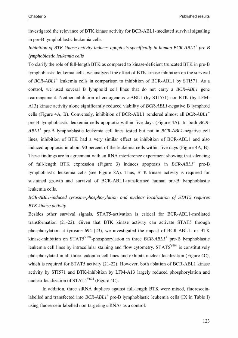
Chapter 5 Published results
123
investigated the relevance of BTK kinase activity for BCR-ABL1-mediated survival signaling
in pre-B lymphoblastic leukemia cells.
Inhibition of BTK kinase activity induces apoptosis specifically in human BCR-ABL1+ pre-B
lymphoblastic leukemia cells
To clarify the role of full-length BTK as compared to kinase-deficient truncated BTK in pre-B
lymphoblastic leukemia cells, we analyzed the effect of BTK kinase inhibition on the survival
of BCR-ABL1+ leukemia cells in comparison to inhibition of BCR-ABL1 by STI571. As a
control, we used several B lymphoid cell lines that do not carry a BCR-ABL1 gene
rearrangement. Neither inhibition of endogenous c-ABL1 (by STI571) nor BTK (by LFM-
A13) kinase activity alone significantly reduced viability of BCR-ABL1-negative B lymphoid
cells (Figure 4A, B). Conversely, inhibition of BCR-ABL1 rendered almost all BCR-ABL1+
pre-B lymphoblastic leukemia cells apoptotic within five days (Figure 4A). In both BCR-
ABL1+ pre-B lymphoblastic leukemia cell lines tested but not in BCR-ABL1-negative cell
lines, inhibition of BTK had a very similar effect as inhibition of BCR-ABL1 and also
induced apoptosis in about 90 percent of the leukemia cells within five days (Figure 4A, B).
These findings are in agreement with an RNA interference experiment showing that silencing
of full-length BTK expression (Figure 3) induces apoptosis in BCR-ABL1+ pre-B
lymphoblastic leukemia cells (see Figure 8A). Thus, BTK kinase activity is required for
sustained growth and survival of BCR-ABL1-transformed human pre-B lymphoblastic
leukemia cells.
BCR-ABL1-induced tyrosine-phosphorylation and nuclear localization of STAT5 requires
BTK kinase activity
Besides other survival signals, STAT5-activation is critical for BCR-ABL1-mediated
transformation (21-22). Given that BTK kinase activity can activate STAT5 through
phosphorylation at tyrosine 694 (23), we investigated the impact of BCR-ABL1- or BTK
kinase-inhibition on STAT5Y694-phosphorylation in three BCR-ABL1+ pre-B lymphoblastic
leukemia cell lines by intracellular staining and flow cytometry. STAT5Y694 is constitutively
phosphorylated in all three leukemia cell lines and exhibits nuclear localization (Figure 4C),
which is required for STAT5 activity (21-22). However, both ablation of BCR-ABL1 kinase
activity by STI571 and BTK-inhibition by LFM-A13 largely reduced phosphorylation and
nuclear localization of STAT5Y694 (Figure 4C).
In addition, three siRNA duplices against full-length BTK were mixed, fluorescein-
labelled and transfected into BCR-ABL1+ pre-B lymphoblastic leukemia cells (IX in Table I)
using fluorescein-labelled non-targeting siRNAs as a control.

Chapter 5 Published results
124
0 20 40 60 80 100 120 140
0
20
40
60
80
100
120
A120
100
80
60
40
20
0
0 20 40 60 80 100 120 140
Per
cent
livin
gce
lls
Time in hours
No BCR-ABL1
B
0 20 40 60 80 100 120 140
0
20
40
60
80
100
120120
100
80
60
40
20
0
0 20 40 60 80 100 120 140 Time in hours
BCR-ABL1p210
0 20 40 60 80 100 120 140
0
20
40
60
80
100
120120
100
80
60
40
20
0
0 20 40 60 80 100 120 140 Time in hours
BCR-ABL1p190
Survival [%] Cell line Cell type Translocation Genes involved
85.2 + 9.6 MHH-PREB-1 Burkitt‘s Lymphoma t(8;14)(q24;q32) MYC-IGH
88.4 + 3.4 KASUMI-2 pre-B cell leukemia t(1;19)(q23;p13) E2A-PBX1
66.8 + 8.4 697 pre-B cell leukemia t(1;19)(q23;p13) E2A-PBX1
76.2 + 7.0 KARPAS-422 Diffuse Large B Cell Lymphoma t(14;18)(q32;q21) BCL2-IGH
93.6 + 0.6 RS4;11 pro-B cell leukemia t(4;11)(q21;q23) MLL-AF4
49.4 + 7.6 REH pre-B cell leukemia t(12;21)(p12;q22) TEL-AML1
97 + 5.1 SEM pro-B cell leukemia t(4;11)(q21;q23) MLL-AF4
89.2 + 1.3 LCL polyclonal EBV+ mature B cells no chromosomal aberration
7.3 + 4.1 BV173 pre-B cell leukemia t(9;22)(q34;q11) BCR-ABL1
19.3 + 15.5 SUP-B15 pre-B cell leukemia t(9;22)(q34;q11) BCR-ABL1
Relative survival [%] average: 80.7 + 5.4 in non-BCR-ABL1+ B cell lineage cells13.3 + 9.8 in BCR-ABL1+ pre-B cell leukemia
+STI571 +LFM-A13
STAT5pY694
STAT5pY694
Control +STI571 +LFM-A13
Control
+ STI571
+ LFM-A13
BCR-ABL1+ B cell precursor leukemia cell lines:
VII VIII IX
non-targeting siRNA +STI571
non-targeting siRNA
BTKfull-length
siRNA
Red: STAT5pY694
Green: siRNA
C
Control
STI571
LFMA13
36 33 30 27PCR-cycles 36 33 30 27 36 33 30 27
BCLXL
GAPDH
BCLXL
GAPDH
BCLXL
GAPDH
BCR-ABL1+ B cell precursor leukemia cell lines:
VII VIII IX
D
0 20 40 60 80 100 120 140
0
20
40
60
80
100
120
A120
100
80
60
40
20
0
0 20 40 60 80 100 120 140
Per
cent
livin
gce
lls
Time in hours
No BCR-ABL1
B
0 20 40 60 80 100 120 140
0
20
40
60
80
100
120120
100
80
60
40
20
0
0 20 40 60 80 100 120 140 Time in hours
BCR-ABL1p210
0 20 40 60 80 100 120 140
0
20
40
60
80
100
120120
100
80
60
40
20
0
0 20 40 60 80 100 120 140 Time in hours
BCR-ABL1p190
Survival [%] Cell line Cell type Translocation Genes involved
85.2 + 9.6 MHH-PREB-1 Burkitt‘s Lymphoma t(8;14)(q24;q32) MYC-IGH
88.4 + 3.4 KASUMI-2 pre-B cell leukemia t(1;19)(q23;p13) E2A-PBX1
66.8 + 8.4 697 pre-B cell leukemia t(1;19)(q23;p13) E2A-PBX1
76.2 + 7.0 KARPAS-422 Diffuse Large B Cell Lymphoma t(14;18)(q32;q21) BCL2-IGH
93.6 + 0.6 RS4;11 pro-B cell leukemia t(4;11)(q21;q23) MLL-AF4
49.4 + 7.6 REH pre-B cell leukemia t(12;21)(p12;q22) TEL-AML1
97 + 5.1 SEM pro-B cell leukemia t(4;11)(q21;q23) MLL-AF4
89.2 + 1.3 LCL polyclonal EBV+ mature B cells no chromosomal aberration
7.3 + 4.1 BV173 pre-B cell leukemia t(9;22)(q34;q11) BCR-ABL1
19.3 + 15.5 SUP-B15 pre-B cell leukemia t(9;22)(q34;q11) BCR-ABL1
Relative survival [%] average: 80.7 + 5.4 in non-BCR-ABL1+ B cell lineage cells13.3 + 9.8 in BCR-ABL1+ pre-B cell leukemia
+STI571 +LFM-A13
STAT5pY694
STAT5pY694
Control +STI571 +LFM-A13
Control
+ STI571
+ LFM-A13
BCR-ABL1+ B cell precursor leukemia cell lines:
VII VIII IX
non-targeting siRNA +STI571
non-targeting siRNA
BTKfull-length
siRNA
Red: STAT5pY694
Green: siRNA
C
Control
STI571
LFMA13
36 33 30 27PCR-cycles 36 33 30 27 36 33 30 27
BCLXL
GAPDH
BCLXL
GAPDH
BCLXL
GAPDH
BCR-ABL1+ B cell precursor leukemia cell lines:
VII VIII IX
D
Figure 4: Inhibition of BTK induces apoptosis specifically in BCR-ABL1+
pre-B lymphoblastic leukemia cells

Chapter 5 Published results
125
Legend to Figure 4:
BCR-ABL1-negative B lymphoid leukemia and lymphoma cell lines (A, left panel) and two BCR-ABL+
leukemia cell lines (A, middle and right panel) were incubated for the times indicated under control conditions, with STI571 (solid line) or with the BTK-inhibitor LFM-A13 (dashed line; A). Relative viability (percent living cells) was calculated as the ratio of viability in the presence of STI571 or LFM-A13 and viability in untreated cells. Two BCR-ABL1+ and eight BCR-ABL1-negative B lymphoid cell lines were incubated in the presence of LFM-A13 or under control conditions for 114 hours (B). Relative survival of the treated cells as compared to control conditions is given as the mean of three independent experiments + S.D.
To analyze BCR-ABL-mediated and BTK-dependent tyrosine-phosphorylation of STAT5 (C), three BCR-ABL1+ B cell precursor cell lines (VII, VIII and IX in Table I) were treated with the BCR-ABL1 kinase-inhibitor STI571 (dotted line), with the BTK-inhibitor LFM-A13 (grey solid line) or incubated under control conditions (shaded histograms; C). Nuclear localization of activated STAT5 (red) in treated and untreated leukemia cells was visualized by confocal laser microscopy (C; red stain, middle panel). BCR-ABL1+ pre-B lymphoblastic leukemia cells were transfected with a mixture of fluorescein-labeled siRNAs against full-length BTK as shown in Figure 3. Fluorescein+ cells carrying siRNA molecules (green) were stained for tyrosine-phosphorylated STAT5 (red) and analyzed by confocal laser microscopy (C, lower panel). As a negative control, non-targeting siRNA-duplices were used. Next, mRNA levels of BCLXL were studied in BCR-ABL1+ pre-B lymphoblastic leukemia cells in the presence or absence of STI571 or LFM-A13. Total RNA was isolated from the leukemia cells and subjected to semiquantitative RT-PCR analysis for mRNA levels of BCLXL (D). cDNA amounts were normalized by amplification of a GAPDH cDNA fragment.
After two transfections, fluorescein+ leukemia cells were sorted and analyzed for
STAT5-phosphorylation (Figure 4C, lower panel). Efficiency and specificity of BTK full-
length silencing was controlled by RT-PCR (Figure 3). In agreement with BTK-inhibition by
LFM-A13, silencing of full-length BTK by RNA interference largely attenuated STAT5Y694-
phosphorylation as shown by confocal lasermicroscopy (Figure 4C) and intracellular FACS-
staining (Figure 8B).
BTK-mediated transcriptional activation of BCLXL in BCR-ABL1+ pre-B lymphoblastic
leukemia cells
In BCR-ABL1+ leukemia cells, STAT5 mainly accomplishes its anti-apoptotic effect by
transcriptional activation of BCLXL (24). Comparing the frequency of SAGE-tags that
distinguish between anti-apoptotic BCLXL and its pro-apoptotic isoform BCLXS in 19
genome-wide SAGE-libraries, SAGE-tags specific for BCLXL were particularly frequent in
BCR-ABL1+ pre-B lymphoblastic leukemia cells (supplementary Table III).
Analyzing mRNA levels for BCLXL in BCR-ABL1+ pre-B lymphoblastic leukemia
cells in the presence or absence of STI571 or LFM-A13, we found that inhibition of BCR-
ABL1 or BTK leads to a considerable reduction of BCLXL expression in the leukemia cells
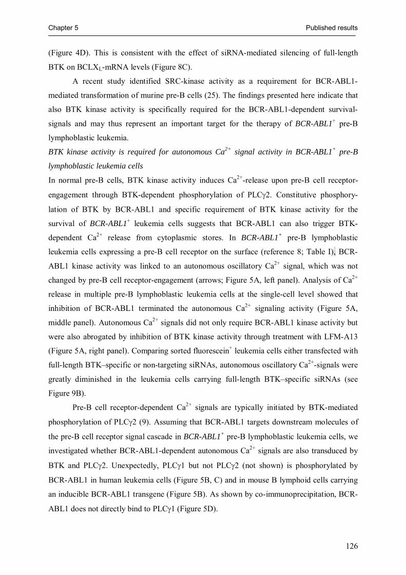
Chapter 5 Published results
126
(Figure 4D). This is consistent with the effect of siRNA-mediated silencing of full-length
BTK on BCLXL-mRNA levels (Figure 8C).
A recent study identified SRC-kinase activity as a requirement for BCR-ABL1-
mediated transformation of murine pre-B cells (25). The findings presented here indicate that
also BTK kinase activity is specifically required for the BCR-ABL1-dependent survival-
signals and may thus represent an important target for the therapy of BCR-ABL1+ pre-B
lymphoblastic leukemia.
BTK kinase activity is required for autonomous Ca2+ signal activity in BCR-ABL1+ pre-B
lymphoblastic leukemia cells
In normal pre-B cells, BTK kinase activity induces Ca2+-release upon pre-B cell receptor-
engagement through BTK-dependent phosphorylation of PLCγ2. Constitutive phosphory-
lation of BTK by BCR-ABL1 and specific requirement of BTK kinase activity for the
survival of BCR-ABL1+ leukemia cells suggests that BCR-ABL1 can also trigger BTK-
dependent Ca2+ release from cytoplasmic stores. In BCR-ABL1+ pre-B lymphoblastic
leukemia cells expressing a pre-B cell receptor on the surface (reference 8; Table I), BCR-
ABL1 kinase activity was linked to an autonomous oscillatory Ca2+ signal, which was not
changed by pre-B cell receptor-engagement (arrows; Figure 5A, left panel). Analysis of Ca2+
release in multiple pre-B lymphoblastic leukemia cells at the single-cell level showed that
inhibition of BCR-ABL1 terminated the autonomous Ca2+ signaling activity (Figure 5A,
middle panel). Autonomous Ca2+ signals did not only require BCR-ABL1 kinase activity but
were also abrogated by inhibition of BTK kinase activity through treatment with LFM-A13
(Figure 5A, right panel). Comparing sorted fluorescein+ leukemia cells either transfected with
full-length BTK–specific or non-targeting siRNAs, autonomous oscillatory Ca2+-signals were
greatly diminished in the leukemia cells carrying full-length BTK–specific siRNAs (see
Figure 9B).
Pre-B cell receptor-dependent Ca2+ signals are typically initiated by BTK-mediated
phosphorylation of PLCγ2 (9). Assuming that BCR-ABL1 targets downstream molecules of
the pre-B cell receptor signal cascade in BCR-ABL1+ pre-B lymphoblastic leukemia cells, we
investigated whether BCR-ABL1-dependent autonomous Ca2+ signals are also transduced by
BTK and PLCγ2. Unexpectedly, PLCγ1 but not PLCγ2 (not shown) is phosphorylated by
BCR-ABL1 in human leukemia cells (Figure 5B, C) and in mouse B lymphoid cells carrying
an inducible BCR-ABL1 transgene (Figure 5B). As shown by co-immunoprecipitation, BCR-
ABL1 does not directly bind to PLCγ1 (Figure 5D).

Chapter 5 Published results
127
A
PLCγ1pY783
EIF-4E
LFM-A13
STI571 − + − − + − − + −− − + − − + − − +
BCR-ABL1+ B cell precursorleukemia cell lines:
VII VIII IX
B Red: PLCγ1pY783
Green: CD10Red: PLCγ1pY783
Green: CD10
−STI571
+STI571
C
ABL1
PLCγ1pY783
CoIP: BCR
p210 p190
BCR-ABL1+ B cellprecursor cell lines:
VII IX
WB:
77 kD
EIF-4E
BTKpY223
PLCγ1pY783
IL-3
DO
X
IL-3
, DO
X,
STI
571
0 50 100 150 200 250 300 350
30
60
90
120
150
180
210
0 50 100 150 200 250 300 350
60
90
120
150
180
210
240+LFM-A13
Time in seconds
0 50 100 150 200 250 300 350
60
90
120
150
180
210
240
Time in seconds
Control
0 50 100 150 200 250 300 350
210
180
150
120
90
60
30
Ca2
+ i [n
mol
/l]
0 50 100 150 200 250 300 350
210
180
150
120
90
60
30
+STI571
Time in seconds
0 50 100 150 200 250 300 350
210
180
150
120
90
60
30
non-targetingsiRNA
non-targetingsiRNA + STI571
BTKfull-length
siRNA
Red: PLCγ1pY783
Green: siRNA
D
Figure 5: BTK links BCR-ABL1 to PLCγ1-mediated Ca2+ signals in pre-B lymphoblastic
leukemia cells

Chapter 5 Published results
128
Legend to Figure 5:
Cytoplasmic Ca2+ levels were measured in single leukemia cells by confocal laser-microscopy. Before Ca2+ measurement, cells were either kept under control conditions or treated for 24 hours with STI571 or LFM-A13 (A). Pre-B cell receptor engagement by addition of µ-heavy chain-specific antibodies (arrows) did not change cytoplasmic Ca2+ concentrations. While PLCγ1Y783 is phosphorylated in the presence but not in the absence (+ STI571) of BCR-ABL1 kinase activity (B, left panels), PLCγ2 was not tyrosine-phosphorylated by BCR-ABL1 (not shown). As a control for non-specific side-effects of STI571, murine B lymphoid cells carrying a doxycycline-inducible BCR-ABL1 transgene were analyzed in the presence or absence of 1 µg/ml doxycycline (DOX). These cells remain viable in the absence of BCR-ABL1 expression if the cell culture fluid is supplemented with 2 ng/ml murine recombinant IL-3. Protein extracts from these cells were subjected to Western blot using human- and mouse-reactive antibodies (B, right panel). Tyrosine-phosphorylation of PLCγ1 was studied in BCR-ABL1+ pre-B lymphoblastic leukemia cells in the presence or absence of STI571 or LFM-A13. EIF-4E was used as a loading control in both Western blots (B, C). Expression of full-length BTK was silenced by RNA interference in a BCR-ABL1+ pre-B lymphoblastic leukemia cell line (IX in Table I). Sorted cells carrying fluorescein-labeled siRNAs (green) were permeabilized, stained for PLCγ1pY783 (red) and analyzed by confocal laser microscopy (C, lower panel). As a negative control non-targeting siRNA duplices were used. BCR-ABL1-binding proteins were co-immunoprecipitated using an antibody against BCR. The immunoprecipitation was controlled by a Western blot using an ABL1-specific antibody (D).
Phosphorylation of PLCγ1 was sensitive to inhibition of BTK by LFM-A13 (Figure
5C) indicating that PLCγ1 is activated through BTK as in normal lymphocytes and not
directly by BCR-ABL1. Silencing of full-length BTK by RNA interference using fluorescein-
labeled full-length BTK-specific siRNAs also results in a marked reduction of PLCγ1Y783-
phosphorylation as assessed by confocal lasermicroscopy (Figure 5C) and intracellular FACS-
staining (Figure 9A).
PLCγ1 contributes to survival signals in BCR-ABL1+ pre-B lymphoblastic leukemia cells
Whereas the role of STAT5 in BCR-ABL1-mediated survival signaling is well established
(21, 22, 24), a possible role of PLCγ1 to promote survival of BCR-ABL1+ pre-B lymphoblastic
leukemia cells was not investigated so far.
To determine whether BCR-ABL1- and BTK-dependent activation of PLCγ1 and
PLCγ1-mediated Ca2+ release contributes to BCR-ABL1-mediated survival signals, we
silenced PLCγ1 mRNA expression by RNA interference in three pre-B lymphoblastic
leukemia cell lines carrying a BCR-ABL1 gene rearrangement (VII to IX, Table I) and four
cell lines carrying other chromosomal translocations (MLL-AF4, X; TEL-AML1, XII; E2A-
PBX1, XIII; TEL-PDGFRB, XV; Table I). As a control, non-targeting siRNAs were used.
Cells were transfected twice with fluorescein-labeled siRNAs. To asses viability, fluorescein+
cells were analyzed for propidium iodide uptake and Annexin V-staining after 48 hours.

Chapter 5 Published results
129
Among the three BCR-ABL1+ pre-B lymphoblastic leukemia cell lines, viability (mean of
three experiments + S.D.) was 90.3 + 2.7 percent with non-targeting siRNAs and 18.6 + 13.7
percent with PLCγ1-specific siRNAs. Among four leukemia cell lines carrying other gene
rearrangements, viability was 84.3 + 12.2 percent with non-targeting siRNAs and 67.6 + 16.9
percent with PLCγ1-specific siRNAs. Unlike BCR-ABL1-negative leukemia cells, silencing of
PLCγ1 in BCR-ABL1+ pre-B lymphoblastic leukemia cells results in a 5-fold reduction of
viability. Thus, PLCγ1 contributes to survival signaling specifically in BCR-ABL1+ pre-B
lymphoblastic leukemia cell lines.
C-terminally truncated BTK functions as a linker between full-length BTK and BCR-ABL1
BCR-ABL1-dependent phosphorylation of BTK suggests that BTK represents a substrate of
the BCR-ABL1 kinase. However, previous work demonstrated that BCR-ABL1 cannot
directly phosphorylate full-length BTK but the N-terminal BTK-SH3 domain fused to GST
(26).
To determine how BCR-ABL1 can induce phosphorylation of full-length BTK, we
studied whether full-length BTK or BTK splice variants are part of the BCR-ABL1-
signalosome. Using protein extracts of two BCR-ABL1+ pre-B lymphoblastic leukemia cell
lines, BCR-ABL1 signaling complexes were immunoprecipitated with a BCR-specific
antibody. Immunoprecipitation was controlled using an anti-ABL1 antibody. Western blot
showed that low amounts of full-length BTK indeed co-immunoprecipitates with BCR-ABL1
(Figure 6A). As a control for quantitative distribution of full-length BTK, BTKp65 and BTKp52
before co-immunoprecipitation with BCR-ABL1, whole cell lysates (WCL) from two BCR-
ABL1+ leukemia cell lines were subjected to Western blot (Figure 6A). While full-length
BTK, BTKp65 and BTKp52 are expressed at similar levels in the leukemia cells (WCL, Figure
6A), the amount of full-length BTK co-immunoprecipitating with BCR-ABL1 was
approximately 20-fold less than the amount of BTKp52 co-immunoprecipitating with BCR-
ABL1. While BTKp65 is clearly expressed in the leukemia cells (WCL, Figure 6A), co-
immunoprecipitation of BTKp65 with BCR-ABL1 could not be detected (Figure 6A).
Preferential binding of BCR-ABL1 to BTKp52 lacking the C-terminus but retaining the
N-terminal BTK SH3-domain is consistent with previous findings demonstrating that BCR-
ABL1 cannot directly phosphorylate full-length BTK but a BTK-SH3 domain-GST fusion
molecule (26). However, binding of BTKp52 to BCR-ABL1 may also be indirect in this case.
That the N-terminal BTK SH3-domain but not full-length BTK can be readily phosphorylated
by BCR-ABL1 suggests that the C-terminus of BTK may interfere with binding of BCR-
ABL1 to BTK.

Chapter 5 Published results
130
Figure 6: C-terminally truncated BTK functions as a linker between full-length BTK
and BCR-ABL1
Likewise, BTKp65 molecules that harbor an in-frame deletion but retain the BTK C-terminus
do not immunoprecipitate with BCR-ABL1. In this regard, the expression of truncated BTK
splice variants lacking the C-terminus of BTK may represent a mechanism to facilitate
phosphorylation of full-length BTK by BCR-ABL1. As BTK molecules can also self-
associate through N-terminal SH3 domains (27), BTKp52 may bind to BCR-ABL1 as well as
to full-length BTK acting as linker between the two kinases.
We next investigated whether full-length BTK can bind to BTKp52. Using an antibody
specific for C-terminal BTK (only recognizing full-length BTK) for immunoprecipitation and
an N-terminal BTK-antibody (recognizing both full-length BTK and BTKp52) for subsequent
Western blotting, we observed that BTKp52 co-immunoprecipitates with full-length BTK
Non
-targ
etin
gsi
RN
A
Non
-targ
etin
gsi
RN
A
BTKfull-length
BTKp52
BTKp65
BTKpY223
EIF4e
BTK
p52
siR
NA
BTK
p52
siR
NA
ABL1
WB:
N-terminalBTK
IgH
WCL CoIP: BCR
BTKfull-length
BTKp52
BTKp65
BCR-ABL1
A BCoIP: C-terminalBTK
Exp. 1 Exp. 2
Non
-targ
etin
gsi
RN
A
Non
-targ
etin
gsi
RN
A
BTKfull-length
BTKp52
BTKp65
BTKpY223
EIF4e
BTK
p52
siR
NA
BTK
p52
siR
NA
ABL1
WB:
N-terminalBTK
IgHIgH
WCL CoIP: BCR
BTKfull-length
BTKp52BTKp52
BTKp65
BCR-ABL1
A BCoIP: C-terminalBTK
Exp. 1 Exp. 2
Proteins binding to BCR-ABL1 were co-immunoprecipitated with an anti-BCR antibody (A). Immunoprecipitation was controlled by an anti-ABL1-specific Western blot showing the characteristic BCR-ABL1 fusion protein expressed by the leukemia cell line. Proteins binding to full-length BTK were co-immunoprecipitated with an antibody against C-terminal BTK. Immunoprecipitation was controlled by Western blot using an antibody against N-terminal BTK. Full-length BTK and BTKp52 proteins co-immunoprecipitating with either BCR-ABL1 or full-length BTK were visualized by Western blot using an antibody against N-terminal BTK (A). As a control for quantitative distribution of full-length BTK and BTKp52 before co-immunoprecipitation with BCR-ABL1 or full-length BTK, whole cell lysates (WCL) were used (A). To analyze the effect of BTKp52 on BCR-ABL1-dependent phosphorylation of full-length BTK, BTKp52 expression was inhibited by RNA interference as described in Figure 3. As a control, non-targeting siRNA duplices were used. siRNAs were fluorescein-labeled and fluorescein+ cells were sorted and subjected to Western blot using antibodies specific for tyrosine-phosphorylated BTKY223 (B). As patterns of tyrosine-phosphorylation varied between replicate experiments, Western blots of two representative experiments (Exp. 1 and 2) are shown. EIF4e was used as a loading control.
BTKfull-length
BTKp65
BTKp52
BCR-ABL1
BTKfull-length
BTKp65
BTKp52
IgH

Chapter 5 Published results
131
(Figure 6A). These findings suggest a role for BTKp52 as a linker between BCR-ABL1 and
full-length BTK.
In this case, BTKp52 would also facilitate tyrosine phosphorylation of full-length BTK
by BCR-ABL1. Therefore, we tested whether silencing of BTKp52 in BCR-ABL1+ pre-B
lymphoblastic leukemia cells results in decreased tyrosine-phosphorylation of full-length
BTK. To this end, BCR-ABL1+ pre-B lymphoblastic leukemia cells were transfected with
either non-targeting or BTKp52-specific fluorescein-labeled siRNAs (Figure 3). Fluorescein+
cells were sorted and analyzed for expression of tyrosine-phosphorylated BTK by Western
blot (Figure 6B). Silencing of BTKp52 mRNA expression by siRNAs did not interfere with
mRNA expression of full-length BTK (Figure 3), yet tyrosine-phosphorylation of full-length
BTK was decreased by more than 90 percent (Figure 6B).
Given that i.) BCR-ABL1 and full-length BTK cannot directly bind to each other, ii.)
BTKp52 can bind to both BCR-ABL1 and full-length BTK and iii.) expression of BTKp52 is
required for efficient tyrosine-phosphorylation of full-length BTK, we conclude that BTKp52
can act as a linker between BCR-ABL1 and full-length BTK. This is in agreement with
previous work suggesting that kinase-deficient BTK may act as a linker molecule between
upstream kinases (e.g. SYK) and downstream effector molecules (e.g. PLCγ2; reference 11).
C-terminally truncated BTK can link full-length BTK to BCR-ABL1 through its SH3 domain
Based on this assumption, one would predict that the coexpression of BTKp52 with full-length
BTK can facilitate the interaction between BTK and BCR-ABL1 and, hence, the
phosphorylation of downstream signaling molecules. To test this possibility, BCR-ABL1,
BTK and its truncated variant BTKp52 were expressed in 293T embryonic kidney cells either
alone, in various combinations or in the presence of BTK- (LFM-A13) or BCR-ABL1-
(STI571) kinase inhibitors. As a readout of this experiment, tyrosine phosphorylation of
PLCγ1 and STAT5 was measured. According to a previous study, 293T cells do not express
BTK but both PLCγ1 and STAT5 (28).
The expression of either full-length BTK, BTKp52 or BCR-ABL1 alone had no effect
on tyrosine-phosphorylation of downstream molecules in general. However, to some extent,
single transfection with full-length BTK induced activation of PLCγ1. Likewise, BCR-ABL1
alone induced to some extent tyrosine phosphorylation of STAT5 (Figure 7AB), indicating
that BCR-ABL1 can partially activate STAT5 in the absence of BTK. Co-expression of
BTKp52 lacking a functional kinase domain together with BCR-ABL1 did not increase
tyrosine-phosphorylation as compared to expression of BCR-ABL1 alone. However, co-
expression of BTKp52 with full-length BTK and BCR-ABL1 drastically upregulated both

Chapter 5 Published results
132
PLCγ1 and STAT5-phosphorylation (Figure 7A). As assessed by intracellular staining,
phosphorylated PLCγ1 and STAT5 was distributed in the cytoplasm and in the nucleus,
respectively (Figure 7B).
Activation of PLCγ1 and STAT5 upon triple-transfection with full-length BTK,
BTKp52 and BCR-ABL1 was largely sensitive to BTK- (LFM-A13) or BCR-ABL1-
(STI571) kinase inhibition. Hence, enzymatic activity of both kinases is required for
activation of PLCγ1 and STAT5. As a control, the enzymatic activity of BTK expressed in
transfected 293T cells was confirmed in an in vitro kinase assay as described in the Materials
and Methods section. The kinase activity of BTK immunoprecipitated from 1 x 10⁵
transfected 293T cells was roughly comparable to that of 25 ng (0.008 U) recombinant BTK
(Figure 7C).
As BTK-SH3 domains can directly bind to BCR-ABL1 (26) and also bind to proline-
rich regions of other BTK molecules (27), we tested whether the BTK-SH3 domain is
sufficient to link full-length BTK to BCR-ABL1. In fact, transfection of 293T cells with a
vector encoding only the BTK-SH3 domain had a similar effect compared to transfection with
BTKp52 comprising PH, TH, SH3, SH2 and a truncated kinase domain (Figure 2C). Indeed,
the SH3 domain was sufficient to enable BCR-ABL1-driven activation of PLCγ1 and STAT5
in the presence of active full-length BTK (Figure 7A). We conclude that BTKp52, mainly
through its SH3 domain, can act as a linker between full-length BTK and BCR-ABL1.
BTKp52 but not BTKp65 cooperates with full-length BTK to transduce BCR-ABL1-dependent
survival signals
To clarify the specific contribution of BTKp52 and BTKp65 to BCR-ABL1-downstream
survival signaling, Annexin V expression, STAT5-phosphorylation and BCLXL mRNA levels
were measured in leukemia cells after transfection with fluorescein-labelled siRNAs against
BTKp52 and BTKp65 and non-targeting siRNAs (Figure 8 A-C). Transfection of leukemia cells
with non-targeting siRNAs or siRNAs against BTKp65 had no effect on viability as assessed
by Annexin V-staining (Figure 8A), tyrosine-phosphorylation of STAT5 (Figure 8B) and
BCLXL mRNA levels (Figure 8C). In contrast, siRNA-mediated silencing of BTKp52 induced
apoptosis in BCR-ABL1+ pre-B lymphoblastic leukemia cells to a similar extent as full-length
BTK (Figure 8A). Likewise, silencing of either full-length BTK or BTKp52 decreased
tyrosine-phosphorylation of STAT5 (Figure 8B) and reduced mRNA levels of BCLXL in the
leukemia cells (Figure 8C). These findings collectively indicate, that survival signals in BCR-
ABL1+ pre-B lymphoblastic leukemia cells not only require BTK kinase activity but also
BTKp52 acting as a linker between full-length BTK and BCR-ABL1.

Chapter 5 Published results
133
Figure 7: BTKp52 facilitates BTK- and BCR-ABL1-dependent activation of PLCγ1 and
STAT5 through its SH3-domain
BTK BTK none none IP (293T cells)
none IP BTK BTK-SH3 Kinase Assay (PLCγ1)
BTK PLCγ1Y783 PLCγ1Y783 PLCγ1Y783 WB
77 kD
61 kD
IgH
+ BCR-ABL1 − − + BCR-ABL1 + BCR-ABL1 + BCR-ABL1 + BCR-ABL1 − + BTKfull-length − + BTKfull-length − + BTKfull-length + BTKfull-length
− − + BTKp52 − + BTKp52 + BTKp52 + BTKp52
− − − − − − + LFM-A13
PLCγ1Y783
STAT5Y694
GFP
BCR−ABL1
BTKp52
BTK−SH3
BTKfull-length
BCR−ABL1 + BTKp52
BCR−ABL1 + BTK−SH3
BCR−ABL1 + BTKfull-length
BCR−ABL1 + BTKfull-length + BTKp52
BCR−ABL1 + BTKfull-length + BTK−SH3
BCR−ABL1 + BTKfull-length + BTKp52 + STI571
BCR−ABL1 + BTKfull-length + BTKp52 + LFMA13
PLCγ1Y783 STAT5Y694
A
B
C BTK BTK none none IP (293T cells)
none IP BTK BTK-SH3 Kinase Assay (PLCγ1)
BTK PLCγ1Y783 PLCγ1Y783 PLCγ1Y783 WB
77 kD
61 kD
IgH
+ BCR-ABL1 − − + BCR-ABL1 + BCR-ABL1 + BCR-ABL1 + BCR-ABL1 − + BTKfull-length − + BTKfull-length − + BTKfull-length + BTKfull-length
− − + BTKp52 − + BTKp52 + BTKp52 + BTKp52
− − − − − − + LFM-A13
PLCγ1Y783
STAT5Y694
GFP
BCR−ABL1
BTKp52
BTK−SH3
BTKfull-length
BTKp52
BTK−SH3
BTKfull-length
BCR−ABL1 + BTKp52
BCR−ABL1 + BTK−SH3
BCR−ABL1 + BTKfull-length
BCR−ABL1 + BTKp52
BCR−ABL1 + BTK−SH3
BCR−ABL1 + BTKfull-length
BCR−ABL1 + BTKfull-length + BTKp52
BCR−ABL1 + BTKfull-length + BTK−SH3
BCR−ABL1 + BTKfull-length + BTKp52 + STI571
BCR−ABL1 + BTKfull-length + BTKp52
BCR−ABL1 + BTKfull-length + BTK−SH3
BCR−ABL1 + BTKfull-length + BTKp52 + STI571
BCR−ABL1 + BTKfull-length + BTKp52 + LFMA13
PLCγ1Y783 STAT5Y694
A
B
C

Chapter 5 Published results
134
Legend to Figure 7:
BCR-ABL1, full-length BTK, BTKp52 and the SH3-domain of BTK were expressed in 293T embryonic kidney cells either alone or in various combinations in the presence or absence of STI571 or LFM-A13 as indicated. As a readout, cells were harvested, subjected to intracellular staining for tyrosine-phosphorylated PLCγ1 or STAT5 and analyzed by flow cytometry (A). To visualize cytoplasmic localization of tyrosine phosphorylated PLCγ1 and nuclear localization of activated STAT5, the stained cells were also subjected to analysis by confocal laser-microscopy (B). 1 x 10⁶ 293T cells were transiently transfected with full-length BTK for 24 hours and subjected to immunoprecipitation of full-length BTK. Immunoprecipitation was controlled by an BTK-specific Western blot (C). Kinase activity of immunoprecipitated BTK was analyzed in an in vitro kinase assay using 150 ng of a PLCγ1 fragment (amino acids 530 to 850) as substrate. In parallel, 25 ng of recombinant active BTK and 100 ng of kinase-deficient SH3-domain of BTK were used in kinase assays as positive and negative controls, respectively.
Requirement of BTKp52 for BCR-ABL1-driven autonomous Ca2+ signaling activity
Autonomous oscillatory Ca2+ signals in BCR-ABL1+ pre-B lymphoblastic leukemia cells
require kinase activity of both BCR-ABL1 and BTK (Figure 5A) as well as PLCγ activity (9).
Therefore, we investigated the impact of specific silencing of full-length BTK, BTKp52 and
BTKp65 on PLCγ1 activation (Figure 9A). siRNA-mediated knockdown of full-length BTK-
and BTKp52-expression similarly reduced tyrosine-phosphorylation of PLCγ1, whereas
siRNAs against BTKp65 had no effect (Figure 9A). In agreement with this, siRNA-mediated
inhibition of either full-length BTK or BTKp52 expression but not expression of BTKp65
largely reduced the amplitude of autonomous Ca2+ oscillations in BCR-ABL1+ pre-B
lymphoblastic leukemia cells (Figure 9B). We could not identify a specific function of
BTKp65 with respect to BCR-ABL1-mediated transformation in pre-B lymphoblastic leukemia
cells.

Chapter 5 Published results
135
non-targetingsiRNA
non-targetingsiRNA +STI571
BTKfull length
siRNABTKp52
siRNABTKp65
siRNA
BCLXL
27 30 33 27 30 33 27 30 33 27 30 33 27 30 33 PCR cycles
C
Anne
xin
V
siRNAs
STAT
5pY69
4
non-targetingsiRNA
BTKfull length
siRNABTKp52
siRNABTKp65
siRNATransfection:A
siRNAs
B non-targetingsiRNA
BTKfull length
siRNABTKp52
siRNABTKp65
siRNATransfection:
COX6B
non-targetingsiRNA
non-targetingsiRNA +STI571
BTKfull length
siRNABTKp52
siRNABTKp65
siRNA
BCLXL
27 30 33 27 30 33 27 30 33 27 30 33 27 30 33 PCR cycles
C
Anne
xin
V
siRNAs
STAT
5pY69
4
non-targetingsiRNA
BTKfull length
siRNABTKp52
siRNABTKp65
siRNATransfection:A
siRNAs
B non-targetingsiRNA
BTKfull length
siRNABTKp52
siRNABTKp65
siRNATransfection: non-targeting
siRNABTKfull length
siRNABTKp52
siRNABTKp65
siRNATransfection:
COX6B
Figure 8: BTKp52 but not BTKp65 promotes cell survival, tyrosine-phosphorylation of
STAT5 and upregulation of BCLXL
BCR-ABL1+ pre-B lymphoblastic leukemia cells (IX, Table 1) were transfected with fluorescein-labeled siRNA duplices against full-length BTK, BTKp52 or BTKp65 as described above (see Figure 3) and analyzed by flow cytometry for Annexin V expression (A). As a negative control, non-targeting siRNA duplices were used. Likewise, siRNA-treated leukemia cells were subjected to intracellular staining for tyrosine-phosphorylated STAT5 and analyzed by flow cytometry (B). For the analysis of mRNA levels of BCLXL, BCR-ABL1+ pre-B lymphoblastic leukemia cells were transfected with a non-targeting siRNA duplex in the presence or absence of STI571 (for 12 hours), as well as with siRNAs against full-length BTK, BTKp52 or BTKp65. 1 x 10⁵ leukemia cells carrying fluorescein-labeled siRNAs sorted by FACS and subjected to semiquantitative RT-PCR analysis for mRNA levels of BCLXL (C). mRNA levels for COX6B (C) and HPRT (see Figure 3) remained stable in all experiments.

Chapter 5 Published results
136
Figure 9: Specific silencing of BTKfull-length or BTKp52 reduces PLCγ1 phosphorylation and
autonomous Ca2+ oscillations in BCR-ABL+ pre-B lymphoblastic leukemia cells
BCR-ABL1+ pre-B lymphoblastic leukemia cells were transfected with non-targeting siRNAs or siRNAs against full-length BTK, BTKp52 or BTKp65 (A), then subjected to intracellular staining for tyrosine-phophorylated PLCγ1 and analyzed by flow-cytometry (A). Cytoplasmic Ca2+ levels [nmol/l] were measured in single siRNA-containing cells by confocal laser-scanning microscopy by continuous scanning for six minutes (B). Prior to analysis of Ca2+ levels, leukemia cells carrying fluorescein-labeled siRNAs were sorted by FACS. For each condition, approximately 50 individual cells were recorded. As a control, cytoplasmic Ca2+ levels were measured in cells with non-targeting siRNAs (red line).
B
0 60 120 180 240 300 360
0
30
60
90
120
150
180
non-targeting siRNA
BTKp65 siRNA
BTKp52 siRNA
BTKfull-length siRNA
Time in seconds
Ca2+
i [nm
ol/l]
PLC
γ1pY
783
siRNA
Anon-targetingsiRNA
BTKfull length
siRNABTKp52
siRNABTKp65
siRNATransfection:
B
0 60 120 180 240 300 360
0
30
60
90
120
150
180
non-targeting siRNA
BTKp65 siRNA
BTKp52 siRNA
BTKfull-length siRNA
Time in seconds
Ca2+
i [nm
ol/l]
PLC
γ1pY
783
siRNA
Anon-targetingsiRNA
BTKfull length
siRNABTKp52
siRNABTKp65
siRNATransfection:

Chapter 5 Published results
137
Discussion
The leukemogenic BCR-ABL1 kinase mimicks a constitutively active pre-B cell receptor in
pre-B lymphoblastic leukemia cells. While the leukemia cells frequently carry only non-
functional IGH-alleles (8), pre-B cell receptor signaling is even compromised in the few
cases, in which the leukemia cells express a pre-B cell receptor on their surface: Important
components of the pre-B cell receptor signaling cascade including SYK, SLP65 and BTK are
not phosphorylated in response to pre-B cell receptor-engagement, and BTK is constitutively
phosphorylated by BCR-ABL1. BCR-ABL1-dependent activation of BTK is critical for
autonomous survival signals that would otherwise arise from the pre-B cell receptor. We
conclude that BTK kinase activity is no longer responsive to pre-B cell receptor-dependent
signals but contributes to multiple aspects of BCR-ABL1-driven survival signaling in pre-B
lymphoblastic leukemia cells.
While BCR-ABL1 cannot directly interact with full-length BTK (26), however, BCR-
ABL1-induced aberrant splicing of BTK pre-mRNA leads to the expression of a truncated
splice variant of BTK acting as a linker molecule between the two kinases. Acting as a linker,
truncated BTKp52 can not only facilitate BCR-ABL1-dependent activation of full-length BTK
but also of downstream molecules including PLCγ1, STAT5 and BCLXL.
The interaction between BCR-ABL1 and BTK is critical because not only inhibition of
BCR-ABL1 by STI571 but also interference with BTK kinase activity specifically induces
apoptosis in BCR-ABL1+ B lymphoid leukemia cells. As resistance to STI571 is frequent in
the therapy of this leukemia entity (15), inhibition of BTK or its truncated splice variant
potentially represents a therapeutic approach to circumvent STI571-resistance of BCR-ABL1+
pre-B lymphoblastic leukemia cells.
Acknowledgements:
We would like to thank Martin Krönke and Klaus Rajewsky for continuous support and discussions and Stefanie
Jauch and Peter Wurst for excellent technical assistance. N.F. is supported by a fellowship of the José-Carreras-
Leukemia Foundation. F.K. is supported by the Studienstiftung des deutschen Volkes. M.M. is supported by the
Deutsche Forschungsgemeinschaft through the Emmy-Noether-Programm and through grants MU1616/2-1,
MU1616/3-1 (to M.M.), the German José-Carreras-Leukemia-Foundation (grant to M.M.), the Deutsche
Krebshilfe through Molecular Mechanisms of Malignant Lymphoma (to M.M.) and the Ministry of Science and
Research for North Rhine-Westphalia through the Stem Cell Network NRW (to M.M.). The authors have no
conflicting financial interests.

Chapter 5 Published results
138
Materials and Methods
Patient samples and cell lines
Clinical data on patient samples and cytogenetic data on cell lines used are summarized in
Table I and are described in detail in supplementary information. All studies on human
materials were performed on samples provided in compliance with Institutional Review Board
regulations (Department of Hematology, University of Frankfurt, Frankfurt/Main, Germany).
Sequence analysis of BTK and semiquantitative RT-PCR
In a search for BTK isoforms, fragments of the BTK cDNA were amplified covering the
entire coding region using the PCR primer pairs listed in supplementary Table I. BTK
amplification products were sequenced as previously described (29). Primers used for
semiquantitative RT-PCR analysis of human BCR-ABL1, BCLXL, GAPDH, HPRT and
COX6B transcripts are listed in supplementary Table I.
Transient expression of BCR-ABL1 in pre-B lymphoblastic leukemia cells
pre-B lymphoblastic leukemia cells (697 cell line) carrying an E2A-PBX1 but no BCR-ABL1
gene rearrangement were transiently transfected by electroporation (250 V and 950 μF) with
pMIG-GFP or pMIG-GFP_BCR-ABL1 vectors encoding either GFP only or GFP and BCR-
ABL1 as previously described (30). For both transfections, GFP+ and GFP cells were sorted
after 24 hours and subjected to mRNA isolation.
RNA interference with BTK and PLCγ1 expression
For each target, three different siRNA duplices were synthesized (MWG Biotech, Ebersberg,
Germany). Sequences of siRNA duplices are listed in supplementary Table I.
For knockdown of PLCγ1 mRNA expression, a pool of three validated siRNAs targeting
different PLCγ1 exons was used (Upstate, Lake Placid, NY). As a control, a non-targeting
siRNA duplex was used that does not match a known mRNA sequence. All siRNA duplices
were labeled with fluorescein using an siRNA labeling kit (Ambion, Austin, TX) according to
the manufacturer’s protocol. BCR-ABL1+ pre-B lymphoblastic leukemia cells were transfected
with a mixture of three fluorescein-labeled siRNAs for each target sequence at a concentration
of 100 nmol/l using Oligofectamine in Opti-MEM1 medium (Invitrogen). After 24 hours,
leukemia cells were re-transfected with labeled siRNAs and subsequently incubated for
further 24 hours. Transfection efficiency was controlled by fluorescence microscopy and by
FACS. The silencing effect of siRNAs for specific BTK isoforms was controlled by RT-PCR
analysis of BTK splice variants in sorted of fluorescein+ cells. siRNA-containing fluorescein+
cells were sorted using a FACStar 440 cell sorter. For each condition, 5 x 10⁵ cells were
sorted and subjected to RNA isolation and cDNA synthesis.

Chapter 5 Published results
139
Western blotting
For the detection of signaling molecules by Western blot, antibodies against BTK, PLCγ1,
PLCγ2, SLP65 and SYK (Cell Signaling Technology, Beverly, MA), as well as
phosphotyrosine-specific antibodies against BTKY223, PLCγ1Y783, PLCγ2Y1217, SLP65Y96 and
SYKY323(Cell Signaling Technology) were used together with the WesternBreeze
immunodetection system (Invitrogen). Mouse monoclonal antibodies against tyrosine-
phosphorylated BTKY223 and BTKY551 were kindly provided by Dr. Owen N. Witte, Los
Angeles, CA.
Co-immunoprecipitation
For immunoprecipitation of BCR-ABL1 or BTK, 1.7 mg protein lysate in a volume of 800 μl
was incubated with 30 μl rProtein G Agarose beads (Invitrogen) for two hours at 4°C on a
shaker. The supernatant was separated from the agarose beads and incubated with 6 µg of an
antibody against BCR (Santa Cruz Biotechnology) or with 15 µg of an antibody against C-
terminal BTK (kindly provided by Dr. Owen N. Witte, Los Angeles, CA). After 8 hours, 30
μl agarose beads were added and incubation was continued overnight. Afterwards, the agarose
beads were washed twice with lysis buffer, resuspended in 35 μl of lysis buffer and subjected
to Western blotting.
Flow cytometry
For FACS analysis of BCR-ABL1+ pre-B lymphoblastic leukemia cells, antibodies against
tyrosine-phosphorylated BTKY223, tyrosine-phosphorylated PLCγ1Y783 and tyrosine-
phosphorylated STAT5Y694 (Cell Signaling Technologies) were used. Apoptotic or dead cells
were identified by propidium iodide or PE-labelled Annexin V (BD Biosciences). As
secondary antibody, anti-rabbit or anti-mouse IgG-Cy3 (Jackson Immunoresearch) was used.
Immunofluorescence and confocal lasermicroscopy
Nuclear, cytoplasmic or membrane localization of phosphorylated BTKY223, PLCγ1Y783 and
STAT5Y694 was analyzed using primary antibodies from Cell Signaling Technology together
with anti-rabbit IgG-Cy5 (Jackson Immunoresearch) as secondary antibody, respectively.
Cells were fixed with 0.4% para-formaldehyde and incubated for 10 minutes in 90% methanol
on ice and subjected to confocal laser-scanning microscopy as previously described (14).
Expression of BCR-ABL1, full-length BTK, BTKp52 and the BTK-SH3 domain in 293T cells
1 x 10⁶ 293T embryonic kidney cells were plated on a 24-well plate 24 hours prior to
transfection. Different combinations of expression vectors were prepared as indicated and 5
µg of each vector was incubated with 8 µl of FuGENE6 (Roche) and 50 µl of serum-and
antibiotic-free RPMI medium for 15 minutes at room temperature. The expression vector

Chapter 5 Published results
140
pMIG-bcr/abl for BCR-ABL1p210 (kindly provided by Dr. David Baltimore, La Jolla, CA) was
used as previously described (30). The expression vector pMIG-flagBtk for human full-length
BTK was generated by cloning of the human BTK cDNA into the pMIG-R vector (30). For
expression of BTKp52 (EMBL/GenBank accession number AJ888378), the expression vector
pcDNA3.1 was used together with the directional TOPO Expression Kit (Invitrogen).
For expression of the BTK-SH3 domain, the pEBG BTK-SH3 vector (kind gift from Dr.
Magnus Bäckesjø, Karolinska Institute, Huddinge, Sweden) was used as previously described
(26).
Measurement of Ca2+ signals in BCR-ABL1+ pre-B lymphoblastic leukemia cells
Pre-B lymphoblastic leukemia cells carrying a BCR-ABL1 gene rearrangement were cultured
in the presence or absence of the BCR-ABL1-inhibitor STI571 (from Novartis, Basel,
Switzerland) or the BTK-inhibitor LFM-A13 (Calbiochem, Darmstadt, Germany) for the
times indicated. In a different set of experiments, cells were transfected with fluorescein-
labeled siRNA duplices and fluorescein+ cells were sorted and conditioned in RPMI-medium
at 37°C before measurement of cytoplasmic Ca2+ concentrations. Cells were washed and
stained with Fluo-3 dye (Calbiochem) for 30 minutes. Changes of cytosolic Ca2+ were
measured by confocal laser microscopy (31).
For each condition, Ca2+-release of approximately 50 individual cells was recorded.
In vitro kinase assay
BTK immunoprecipitated from transfected 293T cells was used in a kinase assay including a
Mg2+/ATP cocktail (100 μmol/l) and a 61 kD fragment of PLCγ1 (Santa Cruz Biotechnology)
as a substrate of BTK. Equal amounts of PLCγ1 peptide (150 ng) and ATP were incubated for
10 minutes at 30°C with BTK-immunoprecipitates from 1 x 10⁵ transfected 293T cells, 25 ng
active human recombinant BTK (Upstate) as a positive control or 100 ng of a peptide
corresponding to the BTK-SH3 domain (Labvision, Westinghouse, CA; lacking kinase
activity) as a negative control. The kinase activity of BTK immunoprecipitated from 5 x 10⁵
transfected 293T cells was roughly comparable to that of 25 ng (0.008 U) recombinant BTK
(Figure 7C).
Online supplemental material
A detailed description of patient samples and cell lines used in this paper is available as
Supplementary Information. Supplementary Table I, lists all PCR-primers and siRNA
duplices used. Supplementary Table II describes all aberrant splice variants of BTK amplified
from BCR-ABL1+ pre-B lymphoblastic leukemia cells. Supplementary Table III depicts
SAGE-data on mRNA levels of BCLX and BTK isoforms in normal B cell subsets and BCR-

Chapter 5 Published results
141
ABL1+ leukemia cells. Supplementary information and Tables I to III are available at
http://www.jem.org/cgi/content/full/jem/(URL pending).
Supplementary Table I: PCR primers used for BTK sequence analysis, semiquantitative
RT-PCR and RNA interference
PCR-primers BTK exon 2 forward 5’-ATCCCAACAGAAAAAGAAAACAT-3’ BTK exon 8 reverse 5’-GTTGCTTTCCTCCAAGATAAAAT-3’ BTK exon 8 forward 5’-ATCTTGAAAAAGCCACTACCG-3’ BTK exon 12 forward 5’-TGATACGTCATTATGTTGTGTGTT-3’ BTK exon 13 forward 5’-ACTCATATCCAGGCTCAAATATC-3’ BTK exon 14 reverse 5’-ATCATGACTTTGGCTTCTTCAAT-3’ BTK exon 14 forward 5’-AATTGATCCAAAGGACCTGAC-3’ BTK exon 15 forward 5’-GAGAAGCTGGTGCAGTTGTAT-3’ BTK exon 16 reverse .1 5’-GGCCGAAATCAGATACTTTAAC-3’ BTK exon 16 reverse.2 5’-CTTTAACAACTCCTTGATCGTTT-3’ BTK exon 17 reverse 5’-ATTTGCTGCTGAACTTGCTATAC-3’ BTK exon 18 reverse 5’-TATACCTTCTCTGAAGCCAGATG-3’ BTK exon 19 reverse.1 5’-AAATTTCAGGCACAATAATTTCT-3’ BTK exon 19 reverse.2 5’-GGATTCTTCATCCATGACATCTA-3’ BTKp52 forward 5’CTTCCTCTCTGGACTGTAAGAAT-3’ BTKp52 reverse 5’-ATACAACTGCACCAGCTTCTC-3’ BCR-ABL1 forward 5’-ACCTCACCTCCAGCGAGGAGGACTT-3' BCR-ABL1 reverse 5'-TCCACTGGCCACAAAATCATACAGT-3' BCLXL forward 5’-AATCTTATCTTGGCTTTGGAT-3’ BCLXL reverse 5’-GTTCTCTTCCACATCACTAAA-3’ COX6B forward 5’-AACTACAAGACCGCCCCTTT-3’ COX6B reverse 5’-GCAGCCAGTTCAGATCTTCC-3’ GAPDH forward 5’-TTAGCACCCCTGGCCAAG-3’ GAPDH reverse 5’-CTTACTCCTTGGAGGCCATG-3’ HPRT forward 5’-CCTGCTGGATTACATCAAAGCACTG-3’ HPRT reverse 5’-CACCAGCAAGCTTGCGACC-3’ siRNA duplices Non-targeting siRNA 5’-UUGUACCUAAUUUCGUCCCAC-3’ 3’-CAUGGAUUAAAGCAGGGUGUU-5’ BTKfull-length siRNA.1 5’-UUGCUGGUGCAGUUGUAUGGC-3’ 3’-CGACCACGUCAACAUACCGUU-5’ BTKfull-length siRNA.2 5’-UUGUUGUAUGGCGUCUGCACC-3’ 3’-CAACAUACCGCAGACGUGGUU-5’ BTKfull-length siRNA.3 5’-UUGUACAUGGCCAAUGGCUGC-3’ 3’-CAUGUACCGGUUACCGACGUU-5’ BTKp65 siRNA.1 5’-UUUCAUGAUGUAUGUCCUGGA-3’ 3’-AGUACUACAUACAGGACCUUU-5’ BTKp65 siRNA.2 5’-UUAAGUCAUGAUGUAUGUCCU-3’ 3-‘UUCAGUACUACAUACAGGAUU-5’ BTKp65 siRNA.3 5’-UUCCAAAGUCAUGAUGUAUGU-3’ 3’-GGUUUCAGUACUACAUACAUU-5’

Chapter 5 Published results
142
BTKp52 siRNA.1 5’-UUUCAUGAUGCAGCUCGAAAC-3’ 3’-AGUACUACGUCGAGCUUUGUU-5’ BTKp52 siRNA.2 5’-UUAGUCAUGAUGCAGCUCGAA-3’ 3’-UCAGUACUACGUCGAGCUUUU-5’ BTKp52 siRNA.3 5’-UUCCAAAGUCAUGAUGCAGCU-3’ 3’-GGUUUCAGUACUACGUCGAUU-5’

Chapter 5 Published results
143
Supp
lem
enta
ry T
able
II:
Seq
uenc
e an
alys
is o
f abe
rrant
spl
ice
varia
nts
of B
TK in
BC
R-A
BL1+ p
re-B
lym
phob
last
ic le
ukem
iaa
Exo
ns d
elet
ed
Spl
ice
Tran
scrip
t
Tran
slat
ion
Wes
tern
Kin
ase
C-te
rmin
us
Freq
uenc
y
site
s
blot
activ
ityc
Ex
on 1
4
Cry
ptic
Δ
Exon
14,
Δ62
bp
exon
13,
Tr
unca
ted
SH2
and
kina
se d
omai
n n.
d.
−
−
uniq
ue (1
/9)
Δ13
3 bp
exo
n 15
Ex
on 1
5
Reg
ular
Δ
Exon
15
Trun
cate
d ki
nase
dom
ain
52 k
D p
rote
in
− −
re
curr
ent (
9/9)
Ex
ons
14 a
nd 1
5 C
rypt
ic
ΔEx
on 1
5, Δ
41 b
p ex
on 1
4 Tr
unca
ted
kina
se d
omai
n
n.d.
− −
un
ique
(1/9
)
Ex
ons
15 a
nd 1
6b R
egul
ar
ΔEx
ons
15, 1
6
In-fr
ame
dele
tion
in k
inas
e do
mai
n 65
kD
pro
tein
−
+
recu
rren
t (9/
9)
Exon
s 15
to 1
8
Cry
ptic
Δ
Exon
s 15
-18,
Δ79
bp
exon
14,
Tr
unca
ted
kina
se d
omai
n
n.d.
− −
un
ique
(1/9
)
Slip
page
In
s 3
bp, Δ
24 b
p ex
on 1
9 Ex
ons
13 to
17
Cry
ptic
Δ
exon
s 13
-17,
Δ8
bp e
xon
12,
Trun
cate
d SH
2 an
d ki
nase
dom
ain
n.d.
− −
un
ique
(1/9
)
Δ95
bp
exon
18
N
otes
: a.
: Seq
uenc
e da
ta a
re a
vaila
ble
from
EM
BL/G
enBa
nk u
nder
acc
essi
on n
umbe
rs A
J888
376,
AJ8
8837
7, A
J888
378,
AJ8
8837
9, A
J888
380,
AJ8
8838
1 b.
: Thi
s sp
lice
varia
nt w
as p
revi
ousl
y de
scrib
ed b
y G
oodm
an e
t al.,
200
3 (s
ee re
fere
nce
18)
c.: K
inas
e ac
tivity
pre
dict
ed b
ased
on
phen
otyp
e of
XLA
pat
ient
s (h
ttp://
bioi
nf.u
ta.fi
/BTK
base
/BTK
base
brow
ser.h
tml )
SH2:
SR
C h
omol
ogy
dom
ain
2; In
s: In
serti
on; Δ
: Del
etio
n

Chapter 5 Published results
144
Supplementary Table III: Frequency of BTK and BCLX splice variants in normal bone marrow progenitors and B cell subsets, and B lymphoid leukemia and lymphoma cases
SAGE analysis To identify differentially expressed genes between BCR-ABL1+ pre-B lymphoblastic leukemia cells and normal pre-B cells that play a role in malignant transformation of pre-B cells by BCR-ABL1, we analyzed gene expression profiles generated by the serial analysis of gene expression (SAGE) method. A total of 887,000 SAGE tags were collected for 19 SAGE profiles. CD34+ hematopoietic progenitor cells, CD15+ common myeloid progenitor cells, T cell progenitors, CD10+ CD19+ pre-B cells, CD5+ CD19+ B1 cells, CD19+ CD27− naïve B cells, CD20+ CD77+ germinal center B cells, CD19+ CD27+ memory B cells, CD19+ CD138+ plasma cells, pre-B lymphoblastic leukemia cells carrying a BCR-ABL1, MLL-AF4 or TEL-AML1 gene rearrangement and CCND1-IGH+ mantle zone B cell lymphoma cells were analyzed by SAGE (supplementary Table II). In order to identify isoforms of BTK and BCLX, SAGE-tags were analyzed that distinguish between full-length BTK, BTKp65 and BTKp52 as well as between BCLXL and BCLXS. Based on SAGE-tag counts/100,000 SAGE-tags, the relative frequency of splice variants of BTK and BCLX was analyzed.
BTKfull-length BTKp65 BTKp52 BCLXL BCLXS
SAGE-Tag GATGAAGAAT ATGTATGTCC ATGCAGCTCG CCTGTCCAGC ACTGTGGCCG
Cell Type
CD34+ Hematopoietic progenitors 3 7 2CD15+ Myeloid progenitors 5 2CD7+ CD10+ T cell progenitors 4 3MLL-AF4 pro-B ALL case I 4 4MLL-AF4 pro-B ALL case II 4MLL-AF4 pro-B ALL case III 4 2MLL-AF4 pro-B ALL case IV 3TEL-AML1 pre-B ALL case I 4 3TEL-AML1 pre-B ALL case II 3BCR-ABL1 pre-B ALL case I 4 4 11 15BCR-ABL1 pre-B ALL case II 4 16 12BCL1-IGH MCL case I 4 4BCL1-IGH MCL case II 6CD10+ CD19+ pre-B cells 71 3 6CD5+ CD19+ B1 cells 7 4CD19+ CD27− Naive B cells 4 4CD20+ CD77+ Germinal center 17 3 3CD19+ CD27+ Memory B cells 27 11CD19+ CD138+ Plasma cells 8 4
Notes:All SAGE-tag counts are normalized to 100,000 total SAGE-tags.BTKp51 was not expressed in the two cases of BCR-ABL1+ pre-B lymphoblastic leukemia cases studied by SAGEALL, acute lymphoblastic leukemia; MCL, mantle zone B cell lymphomaMLL-AF4, TEL-AML1, BCR-ABL1 and BCL1-IGH denote gene rearrangements resulting from chromosomalt(4;11)(q21;q23), t(12;21)(p13;q22), t(9;22)(q34;q11) and t(11;14)(q13;q32) translocations, respectively

Chapter 5 Published results
145
References
1. Look A.T. Oncogenic transcription factors in the human acute leukemias. Science. 1997; 278:
1059-1064.
2. Huettner C.S., P. Zhang, R.A. Van Etten, D.G. Tenen. Reversibility of acute B-cell leukaemia
induced by BCR-ABL1. Nat Genet. 2000; 24: 57-60.
3. Rajewsky K. Clonal selection and learning in the antibody system. Nature. 1996; 381: 751-
758.
4. Kato I., T. Takai, A. Kudo The pre-B cell receptor signaling for apoptosis is negatively
regulated by Fc gamma RIIB. J Immunol. 2002; 168: 534-629.
5. Küppers R., U. Klein, M.L. Hansmann, K. Rajewsky Cellular origin of human B-cell
lymphomas. N Engl J Med. 1999; 341:1520-1529.
6. Kanzler H., R. Küppers, M.L. Hansmann, K. Rajewsky Hodgkin and Reed-Sternberg cells in
Hodgkin's disease represent the outgrowth of a dominant tumor clone derived from (crippled)
germinal center B cells. J Exp Med. 1996; 184:1495-1505.
7. Dykstra M.L., R. Longnecker, S.K. Pierce Epstein-Barr virus coopts lipid rafts to block the
signaling and antigen transport functions of the BCR. Immunity. 2001; 14:57-67.
8. Klein F., N. Feldhahn, L. Harder, H. Wang, M. Wartenberg, W.K. Hofmann, P. Wernet, R.
Siebert, M. Müschen The BCR-ABL1 Kinase Bypasses Selection for the expression of a pre-
B cell receptor in pre-B acute lymphoblastic leukemia cells. J Exp Med. 2004; 199:673-685.
9. Fluckiger A.C., Z. Li, R.M. Kato, M.I. Wahl, H.D. Ochs, R. Longnecker, J.P. Kinet, O.N.
Witte, A.M. Scharenberg, D.J. Rawlings Btk/Tec kinases regulate sustained increases in
intracellular Ca2+ following B-cell receptor activation. EMBO J. 1998; 17: 1973-1985
10. Satterthwaite A.B., O.N. Witte The role of Bruton's tyrosine kinase in B-cell development and
function: a genetic perspective. Immunol Rev. 2000; 175:120-127.
11. Middendorp S., G.M. Dingjan, A. Maas, K. Dahlenborg, R.W. Hendriks Function of Bruton's
tyrosine kinase during B cell development is partially independent of its catalytic activity. J
Immunol. 2003; 171: 5988-5996.
12. Kersseboom R., S. Middendorp, G.M. Dingjan, K. Dahlenborg, M. Reth, H. Jumaa, R.W.
Hendriks Bruton's tyrosine kinase cooperates with the B cell linker protein SLP-65 as a tumor
suppressor in pre-B cells. J Exp Med. 2003; 198: 91-98.
13. Middendorp S., A.J.E. Zijstra, R. Kersseboom, G.M. Dingjan, H. Jumaa, R. Hendriks Tumor
suppressor function of Bruton’s tyrosine kinase is independent of its catalytic activity. Blood.
2005; 105: 259-265.
14. Kitamura D., J. Roes, R. Kuhn, K. Rajewsky A B cell-deficient mouse by targeted disruption
of the membrane exon of the immunoglobulin mu chain gene. Nature. 1991;350: 423-426

Chapter 5 Published results
146
15. Druker B.J., C.L. Sawyers, H. Kantarjian, D.J. Resta, S.F. Reese, J.M. Ford, R. Capdeville, M.
Talpaz Activity of a specific inhibitor of the BCR-ABL tyrosine kinase in the blast crisis of
chronic myeloid leukemia and acute lymphoblastic leukemia with the Philadelphia
chromosome. N Engl J Med. 2001; 344: 1038-1042.
16. Klucher K.M., D.V. Lopez, G.Q. Daley Secondary mutation maintains the transformed state in
BaF3 cells with inducible BCR/ABL expression. Blood. 1998; 91:3927-3934.
17. Wahl M.I., A.C. Fluckiger, R.M. Kato, H. Park, O.N. Witte, D.J. Rawlings Phosphorylation of
two regulatory tyrosine residues in the activation of Bruton's tyrosine kinase via alternative
receptors. Proc Natl Acad Sci U S A. 1997; 94: 11526-11533.
18. Goodman P.A., C.M. Wood, A.O. Vassilev, C. Mao, F.M. Uckun Defective expression of
Bruton's tyrosine kinase in acute lymphoblastic leukemia. Leuk Lymphoma. 2003; 44:1008-
1011.
19. Perrotti D., Calabretta B. Post-transcriptional mechanisms in BCR/ABL leukemogenesis: role
of shuttling RNA-binding proteins. Oncogene. 2002; 21: 8577-8583.
20. Salesse S., S.J. Dylla, C.M. Verfaillie p210BCR/ABL-induced alteration of pre-mRNA
splicing in primary human CD34+ hematopoietic progenitor cells. Leukemia. 2004; 18: 727-
733.
21. Nieborowska-Skorska M., MA. Wasik, A. Slupianek, P. Salomoni, T. Kitamura, B.
Calabretta, T. Skorski Signal transducer and activator of transcription (STAT)5 activation by
BCR/ABL is dependent on intact Src homology (SH)3 and SH2 domains of BCR/ABL and is
required for leukemogenesis. J Exp Med. 1999; 189: 1229-1242.
22. Sillaber C., F. Gesbert, D.A. Frank, M. Sattler, J.D. Griffin STAT5 activation contributes to
growth and viability in Bcr/Abl-transformed cells. Blood. 2000; 95:2118-2125.
23. Mahajan S., A. Vassilev, N. Sun, Z. Ozer, C. Mao, F.M. Uckun Transcription factor STAT5A
is a substrate of Bruton's tyrosine kinase in B cells. J Biol Chem. 2001; 276: 31216-31228.
24. Gesbert F., J.D. Griffin Bcr/Abl activates transcription of the Bcl-X gene through STAT5.
Blood. 2000; 96: 2269-2276.
25. Hu Y., Y. Liu, S. Pelletier, E. Buchdunger, M. Warmuth, D. Fabbro, M. Hallek, R.A. Van
Etten, S. Li Requirement of Src kinases Lyn, Hck and Fgr for BCR-ABL1-induced B-
lymphoblastic leukemia but not chronic myeloid leukemia. Nat Genet. 2004; 36: 453-461.
26. Backesjo C.M., L. Vargas, G. Superti-Furga, C.I. Smith Phosphorylation of Bruton's tyrosine
kinase by c-Abl. Biochem Biophys Res Commun. 2002; 299: 505-510.
27. Laederach A., K.W. Cradic, K.N. Brazin, J. Zamoon, B.D. Fulton, X.Y. Huang, A.H.
Andreotti Competing modes of self-association in the regulatory doamins of Bruton’s tyrosine
kinase: Intramolecular contact versus asymmetric homodimerization. Prot Science 2002; 11:
36-45

Chapter 5 Published results
147
28. Ohya K., S. Kajigaya, A. Kitanaka, K. Yoshida, A. Miyazato, Y. Yamashita, T. Yamanaka, U.
Ikeda, K. Shimada, K. Ozawa, H. Mano Molecular cloning of a docking protein, BRDG1, that
acts downstream of the Tec tyrosine kinase. Proc Natl Acad Sci U S A. 1999; 96: 11976-
11981.
29. Klein F., N. Feldhahn, Lee S, H. Wang, F. Ciuffi, M. von Elstermann, M.L. Toribio, H. Sauer,
M. Wartenberg, V.S. Barath, M. Krönke, P. Wernet, J.D. Rowley, M. Müschen T lymphoid
differentiation in human bone marrow. Proc Natl Acad Sci U S A. 2003; 100: 6747-6752.
30. Pear W.S., J.P. Miller, L. Xu, J.C. Pui, B. Soffer, R.C. Quackenbush, A.M. Pendergast, R.
Bronson, J.C. Aster, M.L. Scott, D. Baltimore Efficient and rapid induction of a chronic
myelogenous leukemia-like myeloproliferative disease in mice receiving P210 bcr/abl-
transduced bone marrow. Blood. 1998; 92: 3780-3792.
31. Feldhahn N., I. Schwering, S. Lee, M. Wartenberg, F. Klein, H. Wang, G. Zhou, S.M. Wang,
J.D. Rowley, J. Hescheler, M. Krönke, K. Rajewsky, R. Küppers, M. Müschen Silencing of B
cell receptor signals in human naive B cells. J Exp Med. 2002; 196:1291-130

Chapter 5 Published results
148
5.5 Activation-induced cytidine deaminase acts as a mutator in BCR-ABL1-
induced pre-B lymphoblastic leukemia and lymphoid blast crisis of
chronic myeloid leukemia
submitted to the Journal of Experimental Medicine 2005
By Niklas Feldhahn1*, Nadine Henke1*, Bonaventure Ndikung Bejeng Soh1,
Florian Klein1, Jana L. Mooster1, Mieke Sprangers1, Hassan Jumaa2, Wolf-
Karsten Hofmann3, Jobst Greeve4, Aihong Li5, Janet D. Rowley6 and Markus
Müschen1
1Laboratory for Molecular Stem Cell Biology, Heinrich-Heine-Universität Düsseldorf, 40225 Düsseldorf, Germany; 2Max-Planck Institute for Immunobiology, Freiburg, Germany; 3Department of Hematology and Oncology, University Hospital Benjamin Franklin, 12200 Berlin, Germany; 4Department of Internal Medicine, Inselspital Bern, 3010 Bern, Switzerland; 5Department of Medical Biosciences, Pathology, Umea University, Umea, Sweden;
6Department of Medicine, The University of Chicago, Chicago 60637 IL; * N.F. and N.H. equally contributed to this manuscript.
Summary
The Philadelphia chromsome (Ph) encoding the oncogenic BCR-ABL1 kinase
defines a subset of pre-B acute lymphoblastic leukemia (ALL) with a
particularly unfavorable prognosis. Here we show that oncogenic BCR-ABL1
kinase activity induces aberrant somatic hypermutation in Ph-positive ALL
cells. In mature germinal center B cells, activation induced cytidine deaminase
(AID) introduces somatic mutations into immunoglobulin variable (V) region
genes. Comparing Ph-positive and Ph-negative ALL cells, AID expression was
found in 23 of 26 Ph-positive but only 2 of 74 Ph-negative ALLs. Consistent
with aberrant AID expression in Ph-positive ALL, immunoglobulin V region
genes were mutated in most Ph-positive but unmutated in Ph-negative cases.
Forced expression of BCR-ABL1 in Ph-negative pre-B cells and usage of the
BCR-ABL1-kinase inhibitor STI571 revealed that BCR-ABL1 kinase activity
causes aberrant expression of AID.
Of note, aberrantly expressed AID in Ph-positive ALL also targets non-
immunoglobulin genes. For instance, AID introduced DNA-single-strand
breaks within the tumor suppressor gene CDKN2B in Ph-positive ALL cells,

Chapter 5 Published results
149
which was sensitive to both inhibition of BCR-ABL1 kinase activity and
silencing of AID expression by RNA interference.
These findings identify AID as a BCR-ABL1-induced mutator in B
lymphoid leukemia cells.
Introduction
Somatic hypermutation (SHM) and class-switch recombination (CSR) represent physiological
processes that modify variable (V) and constant (C) regions of immunoglobulin genes in
mature germinal center B cells (1). Both SHM and CSR critically depend on expression of
activation-induced cytidine deaminase (AID), which introduces single-strand breaks into
target DNA (2). AID-mediated DNA single-strand breaks leading to SHM and CSR are
specifically introduced into V or C regions of immunoglobulin genes, respectively. At much
lower frequency, however, AID can also target non-immunoglobulin genes in germinal center
B cells (3) and may even act as a genome-wide mutator (4). Such targeting errors carry the
risk of oncogenic mutation leading to the transformation of a germinal center B cell, which
may give rise to B cell lymphoma. For instance, aberrant SHM or CSR may lead to
chromosomal translocation of proto-oncogenes including MYC, BCL2, BCL6 and CCND1 and
cause various types of B cell lymphoma (3). Therefore, tight regulation of AID expression in
germinal center B cells and control of DNA strand breaks related to SHM and CSR is critical
to prevent B cell malignancy.
Results
Aberrant AID expression correlates with the Philadelphia chromosome in pre-B acute
lymphoblastic leukemia
Pre-B cells represent the normal precursor of pre-B acute lymphoblastic leukemia (ALL), do
not express AID (Table 1; Figure 1A) and carry immunoglobulin genes that have neither
undergone SHM nor CSR (5). Therefore, it was unexpected that AID is expressed in a subset
of ALL (Table 1, Figure 1B). Interestingly, AID expression correlates with the presence of
t(9;22)(q34;q11), the so-called Philadelphia chromosome (Ph). Ph encodes the oncogenic
BCR-ABL1 kinase and defines a subgroup of ALL with a particularly unfavorable prognosis
(6). Studying AID mRNA expression in 100 cases of pre-B ALL, AID mRNA was detected in
23 of 26 cases of Ph-positive ALL but only in 2 of 74 cases of Ph-negative ALL
(Supplementary Figure 1). Compared to normal germinal center B cells, mRNA levels for
AID are lower in some but similar in other cases of Ph-positive ALL (Figure 1B).

Chapter 5 Published results
150
Tabl
e 1:
S
omat
ic h
yper
mut
atio
n an
d cl
ass-
switc
h re
com
bina
tion
in P
h-po
sitiv
e pr
e-B
acu
te ly
mph
obla
stic
leuk
emia
cel
ls
Cel
l typ
e
Phen
otyp
e M
utat
eda V
H
Mut
atio
n fr
eque
ncy
[x 1
0-3 b
p]
CH re
gion
s
A
ID
a
ll V H
mut
ated
a VH
Cla
ss s
witc
hb
m
RN
A e
xpre
ssio
nc
Bon
e m
arro
w p
re-B
cel
ls
C
D19
+ Vpr
eB+
1/ 3
6 cl
ones
4
.4 ±
4
11
No;
Cμ
only
N
od
Per
iphe
ral b
lood
nai
ve B
cel
ls
IgD
+ CD
27-
0/ 1
2 cl
ones
3
.1 ±
1
no
t app
licab
le
No;
Cμ
and
Cδ
Nod
Tons
illar
ger
min
al c
ente
r B c
ells
C
D19
+ CD
38+
12/ 1
4 cl
ones
4
0.2
± 12
46
.5 ±
8
Y
es; C
γ1, C
γ2, C
γ3, C
ε, C
α
Y
esd
Per
iphe
ral b
lood
mem
ory
B c
ells
C
D19
+ CD
27+
52/ 5
4 cl
ones
4
5.2
± 9
46
.8 ±
8
Y
es; C
γ1, C
γ2, C
γ3, C
ε, C
α
N
od
Ph-
nega
tive
pre-
B A
LL
C
D10
+ CD
19+
4/ 4
8 ca
ses
4.3
± 5
29.1
± 7
0/ 1
0 ca
ses;
Cμ
only
2/ 7
4 ca
ses
Ph-
posi
tive
pre-
B A
LL
C
D10
+ CD
19+
28/ 3
2 ca
ses
36.
1 ±
29
40.1
± 8
5/ 2
1 ca
ses;
Cγ1
, Cγ2
, Cγ3
, Cα
23
/ 26
case
s
Not
es:
a.: A
ll V
H s
eque
nces
car
ryin
g m
ore
than
10
x 10
-3 b
p m
ism
atch
es to
ger
mlin
e V H
gen
es a
re c
onsi
dere
d m
utat
ed.
b.: F
or n
orm
al B
cel
l sub
sets
, rea
rran
ged
VH a
nd C
H re
gion
s w
ere
ampl
ified
from
bul
k po
pula
tions
usi
ng V
H- t
oget
her w
ith C
μ, C
δ, C
γ1, C
γ2, C
γ3, C
γ4, C
ε an
d C
α-s
peci
fic p
rimer
s in
45
cycl
es. F
or le
ukem
ia
cells
, ind
ivid
ual c
ases
wer
e an
alys
ed. A
ll R
T-P
CR
pro
duct
s w
ere
conf
irmed
by
sequ
ence
ana
lysi
s.
c.: A
ID tr
ansc
ripts
wer
e am
plifi
ed fr
om n
orm
al B
cel
l pop
ulat
ions
and
leuk
emia
cel
ls in
45
cycl
es o
f RT-
PC
R. F
or e
ach
popu
latio
n, c
DN
A a
mou
nts
corr
espo
ndin
g to
105 c
ells
wer
e us
ed.
d.: F
or R
T-P
CR
ana
lysi
s of
AID
exp
ress
ion
in n
orm
al B
cel
l sub
sets
, see
Fig
ure
1A.

Chapter 5 Published results
151
Table 2A: VH region genes in human Ph-negative pre-B lymphoblastic leukemia cells
Case VH DH JH Number of mutations Cytogenetics Genes involved [x 10-3 bp] Philadelphia chromosome-negative pre-B lymphoblastic leukemia 1 V6-1 D2-2 J4 0 Hyperdiploid N.N. 2 V4-61 none J4 7 (26) t(9;15)(p2;q1) N.N. 3 V3-13 D2-15 J5 0 Hyperdiploid N.N. 4 V3-66 none J4 18 (67) Hyperdiploid ; del(6)(q2) N.N. 5 V4-34 none J6 0 Hyperdiploid N.N. 6 V4-34 D3-22 J4 8 (30) del(6)(q2q5) N.N. 7 V4-39 D3-22 J6 1 (4) Hyperdiploid, trisomy 14 N.N. 8 V4-59 D3-10 J6 0 Hyperdiploid N.N. 9 V3-64 none J5 5 (19) t(17;22)(q25;q11) BCR locus V7-4 D6-19 J2 6 (22) V1-18 D3-3 J6 3 (11) 10 V3-33 D2-2 J6 0 t(3;12)(p11;p13) TEL rearranged V4-4 none J1 1 (4) 11 V4-34 D1-20 J4 0 Hyperdiploid N.N. 12 V1-3 D6-6 J5 0 Hyperdiploid N.N. 13 V6-1 D2-8 J6 1 (4) Hyperdiploid N.N. 14 V5-a D2-15 J6 1 (4) t(4;11)(q21;q23) MLL-AF4 15 V3-74 D2-2 J4 0 t(1;12)(p32;p12),t(7;7)(q22;q36) TEL rearranged 16 V3-33 none J6 0 no gross abnormality N.N. 17 V6-1 D2-21 J6 0 t(2;14)(q23;q32); t(12;21)(p13;q22) IGH locus; TEL-AML1 18 V6-1 D3-10 J6 0 Hyperdiploid N.N. 19 V4-34 D3-16 J4 1 (4) Hyperdiploid N.N. 20 V1-3 none J4 1 (4) del1(q?) N.N. 21 V2-5 D5-12 J5 2 (7); 69-bp deletion in VH Hyperdiploid N.N. 22 V2-70 D3-10 J4 1 (4) der(9)t(9;22)(p11;q11) BCR locus 23 V3-9 D3-3 J6 2 (7) Hyperdiploid N.N. 24 V1-58 none J6 1 (4), 70-bp deletion in VH Hyperdiploid N.N. 25 V2-5 D2-15 J4 0 Hyperdiploid N.N. 26 V2-5 D3-10 J6 2 (7) t(11;19)(q23;p13.3) MLL-ENL 27 V3-23 D2-2 J6 1 (4) Hyperdiploid N.N. 28 V2-5 D3-10 J6 0 Hyperdiploid N.N. 29 V4-34 D6-19 J4 1 (4) Hyperdiploid N.N. 30 V3-53 D2-21 J4 1 (4) Hyperdiploid N.N. 31 V6-1 D2-2 J6 0 Hyperdiploid N.N. 32 V4-34 D3-9 J6 0 no gross abnormality N.N. 33 V1-8 D3-3 J4 1 (4) t(1;19)(q23;p13) E2A-PBX1
V3-66 D2-2 J4 0 34 V2-70 D3-10 J6 0 der(12)t(12;14)(p12;q11) TEL rearranged 35 V6-1 D2-2 J6 0 del(10)(p11) N.N. 36 V3-21 none J6 0 del(9)(p11),del(17)(p11) N.N. 37 V3-9 D2-15 J4 0 Hyperdiploid N.N. 38 V3-30 D3-10 J6 0 Hyperdiploid N.N. 39 V2-5 D2-2 J3 0 t(12;21)(p13;q22) TEL-AML1 V3-15 D3-19 J6 1 (4) 40 V4-34 D2-15 J6 1 (4) t(12;21)(p13;q22) TEL-AML1 V4-39 none J6 0 41 V3-13 D3-22 J6 2 (7) t(4;11)(q21;q23) MLL-AF4 V2-5 D2-2 J6 0 42 V3-20 D2-8 J5 0 t(4;11)(q21;q23) MLL-AF4 V6-1 D1-7 J4 0 43 V3-15 D3-10 J6 1 (4) t(12;21)(p13;q22), t(16;21)(q24 ;q22) TEL-AML1 44 V4-34 D3-22 J2 0 t(1;19)(q23;p13), del(6)(q21) E2A-PBX1 V2-26 D2-2 J4 1 (4) 45 V3-7 D3-10 J4 1 (4) t(1;19)(q23;p13), t(3;14)(q27;q24) E2A-PBX1 46 V3-15 D3-16 J5 0 t(1;19)(q23;p13); t(9;9)(q21;q11) E2A-PBX1 47 V1-69 D3-10 J6 2 (7) t(5 ;12)(q33 ;p13) TEL-PDGFRB 48 V6-1 D5-5 J6 0 Hyperdiploid N.N. V4-59 D2-8 J6 0 Mean: 1.2 ± 2 (4.3 ± 5)
Notes: Cases 1-9 were previously described in Height et al., 1996 (reference 7). Sequence data were re-analysed based on the full sequence of human germline immunoglobulin heavy chain V genes (reference 8). Cases 1-40 are primary cases of Ph-negative leukemia. Cases 41-48 represent leukemia cell lines BEL1 (41), RS4;11 (42), REH (43), 697 (44), Kasumi-2 (45), MHH-CALL3 (46), Nalm-6 (47) and HPB-Null (48). Deletions were counted as one mutation.

Chapter 5 Published results
152
Table 2B: VH region genes in human Ph-positive pre-B lymphoblastic leukemia cells
Case VH DH JH Number of mutations Cytogenetics Genes involved
[x 10-3 bp]
Philadelphia chromosome-positive pre-B lymphoblastic leukemia 49 V3-66 D3-3 J4 12 (44) t(9;22)(q34;q11) BCR-ABL1 50 V3-49 D2-21 J4 21 (78) t(9;22)(q34;q11) BCR-ABL1
V3-48 D6-13 J4 49 (181) 51 V4-30 D4-23 J6 7 (26) t(9;22)(q34;q11) BCR-ABL1 52 V2-70 D2-2 J6 8 (30); 87-bp deletion in VH t(9;22)(q34;q11) BCR-ABL1 53 V4-59 D3-9 J4 0 t(9;22)(q34;q11) BCR-ABL1 54 V1-18 D3-3 J3 4 (15) t(9;22)(q34;q11) BCR-ABL1
V3-74 D4-17 J4 3 (11) V3-7 D6-13 J4 10 (37) V3-53 D3-22 J6 16 (59)
55 V4-31 D2-2 J4 13 (48) t(9;22)(q34;q11) BCR-ABL1 56 V4-34 D5-12 J2 17 (63) t(9;22)(q34;q11) BCR-ABL1
V4-31 D2-2 J4 13 (48) 57 V3-7 D6-13 J5 4 (15) t(9;22)(q34;q11) BCR-ABL1
V1-2 none J5 4 (15) V3-74 D3-3 J5 13 (48)
58 V3-49 none J5 21 (78) t(9;22)(q34;q11) BCR-ABL1 V1-46 D5-5 J4 6 (22)
59 V3-49 D7-27 J6 22 (81) t(9;22)(q34;q11) BCR-ABL1 60 V4-34 D6-13 J5 4 (15) t(9;22)(q34;q11) BCR-ABL1 61 V4-61 D2-2 J4 0 t(9;22)(q34;q11) BCR-ABL1 62 V3-33 D3-22 J4 0 t(9;22)(q34;q11) BCR-ABL1 63 V5-51 D3-22 J4 4 (15) t(9;22)(q34;q11) BCR-ABL1 V5-51 D6-19 J3 3 (11) t(9;22)(q34;q11) BCR-ABL1 64 V5-51 D1-26 J1 5 (19) t(9;22)(q34;q11) BCR-ABL1 65 V3-30 D3-22 J3 4 (15) t(9;22)(q34;q11) BCR-ABL1 66 V1-46 D6-13 J4 0 t(9;22)(q34;q11) BCR-ABL1 V1-2 D3-10 J5 4 (15) t(9;22)(q34;q11) BCR-ABL1 67 V2-5 D3-22 J3 3 (11) t(9;22)(q34;q11) BCR-ABL1 68 V1-46 D3-10 J5 6 (22) t(9;22)(q34;q11) BCR-ABL1 69 V1-18 D2-2 J6 7 (26); intraclonal diversity t(9;22)(q34;q11) BCR-ABL1 V3-21 D6-25 J6 4 (15) 70 V1-3 D5-18 J6 0 t(9;22)(q34;q11) BCR-ABL1 V3-7 D2-2 J6 4 (15); intraclonal diversity V3-30 D5-5 J4 11 (41) 71 V1-8 D5-18 J6 0 t(9;22)(q34;q11) BCR-ABL1 V1-46 D2-21 J5 36 (133); intraclonal diversity 72 V3-33 D2-2 J4 24 (89); intraclonal diversity t(9;22)(q34;q11) BCR-ABL1 V3-30 D6-6 J4 21 (78); intraclonal diversity V3-23 D2-2 J4 14 (52) V3-30 D2-15 J6 17 (63); intraclonal diversity 73 V2-5 none J5 2 (7) t(9;22)(q34;q11) BCR-ABL1 74 V3-21 D2-15 J3 6 (22); intraclonal diversity t(9;22)(q34;q11) BCR-ABL1 75 V4-34 D2-8 J2 6 (22) t(9;22)(q34;q11) BCR-ABL1 V3-74 none J5 37 (137); intraclonal diversity 76 V3-43 none J4 3 (11) t(9;22)(q34;q11) BCR-ABL1 V6-1 D3-9 J6 6 (22) 77 V1-8 none J2 10 (37); intraclonal diversity t(9;22)(q34;q11) BCR-ABL1 V2-5 D3-16 J4 3 (11) V2-70 D3-16 J4 7 (26); intraclonal diversity V3-9 D2-21 J5 6 (22); intraclonal diversity V4-31 D3-16 J4 7 (26); intraclonal diversity V4-59 D3-16 J4 6 (22); intraclonal diversity 78 V1-46 none J4 6 (22) t(9;22)(q34;q11) BCR-ABL1 V3-30 none J5 12 (44) V3-30 D6-13 J3 11 (41); intraclonal diversity V4-34 none J5 14 (52) 79 V1-2 D2-2 J6 19 (70); intraclonal diversity t(9;22)(q34;q11) BCR-ABL1 V3-53 D2-8 J6 3 (11) V4-4 none J6 12 (44) 80 V1-69 D3-22 J6 4 (15) t(9;22)(q34;q11) BCR-ABL1 V4-55 D3-22 J6 2 (7)
Mean: 9.7 ± 8 (36.1 ± 29)
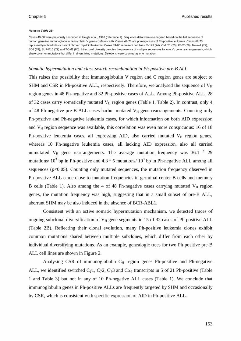
Chapter 5 Published results
153
Notes to Table 2B:
Cases 49-58 were previously described in Height et al., 1996 (reference 7). Sequence data were re-analysed based on the full sequence of human germline immunoglobulin heavy chain V genes (reference 8). Cases 49-73 are primary cases of Ph-positive leukemia. Cases 69-73 represent lymphoid blast crisis of chronic myeloid leukemia. Cases 74-80 represent cell lines BV173 (74), CMLT1 (75), K562 (76), Nalm-1 (77), SD1 (78), SUP-B15 (79) and TOM1 (80). Intraclonal diversity denotes the presence of multiple sequences for one VH gene rearrangements, which share common mutations but differ in diversifying mutations. Deletions were counted as one mutation.
Somatic hypermutation and class-switch recombination in Ph-positive pre-B ALL
This raises the possibility that immunoglobulin V region and C region genes are subject to
SHM and CSR in Ph-positive ALL, respectively. Therefore, we analysed the sequence of VH
region genes in 48 Ph-negative and 32 Ph-positive cases of ALL. Among Ph-positive ALL, 28
of 32 cases carry somatically mutated VH region genes (Table 1, Table 2). In contrast, only 4
of 48 Ph-negative pre-B ALL cases harbor mutated VH gene rearrangements. Counting only
Ph-positive and Ph-negative leukemia cases, for which information on both AID expression
and VH region sequence was available, this correlation was even more conspicuous: 16 of 18
Ph-positive leukemia cases, all expressing AID, also carried mutated VH region genes,
whereas 10 Ph-negative leukemia cases, all lacking AID expression, also all carried
unmutated VH gene rearrangements. The average mutation frequency was 36.1 + 29
mutations/ 103 bp in Ph-positive and 4.3 + 5 mutations/ 103 bp in Ph-negative ALL among all
sequences (p<0.05). Counting only mutated sequences, the mutation frequency observed in
Ph-positive ALL came close to mutation frequencies in germinal center B cells and memory
B cells (Table 1). Also among the 4 of 48 Ph-negative cases carrying mutated VH region
genes, the mutation frequency was high, suggesting that in a small subset of pre-B ALL,
aberrant SHM may be also induced in the absence of BCR-ABL1.
Consistent with an active somatic hypermutation mechanism, we detected traces of
ongoing subclonal diversification of VH gene segments in 15 of 32 cases of Ph-positive ALL
(Table 2B). Reflecting their clonal evolution, many Ph-positive leukemia clones exhibit
common mutations shared between multiple subclones, which differ from each other by
individual diversifying mutations. As an example, genealogic trees for two Ph-positive pre-B
ALL cell lines are shown in Figure 2.
Analysing CSR of immunoglobulin CH region genes Ph-positive and Ph-negative
ALL, we identified switched Cγ1, Cγ2, Cγ3 and Cα2 transcripts in 5 of 21 Ph-positive (Table
1 and Table 3) but not in any of 10 Ph-negative ALL cases (Table 1). We conclude that
immunoglobulin genes in Ph-positive ALLs are frequently targeted by SHM and occasionally
by CSR, which is consistent with specific expression of AID in Ph-positive ALL.
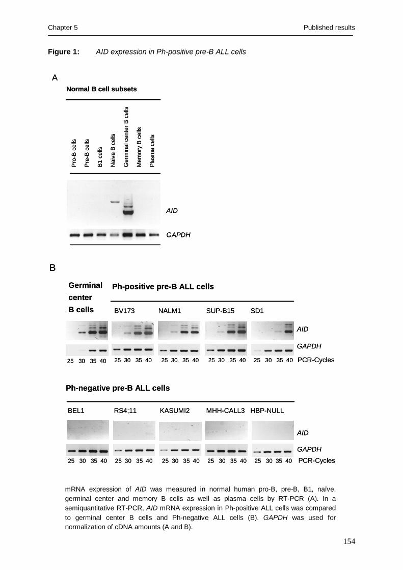
Chapter 5 Published results
154
Figure 1: AID expression in Ph-positive pre-B ALL cells
Pro
-B c
ells
Pre
-B c
ells
B1
cells
Nai
ve B
cel
ls
Ger
min
alce
nter
B c
ells
Mem
ory
B c
ells
Pla
sma
cells
Normal B cell subsetsA
AID
GAPDH
Pro
-B c
ells
Pre
-B c
ells
B1
cells
Nai
ve B
cel
ls
Ger
min
alce
nter
B c
ells
Mem
ory
B c
ells
Pla
sma
cells
Normal B cell subsetsA
AID
GAPDH
Germinalcenter B cells NALM1 SD1BV173 SUP-B15
Ph-positive pre-B ALL cells
GAPDH
AID
25 30 35 40 25 30 35 40 25 30 35 40 25 30 35 40 PCR-Cycles
25 30 35 40 25 30 35 40 25 30 35 40 25 30 35 40 25 30 35 40 PCR-CyclesGAPDH
AID
RS4;11 MHH-CALL3BEL1 KASUMI2
Ph-negative pre-B ALL cells
HBP-NULL
25 30 35 40
B
mRNA expression of AID was measured in normal human pro-B, pre-B, B1, naïve, germinal center and memory B cells as well as plasma cells by RT-PCR (A). In a semiquantitative RT-PCR, AID mRNA expression in Ph-positive ALL cells was compared to germinal center B cells and Ph-negative ALL cells (B). GAPDH was used for normalization of cDNA amounts (A and B).

Chapter 5 Published results
155
V H4-
59 D
H3-
16 J
H4
117
117a
117b
117a
117b
24
117a
117b
111
117a
117b
24 23 117a
117b
24 23 61 104
61
61 118
119
122
123
61 51
V H3-
9 D
H2-
21 J
H3
61 76
61 118
119
122
123
76
VH2-
70 D
H3-
16 J
H4
2039
20 3920 42 55 67 81 12
5a12
5b
39 100
125a
39 100
125a
76
VH3-
21 D
H2-
15 J
H3
7212
5
72 39 76 78
125
8612
512
712
8
Figu
re2:
Trac
ing
the
clon
alev
olut
ion
of P
h-po
sitiv
e pr
e-B
ALL
cel
ls b
y V
Hre
gion
gen
e m
utat
ions
NA
LM1
cells
BV
173
cells
61 51 104
125
61 76 86 101
39 100
125a
76 86
39 100
125a
76 39 51
125
127
128
61 86
125
127
128
51 76 88
A g
enea
logi
c tre
e of
ong
oing
som
atic
hyp
erm
utat
ion
of V
H g
ene
segm
ents
in th
ree
subc
lone
s of
the
Ph-
posi
tive
pre-
B A
LL c
ell l
ine
NA
LM1
and
in
the
Ph-
posi
tive
pre-
B A
LL c
ell l
ine
BV
173
is s
how
n. N
umbe
rs in
dica
te th
e m
utat
ed c
odon
s w
ithin
the
rear
rang
ed v
aria
ble
regi
on. E
ach
grou
p of
co
don
num
bers
repr
esen
ts o
ne V
H s
eque
nce,
a a
nd b
den
ote
dist
inct
mut
atio
ns w
ithin
the
sam
e co
don.

Chapter 5 Published results
156
BCR-ABL1-induced AID expression in Ph-positive pre-B ALL
We next investigated whether the Ph-encoded BCR-ABL1 kinase contributes to aberrant AID
expression in Ph-positive ALL. As shown in Figure 3, inhibition of BCR-ABL1 kinase
activity by STI571 downregulates AID expression in Ph-positive ALL at the mRNA (Figure
3A) and protein level (Figure 3B). Conversely, activation of transgenic expression of BCR-
ABL1 in a murine pro-B cell line (Figure 3C) and forced retroviral expression of BCR-ABL1
in a Ph-negative ALL (Figure 3D) induces de novo expression of AID. To validate the
causative link between BCR-ABL1 kinase activity and aberrant AID expression in patients
suffering from Ph-positive ALL, we compared matched samples pairs of Ph-positive ALL
before the onset and during continued therapy with the BCR-ABL1 kinase inhibitor STI571.
Confirming in vitro observations, primary patient-derived Ph-positive ALL cells
downregulate AID in the presence but not in the absence of the BCR-ABL1 kinase inhibitor
STI571 (Figure 3E). We conclude that BCR-ABL1 kinase activity is required for aberrant
AID expression in Ph-positive ALL cells.
BCR-ABL1 represses ID2, a negative regulator of AID
Previous studies demonstrated that AID expression is tightly regulated by the transcription
factor E2A and its inhibitors ID2 and ID3 (9, 10). Interestingly, previous work showed that
ID2 is among the genes that are transcriptionally silenced by v-abl in murine Abelson-virus-
transformed pre-B cells (11). We therefore investigated the relationship between BCR-ABL1
kinase activity, ID2 and AID expression in four Ph-positive ALL cell lines. In untreated Ph-
positive ALL cells, ID2 was repressed whereas AID was clearly detectable at the protein level
(Supplementary Figure 2). Inhibition of BCR-ABL1 kinase by STI571, however, relieved
repression of ID2 while AID expression decreased below the detection limit (Supplementary
Figure 2). Consistent with the previously described role of ID2 as a repressor of AID, these
findings identify an inverse relationship between ID2 and AID, which depends on BCR-
ABL1 kinase activity.
Lineage-specific regulation of AID in Ph-positive leukemia
Given that Ph-induced leukemia not only comprises ALL but also chronic myeloid leukemia
(CML), which derives from myeloid lineage progenitors, we investigated whether BCR-
ABL1 kinase activity drives aberrant expression of AID irrespective of lineage derivation in
both Ph-positive ALL and CML. To this end we sorted B cell, T cell and myeloid lineage
subclones from one multilineage CML cell line (CML-T1) and five primary cases of CML in
multilineage blast crisis and analyzed B- and T lymphoid as well as myeloid subclones for
AID expression (Figure 4).

Chapter 5 Published results
157
Figure 3: AID expression in Ph–positive pre-B ALL cells depends on BCR-ABL1 kinase
activity
Before STI571 During STI571
PCR-cycles: 25 30 35 40 25 30 35 40
Ph+ ALL1
Ph+ ALL2
Ph+ ALL3
Ph+ ALL4
Ph+ ALL5
AID
GAPDH
AID
GAPDH
AID
GAPDH
AID
GAPDH
AID
GAPDH
27 31 35 39 45 27 31 35 39 45
BV173 cells
Control + STI571
A
NALM1 cells
Control + STI571
D
AID
GAPDH
+MIG_ +MIG_GFP/
GFP BCR_ABL1
B
STI571
AID
EIF4e
− +
Ph-positive pre-B ALL cells
C
Aid
Oct2
Obf1
Inducible BCR-ABL1 –transgene cells
+ IL3+ Dox
+ IL3
Ph-negative pre-B ALL cells
E
Before STI571 During STI571
PCR-cycles: 25 30 35 40 25 30 35 40
Ph+ ALL1
Ph+ ALL2
Ph+ ALL3
Ph+ ALL4
Ph+ ALL5
AID
GAPDH
AID
GAPDH
AID
GAPDH
AID
GAPDH
AID
GAPDH
Before STI571 During STI571
PCR-cycles: 25 30 35 40 25 30 35 40
Ph+ ALL1
Ph+ ALL2
Ph+ ALL3
Ph+ ALL4
Ph+ ALL5
AID
GAPDH
AID
GAPDH
AID
GAPDH
AID
GAPDH
AID
GAPDH
27 31 35 39 45 27 31 35 39 45
BV173 cells
Control + STI571
A
NALM1 cells
Control + STI571
D
AID
GAPDH
+MIG_ +MIG_GFP/
GFP BCR_ABL1
B
STI571
AID
EIF4e
− +
Ph-positive pre-B ALL cells
C
Aid
Oct2
Obf1
Inducible BCR-ABL1 –transgene cells
+ IL3+ Dox
+ IL3
Ph-negative pre-B ALL cells
E
27 31 35 39 45 27 31 35 39 45 27 31 35 39 45 27 31 35 39 45

Chapter 5 Published results
158
Legend to Figure 3:
Ph-positive ALL cell lines (BV173 and Nalm1) were treated with or without 10 µmol/l STI571 for 24 hours and subjected to semiquantitative RT-PCR analysis for AID and GAPDH mRNA expression (A). Protein lysates from the same cells were used for Western blotting (B) together with antibodies against AID and EIF4E (used as a loading control). IL3-dependent murine pro-B cells carrying a doxycycline (Dox)-inducible BCR-ABL1 transgene were incubated with or without 1 µg/ ml doxycycline for 24 hours and subjected to RT-PCR analysis of aid, oct2 and obf1 mRNA expression (C). Ph-negative pre-B ALL cells were transfected with a MIG GFP_IRES_BCR-ABL1 and a MIG_GFP vector as a control. After 24 hours, GFP-expressing cells were sorted and subjected to RT-PCR analysis for AID expression (D). Matched sample pairs from five patients with Ph-positive ALL before the onset and during continued treatment with the BCR-ABL1 kinase inhibitor STI571 were analyzed for AID mRNA levels by semiquantitative RT-PCR (E). The content of Ph-positive ALL cells in all samples was normalized by semiquantitative RT-PCR analysis of BCR-ABL1 fusion transcripts (not shown). As a loading control for cDNA amounts, a cDNA fragment of GAPDH was amplified.
AID was highly expressed in B lymphoid subclones, detectable at low levels in T lineage cells
and absent in myeloid subclones. These findings are consistent with a key role of the (B-)
lymphoid transcription factor E2A in the regulation of AID expression (9), which is not active
in myeloid lineage cells (12).
While CML typically develops as a myeloid lineage leukemia in a chronic phase for
up to 15 years, transformation of chronic phase CML into B lymphoid blast crisis
dramatically changes the kinetics of the disease and defines the terminal stage of CML (13).
Therefore, we studied whether AID may contribute to clonal evolution and tumor progression
in Ph-positive ALL and B lymphoid subclones of lymphoid blast crisis CML.
AID-induced DNA-single-strand breaks in Ph-positive leukemia cells
First, we investigated whether transgenic AID expression is sufficient to pre-maturely induce
SHM in bone marrow-derived pre-B cells, which represent the normal precursor of Ph-
positive ALL. To this end, we sorted pre-B cells from the bone marrow of Aid-transgenic
mice and wildtype littermates and analyzed genomic DNA from the pre-B cell populations for
DNA single-strand breaks (SSB) within rearranged Vh gene segments and the myc gene.
DNA-SSB were detected in both Vh and myc loci of Aid-transgenic but not wildtype pre-B
cells (Figure 5A).
To test whether AID expression and introduction of DNA-SSB are causally linked in
Ph-positive ALL, we silenced AID expression in three Ph-positive ALL cell lines by RNA
interference (Figure 5B; left). AID-targeting and non-targeting siRNA duplices were
fluorochrome-labeled and repeatedly transfected into Ph-positive ALL cells. Fluorochrome-
positive cells were sorted by flow cytometry and subjected to further analysis.

Chapter 5 Published results
159
CD19
CD
3
CML-T1 cells, sort:
CD
3+C
D19
−
CD
19+
A
B
AID
GAPDH
Ph+ mixed lineage CML blast crisis,
FACS Sort:
B lymphoid Myeloid CD13− CD19+ CD13+ CD19−
CML-LBC I
AID
GAPDH
CML-LBC II
25 30 35 40 45 PCR cycles25 30 35 40 45
CD19
CD
3
CML-T1 cells, sort:
CD
3+C
D19
−
CD
19+
A
B
AID
GAPDH
Ph+ mixed lineage CML blast crisis,
FACS Sort:
B lymphoid Myeloid CD13− CD19+ CD13+ CD19−
CML-LBC I
AID
GAPDH
CML-LBC II
25 30 35 40 45 PCR cycles25 30 35 40 45
Figure 4: AID expression in Ph-positive multi-lineage blast crisis cells is restricted to
lymphoid cells
CD3+ CD19− T lymphoid and CD19+ B lymphoid subpopulations from the multi-lineage CML blast crisis cell line CMLT1 were sorted by FACS (A) and subjected to cDNA synthesis. Thereafter, AID mRNA expression of the sorted subsets was analyzed by RT-PCR (A; right). FACS-sorted B lymphoid and myeloid subclones from two primary cases of CML in multilineage blast crisis were analyzed for AID mRNA expression by semiquantitative RT-PCR with the cycle numbers indicated (B). As a loading control, a cDNA fragment of GAPDH was amplified (A; B).

Chapter 5 Published results
160
− − + − − − + − − −
BV173
NALM1
SD1
AID
siR
NA
AID
siR
NA
Non
-targ
etin
gsi
RN
A, n
o Li
gase
STI571:
Non
-targ
etin
gsi
RN
A
Non
-targ
etin
gsi
RN
A
CD
KN
2B
Rearranged
VH D
JH
CD
KN2B
-S
SB
CD
KN
2B-SSB
SSB
within
a rearranged
VH D
JH
SS
B within
a rearrangedV
H DJ
H
− − + − − − + − − −
BV173
NALM1
SD1
AID
siR
NA
AID
siR
NA
Non
-targ
etin
gsi
RN
A, n
o Li
gase
STI571:
Non
-targ
etin
gsi
RN
A
Non
-targ
etin
gsi
RN
A
CD
KN
2B
Rearranged
VH D
JH
CD
KN2B
-S
SB
CD
KN
2B-SSB
SSB
within
a rearranged
VH D
JH
SS
B within
a rearrangedV
H DJ
H
Figure 5: AID induces single-strand breaks (SSB) in immunoglobulin and non-
immunoglobulin genes
A
myc-SS
B
myc
VH -S
SB
VH -J
H
wt Aid-tg
myc-SS
B
myc
VH -S
SB
VH -J
H
wt Aid-tgA
myc-SS
B
myc
VH -S
SB
VH -J
H
wt Aid-tg
myc-SS
B
myc
VH -S
SB
VH -J
H
myc-SS
B
myc
VH -S
SB
VH -J
H
wt Aid-tg
myc-SS
B
myc
VH -S
SB
VH -J
H
wt Aid-tg
Aid
Beta-actin
B
A
AID
GAPDH
AID
GAPDH
AID
GAPDH
AID-targeting siRNA
Non-targetingsiRNA
Non-targetingsiRNA +STI571
25 30 35 45 25 30 35 45 25 30 35 45 PCR-cycles
AID
GAPDH
AID
GAPDH
AID
GAPDH
AID-targeting siRNA
Non-targetingsiRNA
Non-targetingsiRNA +STI571
25 30 35 45 25 30 35 45 25 30 35 45 PCR-cycles

Chapter 5 Published results
161
Pre-B cells were sorted from the bone marrow of mice carrying a B cell-specific Aid-transgene (Aid-tg) and from wildtype littermates (wt). Specific expression of Aid in Aid-transgenic pre-B cells but not pre-B cells from wt-littermates was confirmed by RT-PCR (A, right). Genomic DNA was isolated from Aid-tg and wt pre-B cells and analyzed for DNA single-strand breaks (SSB) within rearranged Vh gene segments and the myc locus by LM-PCR. As a loading control, genomic fragments of rearranged Vh region genes and of the myc gene were amplified (A, left).
To establish a causative link between AID function and the occurrence of DNA-SSB, AID mRNA expression was silenced in three Ph-positive ALL cell lines by RNA interference using fluorochrome-labeled siRNAs against AID or non-targeting siRNA duplices as a control. Fluorochrome-labeled cells were sorted and analysed for silencing efficiency and specificity by RT-PCR (B, right panel), and for DNA-SSB within rearranged VH gene segments and the CDKN2B gene by LM-PCR (B, left panel). For BV173, Nalm1 and SD1 cells, DNA-SSB intermediates in rearranged VH3-21, VH3-9 and VH3-30 gene segments were amplified, respectively. As a loading control for genomic DNA, VH gene rearrangements and a genomic fragment of the CDKN2B gene were amplified (B). From three patients with multilineage blast crisis of chronic myeloid leukemia (CML-BC), myeloid (CD13+ CD19−) and B lymphoid (CD13− CD19+) subclones were sorted and subjected to LM-PCR for DNA-SSB within rearranged VH gene segments and CDKN2A and CDKN2B genes (C, middle and bottom panel). In these CML-BC cases, only B lymphoid but not myeloid subclones express AID (C, top panel). As a loading control for genomic DNA, VH gene rearrangements and genomic fragments of the CDKN2A and CDKN2B genes were used. Reflecting their lineage identity, from myeloid subclones of CML-BC no VH gene rearrangements were amplified. Two additional cases of CML-BC (not shown) were not informative for DNA-SSB within CDKN2A and CDKN2B. In these cases, no CDKN2A or CDKN2B fragments could be amplified owing to large bi-allelic deletions at these loci. As a positive control, genomic DNA from normal germinal center B cells was used.
Germinal center B cells
Myeloid Myeloid Myeloid
Germinal center B cells
Myeloid Myeloid Myeloid
CD20+ CD77+
CD
KN
2B-S
SB
CD
KN
2B
CD
KN
2A-S
SB
CD
KN
2A
VH -S
SB
VH -J
H
CD
KN
2B-S
SB
CD
KN
2B
CD
KN
2A-S
SB
CD
KN
2A
VH -S
SB
VH -J
H
CD
KN
2B-S
SB
CD
KN
2B
CD
KN
2A-S
SB
CD
KN
2A
VH -S
SB
VH -J
H
CD
KN2B-SS
B
CD
KN2B
CD
KN2A-SS
B
CD
KN2A
VH -SSB
VH -J
H
B lymphoid B lymphoid B lymphoid
CD
KN
2B-S
SB
C
DK
N2B
C
DK
N2A
-SS
B
CD
KN
2A-S
SB
V
H -SS
B
VH -J
H
CD
KN
2B-S
SB
C
DK
N2B
C
DK
N2A
-SS
B
CD
KN
2A-S
SB
V
H -SS
B
VH -J
H
CD
KN
2B-S
SB
CD
KN
2B
CD
KN
2A-S
SB
CD
KN
2A-S
SB
VH -S
SB
VH -J
H
CD
KN
2B-S
SB
CD
KN
2B
CD
KN
2A-S
SB
CD
KN
2A-S
SB
VH -S
SB
VH -J
H
CCML-LBC VCML-LBC IVCML-LBC III
CD13- CD19+ CD13+ CD19− CD13- CD19+ CD13+ CD19− CD13- CD19+ CD13+ CD19−
B lymphoid Myeloid B lymphoid Myeloid B lymphoid Myeloid
AID
GAPDH
PCR-cycles25 30 35 40 45 25 30 35 40 45 25 30 35 40 45 25 30 35 40 45 25 30 35 40 45 25 30 35 40 45
CCML-LBC VCML-LBC IVCML-LBC III
CD13- CD19+ CD13+ CD19− CD13- CD19+ CD13+ CD19− CD13- CD19+ CD13+ CD19−
B lymphoid Myeloid B lymphoid Myeloid B lymphoid Myeloid
AID
GAPDH
PCR-cycles25 30 35 40 45 25 30 35 40 45 25 30 35 40 45 25 30 35 40 45 25 30 35 40 45 25 30 35 40 45

Chapter 5 Published results
162
RNA interference substantially reduced AID mRNA levels in the three Ph-positive ALL cell
lines (Figure 5B; left). Comparing Ph-positive ALL cells carrying non-targeting siRNAs with
Ph-positive cells carrying AID-specific siRNA duplices, DNA-SSB were detected within
rearranged VH gene segments and also within the CDKN2B gene in the former but not in the
latter case (Figure 5B; right). Likewise, inhibition of BCR-ABL1 kinase activity by STI571
largely reduced the frequency of DNA-SSB within VH and CDKN2B genes (Figure 5B; right).
CDKN2B encodes the p15/INK4B tumor suppressor in ALL and CML and is frequently
deleted in lymphoid but not myeloid lineage blast crisis of CML (14, 15). We also studied the
immediately adjacent CDKN2A locus, which we found already deleted in BV173 and Nalm1
cells and therefore not informative for analysis of DNA-SSB (not shown).
These findings identify AID as a lineage-specific mutator within but also outside the
immunoglobulin gene loci in Ph-positive ALL cells.
To test whether such a mutation mechanism is also active in primary patient-derived
CML multi-lineage blast crisis, we studied DNA-SSB in rearranged VH gene segments, the
CDKN2A and the CDKN2B loci in myeloid and B cell lineage subclones sorted from five
cases of multilineage CML blast crisis (Figure 5C). As a control, tonsillar germinal center B
cells were used. Consistent with differential expression of AID in myeloid and B cell lineage
subclones of CML-blast crisis in these patients (Figure 5C, top), DNA-SSB within rearranged
VH, CDKN2A and CDKN2B were exclusively detected in the B lymphoid but not the myeloid
compartment (Figure 5C). Reflecting their lineage identity, we could not amplify rearranged
VH gene segments from myeloid subclones of CML blast crisis, thus lacking the target for
DNA-SSB at this locus. In two of five cases, the CDKN2A locus or both CDKN2A and
CDKN2B loci were already deleted (not shown). We conclude that an AID-induced, lineage
specific mutation mechanism is indeed active in primary CML blast crisis.
Discussion
In summary, these findings identify AID as a mutator in Ph-positive ALL and lymphoid blast
crisis CML. Aberrant expression of AID leading to somatic hypermutation inside and outside
the immunglobulin loci is driven by oncogenic BCR-ABL1 kinase activity and likely
contributes to the particularly unfavorable prognosis of Ph-positive leukemia (6). Ph-positive
leukemias typically develop resistance to chemotherapy and inhibition of BCR-ABL1 kinase
activity within a short period of time (6). Further studies that address a specific role of AID in
the acquisition of mutations leading to drug resistance in Ph-positive ALL are currently under
way.

Chapter 5 Published results
163
Of note, a small fraction of cases of Ph-negative pre-B ALL carry mutated VH region
genes (and presumably express AID) in the absence of BCR-ABL1. This indicates that other
genetic aberrations besides the Philadelphia chromosome may likewise induce aberrant
somatic hypermutation in pre-B ALL cells. If BCR-ABL1-induced upregulation of AID
indeed mainly results from repression of ID2 and indirect activation of E2A, one would
envision that other factors that influence the balance between ID2 and E2A may have a
similar effect in Ph-negative leukemia cells.
Acknowledgements: We would like to thank Martin Krönke and Klaus Rajewsky for continuous support and discussions and Stefanie Jauch and Peter Wurst for excellent technical assistance. N.F. is supported by a fellowship of the José-Carreras-Leukemia Foundation. F.K. is supported by the Studienstiftung des deutschen Volkes. M.M. is supported by the Deutsche Forschungsgemeinschaft through the Emmy-Noether-Programm and through grants MU1616/2-1, MU1616/3-1 (to M.M.), the German José-Carreras-Leukemia-Foundation (grant to M.M.), the Deutsche Krebshilfe through Molecular Mechanisms of Malignant Lymphoma (to M.M.) and the Ministry of Science and Research for North Rhine-Westphalia through the Stem Cell Network NRW (to M.M.). The authors have no conflicting financial interests.
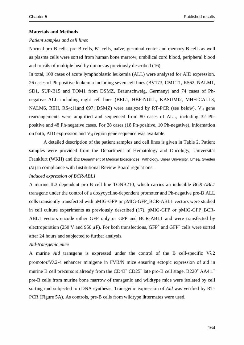
Chapter 5 Published results
164
Materials and Methods
Patient samples and cell lines
Normal pro-B cells, pre-B cells, B1 cells, naïve, germinal center and memory B cells as well
as plasma cells were sorted from human bone marrow, umbilical cord blood, peripheral blood
and tonsils of multiple healthy donors as previously described (16).
In total, 100 cases of acute lymphoblastic leukemia (ALL) were analysed for AID expression.
26 cases of Ph-positive leukemia including seven cell lines (BV173, CMLT1, K562, NALM1,
SD1, SUP-B15 and TOM1 from DSMZ, Braunschweig, Germany) and 74 cases of Ph-
negative ALL including eight cell lines (BEL1, HBP-NULL, KASUMI2, MHH-CALL3,
NALM6, REH, RS4;11and 697; DSMZ) were analyzed by RT-PCR (see below). VH gene
rearrangements were amplified and sequenced from 80 cases of ALL, including 32 Ph-
positive and 48 Ph-negative cases. For 28 cases (18 Ph-positive, 10 Ph-negative), information
on both, AID expression and VH region gene sequence was available.
A detailed description of the patient samples and cell lines is given in Table 2. Patient
samples were provided from the Department of Hematology and Oncology, Universität
Frankfurt (WKH) and the Department of Medical Biosciences, Pathology, Umea University, Umea, Sweden
(AL) in compliance with Institutional Review Board regulations.
Induced expression of BCR-ABL1
A murine IL3-dependent pro-B cell line TONB210, which carries an inducible BCR-ABL1
transgene under the control of a doxycycline-dependent promoter and Ph-negative pre-B ALL
cells transiently transfected with pMIG-GFP or pMIG-GFP_BCR-ABL1 vectors were studied
in cell culture experiments as previously described (17). pMIG-GFP or pMIG-GFP_BCR-
ABL1 vectors encode either GFP only or GFP and BCR-ABL1 and were transfected by
electroporation (250 V and 950 μF). For both transfections, GFP+ and GFP− cells were sorted
after 24 hours and subjected to further analysis.
Aid-transgenic mice
A murine Aid transgene is expressed under the control of the B cell-specific Vλ2
promotor/Vλ2-4 enhancer minigene in FVB/N mice ensuring ectopic expression of aid in
murine B cell precursors already from the CD43+ CD25− late pro-B cell stage. B220+ AA4.1+
pre-B cells from murine bone marrow of transgenic and wildtype mice were isolated by cell
sorting und subjected to cDNA synthesis. Transgenic expression of Aid was verified by RT-
PCR (Figure 5A). As controls, pre-B cells from wildtype littermates were used.

Chapter 5 Published results
165
Cell sorting and flow cytometry
CD19+ CD13− B lymphoid and CD13+ CD19− myeloid lineage subpopulations from five
primary cases of CML in multilineage blast crisis as well as CD3+ CD19− T lymphoid and
CD19+ B lymphoid subpopulations from one multilineage CML cell line (CMLT1) were
stained using antibodies against CD19, CD3 and CD13 (BD Biosciences) and sorted using a
FACStar 440 cell sorter.
Sequence analysis of VH and CH region genes and semiquantitative RT-PCR
In order to characterize the configuration of VH and CH region genes, two primer sets were
used amplifying the variable (V) region alone or the variable (V) region together with the
constant (C) region of rearranged immunoglobulin heavy chain genes as previously described
(18). Mutation frequencies of VH region genes of 10 /103 bp or higher were considered
significantly (p< 0.01) above the frequency of mutations introduced by Taq DNA polymerase
error, which was experimentally determined as 2.5 /103 bp. Primers used for semiquantitative
RT-PCR analysis of human AID and GAPDH, and for RT-PCR analysis of murine Aid, Oct2
and Obf1 transcripts are listed in supplementary Table 1.
Western blotting and immunofluorescence
For the detection of AID by Western blot, an antibody against human AID (L7E7; Cell
Signaling Technology, Beverly, MA), was used together with the WesternBreeze
immunodetection system (Invitrogen, Karlsruhe, Germany). Detection of EIF4e was used as a
loading control (Santa Cruz Biotechnology, Heidelberg, Germany). ID2 and AID expression
were analyzed by immunofluorescence using primary antibodies against human ID2 (Santa
Cruz Biotechnology) and AID (L7E7; Cell Signaling Technology) together with anti-rabbit
IgG-Cy2 and anti-mouse IgG-Cy3 (Jackson Immunoresearch) as secondary antibodies,
respectively. Prior to staining, cells were fixed with 0.4% para-formaldehyde and incubated
for 10 minutes in 90% methanol on ice.
Silencing of AID mRNA expression by RNA interference
For silencing of AID mRNA expression one previously validated siRNA (19) and a non-
targeting siRNA duplex was used. All siRNA duplices (for sequences, see Supplementary
Table 3) were labeled with fluorescein using an siRNA labeling kit (Ambion, Austin, TX)
according to the manufacturer’s protocol. Transfected fluorescein-positive cells were sorted
by FACS as previously described (17). RNA interference was verified by RT-PCR.
Ligation-mediated PCR for detection of DNA single-strand breaks
Genomic DNA from 2.5 x 10⁶ cells containing a nick on the lower strand is denatured for 10
minutes at 95°C, and a gene-specific primer (Supplementary Table 3) is hybridized and

Chapter 5 Published results
166
extended to the position of the nick as previously described (20) (first strand extension),
leaving a blunt end using Vent DNA polymerase (New England Biolabs, Beverly, MA). Next,
a double-stranded linker is ligated to the newly created blunt end using T4 DNA ligase
(Invitrogen) at 14°C overnight. The linker was constructed by annealing of the
oligonucleotides 5'-TTTCTGCTCGAATTCAAGCTTCTAACGATGTACGGGGACATG 3'
and 3’ amino (C7)- GACGAGCTTAAGTTCGAAGATTGCTACATGCCCCT -5’ and
protruding 3' overhangs were removed by 3'→ 5' exonuclease activity of the Klenow fragment
of E. coli DNA polymerase I (Invitrogen). Ligation-mediated PCR (reference 21; LM-PCR)
was carried out with modifications as previously described (22). In two semi-nested rounds of
amplification at an annealing temperature of 59°C, linker-ligated intermediates of DNA-
single-strand breaks within various genes were amplified using gene specific primers together
with two linker specific primers (Supplementary Table 1).

Chapter 5 Published results
167
Tabl
e 3:
Cla
ss-s
witc
h re
com
bina
tion
in P
h-po
sitiv
e A
LL c
ells
Cas
eaV H
DH
J Hre
arra
ngem
ent
C
Hre
gion
70 (F
ACS-
sort:
CD
19+
CD
34+ )
TATGTATTATTGTACGAAAGA
GGG
TTC
AGC
TATG
GCCGACCTGAACTTCTGGGGCCAGGGAACCCTGGTCACCGTCTCCTCAG
CCT
CCAC
CAA
GGG
CCC
AT
Lym
phoi
d bl
ast c
risis
CM
L-G-------C---G-------
--A-A---------
A-T--T-TG---A-----------------------------------------------------
VH3-
30
D
H5-
5
J H
4
Cγ 2
71 (F
ACS-
sort:
CD
19+
CD
34+ )
GCCTTCTATTATTGTGCGCGA
GGT
GAT
ACC
ACAGTCAGCGGGA
CCAACTGGCTCGCCCCCTGGGGCCAGGGAACCCTGGTCACCGTCTCCTCTG
CCTC
CAC
CAA
GGG
CCCA
T
Lym
phoi
d bl
ast c
risis
CM
L---G-G-----C------A--
------TG-
A-------T---A------------------------------------A-------------------
VH1-
46
D
H2-
21
J H
5
Cγ 2
72 (F
ACS-
sort:
CD
19+
CD
34+ )
TCTGTATTACTGTGTGAAAGG A
GAG
TGGGC
CATTGACTACTGGGGCCAGGGAACCCTGGTCACCGTCTCCTCAG
CCTC
CAC
CAAG
GGC
CCA
T
Lym
phoi
d bl
ast c
risis
CM
L-G------------C-----A ----
-T------------------------------------------------------------
VH3-
30
D
H6-
6
J H
4
Cγ 1
GCCGTATATTACTGTGCGAAA TGGTA
CGG
ACA
TTG
CAGTAGTACCACCTGCACGAC
CCTTGACTACTGGGGCCAGGGAACCCTGGTCACCGTCTCCTCAG
CCTC
CAC
CAAG
GGC
CCA
T
---------------------
T---T----
-T------------------------------------------------------------
VH3-
23
D
H2-
2
J H
4
Cγ 1
CGGCCATATATTACTGTGCGA CCACCGA
GGG
GAT
AGC
AGTG
GCT
GGT
ACTTCGT
CTGGGGCCAAGGGACCACGGTCATCGTCTCCTCCG
CCT
CCAC
CAA
GGG
CCC
AT-----G-G-------------
---T-----------------
------------A---CT-----C---------A-------------------
VH3-
53
D
H6-
19
J H
5
Cγ 1
CCGTTTATTACTGTGCGAAA TTGGAACG
TAT
GGT
GAC
TACC
GGTCGGC
GGCCTTCCCCTGGGGCCAGGGAACCCTGGTCACCGTCTCCTCAG
CATC
CCC
GACC
AGC
CCC
A----A---------------
--C----------
-TT-GA--------------------------------------------------------
VH3-
23
DH4-
17
J H5
Cα
2
TGTGTATTACTGTGCGAGAGA
TCG
GGG
GCT
GGCA
GCTACTC
ACTTTGACTCCTGGGGCCAGGGAACCCTGGTCACCGTCTCTTCAG
CAT
CCC
CGAC
CAG
CCC
CA---------------------
G-A-T------T-C
---------A------------------------------C----------------------
VH3-
33
D
H2-
2
J H4
Cα 2
TGTTTATTACTGTGCGAAAGA TGCAG
AAA
ATG
AGG
CAGT
GACTGGTAGGGG
CTACTACGGTATGGACGTCTGGGGCCAAGGGACCACGGTCACCGTCTCCTCAG
CAT
CCCC
GAC
CAG
CCC
CA---G-----------------
-T-T--T--TG----
-----------------------------------------------------------------------
VH3-
30
D
H2-
15
J H
6
Cα
2
78 P
h+ce
lllin
e SD
1GCTGTCTATTATTGTGTGAAA
CCG
ATG
GGA
CCCTACCGCGAG
GCTTTTGATATCTGGGGCCAAGGGACAGTGGTCACCGTCTCTTCAG
CCTC
CAC
CAA
GGGC
CCA
T-
---G-----C----C----
-A-C---T--
---------------------------A------------------------------------
VH3-
30
DH6-
13
J H3
Cγ 1
77
Ph+
cell
line
Nal
m-1
AGTCTGAGAGCTGAGGACACC TGGGGG
TAC
TTT
GAC
TACTGGGGCC
AGGGAACCCTGGTCACCGTCTCCTCAG
CTT
CCAC
CAA
GGG
CCC
AT---------A-----------
--T-------
---------------------------------------------
VH3-
43
DH3-
9
J H
5
Cγ 3
Not
e:a.
Num
bers
for c
ases
refe
r to
Tabl
e 2B
Tabl
e 3:
Cla
ss-s
witc
h re
com
bina
tion
in P
h-po
sitiv
e A
LL c
ells
Cas
eaV H
DH
J Hre
arra
ngem
ent
C
Hre
gion
70 (F
ACS-
sort:
CD
19+
CD
34+ )
TATGTATTATTGTACGAAAGA
GGG
TTC
AGC
TATG
GCCGACCTGAACTTCTGGGGCCAGGGAACCCTGGTCACCGTCTCCTCAG
CCT
CCAC
CAA
GGG
CCC
AT
Lym
phoi
d bl
ast c
risis
CM
L-G-------C---G-------
--A-A---------
A-T--T-TG---A-----------------------------------------------------
VH3-
30
D
H5-
5
J H
4
Cγ 2
71 (F
ACS-
sort:
CD
19+
CD
34+ )
GCCTTCTATTATTGTGCGCGA
GGT
GAT
ACC
ACAGTCAGCGGGA
CCAACTGGCTCGCCCCCTGGGGCCAGGGAACCCTGGTCACCGTCTCCTCTG
CCTC
CAC
CAA
GGG
CCCA
T
Lym
phoi
d bl
ast c
risis
CM
L---G-G-----C------A--
------TG-
A-------T---A------------------------------------A-------------------
VH1-
46
D
H2-
21
J H
5
Cγ 2
72 (F
ACS-
sort:
CD
19+
CD
34+ )
TCTGTATTACTGTGTGAAAGG A
GAG
TGGGC
CATTGACTACTGGGGCCAGGGAACCCTGGTCACCGTCTCCTCAG
CCTC
CAC
CAAG
GGC
CCA
T
Lym
phoi
d bl
ast c
risis
CM
L-G------------C-----A ----
-T------------------------------------------------------------
VH3-
30
D
H6-
6
J H
4
Cγ 1
GCCGTATATTACTGTGCGAAA TGGTA
CGG
ACA
TTG
CAGTAGTACCACCTGCACGAC
CCTTGACTACTGGGGCCAGGGAACCCTGGTCACCGTCTCCTCAG
CCTC
CAC
CAAG
GGC
CCA
T
---------------------
T---T----
-T------------------------------------------------------------
VH3-
23
D
H2-
2
J H
4
Cγ 1
CGGCCATATATTACTGTGCGA CCACCGA
GGG
GAT
AGC
AGTG
GCT
GGT
ACTTCGT
CTGGGGCCAAGGGACCACGGTCATCGTCTCCTCCG
CCT
CCAC
CAA
GGG
CCC
AT-----G-G-------------
---T-----------------
------------A---CT-----C---------A-------------------
VH3-
53
D
H6-
19
J H
5
Cγ 1
CCGTTTATTACTGTGCGAAA TTGGAACG
TAT
GGT
GAC
TACC
GGTCGGC
GGCCTTCCCCTGGGGCCAGGGAACCCTGGTCACCGTCTCCTCAG
CATC
CCC
GACC
AGC
CCC
A----A---------------
--C----------
-TT-GA--------------------------------------------------------
VH3-
23
DH4-
17
J H5
Cα
2
TGTGTATTACTGTGCGAGAGA
TCG
GGG
GCT
GGCA
GCTACTC
ACTTTGACTCCTGGGGCCAGGGAACCCTGGTCACCGTCTCTTCAG
CAT
CCC
CGAC
CAG
CCC
CA---------------------
G-A-T------T-C
---------A------------------------------C----------------------
VH3-
33
D
H2-
2
J H4
Cα 2
TGTTTATTACTGTGCGAAAGA TGCAG
AAA
ATG
AGG
CAGT
GACTGGTAGGGG
CTACTACGGTATGGACGTCTGGGGCCAAGGGACCACGGTCACCGTCTCCTCAG
CAT
CCCC
GAC
CAG
CCC
CA---G-----------------
-T-T--T--TG----
-----------------------------------------------------------------------
VH3-
30
D
H2-
15
J H
6
Cα
2
78 P
h+ce
lllin
e SD
1GCTGTCTATTATTGTGTGAAA
CCG
ATG
GGA
CCCTACCGCGAG
GCTTTTGATATCTGGGGCCAAGGGACAGTGGTCACCGTCTCTTCAG
CCTC
CAC
CAA
GGGC
CCA
T-
---G-----C----C----
-A-C---T--
---------------------------A------------------------------------
VH3-
30
DH6-
13
J H3
Cγ 1
77
Ph+
cell
line
Nal
m-1
AGTCTGAGAGCTGAGGACACC TGGGGG
TAC
TTT
GAC
TACTGGGGCC
AGGGAACCCTGGTCACCGTCTCCTCAG
CTT
CCAC
CAA
GGG
CCC
AT---------A-----------
--T-------
---------------------------------------------
VH3-
43
DH3-
9
J H
5
Cγ 3
Not
e:a.
Num
bers
for c
ases
refe
r to
Tabl
e 2B
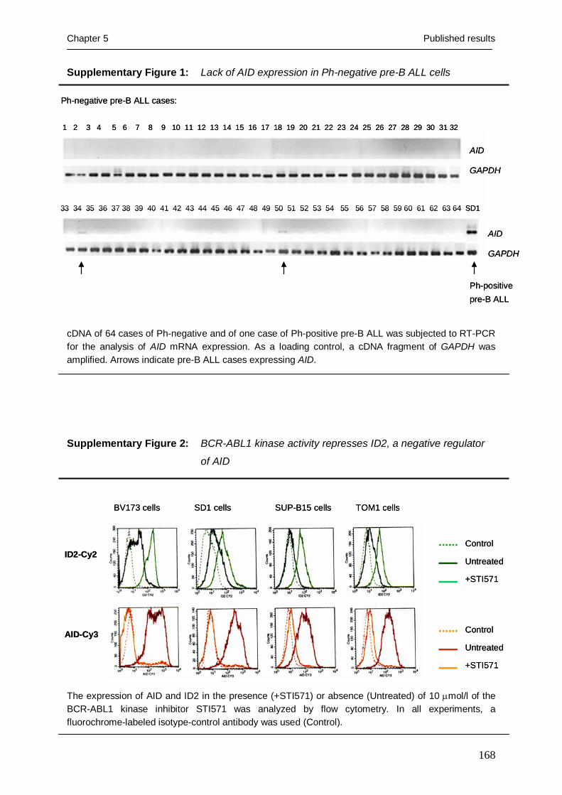
Chapter 5 Published results
168
Supplementary Figure 1: Lack of AID expression in Ph-negative pre-B ALL cells
cDNA of 64 cases of Ph-negative and of one case of Ph-positive pre-B ALL was subjected to RT-PCR for the analysis of AID mRNA expression. As a loading control, a cDNA fragment of GAPDH was amplified. Arrows indicate pre-B ALL cases expressing AID.
Supplementary Figure 2: BCR-ABL1 kinase activity represses ID2, a negative regulator
of AID
The expression of AID and ID2 in the presence (+STI571) or absence (Untreated) of 10 μmol/l of the BCR-ABL1 kinase inhibitor STI571 was analyzed by flow cytometry. In all experiments, a fluorochrome-labeled isotype-control antibody was used (Control).
Ph-positive pre-B ALL
33 34 35 36 37 38 39 40 41 42 43 44 45 46 47 48 49 50 51 52 53 54 55 56 57 58 59 60 61 62 63 64 SD1
Ph-negative pre-B ALL cases:
1 2 3 4 5 6 7 8 9 10 11 12 13 14 15 16 17 18 19 20 21 22 23 24 25 26 27 28 29 30 31 32
AID
GAPDH
AID
GAPDH
Ph-positive pre-B ALL
33 34 35 36 37 38 39 40 41 42 43 44 45 46 47 48 49 50 51 52 53 54 55 56 57 58 59 60 61 62 63 64 SD1
Ph-negative pre-B ALL cases:
1 2 3 4 5 6 7 8 9 10 11 12 13 14 15 16 17 18 19 20 21 22 23 24 25 26 27 28 29 30 31 32
AID
GAPDH
AID
GAPDH
BV173 cells SD1 cells SUP-B15 cells TOM1 cells
ID2-Cy2
AID-Cy3
Control
Untreated
+STI571
Control
Untreated
+STI571
BV173 cells SD1 cells SUP-B15 cells TOM1 cells
ID2-Cy2
AID-Cy3
Control
Untreated
+STI571
Control
Untreated
+STI571

Chapter 5 Published results
169
Supplementary Table 1: Summary of oligonucleotides used for RT-PCR, RNA
interference and ligation-mediated PCR
PCR primers GAPDH forward 5’-TTAGCACCCCTGGCCAAG-3’ GAPDH reverse 5’-CTTACTCCTTGGAGGCCATG-3’ BCR-ABL1 forward 5’-ACCTCACCTCCAGCGAGGAGGACTT-3' BCR-ABL1 reverse 5'-TCCACTGGCCACAAAATCATACAGT-3' mAid forward 5'- AAATGTCCGCTGGGCCAA-3' mAid reverse 5'- CATCGACTTCGTACAAGGG-3' AID forward 5'-TGCTCTTCCTCCGCTACATC-3' AID reverse 5'-CCTCATACAGGGGCAAAAGG-3' mObf1 forward 5'-AGCTCCCTGACCATTGAC-3' mObf1 reverse 5'-CTGTCCCATCCCCCTGTAA-3' mOct2 forward 5'-ATCGAGACGAATGTCCGCTT-3' mOct2 reverse 5'-GTAGCTGGTCGGCTTTCC-3' Cμ_R 5’-AGACGAGGGGGAAAAGGGTT-3’ Cγ1_R 5’-GAGTTTTGTCACAAGATTTGGGCT-3’ Cγ2_R 5’-GCACTCGACACAACATTTGCG-3’ Cγ3_R 5’-TCACCAAGTGGGGTTTTGAGC-3’ Cγ4_R 5’-ATGGGCATGGGGGACCATTT-3’ Cα2_R 5’-TGTTGGCGGTTAGTGGGGTC-3’ Cε_R 5’-GAGGTGGCATTGGAGGGAAT-3’ Cδ_R 5’-AGAGCTGGCTGCTTGTCATG-3’ mJ558 forward 5’-GCAAGGCTTCTGGCTACACA-3’ mJH2/3 reverse 5’-CCAGTAAGCAAACCAGGCACA-3’ siRNA duplices Non-targeting siRNA 5’-UUGUACCUAAUUUCGUCCCAC-3’ 3’-CAUGGAUUAAAGCAGGGUGUU-5’ AID siRNA 5'-UUGCUCUUCCUCCGCUACAUU-3' 3'-CGAGAAGGAGGCGAUGUAAUU-3' LM-PCR primers CDKN2A_F1 5’-CCAGGAATAAAATAAGGGGAATA-3’ CDKN2A_F2 5’-GGAATAAAATAAGGGGAATAGGG-3’ CDKN2A_R 5’-CTTTCCTACCTGGTCTTCTAGG-3’ CDKN2B_F1 5’-GTGAACATTCCCAAAATATTAGC-3’ CDKN2B_F2 5’-AAAATATTAGCCTTGGCTTTACTG-3’ CDKN2B_R 5’-AGACTCCTGTACAAATCTACATCG-3’ V3-21_R 5’-CTCTCGCACAGTAATACACAGC-3’ V1-2_R 5’-CTCTCGCACAGTAATACACGAC-3’ V1-69_R 5’-TCTCTCGCACAGTAATACACG-3’ V3-73_R 5’-GGTTTTCAGGCTGTTCATTT-3’ V3-53_R 5’-CACCTTTTAAAATAGCAACAAGG-3’ V3-30_R 5’-AGCATAGCTACTGAAGGTGAAT-3’ Mmyc_F1 5’-ATCCTGAGTCGCAGTATAAAAGA-3’ Mmyc_F2 5’-TTTTCTGACTCGCTGTAGTAATTC-3’ Mmyc_R1 5’-CTCAGAGACTGGTAAGTCAGAAG-3’

Chapter 5 Published results
170
Linker_F1 5’-CTGCTCGAATTCAAGCTTCT-3’ Linker_F2 5’-GCTTCTAACGATGTACGGGG-3’ Linker_R1 5’-GTACATCGTTAGAAGCTTGAA-3’ Linker_R2 5’-GTTAGAAGCTTGAATTCGAGC-3’
References
1. Rajewsky K. Clonal selection and learning in the antibody system. Nature 1996; 381: 751-
758.
2. Muramatsu M, Kinoshita K, Fagarasan S, Yamada S, Shinkai Y, Honjo T. Class switch
recombination and hypermutation require activation-induced cytidine deaminase (AID), a
potential RNA editing enzyme. Cell. 2000;102:553-63.
3. Pasqualucci L, Neumeister P, Goossens T, Nanjangud G, Chaganti RS, Kuppers R, Dalla-
Favera R. Hypermutation of multiple proto-oncogenes in B-cell diffuse large-cell lymphomas.
Nature. 2001; 412: 341-346.
4. Wang CL, Harper RA, Wabl M. Genome-wide somatic hypermutation. Proc Natl Acad Sci U
S A. 2004; 101: 7352-7356.
5. Bird J, Galili N, Link M, Stites D, Sklar J. Continuing rearrangement but absence of somatic
hypermutation in immunoglobulin genes of human B cell precursor leukemia. J Exp Med.
1988; 168: 229-245.
6. Arico M, Valsecchi MG, Camitta B, Schrappe M, Chessells J, Baruchel A, Gaynon P,
Silverman L, Janka-Schaub G, Kamps W, Pui CH, Masera G. Outcome of treatment in
children with Philadelphia chromosome-positive acute lymphoblastic leukemia. N Engl J Med.
2000; 342: 998-1006.
7. Height SE, Swansbury GJ, Matutes E, Treleaven JG, Catovsky D, Dyer MJ. Analysis of clonal
rearrangements of the Ig heavy chain locus in acute leukemia. Blood. 1996; 87: 5242-5250.
8. Matsuda F, Ishii K, Bourvagnet P, Kuma K, Hayashida H, Miyata T, Honjo T. The complete
nucleotide sequence of the human immunoglobulin heavy chain variable region locus. J Exp
Med. 1998; 188: 2151-2162.
9. Gonda H, Sugai M, Nambu Y, Katakai T, Agata Y, Mori KJ, Yokota Y, Shimizu A. The
balance between Pax5 and Id2 activities is the key to AID gene expression. J Exp Med. 2003;
198: 1427-1437.
10. Sayegh CE, Quong MW, Agata Y, Murre C. E-proteins directly regulate expression of
activation-induced deaminase in mature B cells. Nat Immunol. 2003; 4: 586-593.
11. Muljo SA, Schlissel MS. A small molecule Abl kinase inhibitor induces differentiation of
Abelson virus-transformed pre-B cell lines. Nat Immunol. 2003; 4: 31-37.
12. Ikawa T, Kawamoto H, Wright LY, Murre C. Long-term cultured E2A-deficient
hematopoietic progenitor cells are pluripotent. Immunity. 2004; 20:349-60.

Chapter 5 Published results
171
13. Calabretta B, Perrotti D. The biology of CML blast crisis. Blood. 2004; 103: 4010-4022.
14. Sill H, Goldman JM, Cross NC. Homozygous deletions of the p16 tumor-suppressor gene are
associated with lymphoid transformation of chronic myeloid leukemia. Blood. 1995; 85: 2013-
2016.
15. Haidar MA, Cao XB, Manshouri T, Chan LL, Glassman A, Kantarjian HM, Keating MJ,
Beran MS, Albitar M. p16INK4A and p15INK4B gene deletions in primary leukemias. Blood.
1995; 86: 311-315.
16. Müschen M, Re D, Jungnickel B, Diehl V, Rajewsky K, Küppers R. Somatic mutation of the
CD95 gene in human B cells as a side-effect of the germinal center reaction. J Exp Med. 2000;
192: 1833-1840.
17. Feldhahn N, Klein F, Mooster JL, Hadweh P, Sprangers M, Wartenberg M, Bekhite MM,
Hofmann WK, Herzog S, Jumaa H, Rowley JD, Müschen M. Mimicry of a constitutively
active pre-B cell receptor: Aberrant splicing links Bruton’s tyrosine kinase to BCR-ABL1 in
pre-B lymphoblastic leukemia. J Exp Med. 2005; 201: 1837-52.
18. Klein F., N. Feldhahn, L. Harder, H. Wang, M. Wartenberg, W.K. Hofmann, P. Wernet, R.
Siebert, Müschen M. The BCR-ABL1 Kinase Bypasses Selection for the expression of a pre-
B cell receptor in pre-B acute lymphoblastic leukemia cells. J Exp Med. 2004; 199:673-685.
19. Machida K, Cheng KT, Sung VM, Shimodaira S, Lindsay KL, Levine AM, Lai MY, Lai MM.
Hepatitis C virus induces a mutator phenotype: enhanced mutations of immunoglobulin and
protooncogenes. Proc Natl Acad Sci U S A. 2004; 101: 4262-4267.
20. Arudchandran A, Bernstein RM, Max EE. Single-stranded DNA breaks adjacent to cytosines
occur during Ig gene class switch recombination. J Immunol. 2004; 173: 3223-3229.
21. Schlissel M, Constantinescu A, Morrow T, Baxter M, Peng A. Double-strand signal sequence
breaks in V(D)J recombination are blunt, 5'-phosphorylated, RAG-dependent, and cell cycle
regulated. Genes Dev 1993; 7: 2520-2532.
22. Klein F, Feldhahn N, Lee S, Wang H, Ciuffi F, von Elstermann M, Toribio ML, Sauer H,
Wartenberg M, Barath VS, Krönke M, Wernet P, Rowley JD, Müschen M. T lymphoid
differentiation in human bone marrow. Proc Natl Acad Sci U S A. 2003; 100: 6747-6752.

Chapter 5 Published results
172
5.6 Deficiency of Bruton’s tyrosine kinase in B cell precursor leukemia cells
published in the Proceedings of the national academy of science USA; 2005;
102: 13266-13271
By Niklas Feldhahn1, Paula Río2,3, Bonaventure Ndikung Bejeng Soh1,
Stefanie Liedtke1, Mieke Sprangers1, Florian Klein1, Peter Wernet1, Hassan
Jumaa4, Wolf-Karsten Hofmann5, Helmut Hanenberg2, Janet D. Rowley6 &
Markus Müschen1
1Laboratory for Molecular Stem Cell Biology, Institute for Transplantation Diagnostics and Cell Therapeutics, Heinrich-Heine-Universität Düsseldorf, 40225 Düsseldorf, Germany; 2Department of Pediatric Hematology and Oncology, Children’s Hospital Heinrich-Heine-Universität Düsseldorf, 40225 Düsseldorf, Germany; 3Hematopoietic Gene Therapy Program, CIEMAT/Marcelino Botin Foundation, 28040 Madrid, Spain; 4Max-Plack-Institute for Immunobiology, 79108 Freiburg, Germany; 5Department of Hematology and Oncology, University Hospital "Benjamin Franklin”, 12200 Berlin, Germany; 6Hematology and Oncology, Department of Medicine, Division of Biological Sciences, University of Chicago, Chicago, 60637 IL
Summary
Bruton’s tyrosine kinase (BTK)-deficiency results in a differentiation block at
the pre-B cell stage. Likewise, acute lymphoblastic leukemia cells are typically
arrested at early stages of B cell development. We therefore investigated BTK
function in B cell precursor leukemia cells carrying a BCR-ABL1, E2A-PBX1,
MLL-AF4, TEL-AML1 or TEL-PDGFRB gene rearrangement. While somatic
mutations of the BTK gene are rare in B cell precursor leukemia cells, we
identified kinase-deficient splice variants of BTK throughout all leukemia
subtypes. Unlike infant leukemia cells carrying an MLL-AF4 gene
rearrangement, where expression of full-length BTK was only detectable in
four out of eight primary cases, in leukemia cells harboring other fusion genes
full-length BTK was typically coexpressed with kinase-deficient variants. As
shown by overexpression experiments, kinase-deficient splice variants can act
as a dominant-negative BTK in that they suppress BTK-dependent
differentiation and pre-B cell receptor responsiveness of the leukemia cells. On
the other hand, induced expression of full-length BTK rendered the leukemia
cells particularly sensitive to apoptosis. Comparing BTK expression in
surviving or pre-apoptotic leukemia cells after 10 Gy γ-radiation, we observed

Chapter 5 Published results
173
selective survival of leukemia cells that exhibit expression of dominant-
negative BTK forms. These findings indicate that lack of BTK expression or
expression of dominant-negative splice variants in B cell precursor leukemia
cells i.) can inhibit differentiation beyond the pre-B cell stage and ii.) protect
from radiation-induced apoptosis.
Introduction
Recent findings by us and others suggested a role for the pre-B cell receptor and related
signaling molecules as a tumor suppressor to prevent the development or limit the
proliferation of leukemic cells (1, 2). BCR-ABL1+ pre-B lymphoblastic leukemia cells
frequently exhibit defective expression of the pre-B cell receptor related signaling molecule
SLP65 (1) and acquire independence from pre-B cell receptor-dependent survival signals (2).
The analysis of mouse mutants of the pre-B cell receptor-related signaling molecules SLP65
and Bruton’s tyrosine kinase (BTK) demonstrated that SLP65 and BTK cooperate to suppress
leukemic transformation (3).
Pre-B lymphoblastic leukemia cells typically exhibit a differentiation block at the pre-
B cell stage of development (2); likewise BTK-deficiency in humans leading to X-linked
agammaglobulinemia (XLA) results in a breakdown of pre-B cell receptor signals and a
differentiation block at the pre-B cell stage (4). To elucidate a possible role for BTK in
leukemic transformation of human B cell precursors, we investigated BTK function in pre-B
acute lymphoblastic leukemia cells.
Results and Discussion
Rare occurrence of somatic mutations of the BTK gene in B cell precursor leukemia cells
Pre-B lymphoblastic leukemia cells frequently exhibit defective expression of the pre-B cell
receptor-related linker molecules SLP65 (1). Comparing single slp65-/- and slp65-/-/btk-/-
double mutant mice, Btk was identified as a critical co-factor in preventing leukemic
transformation of murine B cell precursors (3). We therefore searched for somatic mutations
of the BTK gene in 12 cases of B cell precursor leukemia including four cases with MLL-AF4,
three cases with E2A-PBX1, four cases with BCR-ABL1 and one case with a TEL-AML1 gene
rearrangement. Because the majority of the BTK gene mutations in the germline leading to X-
linked agammaglobulinemia (XLA), have been found in the BTK kinase domain (see:
http://bioinf.uta.fi/BTKbase/BTKbasebrowser.html), we focused our analysis on this region

Chapter 5 Published results
174
Tab
le 1
: Se
quen
ce a
naly
sis o
f abe
rran
t spl
ice
varia
nts o
f BTK
in p
re-B
acu
te ly
mph
obla
stic
leuk
emia
a R
ecur
rent
BTK
spl
ice
varia
nts
Gen
e re
arra
ngem
ent
C
ases
d
Exon
s in
volv
ed
Sp
lice
site
Tra
nscr
ipt
Tran
slat
ion
Wes
tern
Blo
t Fr
eque
ncy
M
LL-A
F4
SEM
, RS4
11, B
EL1,
Ex
ons
15 a
nd 1
6 b
Reg
ular
Δ
Exon
s 15
, 16
In-fr
ame
dele
tion
in K
D
65 k
D p
rote
in
14/2
9 BC
R-A
BL1
IX
, X, X
I, X
IV,
E2A-
PBX
1
BV
173,
NAL
M1,
TE
L-AM
L1
SU
P-B1
5, S
D1,
TE
L-PD
GFR
B
KA
SUM
I2, M
HH
- C
ALL3
, NAL
M6
BCR
-ABL
1
IX
, X, X
I, XI
V,
Exon
15 c
Reg
ular
Δ
Exon
15
Trun
cate
d KD
52 k
D p
rote
in
8/29
BV17
3, N
ALM
1,
SU
P-B1
5, S
D1
MLL
-AF4
VI
I and
IX
Ex
ons
14 a
nd 1
5
Cry
ptic
Δ
Exon
15,
Δ41
bp
exon
14
Tr
unca
ted
KD
n.
d.
2/
29
BCR
-ABL
1
Uni
que
BTK
spl
ice
varia
nts
Gen
e re
arra
ngem
ent
C
ase
d
Exon
s in
volv
ed
Sp
lice
site
Tra
nscr
ipt
Tran
slat
ion
Wes
tern
Blo
t
M
LL-A
F4
V
Ex
on 1
8
C
rypt
ic
Δ33
bp
In
fram
e de
letio
n in
KD
n.
d.
SEM
Exon
s 16
and
17
C
rypt
ic
ΔEx
ons
16, 1
7, Δ
119
bp
Trun
cate
d KD
n.d.
ex
on 1
5, Δ
136
bp e
xon
18
BCR
-ABL
1
X
Ex
ons
16 b
Reg
ular
Δ
Exon
16
Trun
cate
d KD
n.d.
XI
Ex
ons
15 to
18
C
rypt
ic,
ΔEx
ons
15-1
8, Δ
79 b
p ex
on 1
4,
Trun
cate
d KD
n.d.
Sl
ippa
ge
Ins
3 bp
, Δ24
bp
exon
19
XV
Ex
on 1
4
C
rypt
ic
ΔEx
on 1
4, Δ
62 b
p ex
on 1
3,
Trun
cate
d SH
2 an
d K
D
n.d.
Δ
133
bp e
xon
15
TE
L-AM
L1
XV
III
Ex
ons
13 to
18
C
rypt
ic,
ΔEx
ons
14-1
8, Δ
31 b
p ex
on 1
2,
Trun
cate
d SH
2 an
d K
D
n.d.
Sl
ippa
ge
Δ22
bp
exon
13,
212
bp
in e
xon
19 a
nd 3
’UTR
Ex
ons
14 to
16
R
egul
ar
ΔEx
ons
14-1
6
Tr
unca
ted
KD
47
kD
pro
tein
XIX
Exon
s 13
to 1
8
Cry
ptic
, Δ
Exon
s 13
-18,
Ins
of a
106
bp
Trun
cate
d SH
2 an
d K
D
n.d.
Sl
ippa
ge
fragm
ent
of In
tron1
8
Ex
ons
14 to
17
R
egul
ar
Loss
of e
xons
14-
17
In
-fram
e de
letio
n in
KD
56
kD
pro
tein
E2A-
PBX
1
KA
SUM
I2
Ex
ons
15 to
17
C
rypt
ic,
ΔEx
ons
15-1
7, Δ
18 b
p ex
on 1
4,
Trun
cate
d KD
n.d.
Sl
ippa
ge
Ins
1 bp
, Δ33
bp
exon
18
M
HH
-CAL
L3
Exon
s 13
to 1
7
Cry
ptic
Δ
Exon
s 13
-17,
Δ13
bp
In
-fram
e de
letio
n in
KD
n.
d.
ex
on 1
2, Δ
92 b
p ex
on 1
8
Not
es:
a.
: Seq
uenc
e da
ta a
re a
vaila
ble
from
EM
BL/G
enBa
nk u
nder
AM
0512
75-A
M05
1286
num
bers
(pen
ding
) b.
: The
se s
plic
e va
riant
s w
ere
prev
ious
ly d
escr
ibed
by
Goo
dman
et a
l., 2
003
(ref
eren
ce 1
5)
c.: T
he s
peci
fic fu
nctio
n of
this
spl
ice
varia
nt in
BC
R-A
BL1+ p
re-B
lym
phob
last
ic le
ukem
ia w
as in
vest
igat
ed b
y Fe
ldha
hn e
t al.,
200
5 (r
efer
ence
6)

Chapter 5 Published results
175
using PCR primers for BTK exons 12 to 19. Deleterious somatic mutations of the BTK gene
were found in one out of 12 cases. In this case, the leukemia cells in a female patient carry an
MLL-AF4 gene rearrangement and deleterious mutations of the BTK gene on both alleles
(both X chromsomes). One BTK allele is inactivated by a mutation Lys→Stop at codon 420
(AAA→TAA), while the second allele harbors a frameshift mutation due to a 1-bp deletion in
codon 386. In the 11 other cases studied, no clonal replacement mutations of the BTK gene
were detected. This analysis does not exclude deleterious mutations in other regions of the
BTK gene, yet indicates that inactivation of the BTK gene by somatic mutation is rare in B
cell precursor leukemia cells.
Expression of kinase-deficient splice variants of BTK in pre-B lymphoblastic leukemia cells
We next examined BTK mRNA expression in 29 human leukemias including 12 cell lines and
17 primary cases. In a PCR strategy covering the entire coding region of BTK, we identified
14 aberrant mRNA splice variants (Table 1). As a control, BTK cDNA fragments were
amplified from normal human pro- and pre-B cells (isolated from bone marrow) and mature B
cell subsets isolated from peripheral blood (Figure 1). At least one aberrant splice variant was
co-amplified with full-length BTK in 21 of 29 cases (Table 1). In only four out of 29 cases,
full-length BTK was exclusively expressed. Of note, absence of BTK expression was found in
four out of 29 cases which all harbor an MLL-AF4 rearrangement. In the remaining cases of
primary B lymphoid leukemia carrying an MLL-AF4 gene rearrangement, three exhibit very
low expression of full-length BTK or high expression of aberrant splice variants while one
shows exclusive expression of full-length BTK (Figure 1). In contrast, full-length BTK could
be detected (not shown) in all three leukemia cell lines with an MLL-AF4 fusion gene (BEL1,
RS4;11, SEM).
The aberrant BTK splice variants all have in common that they encode a truncated
BTK kinase domain (Table 1, supplementary Figure 1). From in vitro kinase assays of BTK
mutants derived from BTK-deficient XLA patients, it is known that truncation of the BTK
kinase domain at amino acid 520 and even replacement mutations in the distal portion of the
kinase domain result in a complete loss of BTK kinase activity (4). Hence, only full-length
BTK but none of the truncated BTK splice variants identified here should exhibit BTK kinase
activity (Table 1, supplementary Figure 1). We conclude that BTK function is compromised
at least in the B cell precursor leukemias that either express only kinase-deficient BTK or no
BTK at all. In all these cases, the leukemia cells harbor an MLL-AF4 gene rearrangement.

Chapter 5 Published results
176
In many leukemia cases with other gene rearrangements, however, concomitant expression of
full-length BTK was also detected. Among the 14 splice variants, three were recurrently
amplified and two previously described variants of BTK (17) were also detected in this
Figure 1: BTK mRNA expression in primary B cell precursor leukemia cells
analysis. While a splice variant lacking exon 15 was consistently and specifically amplified
from leukemia cells carrying a BCR-ABL1 fusion gene (Table 1), the expression of other
recurrent splice variants was not linked to a specific chromosomal rearrangement (Table 1).
Of note, aberrant splicing of BTK in the leukemia cells not only leads to the expression of
defective BTK transcripts. The splice mechanism itself also seems to be deranged as cryptic
splice sites were frequently used (9 of 29 cases) and splice site slippage leading to small
nucleotide insertions or deletions was observed in four of 29 cases (Table 1).
As BTK activity is mainly regulated by tyrosine-phosphorylation, we investigated the
expression of tyrosine-phosphorylated BTK protein in B cell precursor leukemia cell lines
carrying MLL-AF4, E2A-PBX1, BCR-ABL1, TEL-AML1 and TEL-PDGFRB gene
rearrangements. Constitutive tyrosine-phosphorylation of full-length BTK was only observed
in BCR-ABL1+ pre-B lymphoblastic leukemia cells (three of the four cell lines tested; Figure
2). Whereas BTK lacking exon 15 alone (52 kD) was heavily phosphorylated specifically in
Pro
-B
Pre
-B
B1
NB
C
MB
C
PC
MLL-AF4 BCR-ABL1
I II III IV V VI VII VIII IX X XI XII XIII XIV XV XVIII XIX N1 N2 N3
Negative-controls
GAPDH
BTKExons14-19
BTK Exons12-19
Full-lengthBTK
Full-lengthBTK
TEL-AML1Normal B cell subsets
pro-B cell origin pre-B cell origin
Pro
-B
Pre
-B
B1
NB
C
MB
C
PC
MLL-AF4 BCR-ABL1
I II III IV V VI VII VIII IX X XI XII XIII XIV XV XVIII XIX N1 N2 N3
Negative-controls
GAPDH
BTKExons14-19
BTK Exons12-19
Full-lengthBTK
Full-lengthBTK
TEL-AML1Normal B cell subsets
pro-B cell origin pre-B cell origin
Using PCR-primers for exons 12, 14 and 19, two regions of the BTK mRNA were analyzed by RT-PCR in normal human pro-B cells, pre-B cells, B1 cells, naïve B cells (NBC), memory B cells (MBC) and plasma cells (PC) as well as in eight primary cases of B cell precursor leukemia with MLL-AF4 gene rearrangement, seven primary cases with BCR-ABL1 and two primary cases with TEL-AML1 fusion genes. As a loading control for all samples, a GAPDH cDNA fragment was amplified.

Chapter 5 Published results
177
BCR-ABL1+ pre-B lymphoblastic leukemia cells, another recurrent BTK variant lacking exons
15 and 16 (65 kD) was also tyrosine-phosphorylated in other leukemias (Figure 2).
Figure 2: Tyrosine-phosphorylation of BTK and kinase-deficient splice variants in pre-B
lypmphoblastic leukemia cells
Expression of kinase-deficient BTK prevents pre-B cell receptor-driven differentiation of B
cell precursor leukemia cells
BTK kinase-deficiency in humans leading to X-linked agammaglobulinemia (XLA) results in
a breakdown of pre-B cell receptor signals and a differentiation block at the pre-B cell stage
(4). In normal pre-B cells, engagement of the pre-B cell receptor using a μ-chain-specific
antibody (arrows, Figure 3A, left) induces a strong Ca2+-signal, which is sensitive to BTK
kinase inhibition by LFM-A13 (Figure 3A, left).
As BTK kinase activity is required for the transduction of pre-B cell receptor-
dependent Ca2+ signals, the kinase-deficient BTK splice variants (BTK-ΔK) expressed in pre-
B lymphoblastic leukemia cells likely cannot contribute to pre-B cell receptor-dependent Ca2+
signals. However, kinase-deficient forms of BTK may also act as a linker in the signal
transduction of B lymphocytes (6, 18). To investigate the function of BTK-ΔK, we expressed
kinase-deficient BTK using a retroviral expression system in 697 cells that exhibit active pre-
77 kD
52 kD
65 kD
BTKpY223
EIF-4E
MLL-AF4 BCR-ABL1 E2A-PBX1 TEL- TEL-AML1 PDGFRB
SEM
RS4
;11
BEL1
BV17
3
Nal
m1
SUP
-B15
SD1
697
Kazu
mi2
MH
H-
CAL
L3
REH
Nal
m6
77 kD
52 kD
65 kD
BTKpY223
EIF-4E
MLL-AF4 BCR-ABL1 E2A-PBX1 TEL- TEL-AML1 PDGFRB
SEM
RS4
;11
BEL1
BV17
3
Nal
m1
SUP
-B15
SD1
697
Kazu
mi2
MH
H-
CAL
L3
REH
Nal
m6
Using an antibody against phosphorylated BTKY233, tyrosine-phosphorylation of BTK (77 kD) and BTK isoforms was analyzed by Western blot in 12 B cell precursor leukemia cell lines. The defining gene rearrangements of the B cell precursor leukemia cases analyzed are indicated. Western analysis of EIF4E expression was used as a loading control.

Chapter 5 Published results
178
B cell receptor signaling (Figure 3A, right). 697 cells transduced with a control vector
responded by vigorous Ca2+ release upon pre-B cell receptor engagement. However, 697 cells
carrying the BTK-ΔK expression vector entirely lost pre-B cell receptor responsiveness
(Figure 3A, right). We conclude that expression of BTK-ΔK suppresses Ca2+ signals in
response to pre-B cell receptor-stimulation.
Figure 3: Expression of kinase-deficient BTK suppresses pre-B cell receptor-dependent
Ca2+ signals and differentiation
Control Vector Control Vector + STI571
BTK-ΔK Vector BTK-ΔK Vector + STI571
IgM
CD
19
BCR-ABL1+ pre-B lymphoblastic leukemia cellsB
normal bone marrow pre-B cells
Ca2
+ i [n
mol
/l]
Time [minutes]
?
5
200 ?
40 ?
?
5
200 ?
40 ?
Control LFM-A13
Time [minutes]
?
5
120 ?
20 ?
?
5
120 ?
20 ?
Control Vector BTK-ΔK Vector
697 cells
Ca2+
i [nm
ol/l]
A
Control Vector Control Vector + STI571
BTK-ΔK Vector BTK-ΔK Vector + STI571
IgM
CD
19
BCR-ABL1+ pre-B lymphoblastic leukemia cellsB
normal bone marrow pre-B cells
Ca2
+ i [n
mol
/l]
Time [minutes]
?
5
200 ?
40 ?
?
5
200 ?
40 ?
?
5
200 ?
40 ?
Control LFM-A13
Time [minutes]
?
5
120 ?
20 ?
?
5
120 ?
20 ?
Control Vector BTK-ΔK Vector
697 cells
Ca2+
i [nm
ol/l]
A
Normal pre-B cells were sorted from bone marrow of four healthy donors using antibodies against CD19 and VpreB. Sorted pre-B cells were incubated in cell culture medium in the presence or absence of the BTK kinase inhibitor LFM-A13 for 12 hours (A, left). The pre-B lymphoblastic leukemia cell line 697 transduced with a retroviral expression vector for kinase-deficient BTK (BTK-ΔK) or a control vector (A, right). Using an anti-µ chain antibody (arrows), Ca2+ release in response to pre-B cell receptor engagement was measured by laser scanning microscopy (A).
To elucidate the function of kinase-deficient BTK splice variants (BTK-ΔK), BCR-ABL1+ pre-B lymphoblastic leukemia SUP-B15 cells were transduced with either a control vector or a BTK-ΔK expression vector. In both cases, SUP-B15 cells were induced to differentiate by inhibition of BCR-ABL1 kinase activity through 10 µmol/l STI571 for four days. Thereafter, leukemia cells were stained using antibodies against CD19 and IgM (B). To clarify the effect of BTK-ΔK expression on STI571-induced differentiation, outgrowth of differentiating subclones in the presence or abscence of BTK-ΔK expression was analyzed by flow cytometry (B). Dead cells were identified and excluded from analysis by propidium iodide uptake.
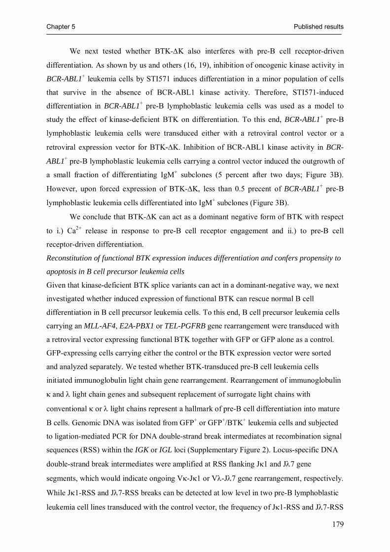
Chapter 5 Published results
179
We next tested whether BTK-ΔK also interferes with pre-B cell receptor-driven
differentiation. As shown by us and others (16, 19), inhibition of oncogenic kinase activity in
BCR-ABL1+ leukemia cells by STI571 induces differentiation in a minor population of cells
that survive in the absence of BCR-ABL1 kinase activity. Therefore, STI571-induced
differentiation in BCR-ABL1+ pre-B lymphoblastic leukemia cells was used as a model to
study the effect of kinase-deficient BTK on differentiation. To this end, BCR-ABL1+ pre-B
lymphoblastic leukemia cells were transduced either with a retroviral control vector or a
retroviral expression vector for BTK-ΔK. Inhibition of BCR-ABL1 kinase activity in BCR-
ABL1+ pre-B lymphoblastic leukemia cells carrying a control vector induced the outgrowth of
a small fraction of differentiating IgM+ subclones (5 percent after two days; Figure 3B).
However, upon forced expression of BTK-ΔK, less than 0.5 precent of BCR-ABL1+ pre-B
lymphoblastic leukemia cells differentiated into IgM+ subclones (Figure 3B).
We conclude that BTK-ΔK can act as a dominant negative form of BTK with respect
to i.) Ca2+ release in response to pre-B cell receptor engagement and ii.) to pre-B cell
receptor-driven differentiation.
Reconstitution of functional BTK expression induces differentiation and confers propensity to
apoptosis in B cell precursor leukemia cells
Given that kinase-deficient BTK splice variants can act in a dominant-negative way, we next
investigated whether induced expression of functional BTK can rescue normal B cell
differentiation in B cell precursor leukemia cells. To this end, B cell precursor leukemia cells
carrying an MLL-AF4, E2A-PBX1 or TEL-PGFRB gene rearrangement were transduced with
a retroviral vector expressing functional BTK together with GFP or GFP alone as a control.
GFP-expressing cells carrying either the control or the BTK expression vector were sorted
and analyzed separately. We tested whether BTK-transduced pre-B cell leukemia cells
initiated immunoglobulin light chain gene rearrangement. Rearrangement of immunoglobulin
κ and λ light chain genes and subsequent replacement of surrogate light chains with
conventional κ or λ light chains represent a hallmark of pre-B cell differentiation into mature
B cells. Genomic DNA was isolated from GFP+ or GFP+/BTK+ leukemia cells and subjected
to ligation-mediated PCR for DNA double-strand break intermediates at recombination signal
sequences (RSS) within the IGK or IGL loci (Supplementary Figure 2). Locus-specific DNA
double-strand break intermediates were amplified at RSS flanking Jκ1 and Jλ7 gene
segments, which would indicate ongoing Vκ-Jκ1 or Vλ-Jλ7 gene rearrangement, respectively.
While Jκ1-RSS and Jλ7-RSS breaks can be detected at low level in two pre-B lymphoblastic
leukemia cell lines transduced with the control vector, the frequency of Jκ1-RSS and Jλ7-RSS
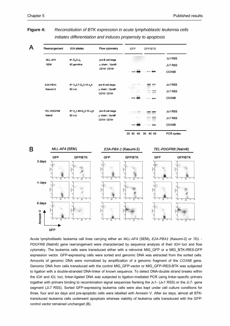
Chapter 5 Published results
180
GFP GFP/BTK
Jκ1 RSS
Jλ7 RSS
COX6B
35 40 45 35 40 45 PCR cycles
Rearrangement IGH alleles Flow cytometry
MLL-AF4 #1 DH3-JH pro-B cell stage
SEM #2 germline μ chain−, VpreB−
CD10+ CD19+
E2A-PBX1 #1 VH3.7-DH3.10-JH4 pre-B cell stage
Kasumi-2 #2 n.d. μ chain+, VpreB+
CD10+ CD19+
TEL-PDGFRB #1 VH1.69-DH3.10-JH6 pre-B cell stage
Nalm6 #2 n.d. μ chain+, VpreB+
CD10+ CD19+
GFP GFP/BTK
MLL-AF4 (SEM)
3 days
4 days
6 days
GFP
Anne
xin
V
E2A-PBX-1 (Kasumi-2) TEL-PDGFRB (Nalm6)
GFP GFP/BTK GFP GFP/BTK
Jκ1 RSS
Jλ7 RSS
COX6B
Jκ1 RSS
Jλ7 RSS
COX6B
A
B
GFP GFP/BTK
Jκ1 RSS
Jλ7 RSS
COX6B
35 40 45 35 40 45 PCR cycles
Rearrangement IGH alleles Flow cytometry
MLL-AF4 #1 DH3-JH pro-B cell stage
SEM #2 germline μ chain−, VpreB−
CD10+ CD19+
E2A-PBX1 #1 VH3.7-DH3.10-JH4 pre-B cell stage
Kasumi-2 #2 n.d. μ chain+, VpreB+
CD10+ CD19+
TEL-PDGFRB #1 VH1.69-DH3.10-JH6 pre-B cell stage
Nalm6 #2 n.d. μ chain+, VpreB+
CD10+ CD19+
GFP GFP/BTK
MLL-AF4 (SEM)
3 days
4 days
6 days
GFP
Anne
xin
V
E2A-PBX-1 (Kasumi-2) TEL-PDGFRB (Nalm6)
GFP GFP/BTK GFP GFP/BTK
Jκ1 RSS
Jλ7 RSS
COX6B
Jκ1 RSS
Jλ7 RSS
COX6B
A
B
Figure 4: Reconstitution of BTK expression in acute lymphoblastic leukemia cells
initiates differentiation and induces propensity to apoptosis
DNA double-strand breaks was increased in leukemia cells transduced with the BTK-IRES-
GFP expression vector (Figure 4A). This indicates that overexpression of functional BTK can
partially relieve the differentiation block of acute lymphoblastic leukemia cells that were
arrested at the pre-B cell stage. However, MLL-AF4+ acute lymphoblastic leukemia cells,
which are arrested at the pro-B cell stage do not initiate immunoglobulin light chain gene
rearrangement in response to BTK overexpression. This might indicate that the initiation
Acute lymphoblastic leukemia cell lines carrying either an MLL-AF4 (SEM), E2A-PBX1 (Kasumi-2) or TEL -PDGFRB (Nalm6) gene rearrangement were characterized by sequence analysis of their IGH loci and flow cytometry. The leukemia cells were transduced either with a retroviral MIG_GFP or a MIG_BTK-IRES-GFP expression vector. GFP-expressing cells were sorted and genomic DNA was extracted from the sorted cells. Amounts of genomic DNA were normalized by amplification of a genomic fragment of the COX6B gene. Genomic DNA from cells transduced with the control MIG_GFP-vector or MIG_GFP-IRES-BTK was subjected to ligation with a double-stranded DNA-linker of known sequence. To detect DNA-double strand breaks within the IGK and IGL loci, linker-ligated DNA was subjected to ligation-mediated PCR using linker-specific primers together with primers binding to recombination signal sequences flanking the Jκ1- (Jκ1 RSS) or the Jλ7- gene segment (Jλ7 RSS). Sorted GFP-expressing leukemia cells were also kept under cell culture conditions for three, four and six days and pre-apoptotic cells were labelled with Annexin V. After six days, almost all BTK-transduced leukemia cells underwent apoptosis whereas viability of leukemia cells transduced with the GFP-control vector remained unchanged (B).

Chapter 5 Published results
181
immunoglobulin light chain gene rearrangement not only requires expression of functional
BTK but also the expression of a functional pre-B cell receptor, which is present in pre-B
lymphoblastic leukemia but missing in pro-B lymphoblastic leukemia cells carrying an MLL-
AF4 gene rearrangement (Figure 4A). While reconstitution of BTK expression expression
initiated immunoglobulin light chain gene rearrangement in three of four cell lines tested, we
could not detect κ or λ light chains on the surface of BTK-GFP-transduced leukemia cells
(not shown). Attempting to establish permanent lines from the control-GFP- or BTK-GFP-
transduced leukemia cells, we noted that reconstitution of expression of functional BTK
substantially increases sensitivity of the leukemia cells to apoptosis. Comparing acute
lymphoblastic leukemia cells carrying control-GFP or BTK-GFP vectors, the BTK-transduced
cells underwent apoptosis spontaneously after six days in cell culture (Figure 4B).
Dominant-negative BTK splice variants can protect pre-B lymphoblastic leukemia cells
against radiation-induced apoptosis
BTK-mediated propensity to apoptosis was observed in MLL-AF4+, E2A-PBX1+ and TEL-
PDFRB+ acute lymphoblastic leukemia cells irrespective of pre-B cell receptor expression
(Figure 4). An earlier study invoked BTK kinase activity as a sensitizer to γ-radiation-induced
apoptosis in chicken DT-40 lymphoma cells (20). Therefore, kinase-deficient dominant-
negative BTK splice variants may have a protective effect against γ-radiation-induced
apoptosis in human acute lymphoblastic leukemia cells. To test this hypothesis, we γ-
irradiated E2A-PBX1+ acute lymphoblastic leukemia cells with 10 Gy. 24 hours after
irradiation, more than 90 percent of the cells had already undergone apoptosis as assessed by
propidium iodide uptake. Among the remaining cells, we sorted Annexin V+ pre-apoptotic
cells and Annexin V− viable cells and compared BTK splice variant expression between pre-
apoptotic and surviving leukemia cells. In two different PCR approaches, the entire kinase
domain (using primers for exons 12 to 19) and at a higher level of resolution, a hotspot-region
of aberrant splicing comprising exons 14 to 17 was amplified. Consistent with a protective
effect against γ-radiation-induced apoptosis, surviving leukemia cells exhibit preferential
expression of kinase-deficient dominant-negative BTK splice variants (Figure 5A). Sequence
analysis of these splice variants revealed that some of the BTK splice variants identified here
in the context of resistance to γ radiation-induced apoptosis were recurrently amplified from
various leukemia subentities (see Table 1). However, novel BTK splice variants were also
amplified (Figure 5B), which again all have in common their lack of a functional kinase
domain.

Chapter 5 Published results
182
Figure 5: Acute lymphoblastic leukemia cells surviving γ-radiation are selected for the
expression of dominant-negative BTK isoforms
Concluding remarks
Taking these findings together, we conclude that somatic mutation of the BTK gene is rare in
B cell precursor leukemia cells. However, in the majority of cases, the leukemia cells exhibit
deranged expression of BTK. While in four out of eight primary cases with an MLL-AF4 gene
rearrangement BTK expression is missing, B cell precursor leukemia cells typically exhibit
co-expression of full-length BTK with kinase-deficient, dominant-negative BTK splice
GAPDH
BTK
Exon 14-17
30 33 36 45 30 33 36 45 PCR-cycles
Annexin V+ Annexin V −
Irradiated acute lymphoblasticleukemia cells:
BTK
Exon 12-19
A
GAPDH
BTK
Exon 14-17
30 33 36 45 30 33 36 45 PCR-cycles
Annexin V+ Annexin V −
Irradiated acute lymphoblasticleukemia cells:
BTK
Exon 12-19
A
B SH2 Kinase
12 13 14 15 16 17 18 19 Full-length BTK
ΔExons 15-16
ΔExon 16
ΔExons 14-16
ΔExons 15-16 (Exon 14 Δ39 bp; Exon 17 Δ48 bp)
ΔExons 15-16 (Exon 14 Δ80 bp; Exon 17 Δ26 bp)
B SH2 Kinase
12 13 14 15 16 17 18 19 Full-length BTK
ΔExons 15-16
ΔExon 16
ΔExons 14-16
ΔExons 15-16 (Exon 14 Δ39 bp; Exon 17 Δ48 bp)
ΔExons 15-16 (Exon 14 Δ80 bp; Exon 17 Δ26 bp)
The pre-B lymphoblastic leukemia cell line Kasumi-2 carrying a E2A-PBX1 gene rearrangement were γ-irradiated with 10 Gy. After 24 hours, more than 90 percent of the leukemia cells were apoptotic. Among the remaining cells, AnnexinV+ pre-apoptotic cells and AnnexinV surviving cells were sorted separately. Sorted cells were subjected to RT-PCR analysis for BTK splice variant expression and cDNA amounts were normalized by amplification of a GAPDH cDNA fragment. Splice variants were detected by amplification of the entire BTK kinase domain using primers for exons 12 and 19 (low resolution; A, top) or for exons 14 and 17 (high resolution, A, bottom). PCR products of this amplification were subjected to sequence analysis (B).
Chapter 5 Published results
183
variants. These dominant-negative BTK splice variants can interfere with pre-B cell receptor-
dependent signal transduction, induce a differentiation block and prevent immunoglobulin κ
or λ light chain gene rearrangement. While overexpression of full-length BTK in B lymphoid
leukemia cells induces propensity to apoptosis, expression of dominant-negative BTK splice
variants can protect against radiation-induced apoptosis.
Acknowledgements:
We would like to thank Klaus Rajewsky and Martin Krönke for continuous support and discussions and Stefanie Jauch and Peter Wurst for excellent technical assistance. N.F. is supported by a fellowship from the German José-Carreras-Leukemia Foundation, F.K. is supported by the Studienstiftung des deutschen Volkes and the Köln Fortune program of the Faculty of Medicine of the University of Cologne. M.M. is supported by the Deutsche Forschungsgemeinschaft through the Emmy-Noether-Programm and through grants MU1616/2-1, MU1616/3-1 (to M.M.), the German José-Carreras-Leukemia-Foundation (grant to M.M.), the Ministry of Science and Research for North Rhine-Westphalia through the Stem Cell Network NRW (to M.M.), the Deutsche Krebshilfe (through grant ‘Molecular Mechanisms of Malignant Lymphoma’ to M.M.).
Materials and Methods
Patient samples, cell lines and cell purification
Normal CD19+ μ-chain− pro-B cells and CD19+ VpreB+ pre-B cells were sorted from human
bone marrow from four healthy donors (purchased from Cambrex, Baltimore, MD) using
immunomagnetic beads against CD19 (Miltenyi Biotech, Bergisch Gladbach, Germany) and
cell sorting using antibodies against CD19, VpreB (BD Biosciences, Heidelberg, Germany)
and the μ-chain (Jackson Immunoresearch, West Grove, PA). Similarly, CD5+ CD19+ B1
cells, IgD+ CD27− naïve B cells, CD19+ CD27+ memory B cells and CD19+ CD138+ plasma
cells were sorted from peripheral blood of four healthy donors using antibodies against CD5,
CD19, CD27, CD138 and IgD (BD Biosciences). In total, 29 B cell precursor leukemias
including 12 cell lines and 17 primary cases were studied. 11 cases of B cell precursor
leukemia with MLL-AF4 gene rearrangement [t(4;11)(q21;q23)] including eight primary cases
(I to VIII, Table 1) and three cell lines (BEL1, RS4;11 and SEM) were analyzed. 11 samples
carrying a BCR-ABL1 gene rearrangement [t(9;22)(q34;q11)] including seven primary cases
(IX to XV, Table 1) and four cell lines (BV173, Nalm1, SD1 and SUP-B15) were studied. In
addition, three leukemia cell lines carrying an E2A-PBX1 gene rearrangement
[t(1;19)(q23;p13); 697, Kasumi2 and MHH-CALL3], three cases of pre-B lymphoblastic
Chapter 5 Published results
184
leukemia with TEL-AML1 fusion gene [t(12;21)(p12;q22)] including two primary cases
(XVIII and XIX, Table 1) and the cell line REH and one pre-B lymphoblastic leukemia cell
line harboring a TEL-PDGFRB gene rearrangement [Nalm6; t(5;12)(q33.2;p13.2)] were
studied. For all cases, fusion transcripts resulting from oncogenic gene rearrangements, were
detected by PCR as previously described (5). Clinical data for all primary cases were
described previously (2).
Western blotting
For the detection of tyrosine-phosphorylated BTK by Western blot, a phosphotyrosine-
specific antibody against BTKY223 and EIF4E (Cell Signaling Technology, Beverly, MA)
were used. Western blot experiments were carried out as previously described (6).
Inhibitors of BCR-ABL1 and BTK
For inhibition of BCR-ABL1 kinase activity, the anti-leukemic drug STI571 (Novartis,
Basel, Switzerland) was used at a concentration of 10 μmol/l. For inhibition of BTK, cells
were incubated with alpha-cyano-beta-hydroxy-beta-methyl-N-(2,5-dibromophenyl)
propenamide (LFM-A13; Calbiochem, Darmstadt, Germany) at a concentration of 50 μg/ml
for the times indicated.
Measurement of pre-B cell receptor responsiveness
Primary human bone marrow pre-B cells were enriched using immunomagnetic MACS beads
as previously described (7). Pre-B lymphoblastic leukemia cells were cultured with 10% fetal
calf serum in RPMI 1640 medium for the times and under the conditions indicated. After pre-
incubation, cells were washed and stained with Fluo-3 dye (Calbiochem, Bad Soden,
Germany) for 30 minutes. Changes of cytosolic Ca2+ were measured by laser scans using
confocal microscopy (8, 9). After 10 to 30 seconds of measurement, antibodies against human
μ-chains (Jackson ImmunoResearch) were added to bone marrow pre-B cells or preincubated
pre-B lymphoblastic leukemia cells (in the presence or absence of LFM-A13). Cytosolic Ca2+
concentrations were determined as previously described (8).
Flow cytometry
Surface expression of IgΜ and CD19 on pre-B lymphoblastic leukemia cell lines and primary
leukemia cells was monitored using antibodies from BD Biosciences, Heidelberg, Germany,
after the incubation times indicated. Pre-apoptotic or dead cells were identified by staining
with Annexin V and propidium iodide (BD Biosciences).
Sequence analysis of BTK and semiquantitative RT-PCR
In a search for BTK isoforms and somatic mutations of the BTK gene, BTK cDNA fragments
covering the entire coding region or genomic DNA fragments of the BTK gene were amplified

Chapter 5 Published results
185
using 5’-ATCCCAACAGAAAAAGAAAACAT-3’ (BTK exon 2 forward), 5’-
GTTGCTTTCCTCCAAGATAAAAT-3’ (BTK exon 8 reverse), 5’-
ATCTTGAAAAAGCCACTACCG-3’ (BTK exon 8 forward),
5'-TGATACGTCATTATGTTGTGTGTT-3' (BTK exon 12 forward), 5’-
TGATACGTCATTATGTTGTGTGTT-3’ (BTK exon 13 forward), 5’-
ATCATGACTTTGGCTTCTTCAAT-3’ (BTK exon 14 reverse), 5’-
CTCAAATATCCAGTGTCTCAACA-3’ (BTK exon 14 forward), 5’-
CTTTAACAACTCCTTGATCGTTT-3’ (BTK exon 17 reverse) and 5’-
GGATTCTTCATCCATGACATCTA-3’ (BTK exon 19 reverse) were used. For
normalization of cDNA and genomic DNA amounts 5’-TTAGCACCCCTGGCCAAG-3’
(GAPDH forward) and 5’-CTTACTCCTTGGAGGCCATG-3’ (GAPDH reverse) and 5’-
AACTACAAGACCGCCCCTTT-3’ (COX6B forward) and 5’-
GCAGCCAGTTCAGATCTTCC-3’ (COX6B reverse) were used.
BTK amplification products were sequenced as previously described (6). Sequences of novel
BTK isoforms are available from Genbank/EMBL (AM051275-AM051286).
Retroviral expression of a kinase-deficient BTK splice variant
All aberrant BTK splice variants identified lack a functional kinase domain (see
supplementary Figure 1). Therefore, we generated a cDNA fragment of human BTK
comprising the entire coding region but lacking the C-terminal portion of the kinase domain
(exons 15 to 19; BTK-ΔK). Following digestion with NotI (New England Biolabs, Frankfurt
am Main, Germany), the PCR-product was ligated into the retroviral S11IN expression vector
(10). This vector is based on the retroviral plasmid SFβ11 (kindly provided by Dr.
Christopher Baum, Hannover, Germany) in which a multicloning site with NotI, EcoRI and
BamHI restriction sites was introduced, followed by an IRES NEO cassette.
293T cells were cultured in DMEM (Invitrogen, Karlsruhe, Germany) supplemented
with 10% fetal calf serum (Invitrogen), 2 mM L-glutamine (Invitrogen), penicillin G (100
units/ml) and streptomycin (100 μg/ml) (Invitrogen). 293T cells were cotransfected with 10
μg of the helper plasmid pHIT60, 10 μg of pczVSV-G envelope (11) and 10 μg of S11IN (as
a control vector) or S11-BTK-ΔK-IN using Fugene 6 (Roche, Basel, Switzerland) following
manufacturer’s instructions. Both vectors are based on SF11 (10) with the 3’LTR of the
spleen focus-forming virus (SFFVp) and internal ribosome entry site (IRES). Alternatively,
MIG-GFP (12) und MIG-BTK-IRES-GFP vectors were used for retroviral delivery of full-
length BTK. For construction of the MIG-BTK-IRES-GFP vector, the cDNA encoding
human full-length BTK was cloned into the MIG vector (12). 24h after transfection, the

Chapter 5 Published results
186
medium was changed for IMDM (Iscove’s modified Medium). 48h after the transfection,
supernatants were filtered through a 0.45 μm filter and used to infect pre-B acute
lymphoblastic leukemia cell lines on plates coated with 2 μg/cm2 of retronectin (Takara
Shuzo, Otsu, Japan; reference 13). Plates were pre-loaded three times with fresh supernatant
(14) and subsequently 2,5 x 10⁵ cells were added to each well. 48 h after infection, cells
expressing the S11IN vector were selected using G418 to a final concentration of 0.5 mg/ml.
Cell sorting
GFP-expressing cells or Annexin V+ and Annexin V− cells were sorted using a FACStar 440
cell sorter. Cells were sorted under sterile conditions and either kept under cell culture
conditions or subjected to RNA isolation and RT-PCR analysis.
Amplification of double-strand recombination signal sequence breaks by ligation-mediated
PCR
Genomic DNA was isolated from about 2.5 x 10⁶ leukemia cells carrying retroviral
expression vectors for either GFP alone or GFP and BTK and ligated to a blunt-end linker
using T4 DNA ligase (Invitrogen, Karlsruhe, Germany) at 14°C overnight. The linker was
constructed by annealing the oligonucleotides 5'-
TTTCTGCTCGAATTCAAGCTTCTAACGATGTACGGGGACATG 3' and 3’ amino (C7)-
GACGAGCTTAAGTTCGAAGATTGCTACATGCCCCT -5’ and protruding 3' overhangs
were removed by 3'→ 5' exonuclease activity of the Klenow fragment of E. coli DNA
polymerase I (Invitrogen, Karlsruhe, Germany). Ligation-mediated PCR (LM-PCR; reference
15) was carried out with modifications as previously described (16). In two semi-nested
rounds of amplification at an annealing temperature of 59°C, RSS-intermediates with a DNA
double-strand break at the 5’ heptamer of Jκ1 gene segments were amplified (see
supplementary Figure 2) using 5’-GTAATTAACATTCAGTCTACTTTC-3’ as external
forward and 5’-TAACATTCAGTCTACTTTCTAAAA-3’ as internal forward primers
together with 5’-TCCCCGTACATCGTTAGAAG–3’ as reverse primer specific for DNA-
ligated linker molecules. To amplify RSS-intermediates with a DNA double-strand break at
the 5’ heptamer of Jλ7 gene segments, 5’-TTCTCACTTCTTCCATGGTGAC-3’ and 5’-
ACTTCTTCCATGGTGACAGTCT-3’ were used in two rounds of PCR amplification as
described above.
γ-radiation
Pre-B lymphoblastic leukemia cells were irradiated with 10 Gy γ-radiation at the Clinic for
Radiotherapy and Radiological Oncology, Heinrich-Heine-Universität Düsseldorf,

Chapter 5 Published results
187
Düsseldorf, Germany. 24 hours after radiation, leukemia cells were stained with Annexin V
and propidium iodide to separately sort pre-apoptotic or surviving cells.

Chapter 5 Published results
188
Supplementary Figure 1a: BTK splice variants in B cell precursor leukemia
PH TH SH2SH3 Kinase
1 138 215 280 377 659
1 2 3 4 5 6 7 8 9 10 11 12 13 14 15 16 17 18 19Full-length
PH TH SH2SH3 Kinase
1 138 215 280 377 442
1 2 3 4 5 6 7 8 9 10 11 12 13 14 16 17 18 19
Stop
ΔExon 15a
PH TH SH2SH3 Kinase
1 138 215 280 377 565
1 2 3 4 5 6 7 8 9 10 11 12 13 14 17 18 19ΔExons 15-16b
PH TH SH2SH3 Kinase
1 138 215 280 377 425
1 2 3 4 5 6 7 8 9 10 11 12 13 14 16 17 18 19
Stop
ΔExons 14-15Exon 14 Δ41 bp
Δ41 bp exon 14
PH TH SH2SH3 Kinase
1 138 215 280 371 384
1 2 3 4 5 6 7 8 9 10 11 12 15 16 17 18 19ΔExon 14Exon 13 Δ62 bpExon 15 Δ133 bp
Δ62 bp exon 13Δ133 bp exon 15
Stop
PH TH SH2SH3 Kinase
1 138 215 280 377 547
1 2 3 4 5 6 7 8 9 10 11 12 13 14 15 18 19
ΔExons 16-17Exon 15 Δ119 bpExon 18 Δ136 bp
StopΔ119 bp exon 15Δ136 bp exon 18
PH TH SH2SH3 Kinase
1 138 215 280 377 648
1 2 3 4 5 6 7 8 9 10 11 12 13 14 15 16 17 18 19
Exon 18 Δ33 bp
Δ33 bp exon 18
PH TH SH2SH3 Kinase
1 138 215 280 377 527
1 2 3 4 5 6 7 8 9 10 11 12 13 14 15 17 18 19
ΔExon 16b
Stop
PH TH SH2SH3 Kinase
1 138 215 280 377 659
1 2 3 4 5 6 7 8 9 10 11 12 13 14 15 16 17 18 19Full-length
PH TH SH2SH3 Kinase
1 138 215 280 377 442
1 2 3 4 5 6 7 8 9 10 11 12 13 14 16 17 18 1917 18 19
StopStop
ΔExon 15a
PH TH SH2SH3 Kinase
1 138 215 280 377 565
1 2 3 4 5 6 7 8 9 10 11 12 13 14 17 18 19ΔExons 15-16b
PH TH SH2SH3 Kinase
1 138 215 280 377 425
1 2 3 4 5 6 7 8 9 10 11 12 13 14 16 17 18 1917 18 19
Stop
ΔExons 14-15Exon 14 Δ41 bp
Δ41 bp exon 14
PH TH SH2SH3 Kinase
1 138 215 280 371 384
1 2 3 4 5 6 7 8 9 10 11 12 15 16 17 18 19ΔExon 14Exon 13 Δ62 bpExon 15 Δ133 bp
Δ62 bp exon 13Δ133 bp exon 15
StopStop
PH TH SH2SH3 Kinase
1 138 215 280 377 547
1 2 3 4 5 6 7 8 9 10 11 12 13 14 15 18 19
ΔExons 16-17Exon 15 Δ119 bpExon 18 Δ136 bp
StopStopΔ119 bp exon 15Δ136 bp exon 18
PH TH SH2SH3 Kinase
1 138 215 280 377 648
1 2 3 4 5 6 7 8 9 10 11 12 13 14 15 16 17 18 19
Exon 18 Δ33 bp
Δ33 bp exon 18
PH TH SH2SH3 Kinase
1 138 215 280 377 527
1 2 3 4 5 6 7 8 9 10 11 12 13 14 15 17 18 19
ΔExon 16b
StopStop

Chapter 5 Published results
189
Supplementary Figure 1b: BTK splice variants in B cell precursor leukemia
PH TH SH2SH3 Kinase
1 138 215 280 377 468
1 2 3 4 5 6 7 8 9 10 11 12 13 18 1918 19
PH TH SH2SH3 K.
1 138 215 280 366 390
1 2 3 4 5 6 7 8 9 10 11 12 1919Intron 18
Intron
Δ2,084 bp Δ1,045 bp
Kinase
ΔExons 14-17
ΔExons 13-18Ins 106 bp Intron 18
PH TH SH2SH3 Kinase
1 138 215 280 377 404
1 2 3 4 5 6 7 8 9 10 11 12 13 17 18 19
Stop
ΔExons 14-16
PH TH SH2SH3 Kinase
1 138 215 280 377 448
1 2 3 4 5 6 7 8 9 10 11 12 13 14 18 19
ΔExons 15-17Exon 14 Δ18 bpIns 1 bpExon 18 Δ33 bp Stop
Δ18 bp exon 14Ins 1 bpΔ33 bp exon 18
PH TH SH2SH3 Kinase
1 138 215 280 377 426
1 2 3 4 5 6 7 8 9 10 11 12 13 14 1919
ΔExons 15-18Exon 14 Δ79 bpIns 3 bpExon 19 Δ24 bp
StopΔ79 bp exon 14Ins 3 bpΔ24 bp exon 19
PH TH SH2SH3 Kinase
1 138 215 280 366 408
1 2 3 4 5 6 7 8 9 10 11 12 18 19
ΔExons 13-17Exon 12 Δ13 bpExon 18 Δ92 bp
Δ13 bp exon 12Δ92 bp exon 18
PH TH SH2SH3
1 2 3 4 5 6 7 8 9 10 11 13 19
Kinase
Δ31 bp exon 12Δ22 bp exon 13Δ212 bp exon 19
Stop
ΔExons 14-18Exon 12 Δ31 bpExon 13 Δ22 bpExon 19 Δ212 bp
12
PH TH SH2SH3
1 2 3 4 5 6 7 8 9 10 11 13 19
Kinase
Δ31 bp exon 12Δ22 bp exon 13Δ212 bp exon 19
Stop
ΔExons 14-18Exon 12 Δ31 bpExon 13 Δ22 bpExon 19 Δ212 bp
12

Chapter 5 Published results
190
Legend to supplementary Figure 1: BTK splice variants in B cell precursor leukemia
BTK isoform expression was analyzed by RT-PCR in 29 cases of B cell precursor leukemia carrying BCR-ABL1, E2A-PBX1, MLL-AF4, TEL-AML1 or TEL-PDGFRB gene rearrangements. 14 aberrant BTK isoforms could be identified in addition to full-length BTK. None of these BTK isoforms carry a functional kinase domain resulting from the usage of regular or cryptic splice sites leading to large deletion and/or the generation of pre-terminal translation-stops. Protein domains (top) and mRNA exons (bottom) are depicted. Untranslated regions are indicated by hatched boxes. Sequence data of
all splice variants is available from EMBL/GenBank under accession numbers (AM051275-AM051286). Supplementary Figure 2: LM-PCR amplification of DNA-double strand break
intermediates at Jκ1 and Jλ7 recombination signal
sequences
RSS
RSS
Jκ1 or Jλ7
LinkerRSS
Jκ1 or Jλ7
Jκ1 or Jλ7
RSS
RSS
Jκ1 or Jλ7
LinkerRSS
Jκ1 or Jλ7
Jκ1 or Jλ7
Immunoglobulin light chain gene rearrangement involves the introduction of DNA double strand breaks at recombination signal sequences (RSS) flanking V and J segments. DNA double strand breaks at RSS can be ligated to double stranded DNA linker molecules. In order to detect DNA double-strand breaks preceding V-J recombination at the IGK or IGL loci, linker-ligated DNA can be amplified using Jk-RSS or Jl-RSS-specific primers together with a linker-specific primer.

Chapter 5 Published results
191
References
1. Jumaa H, Bossaller L, Portugal K, Storch B, Lotz M, Flemming A, Schrappe M, Postila V,
Riikonen P, Pelkonen J, Niemeyer CM, Reth M. Deficiency of the adaptor SLP-65 in pre-B-
cell acute lymphoblastic leukaemia. Nature. 2003; 423: 452-6.
2. Klein F, Feldhahn N, Harder L, Wang H, Wartenberg M, Hofmann WK, Wernet P, Siebert R,
Müschen M. The BCR-ABL1 Kinase Bypasses Selection for the Expression of a Pre-B Cell
Receptor in Pre-B Acute Lymphoblastic Leukemia Cells. J Exp Med. 2004; 199:673-85.
3. Kersseboom R, Middendorp S, Dingjan GM, Dahlenborg K, Reth M, Jumaa H, Hendriks RW.
Bruton's tyrosine kinase cooperates with the B cell linker protein SLP-65 as a tumor
suppressor in Pre-B cells. J Exp Med. 2003; 198: 91-98.
4. Holinski-Feder E, Weiss M, Brandau O, Jedele KB, Nore B, Backesjo CM, Vihinen M,
Hubbard SR, Belohradsky BH, Smith CI, Meindl A. Mutation screening of the BTK gene in
56 families with X-linked agammaglobulinemia (XLA): 47 unique mutations without
correlation to clinical course. Pediatrics. 1998; 101: 276-284.
5. van Dongen JJ, Langerak AW, Bruggemann M, Evans PA, Hummel M, Lavender FL,
Delabesse E, Davi F, Schuuring E, Garcia-Sanz R, van Krieken JH, Droese J, Gonzalez D,
Bastard C, White HE, Spaargaren M, Gonzalez M, Parreira A, Smith JL, Morgan GJ, Kneba
M, Macintyre EA. Design and standardization of PCR primers and protocols for detection of
clonal immunoglobulin and T-cell receptor gene recombinations in suspect
lymphoproliferations. Leukemia. 2003;17:2257-317.
6. Feldhahn N, Klein F, Mooster JL, Hadweh P, Sprangers M, Wartenberg M, Bekhite MM,
Hofmann WK, Herzog S, Jumaa H, Rowley JD, Müschen M. Mimicry of a constitutively
active pre-B cell receptor: Aberrant splicing links Bruton’s tyrosine kinase to BCR-ABL1 in
pre-B lymphoblastic leukemia. J Exp Med, 201: in press (2005)
7. Müschen M, Lee S, Zhou G, Feldhahn N, Barath VS, Chen J, Moers C, Krönke M, Rowley
JD, Wang SM. Molecular portraits of B cell lineage commitment. Proc Natl Acad Sci U S A.
2002; 99: 10014-9.
8. Feldhahn N, Schwering I, Lee S, Wartenberg M, Klein F, Wang H, Zhou G, Wang SM,
Rowley JD, Hescheler J, Krönke M, Rajewsky K, Küppers R, Müschen M. Silencing of B cell
receptor signals in human naive B cells. J Exp Med. 2002; 196:1291-305.
9. Klein F, Feldhahn N, Lee S, Wang H, Ciuffi F, von Elstermann M, Toribio ML, Sauer H,
Wartenberg M, Barath VS, Krönke M, Wernet P, Rowley JD, Müschen M. T lymphoid
differentiation in human bone marrow. Proc Natl Acad Sci U S A. 2003; 100: 6747-52.
10. Hildinger M, Abel KL, Ostertag W, Baum C. Design of 5' untranslated sequences in retroviral
vectors developed for medical use. J Virol 1999; 73: 4083-4089.
11. Pietschmann T, Heinkelein M, Heldmann M, Zentgraf H, Rethwilm A, Lindemann D.

Chapter 5 Published results
192
Foamy virus capsids require the cognate envelope protein for particle export. J Virol 1999; 73:
2613-2621.
12. Pear WS, Miller JP, Xu L, Pui JC, Soffer B, Quackenbush RC, Pendergast AM, Bronson R,
Aster JC, Scott ML, Baltimore D. Efficient and rapid induction of a chronic myelogenous
leukemia-like myeloproliferative disease in mice receiving P210 bcr/abl-transduced bone
marrow. Blood 1998; 92: 3780-3792.
13. Hanenberg H, Xiao XL, Dilloo D, Hashino K, Kato I, Williams DA. Colocalization of
retrovirus and target cells on specific fibronectin fragments increases genetic transduction of
mammalian cells. Nat Med. 1996; 2: 876-882.
14. Hanenberg H, Hashino K, Konishi H, Hock RA, Kato I, Williams DA. Optimization of
fibronectin-assisted retroviral gene transfer into human CD34+ hematopoietic cells. Hum
Gene Ther. 1997; 8: 2193-2206.
15. Schlissel M, Constantinescu A, Morrow T, Baxter M, Peng A. Double-strand signal sequence
breaks in V(D)J recombination are blunt, 5'-phosphorylated, RAG-dependent, and cell cycle
regulated. Genes Dev 1993; 7: 2520-2532.
16. Klein F, Feldhahn N, Mooster JL, Sprangers M, Hofmann WK, Wernet P, Wartenberg M and
Müschen M. Tracing the pre-B to immature B cell transition in human leukemia cells reveals a
coordinated sequence of primary and secondary IGK gene rearrangement, IGK deletion and
IGL gene rearrangement. J Immunol 2005, 174: 367-375
17. Middendorp S, Dingjan GM, Maas A, Dahlenborg K, Hendriks RW. Function of Bruton's
tyrosine kinase during B cell development is partially independent of its catalytic activity. J
Immunol 2003; 171: 5988-5996.
18. Goodman PA, Wood CM, Vassilev AO, Mao C, Uckun FM. Defective expression of Bruton's
tyrosine kinase in acute lymphoblastic leukemia. Leuk Lymphoma 2003; 44: 1011-1008.
19. Muljo SA, Schlissel MS. A small molecule Abl kinase inhibitor induces differentiation of
Abelson virus-transformed pre-B cell lines. Nat Immunol 2003; 4: 31-7
20. Uckun FM, Waddick KG, Mahajan S, Jun X, Takata M, Bolen J, Kurosaki T. BTK as a
mediator of radiation-induced apoptosis in DT-40 lymphoma B cells. Science 1996; 273:1096-
100.

Chapter 6
Appendix

193
6.1 Supplementary information PCR primers GAPDH forward 5’-TTAGCACCCCTGGCCAAG-3’ GAPDH reverse 5’-CTTACTCCTTGGAGGCCATG-3’ BCR-ABL1 forward 5’-ACCTCACCTCCAGCGAGGAGGACTT-3' BCR-ABL1 reverse 5'-TCCACTGGCCACAAAATCATACAGT-3' mAid forward 5'- AAATGTCCGCTGGGCCAA-3' mAid reverse 5'- CATCGACTTCGTACAAGGG-3' AID forward 5'-TGCTCTTCCTCCGCTACATC-3' AID reverse 5'-CCTCATACAGGGGCAAAAGG-3' mObf1 forward 5'-AGCTCCCTGACCATTGAC-3' mObf1 reverse 5'-CTGTCCCATCCCCCTGTAA-3' mOct2 forward 5'-ATCGAGACGAATGTCCGCTT-3' mOct2 reverse 5'-GTAGCTGGTCGGCTTTCC-3' Cμ_R 5’-AGACGAGGGGGAAAAGGGTT-3’ Cγ1_R 5’-GAGTTTTGTCACAAGATTTGGGCT-3’ Cγ2_R 5’-GCACTCGACACAACATTTGCG-3’ Cγ3_R 5’-TCACCAAGTGGGGTTTTGAGC-3’ Cγ4_R 5’-ATGGGCATGGGGGACCATTT-3’ Cα2_R 5’-TGTTGGCGGTTAGTGGGGTC-3’ Cε_R 5’-GAGGTGGCATTGGAGGGAAT-3’ Cδ_R 5’-AGAGCTGGCTGCTTGTCATG-3’ mJ558 forward 5’-GCAAGGCTTCTGGCTACACA-3’ mJH2/3 reverse 5’-CCAGTAAGCAAACCAGGCACA-3’ BTK exon 2 forward 5’-ATCCCAACAGAAAAAGAAAACAT-3’ BTK exon 8 reverse 5’-GTTGCTTTCCTCCAAGATAAAAT-3’ BTK exon 8 forward 5’-ATCTTGAAAAAGCCACTACCG-3’ BTK exon 12 forward 5’-TGATACGTCATTATGTTGTGTGTT-3’ BTK exon 13 forward 5’-ACTCATATCCAGGCTCAAATATC-3’ BTK exon 14 reverse 5’-ATCATGACTTTGGCTTCTTCAAT-3’ BTK exon 14 forward 5’-AATTGATCCAAAGGACCTGAC-3’ BTK exon 15 forward 5’-GAGAAGCTGGTGCAGTTGTAT-3’ BTK exon 16 reverse .1 5’-GGCCGAAATCAGATACTTTAAC-3’ BTK exon 16 reverse.2 5’-CTTTAACAACTCCTTGATCGTTT-3’ BTK exon 17 reverse 5’-ATTTGCTGCTGAACTTGCTATAC-3’ BTK exon 18 reverse 5’-TATACCTTCTCTGAAGCCAGATG-3’ BTK exon 19 reverse.1 5’-AAATTTCAGGCACAATAATTTCT-3’ BTK exon 19 reverse.2 5’-GGATTCTTCATCCATGACATCTA-3’ BTKp52 forward 5’CTTCCTCTCTGGACTGTAAGAAT-3’ BTKp52 reverse 5’-ATACAACTGCACCAGCTTCTC-3’ BCLXL forward 5’-AATCTTATCTTGGCTTTGGAT-3’ BCLXL reverse 5’-GTTCTCTTCCACATCACTAAA-3’ COX6B forward 5’-AACTACAAGACCGCCCCTTT-3’ COX6B reverse 5’-GCAGCCAGTTCAGATCTTCC-3’ HPRT forward 5’-CCTGCTGGATTACATCAAAGCACTG-3’ HPRT reverse 5’-CACCAGCAAGCTTGCGACC-3’

194
LM-PCR primers CDKN2A_F1 5’-CCAGGAATAAAATAAGGGGAATA-3’ CDKN2A_F2 5’-GGAATAAAATAAGGGGAATAGGG-3’ CDKN2A_R 5’-CTTTCCTACCTGGTCTTCTAGG-3’ CDKN2B_F1 5’-GTGAACATTCCCAAAATATTAGC-3’ CDKN2B_F2 5’-AAAATATTAGCCTTGGCTTTACTG-3’ CDKN2B_R 5’-AGACTCCTGTACAAATCTACATCG-3’ V3-21_R 5’-CTCTCGCACAGTAATACACAGC-3’ V1-2_R 5’-CTCTCGCACAGTAATACACGAC-3’ V1-69_R 5’-TCTCTCGCACAGTAATACACG-3’ V3-73_R 5’-GGTTTTCAGGCTGTTCATTT-3’ V3-53_R 5’-CACCTTTTAAAATAGCAACAAGG-3’ V3-30_R 5’-AGCATAGCTACTGAAGGTGAAT-3’ Mmyc_F1 5’-ATCCTGAGTCGCAGTATAAAAGA-3’ Mmyc_F2 5’-TTTTCTGACTCGCTGTAGTAATTC-3’ Mmyc_R1 5’-CTCAGAGACTGGTAAGTCAGAAG-3’ Linker_F1 5’-CTGCTCGAATTCAAGCTTCT-3’ Linker_F2 5’-GCTTCTAACGATGTACGGGG-3’ Linker_R1 5’-GTACATCGTTAGAAGCTTGAA-3’ Linker_R2 5’-GTTAGAAGCTTGAATTCGAGC-3’ siRNA duplices
Non-targeting siRNA 5’-UUGUACCUAAUUUCGUCCCAC-3’ 3’-CAUGGAUUAAAGCAGGGUGUU-5’ AID siRNA 5'-UUGCUCUUCCUCCGCUACAUU-3' 3'-CGAGAAGGAGGCGAUGUAAUU-3' BTKfull-length siRNA.1 5’-UUGCUGGUGCAGUUGUAUGGC-3’ 3’-CGACCACGUCAACAUACCGUU-5’ BTKfull-length siRNA.2 5’-UUGUUGUAUGGCGUCUGCACC-3’ 3’-CAACAUACCGCAGACGUGGUU-5’ BTKfull-length siRNA.3 5’-UUGUACAUGGCCAAUGGCUGC-3’ 3’-CAUGUACCGGUUACCGACGUU-5’ BTKp65 siRNA.1 5’-UUUCAUGAUGUAUGUCCUGGA-3’ 3’-AGUACUACAUACAGGACCUUU-5’ BTKp65 siRNA.2 5’-UUAAGUCAUGAUGUAUGUCCU-3’ 3-‘UUCAGUACUACAUACAGGAUU-5’ BTKp65 siRNA.3 5’-UUCCAAAGUCAUGAUGUAUGU-3’ 3’-GGUUUCAGUACUACAUACAUU-5’ BTKp52 siRNA.1 5’-UUUCAUGAUGCAGCUCGAAAC-3’ 3’-AGUACUACGUCGAGCUUUGUU-5’ BTKp52 siRNA.2 5’-UUAGUCAUGAUGCAGCUCGAA-3’ 3’-UCAGUACUACGUCGAGCUUUU-5’ BTKp52 siRNA.3 5’-UUCCAAAGUCAUGAUGCAGCU-3’ 3’-GGUUUCAGUACUACGUCGAUU-5’

195
Accession numbers:
BTKp52 AJ888378
BTK isoforms in AJ888376-AJ888381
BCR-ABL1-positive
pre-B cell leukemia
BTK isoforms in AM051275-AM051286
BCR-ABL1-negative
pre-B cell leukemia
IK6 AM085310
Sequence data available from GenBank/EMBL/DDBJ under the accession numbers given in the table. 6.2 Abbreviations
ALL, acute lymphoblastic leukaemia
AID, activation-induced cytidine deaminase
BTK, Bruton’s tyrosine kinase
C, constant;
CLP, common lymphoid progenitor
CML, chronic myeloid leukemia
CSR, class-switch recombination
cRSS, cryptic RSS
D, diversity
DAG, diacylglycerol
GL, germline
HSC, hematopoietic stem cell
IGH, immunoglobulin heavy-chain
IGK, immunoglobulin κ light chain

196
IGL, immunoglobulin λ light chain
ITAM, immunoreceptor tyrosine-based activatory motif
ITIM, immunoreceptor tyrosine-based inhibitory motif
J, joining
KDE, κ deleting element
LM-PCR, ligation-mediated PCR
NHEJ, non-homologous end joining
Ph, Philadelphia chromosome
PH, Pleckstrin-homology
PIP2, phosphatidylinositol-4,5-bisphosphate
PIP3, phosphatidylinositol-3,4,5-trisphosphate
PTK, protein tyrosine-kinase
PTP, protein tyrosine-phosphatase
RSS, recombination signal sequence
SAGE, serial analysis of gene expression
siRNA, small interfering RNA
SH2, SRC homology domain 2
SH3, SRC-homology domain 3
SHM, somatic hypermutation
SRC, cellular homolog of Rous sarcoma virus
TH, TEC-homology
V, variable
XLA, X-linked agammaglobulinemia
Xid, X-linked immunodeficiency

197
6.3 Curriculum vitae
Niklas Feldhahn
15.10.1976 Born in Bad Oldesloe (Germany)
1983-1987 Primary school (Lübeck/Germany)
1987-1996 Gymnasium (Lübeck/Germany)
01.06.1996 Abitur disposal (Lübeck/Germany)
1996-1997 Community service
1997-1999 Basic study period of biology
(University of Osnabrück/Germany)
27.08.1999 Intermediate diploma
1999-2002 Main study period of biology
(University of Cologne/Germany)
2001 Assistant at the Science Factory (company for
bioinformatics/Cologne/Germany)
2001-2002 Diploma thesis in the laboratory of PD Dr. Müschen
(Universität zu Köln/Germany)
19.09.2002 Diploma disposal graded with sehr gut (1.0):
“Signale des Antigenrezeptors humaner B Zell
Populationen: eine funktionelle Genomanalyse”
2003-2006 Thesis in the laboratory of Prof. Müschen
(Heinrich-Heine Universität Düsseldorf/Germany)
21.02.2006 Disposal of the Leukemia Clinical Research Award 2006
by the DGHO to Markus Müschen and Niklas Feldhahn
198
Invited talks
2003 First annual B cell meeting (Freiburg/Germany)
2004 Second annual B cell meeting (Berlin/Germany)
2004 Joint Annual Meeting of the German (DGfI) and Dutch
Society (NVvI) of Immunology
(Maastricht/Netherlands)
2005 Joint Annual Meeting of the German (DGfI) and
Scandinavian Society (SSI) of Immunology
(Kiel/Germany)
6.4 List of publications
2002
Müschen M, Lee S, Zhou G, Feldhahn N, Barath VS, Chen J, Moers C,
Krönke M, Rowley JD, Wang SM. Molecular portraits of B cell lineage
commitment. Proc Natl Acad Sci U S A. 2002; 99: 10014-10019
Feldhahn N, Schwering I, Lee S, Wartenberg M, Klein F, Wang H, Zhou G,
Wang SM, Rowley JD, Hescheler J, Krönke M, Rajewsky K, Kuppers R,
Müschen M. Silencing of B cell receptor signals in human naive B cells.
J Exp Med. 2002; 196: 1291-1305
2003
Klein F, Feldhahn N, Lee S, Wang H, Ciuffi F, von Elstermann M, Toribio
ML, Sauer H, Wartenberg M, Barath VS, Krönke M, Wernet P, Rowley JD,
Müschen M. T lymphoid differentiation in human bone marrow.
Proc Natl Acad Sci U S A. 2003; 100: 6747-6752

199
2004
Klein F, Feldhahn N, Harder L, Wang H, Wartenberg M, Hofmann WK,
Wernet P, Siebert R, Müschen M. The BCR-ABL1 kinase bypasses selection
for the expression of a pre-B cell receptor in pre-B acute lymphoblastic
leukemia cells. J Exp Med. 2004; 199: 673-685
Klein F, Feldhahn N, Müschen M. Interference of BCR-ABL1 kinase activity
with antigen receptor signaling in B cell precursor leukemia cells.
Cell Cycle. 2004; 3: 858-860
Kögler G, Sensken S, Airey JA, Trapp T, Müschen M, Feldhahn N, Liedtke S,
Sorg RV, Fischer J, Rosenbaum C, Greschat S, Knipper A, Bender J,
Degistirici O, Gao J, Caplan AI, Colletti EJ, Almeida-Porada G, Muller HW,
Zanjani E, Wernet P. A new human somatic stem cell from placental cord
blood with intrinsic pluripotent differentiation potential. J Exp Med. 2004; 200:
123-35
Su YW, Herzog S, Lotz M, Feldhahn N, Müschen M, Jumaa H. The
molecular requirements for LAT-mediated differentiation and the role of LAT
in limiting pre-B cell expansion. Eur J Immunol. 2004; 34: 3614-3622
2005
Klein F, Feldhahn N, Mooster JL, Sprangers M, Hofmann WK, Wernet P,
Wartenberg M, Müschen M. Tracing the pre-B to immature B cell transition in
human leukemia cells reveals a coordinated sequence of primary and
secondary IGK gene rearrangement, IGK deletion, and IGL gene
rearrangement. J Immunol. 2005; 174: 367-375
Feldhahn N, Klein F, Mooster JL, Hadweh P, Sprangers M, Wartenberg M,
Bekhite MM, Hofmann WK, Herzog S, Jumaa H, Rowley JD, Müschen M.
Mimicry of a constitutively active pre-B cell receptor in acute lymphoblastic
leukemia cells. J Exp Med. 2005; 201: 1837-1852

200
Feldhahn N, Río P, Ndikung Bejeng Soh B, Liedtke S, Sprangers M, Klein F,
Wernet P, Jumaa H, Hofmann WK, Hanenberg H, Rowley JD, Müschen M.
Deficiency of Bruton’s tyrosine kinase in B cell precursor leukemia cells.
Proc Natl Acad Sci USA. 2005; 102: 13266-13271
Soh BN, Klein F, Feldhahn N, Müschen M. B-lymphoid or myeloid lineage
identity of cell lines derived from chronic myeloid leukemia blast crisis.
Cancer Genet Cytogenet. 2005; 161: 187-188
2006
Klein F, Feldhahn N, Herzog S, Sprangers M, Mooster JL, Jumaa H, Müschen
M. BCR-ABL1 induces aberrant splicing of IKAROS and lineage infidelity in
pre-B lymphoblastic leukemia cells. Oncogene. 2006; 25: 1118-24
Sprangers M, Feldhahn N, Herzog S, Hansmann ML, Reppel M, Hescheler J,
Jumaa H, Siebert R, Müschen M. The SRC family kinase LYN redirects B cell
receptor signaling in human SLP65-deficient B cell lymphoma cells.
Oncogene. 2006 Mar 27;
Sprangers M, Feldhahn N, Liedtke S, Jumaa H, Siebert S, Müschen M.
SLP65-deficiency results in perpetual V(D)J-recombinase activity in pre-B
lymphoblastic leukemia and B cell lymphoma cells. Oncogene. 2006;

201
6.5 Specific contribution of Niklas Feldhahn to the publications shown in
chapter 5.1-5.2
In this doctoral thesis, a number of aspects related to BCR-ABL1-mediated survival signals in
pre-B lymphoblastic leukemia cells are described. The published results are presented in
detail in chapter 3.1-3.6 and 5.1-5.6. Besides my own experimental data, this work also
involves the contributions from colleagues and members of the laboratory of Markus
Müschen.
In the following, my own experimental contribution to the specific publications is outlined:
Chapter 5.1: The BCR-ABL1 kinase bypasses selection for the expression of
a pre-B cell receptor in pre-B acute lymphoblastic leukemia
cells By Klein F, Feldhahn N, Harder L, Wang H, Wartenberg M, Hofmann WK,
Wernet P, Siebert R, Müschen M.; J Exp Med. 2004; 199: 673-685
- Generation of gene-expression profiles for naïve and memory B
cells using the SAGE-method for the comparison of BCR-ABL1-
positive pre-B leukemia cells with normal B cell subsets
- Analysis of the configuration of the IGH locus of both alleles of 12
BCR-ABL1-positive pre-B leukemia cases
- Identification of footprints of potential VH-replacement in 9 out of
12 BCR-ABL1-positive pre-B leukemia cases
Chapter 5.2: BCR-ABL1 induces aberrant splicing of IKAROS and lineage
infidelity in pre-B lymphoblastic leukemia cells. By Klein F, Feldhahn N, Herzog S, Sprangers M, Mooster JL, Jumaa H,
Müschen M. Oncogene. 2005; 24: 1-7
- Western blot for the doxycylin-induced expression of the BCR-
ABL1 transgene in the murine pro-B cell line TON-B210

202
- FACS analysis of the expression of B lineage- and myeloid lineage-
specific surface receptors before and during treatment with STI571
- Western blot for the expression of the BCR-ABL1 transgene in
pMIG-BCR-ABL1-IRES-GFP- and pMIG-GFP-transfected 697
cells
- Participation in the RNA-interference-mediated silencing of the
expression of the IKAROS isoform IK6 in BCR-ABL1-positive
pre-B leukemia cells
Chaper 5.3: Tracing the pre-B to immature B cell transition in human
leukemia cells reveals a coordinated sequence of primary and
secondary IGK gene rearrangement, IGK deletion, and IGL
gene rearrangement By Klein F, Feldhahn N, Mooster JL, Sprangers M, Hofmann WK, Wernet P,
Wartenberg M, Müschen M. J Immunol. 2005; 174: 367-375
- Analysis of RAG1 protein expression in BCR-ABL1-positive pre-B
leukemia cells following treatment with STI571
- FACS analysis of immunoglobulin light-chain expression on the
surface of BCR-ABL1-positive pre-B leukemia cells after treatment
with STI571
Chapter 5.4: Mimicry of a constitutively active pre-B cell receptor in acute
lymphoblastic leukemia cells By Feldhahn N, Klein F, Mooster JL, Hadweh P, Sprangers M, Wartenberg M,
Bekhite MM, Hofmann WK, Herzog S, Jumaa H, Rowley JD, Müschen M J Exp
Med. 2005; 201: 1837-1852
- Western blot analysis of protein expression and tyrosine-
phosphorylation of the signaling molecules SYK, SLP65 and BTK
in BCR-ABL1-positive and -negative pre-B leukemia cell lines and
primary cases

203
- RT-PCR analysis of BTK expression in BCR-ABL1-positive pre-B
leukemia cells and subsequent sequence analysis
- Silencing of BTK isoform expression by RNA-interference
- Survival assay of BCR-ABL1-positive and –negative pre-B
leukemia cell lines in the presence or absence of the BCR-ABL1
kinase inhibitor STI571 and/or the BTK kinase inhibitor LFM-A13
- Analysis of Ca2+ signals in BCR-ABL1-positive pre-B cell
leukemia cells in the presence or absence of STI571 and/or LFM-
A13 by confocal laser scan microscopy
- Analysis of the phosphorylation status of STAT5 and PLCγ1 in
BCR-ABL1-positive pre-B leukemia cell lines in the presence or
absence of STI571 and/or LFM-A13 by confocal laser scan
microscopy
- Analysis of PLCγ1- and PLCγ2-phosphorylation in three human
BCR-ABL1-positive pre-B leukemia cell lines and the murine pro-
B cell line TON-B210 by western blot
- Co-immunoprecipitation of BCR-ABL1 and BTK
- Generation of a retroviral expression-vector encoding BTKp52
- Co-transfection of the embryonic kidney cell line 293T with
retroviral expression vectors encoding GFP, BCR-ABL1, full-
length BTK, BTKp52 or BTK-SH3
- Analysis of the phosphorylation status of PLCγ1 and STAT5 in
differentially co-transfected 293T cells
Chapter 5.5: Deficiency of Bruton’s tyrosine kinase in B cell precursor
leukemia cells By Feldhahn N, Río P, Ndikung Bejeng Soh B, Liedtke S, Sprangers M, Klein
F, Wernet P, Jumaa H, Hofmann WK, Hanenberg H, Rowley JD, Müschen M.
Proc Natl Acad Sci USA. 2005; 102: 13266-13271
- RT-PCR analysis of BTK expression in BCR-ABL1-positive and –
negative pre-B leukemia cells
- Detection of BTK tyrosine-phosphorylation in 13 pre-B leukemia
cell lines

204
- Ca2+ flux analysis of a BCR-ABL1-negative pre-B cell line
transfected with a retroviral expression vector encoding GFP or
BTK-ΔK
- FACS-analysis of STI571-induced differentiation of a BCR-ABL1-
positive pre-B leukemia cell line expressing GFP or BTK-ΔK
- Reconstitution of BTK expression in three BTK-deficient pre-B cell
lines
- γ-irradiation of a BTK-deficient pre-B cell leukemia cell line and
subsequent sorting of pre-apoptotic and non-apoptotic cells using
FACS
- Analysis of BTK isoform expression by RT-PCR and sequence
analysis in the γ-irradiated BTK-deficient pre-B cell leukemia cell
line
Chapter 5.6: Activation-induced cytidine deaminase acts as a mutator in
BCR-ABL1-induced pre-B lymphoblastic leukemia and
lymphoid blast crisis of chronic myeloid leukemia By Feldhahn N, Henke N, Soh BNB, Klein F, Mooster JL, Sprangers M, Jumaa
H, Hofmann W, Greeve J, Li A, Rowley JD, Müschen M. submitted for
publication 2005
- Sequence and mutation analysis of the rearranged IGH V region
genes of BCR-ABL1-positive pre-B leukemia cell lines
- Analysis of AID mRNA expression by RT-PCR in FACS-sorted
normal human B cell subsets
- Western blot of AID and EIF4e in BCR-ABL1-positive pre-B
leukemia cells
- FACS-sort of B lymphoid and T lymphoid cells within the CML
cell line CML-T1
- Silencing of AID in three BCR-ABL1-positive pre-B leukemia cell
lines using RNA-interference

205
6.6 Danksagung
Hier am Ende meiner Dissertation, im wortwörtlichsten Sinne, möchte ich allen danken, die
mich über die vergangenen dreieinhalb Jahre unterstützt, motiviert und ausgehalten haben.
Mein herzlichster Dank geht an Markus Müschen, da er mit seiner Motivation und dem
gestellten Thema die Grundlage für das Gelingen meiner Dissertation gegeben hat und auch
als Freund da war, an den Tiefpunkten, die sich im Laufe einer Dissertationsarbeit und im
Privaten ergeben.
Mein Dank gilt zudem Herrn Prof. Detlev Riesner für die Betreuung und Begutachtung
meiner Dissertation und Herrn Prof. Michael Reth für die Begutachtung meiner
Dissertationsarbeit.
Natürlich möchte ich all den Menschen im Labor danken, die mich unterstützt haben in der
Arbeit wie durch die Arbeitsatmosphäre im Labor: Vielen Dank liebe „Steffi“ Jauch, „Bonny,
König von Kamerun“ Bejeng Soh Ndikung, Peter Wurst, „Nadinchen“ Henke, Elke Gülden,
Paul Hadweh, Mieke Sprangers, Steffie Liedtke, „Celinsche“ Bons, Jana Mooster und
natürlich „Sir Simon“ Schulz unseren Systemadministrator! Vielen Dank euch allen!
Vielen Dank auch an Florian „Flo“ Klein und Markus Müschen für die tollen Diskussionen,
Projekte und Ideen, und vor allem für die Zeit neben der Arbeit.
Besonders möchte ich auch all meinen Freunden außerhalb des Labors danken, die mich die
Jahre über ausgehalten und unterstützt haben. Vielen Dank an den „italien guy“ Timmie,
Clausi-Mausi, Tillie, Don Ramon, Karsten, Rastafa-Ralf, Christine, Babak a.k.a. B.B.K., Lars
Weste 130, den T1000 Speedsky, Max, Moe, Jere, Yvonnsche, die süße Nevin, Leila, Anton,
Tobi-Tobsen, Hansen, Amonsen, Henn-Gey und B.R.O..
Nicht zu vergessen ist meine Familie: Meine liebe Mama und mein lieber Papa, meine
Lieblingsschwester Lena „Lenchen“, und meine beiden Brüder Peer und Lasse.
Vielen Dank euch allen!
Niklas

206
Die hier vorgelegte Dissertation habe ich eigenständig und ohne unerlaubte Hilfe angefertigt.
Die Dissertation wurde in der vorgelegten oder in ähnlicher Form noch bei keiner anderen
Institution eingereicht. Ich habe keine erfolglosen Promotionsversuche unternommen.
Düsseldorf, den 10.05.2006