redalyc.characterization of bioactive molecules isolated ... · en el phylum equinodermata, se...
TRANSCRIPT

Revista de Biología Marina y Oceanografía
ISSN: 0717-3326
Universidad de Valparaíso
Chile
Sottorff, Ignacio; Aballay, Ambbar; Hernández, Víctor; Roa, Luis; Muñoz, Lilian X.; Silva, Mario;
Becerra, José; Astuya, Allisson
Characterization of bioactive molecules isolated from sea cucumber Athyonidium chilensis
Revista de Biología Marina y Oceanografía, vol. 48, núm. 1, abril, 2013, pp. 23-35
Universidad de Valparaíso
Viña del Mar, Chile
Disponible en: http://www.redalyc.org/articulo.oa?id=47926382002
Cómo citar el artículo
Número completo
Más información del artículo
Página de la revista en redalyc.org
Sistema de Información Científica
Red de Revistas Científicas de América Latina, el Caribe, España y Portugal
Proyecto académico sin fines de lucro, desarrollado bajo la iniciativa de acceso abierto
23Vol. 48, Nº 1, 2013Revista de Biología Marina y Oceanografía
Revista de Biología Marina y OceanografíaVol. 48, Nº1: 23-35, abril 2013Article
Characterization of bioactive molecules isolatedfrom sea cucumber Athyonidium chilensis
Caracterización de moléculas bioactivas aisladas delpepino de mar Athyonidium chilensis
Ignacio Sottorff1,3, Ambbar Aballay1,2, Víctor Hernández3, Luis Roa4,Lilian X. Muñoz1, Mario Silva3, José Becerra3 and Allisson Astuya1,2
1Cell Culture and Marine Genomics Laboratory, Marine Biotechnology Unit, Natural and Oceanographic Sciences, Universidadde Concepción, PO Box 160-C, Concepción, Chile. [email protected] COPAS Program, Universidad de Concepción, PO Box 160-C, Concepción, Chile3Natural Products Chemistry Laboratory, Botanic Department, Natural and Oceanographic Sciences, Universidad deConcepción. PO Box 160-C, Concepción, Chile4Agri-Food Laboratories-Concepción, SGS Chile Ltda. Américo Vespucio 820, Parque Industrial Las Arucas, Talcahuano, Chile
Resumen.- En la última década se ha incrementado el interés por la búsqueda de moléculas con potencial biomédico y
amigable para el ambiente. Desde esta perspectiva, la búsqueda de principios bioactivos de origen marino ha permitido
valorar la diversidad biológica presente en los ecosistemas acuáticos. En el Phylum Equinodermata, se destaca la familia
Holothuridae, por su capacidad de sintetizar moléculas como saponinas y otros metabolitos secundarios de alto interés
farmacológico debido a su capacidad hemolítica, antitumoral, anti-inflamatoria, antimicrobiana, citostática y
antineoplásica. El interés del presente trabajo se centró en la caracterización de un extracto purificado obtenido desde
Athyonidium chilensis (Holothuria) mediante técnicas cromatográficas y de espectrometría de masas, con la posterior
evaluación de su potencial bioactivo sobre modelos in vitro. Como resultado de estos análisis, se identificaron 2 saponinas.
La primera con un peso molecular de 1522 Da y altamente conjugada con monosacáridos. La segunda con un peso molecular
de 764 Da fue identificada como holoturinósido D. Respecto a los resultados de la actividad biológica del extracto purificado,
éste mostró actividad antibacteriana, antifúngica y citotóxica frente a una línea celular de neuroblastoma. Los resultados
de este estudio se convierten en la primera caracterización de moléculas con actividad biológica desde Athyonidium
chilensis.
Palabras clave: Holothuria, holoturinósido D, saponina
Abstract.- In the last decade, the interest for searching molecules with biomedical potential as well as innocuous for the
environment has been increased. Under this perspective, the search for bioactive principles of marine origin has allowed
to value the biological diversity present in aquatic systems. In phylum Echinodermata, the family Holothuridae is
distinguished by its capacity of synthesizing molecules such as saponins and other secondary metabolites of high
pharmacological interest because of their interesting haemolytic, antitumor, anti-inflammatory, antimicrobial, cytostatic
and antineoplastic capacity. The aim of the present study was focused on the characterization of a purified extract obtained
from Athyonidium chilensis (Holothuria) by chromatographic and mass spectrometric techniques and the later assessment
of its bioactive potential on in vitro models. As a result of these analyses 2 saponins were identified. The first with a
molecular weight of 1522 Da and highly conjugated with monosaccharides. The second with a molecular weight of 764 Da
was identified as holothurinoside D. The biological activity of the purified extract showed antibacterial, antifungal and
cytotoxic activity on a neuroblastoma cell line. Outcomes of this study correspond to the first characterization of molecules
with biological activity from Athyonidium chilensis.
Key words: Holothuria, holothurinoside D, saponins
INTRODUCTION
Advances in technology and chemical sciences of naturalproducts have allowed to researchers get focused on seafloor biological diversity, allowing characterizing speciesand identifying the chemical composition of new
molecules with biopharmacological potential (Munro etal. 1999), in order to increase both scientific andcommercial value of such species (Ruggieri 1976, Scheuer1990, Faulkner 2000, Kelly 2005, Bhakuni & Rawat 2005).
24 Sottorff et al.Bioactive molecules from sea cucumber A. chilensis
There are several examples of bioactive products,naturally produced by marine organisms as defenseagainst a highly competitive environment (Bhakuni &Rawat 2005). Among others, the identification of moleculeswith anticarcinogenic capacity obtained from the BryozoaBugula neritina (Pettit et al. 1996, 2002) can be mentioned,as well as molecules with antitumor and antiviralproperties, from the tunicate Trididemnum solidum(Rinehart et al. 1981, Mayer & Gustafson 2008). Amongthe molecules with greater pharmacological value foundin holothurids, the presence of saponins has beenidentified; an important bioactive substance originallydescribed only in plants (Li et al. 2006), but later isolatedfrom marine organisms such starfishes and sponges(Regalado et al. 2010, Levina et al. 2010). Their productionhas been associated to defensive molecules (Kalinin etal. 1996, Van Dyck et al. 2009).
Holothurids are able to synthesize toxins and diversemetabolites from lanosterol (Makarieva et al. 1993). Thiskind of molecules have pharmacological activitycomparable to other species belonging to the samephylum, with antimicrobial and antifungal properties(Neira et al. 1985, Haug et al. 2002, Ismail et al. 2008) andinteresting antifouling capacity (Selvin & Lipton 2004),as well as powerful molecules with anticarcinogenicproperties (Bryan et al. 1996, Chludil et al. 2002, Mayer &Gustafson 2004). Class Holoturoidea belonging to thephylum Echinodermata is represented by 6 orders and 74
species, from which Athyonidium chilensis is the mostabundant and widely distributed, from Ancón in SouthernPeru and the Southern end of Chile (Larrain 1995).Exploitation of this resource is based exclusively forexport, for which 109 tons yr-1 are extracted (SERNAPESCA2010), destined to Asian countries where it is consumedas a high cost and high nutritional value gastronomicproduct, having a high source of lipids, polyunsaturatedfatty acids and sterols (Drazen et al. 2008). However, thereis a lack of studies on potential bioactive compoundsfound in A. chilensis. For this reason the main objectiveof this study was focused on the identification of potentialbioactive compounds from tissues of A. chilensis bymeans of a number of analytical techniques and the invitro assessment of their pharmacological potential.
MATERIALS AND METHODS
BIOLOGICAL MATERIAL COLLECTION
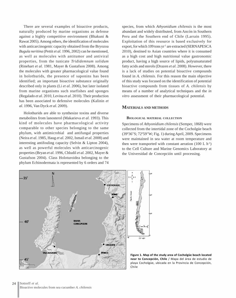
Specimens of Athyonidium chilensis (Semper, 1868) werecollected from the intertidal zone of the Cocholgüe beach(39º36"S; 72º59"W; Fig. 1) during April, 2009. Specimenswere maintained in sea water at room temperature andthen were transported with constant aeration (100 L h-1)to the Cell Culture and Marine Genomics Laboratory atthe Universidad de Concepción until processing.
Figure 1. Map of the study area of Cocholgüe beach located
near to Concepción, Chile / Mapa del área de estudio deplaya Cocholgüe, ubicada en la Provincia de Concepción,
Chile
25Vol. 48, Nº 1, 2013Revista de Biología Marina y Oceanografía
PREPARATION OF PURE EXTRACTS OF A. CHILENSIS,PREPARATIVE CHROMATOGRAPHY AND ANALYSIS BY LC-MS-
MS
Specimens were eviscerated, to avoid contamination, andminced to pieces of 1 cm2 or less. From 5 kg of pooledsample, approximately 1.4 kg were frozen in glass containersof 500 ml capacity. Samples were then lyophilized in a LyphLock 12 Freeze System equipment (Labconco). Oncelyophilization was over, dried tissue was transferred to aheater at 40ºC in order to assure elimination of water.Dehydrated samples were weighted again and the waterpercentage was calculated. Later, samples containing 10-13 g were placed in Whatman filter paper cartridges (11 μmpore size). Extraction was carried out following Soxhletmethod (Bialy et al. 2004) at 60ºC, and two kinds of solventwere used. First extraction was performed usingdichlormethane (CH
2Cl
2) in order to extract fatty or low
polarity compounds (lipophilic) during 3 d through cyclesof 8 h per day. This process was followed by an extractionin methanol (CH
3OH) in order to get the hydrophilic
compounds. Extraction in methanol was carried outfollowing the same method indicated above.
Samples were dissolved in 200 ml of distilled water andloaded per 12 h in an amberlite XAD-7 column (Sigma-Aldrich) and later washed with distilled water to removesalt from the extract and finally eluted using methanol.
Purification of extracted compounds was carried out bymeans of column chromatography, using a 3.5 cm diameterglass column filled with silica gel 60 F
254 (Merck). Samples
were impregnated with 3 g silica gel in a porcelain mortaruntil dryness. Then, samples were carefully mounted inthe column, in order to obtain a development of the sampleand an optimum separation of the mobile phase constitutedby a gradient of chloroform (CHCl
3) and methanol (CH
3OH).
Fractions obtained were monitored through fine layerchromatography, using silica gel 60 F
254 chromatographic
plates, with a mobile phase constituted by 80% chloroform(CHCl
3), 19% methanol (CH
3OH) and 1% water (H
2O). At
the same time, in some cases reverse phasechromatographic phases (RP-18) were used.
CHROMATOGRAPHIC IDENTIFICATION AND DETERMINATION
OF MOLECULAR WEIGHT
LC-MS-MS analysis was carried out in a MDS SCIEXliquid chromatographer (Applied Biosystems), equippedwith an Agilent 1200 series quaternary pump, Agilent 1200vacuum degasser, Agilent Hip-Als, automatic samplerwith temperature control, Agilent 1200 column
thermostatizer, and 3200 Q trap LC-MS-MS Systemdetector. Fractions were injected in the equipment loop,in a volume of 15 μl per sample. As mobile phase aCH
3CN:H
2O gradient (HPLC quality) in C
18 column (Sunfire
Waters Company) was used with a 2.5 μm particle sizeand 21 cm length. In order to improve chromatographicpeaks, 1% formic acid was added in the mobile phase.Determination of molecular weight of the fraction 18 wascarried out by means of an Applied Biosystems Q trap3200 mass spectrometer. This was injected with differentsolvents in order to obtain a comparison of the molecularweights in the different injections. The first one was addedto a CH
3OH:H
2O (50:50%) mixture; the second one, in
CH3OH:H
2O: formic acetate mixture and the third one was
performed in a mixture of CH3OH:H
2O and 1% formic acid.
A direct infusion to the spectrometer was performed at 20μl min-1 of the methanolic fraction in order to obtain firstorder spectra of the fraction (elution having analytes ifinterest) previously evaporated under nitrogen currentat 40ºC. The extract was dissolved in a 1:1 methanol: watermixture, with addition of 5 nM ammonium acetate andfiltered through a 0.22 μm PTFE membrane. Ionizationwas carried out by means of the electrospray mode (ESI)both positive and negative, with parameters of the ionsource of 70, -70 for DP, 10, -10 for EP and 20, -20 for CEP.Capillary voltage was 400 and -4500 Volt in positive andnegative mode, respectively. A scanning between 700 and1700 uma was performed for both ionization modes at50ºC; with a GS1 and GS2 curtain gas of 40 and 50 arbitraryunits respectively. Q1 Quadrapole resolution was of 0.4uma. In the case of the chromatographic analysis 10 μL ofthe extract were injected in a TSK-gel column of 2.1 x 150mm, with 3.5 μm particle size. Used flow was 0.25 μl min-1
and oven temperature of 45ºC. Elution was performed ingradient. With 100% aqueous phase in the beginning,and later linearly increased the organic phase B at 95% at25 min and kept for other 5 min in order to removeimpurities of the column and return to the initial conditionat 35 min for a new injection.
BIOPROSPECTING OF BIOLOGICAL ACTIVITY
HEMOLYTIC TEST
To determine the presence of saponins in the primaryextracts of dichloromethane and methanol, the hemolytictest was used following a method described by Gestetneret al. (1968). Saturated discs with the extracts were placedon blood agar plates for 24 h. Hemolytic halos formedaround the discs were proportional to the hemolytic

26 Sottorff et al.Bioactive molecules from sea cucumber A. chilensis
potential or saponin concentration. Also, this test wasperformed with the fractions purified from the methanolicextract.
ANTIBACTERIAL TEST
The evaluation of the antibacterial activity was performedagainst Gram-positive bacteria: Staphylococcus aureus(ATCC 25923) and Bacillus subtilis (ATCC 6633), andagainst Gram-negative bacteria: Escherichia coli (ATCC25922), Salmonella sp. and Pseudomonas sp., obtainedfrom the bacterial collection of the EnvironmentalMicrobiology Laboratory at the Faculty of BiologicalSciences of the Universidad de Concepción. Bacteria wereincubated at a concentration of 106 UFC ml-1 for 24 h at37ºC in Trypticase soy agar at 37ºC on 10 cm Petri dishes.Extract of A. chilensis was used at 10 mg ml-1, and a mix ofpenicillin and streptomycin (10000 IU and 10 mg ml-1,respectively) was used as a reference antibiotic. Thediameter of the halo of bacteria growth inhibition, or zoneof inhibition, was measured after 24 h of incubation withthe extract.
ANTIFUNGAL TEST
In order to assess antifungal properties Aspergillus sp.,Botrytis sp., Rhizopus sp. and C. albicans coming fromthe fungal culture collection of the Natural ProductsChemistry Laboratory (Universidad de Concepción) wereused. Spores of Aspergillus sp., Botrytis sp. and Rhizopussp. were placed in a H
2O:Tween 20 (10:1) solution and
quantified using a hemocytometer. Standard concentrationused was 1x10⁶ spores per 10 cm Petri dish in HA agar.Purified extract was place in wells of 100 μl at aconcentration of 10 mg ml-1 and incubated at 20ºC. Finally,the zone of inhibition generated around the extract sampleswas measured at 72 h. C. albicans was tested with thesame conditions used for bacteria (incubation in Trypticasesoy agar for 24 h). As positive control 10 mg ml-1
Ketoconazole was used.
CYTOTOXICITY TEST ON MAMMAL CELLS
Neuro 2a (ATCC CCL-131) mouse neuroblastoma cell linewas allowed to grow in RPMI 1640 (Invitrogen) culturemedia, supplemented with 10% fetal bovine serum (FBS,Invitrogen), 0.25 μg ml-1 amphoteromicin B (Invitrogen),100 IU ml-1 100 μg ml-1 penicillin - streptomycin(Invitrogen), 1 mM sodium piruvate (Invitrogen) and 2mM glutamine (Invitrogen). Cells were maintained at 37ºCin an incubator (Thermo Scientifics), with injection of 5%
CO2. In order to determine the cytotoxicity of A. chilensis
extracts on the tumor cell line of mouse neuroblastomaN2a, the inhibition of cell growth was evaluated at 24 hpost-exposure, by means of the MTT (3-(4,5-dimethylthiazol-2-yl)-2,5-diphenyltetrazolium bromide)(Invitrogen) colorimetric assay, according to the methoddescribed by Manger et al. (1993) for determination ofcell viability. A density of 15x104 cells ml-1 in 200 μL ofculture medium supplemented with 5% FBS was grown in96 wells-plates (Nunc) at 37ºC and 5% CO
2 for 24 h. Later,
30 μl of different concentrations of A. chilensis extract(from 3.9 μg ml-1 to 1500 μg ml-1) were added. Also, controlwithout extract was tested. Absorbance was quantifiedat 570 nm, in a Biotek EL-800 plate reader. The cell viabilityof N2a cells was expressed as a percentage of the controlwithout extract. Every value was indicated as the average± SD (n = 8) with 2 experimental replicates.
RESULTS
The first methanolic extract of A. chilensis showedhemolytic activity (data not shown). Therefore, thisextract was under further purification. This purificationwas carried out by means of column chromatography,using chloroform, methanol and water. This methodallowed the separation of the extract in 19 differentfractions, from which only 3 exhibit hemolytic activity,but fraction 18 showed the greatest bioactivity on aspecific test for saponins (blood agar test). Its yieldreached only 0.03% related to the dry weight of the sample.
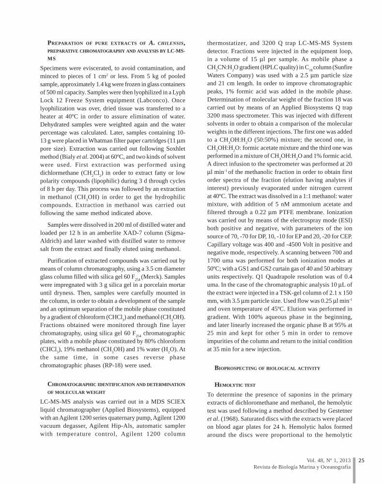
The characterization of the polar compounds from themethanolic fraction of the extraction of A. chilensis bymeans of HPLC showed the presence of 3 compounds,with retention times (RT) of 1.20, 3.25 and 6.06 min, andmolecular weights of 508.5, 1015.8 and 1544 Da,respectively (Fig. 2).
By means of LC-MS-MS, molecules contained infraction 18 were characterized. The first order massspectrum in positive mode (Fig. 3A) presents 5 clusterswith species of the form [M+H]+, with masses of 507 (M1),765 (M2), 1015 (M3), 1272 (M4) and 1523 (M5) Da. Speciesforming adducts with sodium are also observed,generating mass differences of 23 Da for every one ofthem.
In negative electrospray mode (Fig. 3B) the presenceof 5 clusters can also be observed with masses of 566(M1), 823 (M2), 1073.8 (M3), 1330 (M4) and 1581 (M5) Da.Every one of these species corresponds to a number ofadducts with acetate ion (A) present in the ionization
27Vol. 48, Nº 1, 2013Revista de Biología Marina y Oceanografía
vehicle. Masses distribution is correlated with thespectrum in a positive way, where molecular weight ispossible to be assigned (Table 1).
The results from the evaluation of microbiologicalactivity for the chromatographically purified fraction 18from A. chilensis against 5 clinically important
microorganisms showed antimicrobial activity againstGram-positive bacteria such as S. aureus and B. subtilis,with a zone of inhibition of 22 ± 2 and 19 ± 1 mm, respectively.On the other side, there was no evidence of activity againstthe Gram-negative bacteria analyzed (Fig. 4).
Figure 2. Chromatogram of fraction 18 with 3 peaks with RT of 1.20,3.25 and 6.06 min. Gradient scanning methanol: H2O with C-8
column of 2.5 μμμμμm particle size and 21 cm length / Cromatograma
fracción 18 con 3 picos con TR de 1,20, 3,25 y 6,06 min. Corrida engradiente metanol: H2O con columna C-8 de tamaño de partícula
de 2,5 μm y 21 cm de largo
Table 1. Species and molecular masses asigned for fraction 18. Ionization was carried out by
means of the electrospray mode (ESI) both positive and negative / Especies y masas
moleculares asignadas para la fracción 18. La ionización fue llevada a cabo en modoelectrospray (ESI) positivo y negativo

28 Sottorff et al.Bioactive molecules from sea cucumber A. chilensis
A
Figure 3. First order mass spectrum for fraction 18. A) Analysis inpositive electrospray mode. B) Analysis in negative electrospray
mode / Espectro de masas de primer orden de Fracción 18. A)
Representa análisis en modo electrospray positivo B) Análisisen modo electrospray negativo
B

29Vol. 48, Nº 1, 2013Revista de Biología Marina y Oceanografía
Antifungal activity of the extract was evaluated on 4microorganisms medically and commercially important(Zahavi et al. 2000, Pasqualotto & Denning 2008). Fraction18 showed activity against Aspergillus sp., Botrytis sp.and C. albicans, with a zone of inhibition of 24.5 ± 4, 13.5± 3 and 22 ± 1 mm, respectively. There was no activityagainst Rhizopus sp. (Fig. 5).
Finally, the extract from A. chilensis characterized bythe presence of saponins (fraction 18) showed citotoxicactivity on the tumor cell line N2A. A dose-dependingreduction in the cell viability was detected with an IC
50 of
77.34 ± 1.6 μg ml-1 (Fig. 6).
Figure 4. Representation of the antibacterialactivity between fraction 18 (10 mg ml-1, white
bars) and reference antibiotic (Penicillin-
Streptomycin 10 mg ml-1, black bars) /Representación de la actividad antibacteriana
entre la fracción 18 (10 mg ml-1, barras blancas)
y el antibiótico de referencia (Penicilina-Estreptomicina 10 mg ml-1, barras negras)
Figure 5. Representation of the antifungalactivity between fraction 18 (10 mg ml-1, white
bars) and referenced antifungal (10 mg ml-1, black
bars) / Representación de la actividadantifúngica, entre la fracción 18 (10 mg ml-1,
barras blancas) y el antifúngico de referencia(10 mg ml-1, barras negras)
Figure 6. Dose-response curve of the fraction18 on the cell viability of mouse neuroblastoma
N2a cells / Curva de dosis-respuesta de la
fracción 18 sobre la viabilidad celular en lascélulas N2a de neuroblastoma de ratón

30 Sottorff et al.Bioactive molecules from sea cucumber A. chilensis
DISCUSSION
A considerable number of reports have demonstrated thepresence of bioactive metabolites in holothurids, with animportant diversity of glycosylated triterpenes, calledsaponins (Kobayashi et al. 1991, Stonik et al. 1999, Chludilet al. 2003, Kalinin et al. 2005, Antonov et al. 2011). Thesesaponins are described for sea cucumber species collectedin tropical zones of the Pacific and Indian Oceans (Stoniket al. 1999), Mediterranean Sea (Silchenko et al. 2005),North Atlantic (Silchenko et al. 2007), North Pacific(Silchenko et al. 2008) and some sea cucumber from theAntarctic Sea (Maier et al. 2001, Antonov et al. 2008,2009). However, there is a lack of reports about secondarymetabolites and saponins of sea cucumber from SouthAmerican coasts.
The election of solvents and the methodologicalextraction strategy of active metabolites from marineorganisms are fundamental steps for the followingevaluation of biological activity, because it allowsconcentrate, separate and clean the potential bioactivemolecules from the biological matrix of the marineorganisms. Specifically, in marine organisms it has beensuggested the use of alcoholic extractions based onethanol or methanol. However, this kind of extraction isuseful for the preparation of total extracts, which are morecomplex extracts, with low yields of the total sampleweight, and makes difficult to identify the bioactiveprinciples as unique compounds (Silchenko et al. 2008,Avilov et al. 2008). Therefore, the strategy used in thiswork included differentiated extraction with solvents ofincreasing polarity. This methodology can be divided in2 steps: a first non-polar extraction with dichloromethane,in which similar compounds as lipids and non-polarmetabolites are extracted; and a second hydrophilicextraction using methanol as solvent in order to obtainmore polar compounds. This approximation allowed theenrichment of the extracts into highly hydroxylatedmetabolites, which would correspond to the phenoliccompounds described by Mamelona et al. (2007), whodescribed greater yields in the presence of aqueoussolvents, especially from muscle and gonads in seacucumbers from the Atlantic Sea.
IDENTIFICATION OF SAPONINS
Currently, the utilization of LC-MS-MS is emphasized byusing triple quadrupole mass spectrometry withturbulonspray ionization interface in order to obtainmolecular ions and the molecule fragmentation (Van Dyck
et al. 2009). Thus, being know molecular weight and atomsdistribution, a chemical characterization of them can bebuilt.
The major problem in characterizing glycosides is thecapacity of forming adducts with other molecules. Thisinduces the integration of molecules of the analyte withother molecules from the mobile phase, originating clusterswith certain deviation, which are presented as [M+H].Isotopic distribution of the molecules can be observedaround every cluster, as well as the formation of adductswith H+ or Na+. This kind of adduct has been reported byseveral authors for saponin analyses (Van Dyck et al.2009). In the spectrum of the first order masses, onpositive mode, the presence of 5 clusters can be observed.In every one of them, molecular species that correspondto adducts with protons and cations coming from theionization additive used can be noticed. The mostabundant species are those with protons and sodium.Such profile is analogous to that one obtained fromHolothuria forskali (Van Dyck et al. 2009). The firstspecies, denominated M1, of 507 Da, could correspondto 2 different structures and both correspond to a chainof monosaccharides and the aglucon, conjugated with aproton. When it is associated to a sodium atom, its massincreases to 530 Da. Similar behaviours can be observedfor the 4 remaining species, being those species of masses765, 1015, 1272 y 1523 Da, of the form [M+H]+ for M2, M3,M4 and M5 (Table 1). Also, molecular species composedby sodium with mass differences of 23 Da for every oneof them can be observed (Table 1).
In negative electrospray mode, 5 ion clusters, with lowernumber of adducts and intensity can be observed. Thisbehaviour is characteristic in this kind of ionization. Inthis spectrum, the presence of 5 clusters can be observed:566 (M1), 823 (M2), 1073.8 (M3), 1330 (M4) and 1581 (M5)Da. Every one of them corresponds to adducts with ionacetate (A) present in the ionization vehicle. Such massdistribution is correlated to the spectrum in positive mode,being possible to assign the molecular weight to thefragments or molecules after the ionization present infraction 18. Thus, a molecular weight of 764 Da wasassigned to the M2 molecule. Then, species of the form[M2+H]+ and [M2+Na]+ of 765 and 787 Da are confirmedin positive mode with differences of 1 and 23 Da,respectively. In negative mode, the species [M2+A]- of823 Da with a difference of 60 Da is observed. Molecularweights of the other saponins were equally assigned.From the species identified in Table 1, the one with amass of 787 Da corresponds to the saponin

31Vol. 48, Nº 1, 2013Revista de Biología Marina y Oceanografía
holothurinoside D (Fig. 7), isolated from Holothuriaforskali by Rodriguez et al. (1991). The remainingchemical species described would correspond to newsaponins isolated from Athyonidium chilensis. However,the high complexity of the saponins mixtures from theseextracts makes difficult their structural elution and theevaluation of their potential biological role.
The evaluation of the biological activity of purifiedextracts isolated from A. chilensis showed a remarkableactivity against Gram positive bacteria such as S. aureusand B. subtilis, but not against Gram negative bacteriatested in this study. These results are in agreement withobservations described by Haug et al. (2002), whodetermined that most of the extracts prepared fromdifferent organs of the holothurid Cucumaria frondosashowed antibacterial activity against Gram positive andno activity against Gram negative such as E. coli and
Vibrio sp., also suggesting that the antimicrobial activitywould not be exclusively attributed to the presence ofenzymes such as lisozime or other antimicrobial peptides(Haug et al. 2002), but it would be related to the presenceof secondary metabolites like saponin in the extracts fromA. chilensis.
According to the evaluation of the antifungal activity,though they suggest the presence of antagonisticmolecules on proliferation of fungi such as Aspergillussp. and Botrytis sp. did not show activity againstRhizopus sp. This would be explained exclusively on thebasis of the specific inhibition mechanisms, which werenot considered in this study. However, these results arecongruent with what has been reported for holothuridspecies C. frondosa and C. japonica (Attaway &Zaborsky 1993).
Figure 7. Presumptive molecular structure of a derivative holothurinoside D isolated from Athyonidium chilensis / Presumible estructura
molecular de un derivado de holoturinósido D aislado desde la especie Athyonidium chilensis

32 Sottorff et al.Bioactive molecules from sea cucumber A. chilensis
There are reports about the antiproliferative potentialof sphingolipids and branched chain fatty acids obtainedfrom extracts of sea cucumbers on cell lines of peripheraltumours such as prostate cancer cells (Yang et al. 2003)and colon cancer cells (Sugawara et al. 2006). However,there are no records of its evaluation on cell lines fromneuroblastoma. Our results showed that the extractpurified of saponins is capable to reduce bothproliferation and cell viability of the N2a tumour cell lineafter 24 h, with an IC
50 of 77.3 ± 1.6 μg ml-1 (Fig. 6). These
results of cytotoxicity on N2a cells are correlated withthe antiproliferative properties described for total orenriched extracts isolated from other species of seacucumbers in cell lines of human tumours of peripheralorigin such as A549, MCF-7, 1A9, CAKI-1, U-87-MG, PC-3, KB, KB-VIN, SK-MEL-2, HCT-8 (Zou et al. 2006,Sugawara et al. 2006). Antiproliferative activity has beenattributed to the purification or enrichment of certainmolecules like saponins and triterpenic glycosides forwhich different mechanisms are suggested. Among thesemechanisms, selective inhibition by arrest of the cell cycle(Mujoo et al. 2001), specific cytotoxic activity influencedby the glycosylated portion of the saponin structure(Kuroda et al. 2001), and non-specific cytotoxicity causedby detergent action (Mimaki et al. 2001) can be mentioned.Additionally, there are previous information on the basisthat sphingolipids isolated from sea cucumber are able toinduce apoptosis on colon cancer cells (Sugawara et al.2006). On the other side, other molecules such asleptosines are able to produce inhibition of the in vitroproliferation on tumour cell lines by means of the specificinhibition of the protein kinase and topoisomerase II (Jha& Zi-rong 2004).
In conclusion, this study demonstrated that thefraction 18 obtained from A. chilensis contains 2 saponins,which are identified through LC-MS-MS. The firstsaponin has a molecular weight of 1522 Da, whis isfragmented in diverse small molecules, such asmonosaccharides or the aglucon (Fig. 7). The othersaponin that could be identified was of 764 Da and wouldcorrespond to the saponin named holothurinoside D,previously identified in Holothuria forskali by Rodriguezet al. (1991). The other chemical species described in thisstudy would correspond to new saponins identified fromA. chilensis. Additionally, saponin purified extractsshowed an interesting hemolytic, antibacterial andantifungal activity. However, the efficiency of the last 2activities is lower respect to what was observed for othermembers of the family Holothuridae. On the other side, in
relation to the evaluation of the in vitro antiproliferativeproperties of the extracts obtained from A. chilensis, itcan be concluded that it is presented as an interestingfocus of study as potential cytotoxic biomedicines ofmarine origin for the treatment of cancer. An example ofthis is Frondoside A, isolated from sea cucumber whichdemonstrated an important antimetastatic activity (Ma etal. 2012).
Altogether, our results have become into the firstcharacterization of molecules with biological activity fromAnthyonidium chilensis which, in the long term, wouldgive an added value to this resource and it can be proposeit as a potential source of biomedicines. Even though itspoor yield does complex its commercial exploitation, it isa good source of new molecules which can be producedat a bigger scale.
ACKNOWLEDGEMENTS
This research was supported by COPAS Sur-AustralProgram Conicyt (Project PFB-31/2007), Ring project ACT-38 (CONICYT) and the project DIUC 208.112.100-1.Universidad de Concepción, Chile. We are thankful to Dr.María Angélica Mondaca of the Department ofMicrobiology at the Universidad de Concepción for hervaluable helping in microbiological analysis. We wouldlike to make a special mention and a great recognition toMarcela Silva y Zenón Rozas for their collaboration inthis work.
LITERATURE CITED
Antonov AS, SA Avilov, AI Kalinovsky, SD Anastyuk, PSDmitrenok, E Evtushenko, VI Kalinin, AV Smirnov, STaboada, M Ballesteros, C Avila & VA Stonik. 2008.Triterpene glycosides from Antarctic sea cucumbers I.Structure of liouvillosides A1, A2, A3, B1 and B2 from thesea cucumber Staurocucumis liouvillei: new procedure forseparation of highly polar glycoside fractions and taxonomicrevision. Journal of Natural Products 71: 1677-1685.
Antonov AS, SA Avilov, AI Kalinovsky, SD Anastyuk, PSDmitrenok, VI Kalinin, S Taboada, A Bosh, C Avila &VA Stonik. 2009. Triterpene glycosides from Antarctic seacucumbers 2. Structure of achlioniceosides A1, A2 and A3from the sea cucumber Achlionice violaescupidata(=Rhipidothuria racowitzai). Journal of Natural Products72: 33-38.
Antonov AS, SA Avilov, AI Kalinovsky, PS Dmitrenok, VIKalinin, S Taboada, M Ballesteros & C Avila. 2011.Triterpene glycosides from Antarctic sea cucumbers III.Structures of liouvillosides A4 and A5, two minordisulphated tetraosides containing 3-O-methylquinovose
33Vol. 48, Nº 1, 2013Revista de Biología Marina y Oceanografía
as terminal monosaccharide units from the sea cucumberStaurocucumis liouvillei (Vaney). Natural Product Research25: 1324-1333.
Attaway DH. & OR Zaborsky. 1993. Marine biotechnologyIn: Attaway DA & OR Zaborsky (eds). Pharmaceuticaland bioactive natural products 1: 1-524. Plenum Press, NewYork.
Avilov SA, AS Silchenko, AS Antonov, VI Kalinin, AIKalinovsky, AV Smirnov, PS Dmitrenok, EVEvtushenko, SN Fedorov, AS Savina, LK Shubina &VA Stonik. 2008. Synaptosides A and A1, triterpeneglycosides from the sea cucumber Synapta maculatacontaining 3-O-methylglucuronic acid and their cytotoxicactivity against tumor cells. Journal of Natural Products71: 525-531.
Bhakuni DS & DS Rawat. 2005. Bioactive marine toxins. In:Bhakuni DS & DS Rawat (eds). Bioactive marine naturalproducts, pp. 151-207. Springer, New Delhi.
Bialy Z, M Jurzysta, M Mella & A Tava. 2004. Triterpenesaponins from aerial parts of Medicago arabica L. Journalof Agricultural and Food Chemistry 52: 1095-1099.
Bryan PJ, D Rittschof & JB McClintock. 1996. Bioactivityof echinoderm ethanolic body-wall extracts: an assessmentof marine bacterial attachment and macroinvertebrate larvalsettlement. Journal of Experimental Marine Biology andEcology 196: 79-96.
Chludil HD, CC Muniain, AM Seldes & MS Maier. 2002.Cytotoxic and antifungal triterpene glycosides from thePatagonian sea cucumber Hemoiedema spectabilis. Journalof Natural Products 65: 860-865.
Chludil HD, AP Murray, AM Seldes & MS Maier. 2003.Biologically active triterpene glycosides from sea cucumbers(Holothurioidea, Echinodermata). In: Rahman A (ed).Studies in natural products chemistry 28: 587-616. ElsevierScience, Amsterdam.
Drazen JC, CF Phleger, MA Guest & PD Nichols. 2008.Lipid, sterols and fatty acid composition of abyssalholothurians and ophiuroids from the North-East PacificOcean: Food web implications. Comparative Biochemistryand Physiology Part B: Biochemistry and MolecularBiology 151: 79-87.
Faulkner DJ. 2000. Marine pharmacology. Antonie VanLeeuwenhoek 77: 135-145.
Gestetner B, Y Birk & Y Tencer. 1968. Soybean saponins.Fate of ingested soybean saponins and the physiologicalaspect of their hemolytic activity. Journal of Agriculturaland Food Chemistry 16: 1031-1035.
Haug T, AK Kjuul, OB Styrvold, E Sandsdalen, ØM Olsen& K Stensvåg. 2002. Antibacterial activity inStrongylocentrotus droebachiensis (Echinoidea),Cucumaria frondosa (Holothuroidea), and Asterias rubens(Asteroidea). Journal of Invertebrate Pathology 81: 94-102.
Ismail H, S Lemriss, Z Ben-Aoun, L Mhadhebi, A Dellai, YKacem, P Boiron & A Bouraoui. 2008. Antifungal activityof aqueous and methanolic extracts from the Mediterraneansea cucumber, Holothuria polii. Journal of MedicalMycology 18: 23-26.
Jha RK & X Zi-rong. 2004. Biomedical compounds frommarine organisms. Marine Drugs 2: 123-146
Kalinin VI, NG Prokofieva, GN Likhatskaya, EBSchentsova, IG Agafonova, SA Avilov, & OA Drozdova.1996. Hemolytic activities of triterpene glycosides fromthe holothurian Order Dendrochirotida: Some trends in theevolution of this group of toxins. Toxicon 34: 475-483.
Kalinin VI, AS Silchenko, AS Avilov, VA Stonik & AVSmirnov. 2005. Sea cucumbers triterpene glycosides, therecent progress in structural elucidation andchemotaxonomy. Phytochemistry Reviews 4: 221-236.
Kelly MS. 2005. Echinoderms: Their culture and bioactivecompounds. Progress in Molecular and Subcellular Biology39: 139-165.
Kobayashi M, M Hori, K Kan, T Yasuzawa, M Matsusi, ShSuzuki & I Kitagawa. 1991. Marine natural products.XXVII. Distribution of lanostane-type triterpene oligoglycosides in ten kinds of okinawan sea cucumbers. Chemicaland Pharmaceutical Bulletin 39: 2282-2287.
Kuroda M, Y Mimaki, F Hasegawa, A Yokosuka, Y Sashida& H Sakagami. 2001. Steroidal glycosides from the bulbsof Camassia leichtlinii and their cytotoxic activities.Chemical and Pharmaceutical Bulletin 49: 726-731.
Larrain A. 1995. Biodiversidad de equinodermos chilenos:estado actual del conocimiento y sinopsis biosistematica.Gayana Zoología 59: 73-96.
Levina EV, AI Kalinovsky, PS Dmitrenok, EA Martyyas &VA Stonik. 2010. Two new steroidal saponins, hylodosideA and novaeguinoside Y, from the starfish Leptasteriashylodes reticulata and Culcita novaeguineae (juvenile).Natural Products Communications 5: 1737-1742.
Li R, Y Zhou, Z Wu & L Ding. 2006. ESI-QqTOFMS / MSand APCI-IT-MS/MS analysis of steroid saponins fromthe rhizomes of Dioscorea panthaica. Journal of MassSpectrometry 41: 1-22.
Ma X, K Namita, PD Collin, O Goloubeva & AM Fulton.2012. Frondoside A inhibits breast cancer metastasis andantagonizes prostaglandin E receptors EP4 and EP2. BreastCancer Research and Treatment 132: 1004-1008.
Maier MS, AJ Roccatagliata, A Kurriss, H Chludil, AMSeldes, CA Pujiol & EB Damonte. 2001. Two newcytotoxic and virucidal trisulfated glycosides from theAntarctic sea cucumber Staurocucumis liouvillei. Journalof Natural Products 64: 732-736.
34 Sottorff et al.Bioactive molecules from sea cucumber A. chilensis
Makarieva TN, VA Stonik, II Kapustina, VM Boguslavsky,AS Dmitrenoik, VI Kalinin, ML Cordeiro & C Djerassi.1993. Biosynthetic studies of marine lipids. 42. Biosynthesisof steroid and triterpenoid metabolites in the sea cucumberEupentacta fraudatrix. Steroids 58: 508-517.
Mamelona J, E Pelletier, KG Lalancette, J Legault, SKarboune & S Kermasha. 2007. Quantification ofphenolic contents and antioxidant capacity of Atlantic seacucumber, Cucumaria frondosa. Food Chemistry 104:1040-1047.
Manger R, L Leja, S Lee, J Hungerford & M Wekell. 1993.Tetrazolium-based cell bioassay for neurotoxins active onvoltage-sensitive sodium channels: semiautomated assayfor saxitoxins, brevetoxins, and ciguatoxins. AnalyticalBiochemistry 214: 190-194.
Mayer AMS & KR Gustafson. 2004. Marine pharmacologyin 2001-2: antitumour and cytotoxic compounds. EuropeanJournal of Cancer 40: 2676-2704.
Mayer AM & KR Gustafson. 2008. Marine pharmacology in2005-6: antitumour and cytotoxic compounds. EuropeanJournal of Cancer 44: 2357-2387.
Mimaki Y, A Yokosuka, M Kuroda & Y Sashida. 2001.Cytotoxic activities and structure-cytotoxic relationshipsof steroidal saponins. Biological and Pharmaceutical Bulletin24: 1286-1289.
Mujoo K, V Haridas, JJ Hoffmann, GA Wachter, LK Hutter,Y Lu, ME Blake, GS Jayatilake, D Bailey, GB Mills &JU Gutterman. 2001. Triterpenoid saponins from Acaciavictoriae (Bentham) decrease tumor cell proliferation andinduce apoptosis. Cancer Research 61: 5486-5490.
Munro MHG, JW Blunt, EJ Dumdei, SJH Hickford, RELill, SX Li, CN Battershill & AR Duckworth. 1999.The discovery and development of marine compoundswith pharmaceutical potential. Journal of Biotechnology70: 15-25.
Neira C, M Hoeneisen, M Silva & L Minale. 1985. Marineorganic chemistry, IV. Structure of the principal aglyconesfrom the starfish Meyenaster gelatinosus. Journal of NaturalProducts 48: 848-848.
Pasqualotto AC & DW Denning. 2008. New and emergingtratments for fungal infections. Journal of AntimicrobialChemotherapy 61: i19-i30.
Pettit GR, F Gao, PM Blumberg, CL Herald, JC Coll, YKamano, NE Lewin, JM Schmidt & JC Chapuis. 1996.Antineoplastic Agents. 340. Isolation and structuralelucidation of bryostatins 16-18. Journal of NaturalProducts 59: 286-289.
Pettit GR, CL Herald, DL Doubek, DL Herald, E Arnold &J Clardy. 2002. Isolation and structure of bryostatin.Journal of the American Chemical Society 104: 6846-6848.
Regalado EL, D Tasdemir, M Kaiser, N Cachet, P Amade &OP Thomas. 2010. Antiprotozoal steroidal saponins fromthe marine sponge Pandaros acanthifolium. Journal ofNatural Products 73: 1404-1410.
Rinehart KL, JB Gloer Jr, RG Hughes Jr, HE Renis, JPMcGovren, EB Swynenberg, DA Stringfellow, SLKuentzel & LH Li. 1981. Didemnins: antiviral andantitumor depsipeptides from a Caribbean tunicate. Science212: 933-935.
Rodriguez J, R Castro & R Riguera. 1991. Holothurinosides:new antitumour non sulphated triterpenoid glycosides fromthe sea cucumber Holothuria forskali. Tetrahedron 47: 4753-4762.
Ruggieri G. 1976. Drugs from the sea. Science 194: 491-497.
Scheuer P. 1990. Some marine ecological phenomena: chemicalbasis and biomedical potential. Science 248: 173-177.
Selvin J & AP Lipton. 2004. Antifouling activity of bioactivesubstances extracted from Holothuria scabra. Hydrobiologia513: 251-253.
SERNAPESCA. 2010. Anuario estadístico de pesca. ServicioNacional de Pesca, Valparaíso. <http://www.sernapesca.cl>
Silchenko AS, VA Stonik, SA Avilov, VI Kalinin, AIKalinovsky, AM Zaharenko, AV Smirnov, E Mollo & GCimino. 2005. Holothurins B2, B3 and B4, new triterpeneglycosides from Mediteranean sea cucumbers of the genusHolothuria. Journal of Natural Products 68: 564-567.
Silchenko AS, SA Avilov, AI Kalinovsky, PS Dmitrenok, VIKalinin, J Morre, ML Deinze, C Woodward & PD Collin.2007. Glycosides from the North Atlantic sea cucumberCucumaria frondosa V: structures of five new minortrisulfated triterpene oligoglycosides, frondosides A7-1, A7-3, A7-4, and isofrondoside C. Canadian Journal ofChemistry 85: 626-636.
Silchenko AS, SA Avilov, VI Kalinin, AI Kalinovsky, PSDmitrenok, SN Fedorov, VG Stepanov, Z Dong & VAStonik. 2008 . Constituents of the sea cucumberCucumaria okhotensis. Structures of okhotosides B1–B3and cytotoxic activities of some glycosides from thisspecies. Journal of Natural Products 71: 351-356.
Stonik VA, VI Kalinin & SA Avilov. 1999. Toxins from thesea cucumbers (Holothuroids): chemical structures,properties, taxonomic distribution, biosynthesis andevolution. Journal of Natural Toxins 8: 235-248.
Sugawara T, N Zaima, A Yamamoto, S Sakai, R Noguchi& T Hirata. 2006. Isolation of sphingoid bases of seacucumber cerebrosides and their cytotoxicity againsthuman colon cancer cells. Bioscience, Biotechnology andBiochemistry 70: 2906-2912.

35Vol. 48, Nº 1, 2013Revista de Biología Marina y Oceanografía
Van Dyck S, P Gerbaux & P Flammang. 2009. Elucidation ofmolecular diversity and body distribution of saponins inthe sea cucumber Holothuria forskali (Echinodermata) bymass spectrometry. Comparative Biochemistry andPhysiology Part B: Biochemistry and Molecular Biology152: 124-134.
Yang P, P Collin, T Madden, D Chan, B Sweeney-Gotsch,D McConkey & RA Newman. 2003. Inhibition ofproliferation of PC3 Cells by the branched-chain fattyacid,12-methyltetradecanoic acid, is associated withinhibition of 5-lipoxygenase. The Prostate 55: 281-291.
Zahavi T, L Cohen, W Batia, L Schena, A Daus, T Kaplunov,J Zutkin, R Ben-Arie & S Droby. 2000. Biological controlof Botrytis, Aspergillus and Rhizopus rots on table and winegrapes in Israel. Postharvest Biology and Technology 20:115-124.
Zou Z, Y Yi, H Wu, X Yao, L Du, W Jiuhong, CC Liaw &KH Lee. 2006. Intercedensides D-I, cytotoxic triterpeneglycosides from the sea cucumber Mensamaria intercedensLampert. Journal of Natural Products 69: 1530-1530.
Received 28 May 2012 and accepted 17 December 2012
Editor: Claudia Bustos D.