of 28 analytical detection and biological assay of antileukemic drug using
TRANSCRIPT

Page 1 of 28
ANALYTICAL DETECTION AND BIOLOGICAL ASSAY OF ANTILEUKEMIC DRUG USING GOLD NANOPARTICLES
V. Selvaraj1, M. Alagar1,* and I. Hamerton2
1 Department of Chemical Engineering, Alagappa College of Technology, Anna University, Chennai - 600 025, India 2 Chemistry Division, School of Biomedical and Molecular Sciences, University of Surrey, Guildford, Surrey, GU2 7XH, United Kingdom * To whom correspondence should be addressed Introduction
Materials in the nanometre size range may possess beneficial properties which can
influence the development of remarkable industrial and engineering applications including
biotechnological systems. The formation of hybrid systems comprising nanoparticles and
biomolecules paves the way from cell markers to biosensing1,2, bioimaging3 and targeted drug
delivery4,5. Nanoparticles have been found to be useful in the development of systemic, oral,
pulmonary, transdermal and other administration routes to study drug targeting, the enhancement
of drug bioavailability and protection of drug bioactivity and stability6-8. For instance,
nanoparticles can be used to enhance oral delivery6,9 by improving the bioavailability of poorly
absorbed drugs. They are able to penetrate the cells to facilitate cellular internalization and
connective tissue permeation, thus enabling the drugs to be delivered efficiently to the targeted
tissue without clogging capillaries10,11. Consequently, nanotechnology is now used as potential
route to study the delivery of anti-leukaemic drugs and used to improve drug diffusion through the
blood/brain barrier. Nanoparticles can also act as drug carriers during intravenous injection, which
allows the carriers to penetrate the membranes of the cells and deliver the drugs to cancerous
tomours. A recent literature review has indicated that some nanoparticle-bound antitumour agents
showed prolonged drug retention in tumours, reduction in tumour growth and prolonged survival
of tumour bearing animals12-15. Optical16 and electrochemical17 sensing of biomolecules using
colloidal gold is widely studied by the conjugation of various biomolecules such as protein A,
avidin, streptavidin, glucose oxidase, horseradish peroxidise and immunoglobins, etc..
Page 2 of 28
6-Mercaptopurine (6-MP) has achieved a special place in biomedicine, owing to its activity in
cancer chemotherapy21. It is one of the oldest antineoplastic drug currently in use and as there is a
good correlation between the drug’s coordination chemistry (it contains sulphur and nitrogen donor
sites with potential for binding at N-1, N-3, N-7 and N-9) and its chemotherapeutic activity, it has
attracted much attention22-24. The activity of 6-MP on tumour cells is believed to be due to its
ability to convert these into the corresponding ribosides. In order to suppress the undesirable side
effects associated with anti-tumour drugs, metal-drug complexes are currently used as slow release
prodrugs of 6-MP. The latter employ coordinated components of certain transition metals like
platinum and palladium25. A number of detection methods have been suggested for the
identification of 6-MP, which include potentiometric titration for tablet assay26, amperometric
detection at a phthalocyanine-modified electrode27 and spectrophotometry28. Whilst Zima et al.29
reported the determination of 6-MP using tast polarography and differential pulse polarography at
the dropping mercury electrode (DME), Basina et al. studied the electrochemical behaviour of 6-
MP using catalytic cathodic stripping voltammetry30 and found that 6-MP-modified mercury
electrodes act as promoters for the redox exchange of Cytochrome C.
In the present work, gold nanoparticles have been used as probes for the detection of the anticancer
drug 6-MP and evidence for their complexation has been obtained using UV-Visible spectroscopy,
cyclic voltammetry, transmission electron microscopy (TEM), and fluorescent spectroscopy.
Furthermore, the efficacy of the complexes of 6-MP and colloidal gold is examined with biological
assays in order to evaluate their antibacterial and antifungal activities.
Experimental
Apparatus
Samples were characterized by ultraviolet (U.V.)-visible spectroscopy (Perkin-Elmer Lambda 25).
The path length was 1 cm and matched 1 x 1 cm2 cuvettes were used. Transmission electron
microscopy (TEM) was undertaken employing a Tec NIE 10 instrument with an accelerating
voltage of 120 kV; samples was prepared by mixing aqueous solution of MP (5mM) and gold
solution (1mM). Fourier transform infrared (FTIR) spectroscopy was performed using a PE IR

Page 3 of 28
SPECTRUM ASCII PEDS 1.60 spectrometer and samples were presented as KBr pellets. Spectra
were acquired at room temperature at a resolution of 4 cm-1. Optical emission spectroscopy was
carried out using Fluoromax-2, Gram 386 spectrometer. Cyclic voltammetric studies was
performed using an Autolab PG Stat 12 electrochemical analyzer in a three electrode cell, using
platinum wire as the counter electrode and a 50 mM KCl calomel reference electrode. The scans
were initiated at – 0.4 V and all electrochemical measurements were performed in a static nitrogen
atmosphere. The working electrode used in this work was indium-tin-oxide (ITO) coated with
colloidal gold and this was prepared in the following manner. The ITO slides were cleaned
thoroughly by ultrasonication in water, followed by acetone. The cleaned glass slides were
immersed in a solution (2% v/v) of 3-aminopropyl trimethoxysilane (APTMS) in methanol for 20
hours and subsequently immersed in freshly prepared colloidal gold for another 15 hours to create
a submonolayer of gold nanoparticles. For electrode modification, freshly prepared gold plates
were immersed for 12 hours in 6-MP solution (1 mM in 0.01 M HClO4), to avoid sulphide
contamination. This process was repeated for all the electrochemical studies (Scheme 1).
Insert Scheme 1
Materials
HAuCl 4.3H2O (98 %) and trisodium citrate (99 %, AR) were purchased from CDH and Analytical
Rasayan respectively. 6 -Mercaptopurine (6-MP) was synthesized according to a modified,
previously reported procedure32,33 and the purity of the sample ascertained by comparing it with the
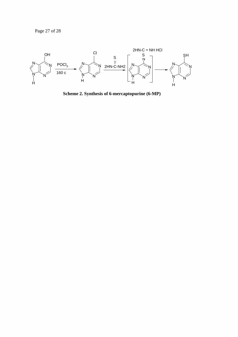
commercial sample obtained from Aldrich. The reaction scheme is given below (Scheme.2)
Insert Scheme 2
Synthesis of 6-chloropurine To phosphoryl chloride (200 cm3, 2.2 mol.) was added drop wise water (20 cm3) and, after all the
water had been added, the mixture was boiled for 1.5 hours to dispel the hydrogen chloride. The
mixture was subsequently cooled and the top layer was used for the chlorination of hypoxanthine
to form 6-dichloropurine. A mixture of hypoxanthine (8 g) and pyrophosphoryl chloride (64 cm3)
was heated in a sealed glass tube at 165 ºC for 19 hours. After cooling, the brown solution was

Page 4 of 28
decanted from the solid residue in the tube and the volatile material was removed under reduced
pressure. The syrupy residue was poured on to crushed ice (200 g), a small amount of tan
precipitate removed and the filtrate was repeatedly extracted with portions of diethyl ether (6 x 350
cm3). The ethereal solution was allowed to stand over anhydrous potassium carbonate for one hour
and then over calcium sulphate overnight. On evaporation of diethyl ether, the crude product (4.3
g., 43 %), m.p. 175-177 ºC, was obtained. A small portion was recrystallized from boiling water
(150 cm3) and was allowed to stand at 0 ºC for 1.5 hours and then filtered through a sintered glass
funnel.
Preparation of 6-mercaptopurine (6-MP) A suspension of 6-chloropurine (0.98 g, 6.3 mmol.) and an equimolar quantity of thiourea in
absolute ethanol (14 cm3) was heated to reflux; the solids dissolved and soon a yellow crystalline
product precipitated. After refluxing for one hour, the mixture was chilled and crude 6-MP (0.64
g) was collected, the product was dried over P2O5 in vacuo at 100°C.
Preparation of citrate capped gold nanoparticles
HAuCl4.6H2O (1 mM, 500 cm3) was heated and to the boiling solution was added trisodium citrate
(38.8 mM, 50 cm3) as one portion. After the addition, the previously yellow solution of gold
chloride turned wine red in colour and gave a characteristic absorbance at 518 nm in the U.V-
Visible spectrum. From the TEM measurements, the average diameter of the gold nanoparticles
was found to be in the range 17-18 nm.
Preparation of 6-mercaptopurine (6-MP) coated gold nanoparticles
Citrate-stabilized gold nanoparticles (1mM, 50 cm3) were mixed with 6-MP (5 mM) in 2-propanol
(25 cm3) and stirred effectively for 5 hours until the wine red colour became blue. The 6-MP
coated gold nanoparticles were obtained after centrifugation.
Details of Microbial Assay
Antibacterial and antifungal activities were studied using a disk diffusion method, wherein a
suspension of both gramme positive and gramme negative organisms were added to sterile nutrient

Page 5 of 28
agar at 45 ºC and the mixture was solidified on a Petri dish. Disks made from filter paper dipped
in 6-MP and 6-MP-gold were placed on agar plates and the plates were left for one hour at 25 ºC to
allow a period of pre-incubation diffusion in order to minimize the effects of variation in time
between the applications of different solutions. The plates were again incubated, this time at 37 ºC
for 24 hours, and observed for antibacterial activity by determining the diameters of the zones of
inhibition for each of the samples.
Results and Discussion
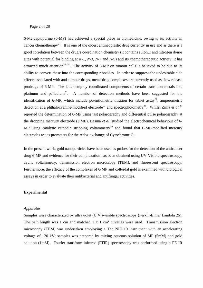
The plasmon band observed for the wine red colloidal gold at 518 nm in the U.V-Visible spectrum
is characteristic of gold nanoparticles. Pure drug shows a maxima at 321 nm The addition of 6-MP
to colloidal gold results in a reduction in the intensity of this absorption band at 518 nm and 321
nm corresponding to Au(0) and drug moiety and is accompanied by the emergence of an additional
peak at 670 nm (Figure 1).
Insert Figure 1 The latter can be verified by a colour change from purple to blue with the addition of drug to
colloidal gold. The time dependent UV-Visible spectra were obtained after mixing (0.5 mM) 6-
MP with gold nanoparticles at timed interval of 30 minutes (curves a-d); curve (e) was obtained
after 5 hours. The appearance of a new peak is due to the aggregation of gold nanoparticles and
the replacement of citrate by 6-MP leading to the formation of gold-drug complex.
These observations are in agreement with the data obtained using FT-IR spectroscopy. 6-MP
exists as tautomer form in the solid state as C=S group and the IR spectrum of free (uncomplexed)
6-MP (Figure 2) displays absorption bands at 1275 cm-1 due the presence of the C=S group.
Insert Figure 2
This peak was absent in the spectrum of the adsorbed form (Figure 3) apparently confirming the
existence of complex formation of the 6-MP through the sulphur atom.

Page 6 of 28
Insert Figure 3
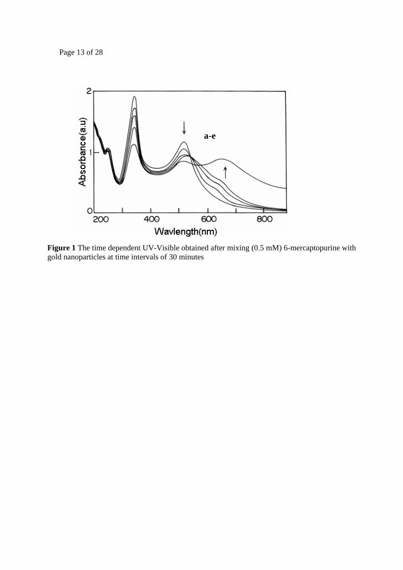
Fluorescence studies were also undertaken to ascertain the binding of the drug moiety with the
gold nanoparticles. Thus, free 6-MP has a broad emission centred at ca. 380 nm when excited at a
wavelength of 320 nm. The non-fluorescent gold nanoparticles are transformed into a markedly
fluorescent species when added to 6-MP, the intensity of fluorescence being decreased when
colloidal gold is added to the drug (Figure 4).
Insert Figure 4
Insert Figure 5
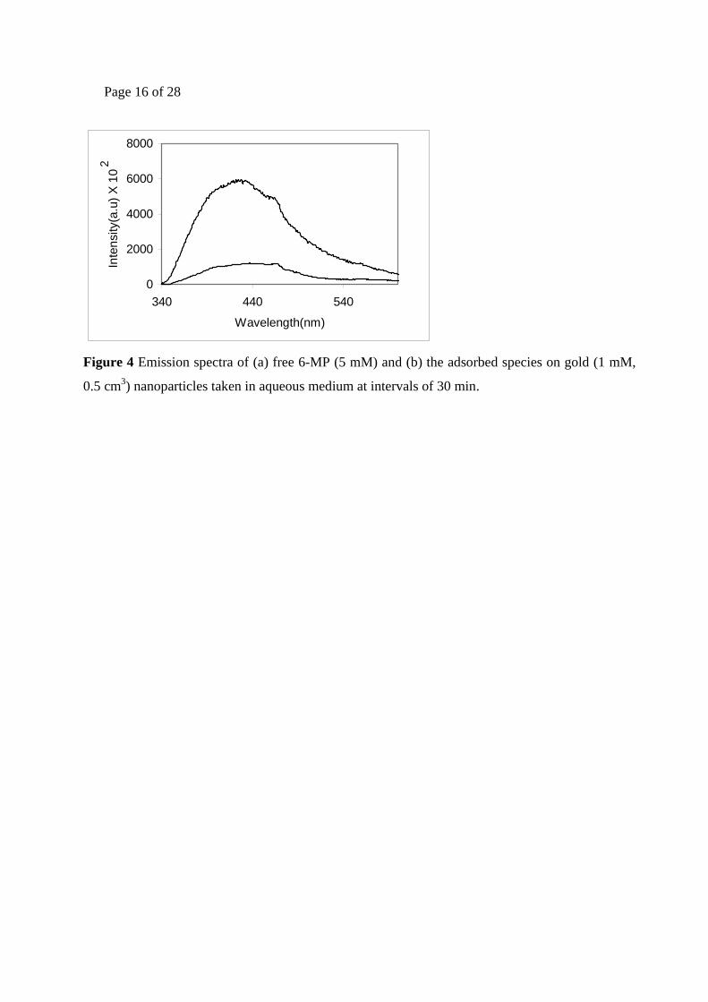
The fluorescence spectrum of 6-MP was studied with different concentrations of gold
nanoparticles (Figure 6).
Insert Figure 6
With a gradual increase in concentration of Au(0), (Figure 6b-e), the intensity of 6-MP (Figure 6)
decreases concomitantly. Throughout this experiment the concentration of 6-MP remained
constant and displayed peaks at 424 nm and 368 nm in the emission and excitation spectra
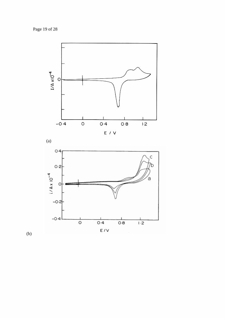
respectively. Cyclic voltammetry (cv) was employed using gold nanoparticle-modified electrodes,
to ascertain the nature of the bond formation and the cyclic voltammograms of the gold
nanoparticle-modified ITO plate immersed in 0.01 M HClO4 solutions are shown (Figure 7). The
scans were initiated at –0.4 V and a reduction peak was observed at +0.69 V, which corresponds to
the formation of gold oxide.
Insert Figure 7
The electrochemical evaluation of 6-MP capped gold nanoparticles is also shown in Figure 7b.
Peak 1 shows the cv response for the ITO electrode (modified with gold nanoparticles) in 1 mM
K4Fe(CN)6 at a scan rate of 10 mv/s. After the exposure of gold electrode in the 6-MP solution, a

Page 7 of 28
reduction in the peak current is observed due to the binding of 6-MP with the nanoparticles and
more pronounced changes in the electrochemical profile of the gold electrode occur as the 6-MP
concentration increases (Figure 7b). In fact, the onset of gold oxide formation is heralded by the
change to a positive potential, indicating that the 6-MP molecule has been spontaneously adsorbed
on the gold surface. However at more positive potentials, a large anodic current was obtained,
which should correspond to the oxidative desorption of the previously adsorbed molecule35, and a
reduction peak was absent when the gold electrode surface was completely adsorbed by 6-MP.
The shape of the voltammograms for [Fe(CN)6]3- and [Fe(CN)6]4- at the coated and uncoated
substrates indicates that the current is primarily controlled by linear diffusion and suggest that the
monolayer does not completely block electron transfer, as these small molecules provide only a
partial barrier. This decrease in both anodic and cothodic current is due to blocking behaviour of
MP on gold modified electrode (Scheme 3).
Insert Scheme 3
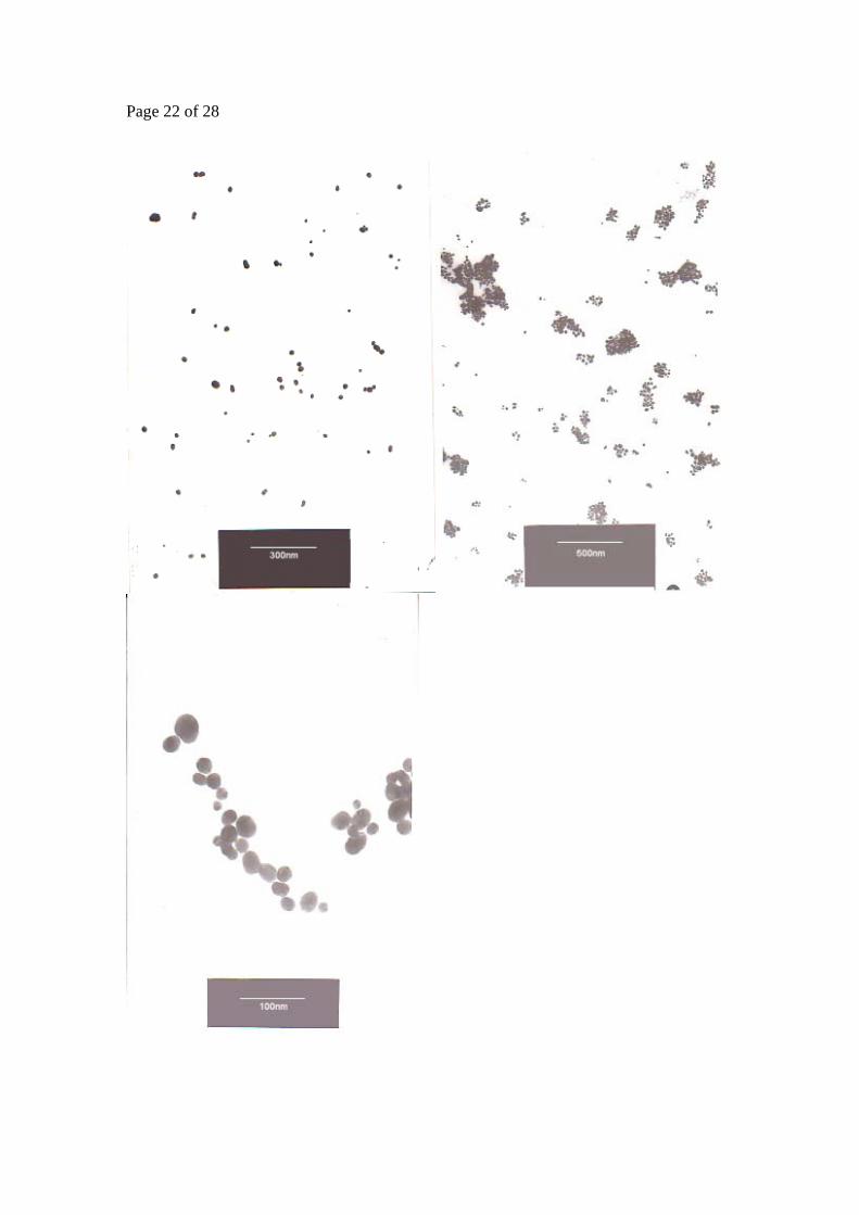
The TEM image (Figure 8) displays clearly aggregates of gold nanoparticles: colloidal gold has an
average diameter of ca. 18 nm. On the addition of gold, aggregation of the gold takes place (this
phenomenon was previously illustrated in Schemes 2 and 3).
Insert Figure 8
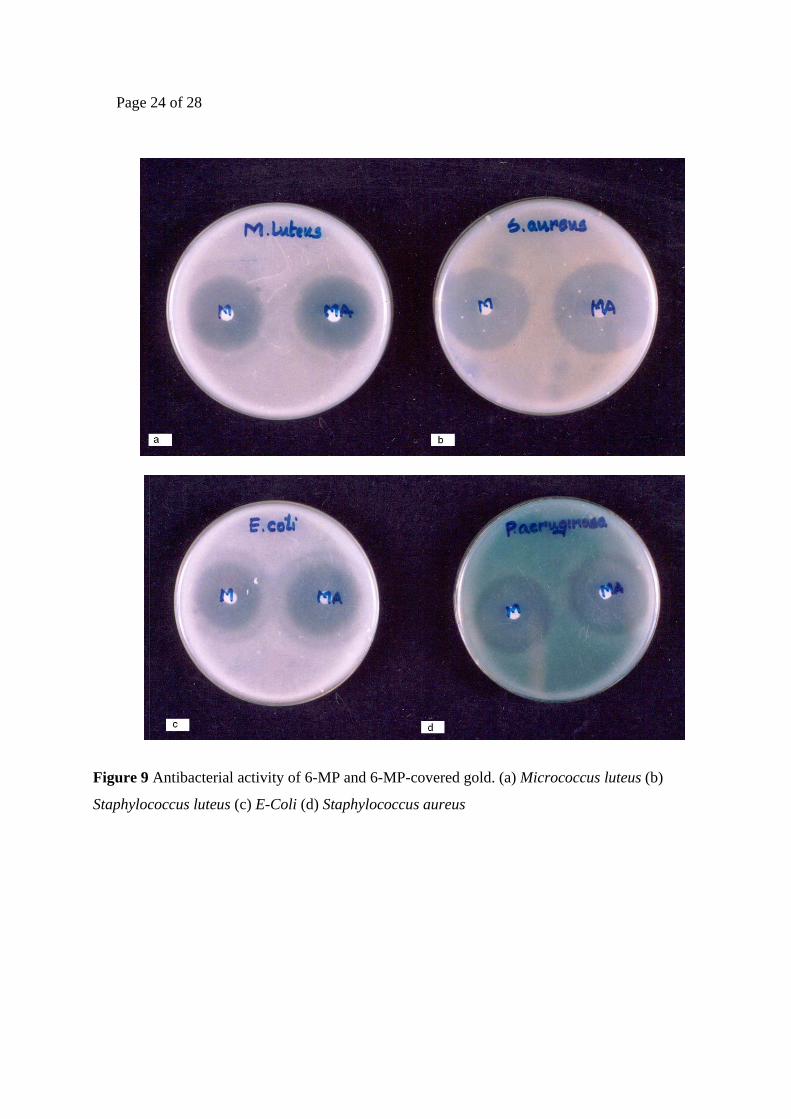
Determination of the antibacterial and antifungal activity of the 6-MP-colloidal gold complex
Table 1 details the growth inhibition effected by drug-coated gold nanoparticles against both
gramme positive and gramme negative organisms viz Micrococcus luteus, Staphylococcus aureus,
Pseudomonas aeruginosa and E-Coli. It was observed that the coated colloid was most effective
for gramme negative organisms and the levels of inhibition can be seen clearly in Figure 9.
Figure 9 Antibacterial activity of 6-MP and 6-MP-covered gold. (a) Micrococcus luteus (b)
Staphylococcus luteus (c) E-Coli (d) Staphylococcus aureus

Page 8 of 28
Table 1 Antibacterial activity of 6-mercaptopurine (6-MP)
6-MP also shows a good antifungal activity against Aspergillus fumigatus and Aspergillus niger
and these activity levels were found to be enhanced on the addition of colloidal gold (Table 2 and
Figure 10). The colloidal gold solution alone does not show any appreciable antifungal and
antibacterial activities.
Insert Figure 10
Table 2 Antifungal activity of 6-mercaptopurine (6-MP)
Although these preliminary studies demonstrate the effectiveness of the gold-drug complex, the
precise mechanism by which it operates is not yet known and work continues to investigate this
further. Some gold-drugs, namely aurothiomalate and aurothioglucose, have already been
evaluated for activity against human immunodeficiency virus (HIV) for the treatment of AIDS44,45
and, in the same way, gold-drugs have been explored for their effectiveness as antifungal,
antibacterial and anticancer agents. Gold-drugs are used more as last-line modes of treatment for
severe cases of rheumatoid arthritis in favour of organic drugs. It gives more potent and reduced
toxic side effects. These properties may be due to geomentry of the complex. The normal mode of
Levels of Zone of inhibition
Micro organisms
Nature of organisms
Pure 6-MP 6-MP-coated gold nanoparticles
Micrococcus luteus Gramme Positive 41 45 Staphylococcus aureus Gramme Positive 49 57
Pseudomonas aeruginosa Gramme Negative 51 63 E-Coli Gramme Negative 60 72
Levels of Zone of inhibition
Fungal Organisms
Pure 6-MP 6-MP-coated gold nanoparticles
Aspergillus fumigatus 45 59 Aspergillus niger 32 37

Page 9 of 28
metabolite pathway and the release mechanism may be altered (favourably in some cases) by the
presence of metal nanoparticles, to attain a greater efficiency and reduced side effects.
Conclusions The binding of 6-mercaptopurine (6-MP) to colloidal gold via complexation through the thiol
group was studied using different analytical techniques (e.g. U.V-Visible spectroscopy, cyclic
voltammetry, transmission electron microscopy, fluorescence spectroscopy and IR spectroscopy).
Fluorescence and electrochemical studies have shown the nature of interaction and will be used in
future studies. The combination of gold with 6-MP results in a more potent complex compared
with the individual parts.
Acknowledgement
The authors thank Dr Brendan J. Howlin (Chemistry Division, University of Surrey) for useful
discussions in the preparation of this manuscript.
References
1. Bruchez M Jr, Moronne M, Gin P, Weiss S; Alivistas AP. Science 1998;281:2013-2016
2. Lahav M, Shipway AN, Willner, I. J Chem Soc Perkin Trans 2 1999;1925-1931
3. Djalali R, Chen Y, Matsui, H. J Am Chem Soc 2002;124:13660-13661
4. West JL, Halas NJ. Curr Opin Biotechnol 2000;11:215-217
5. Hermanson GT, editor. Bioconjugate Techniques. Academic Press: San Diego, 1996
6. Maincent P, Le Verge R, Sade P, Couvreur P, Devissaguet JP. J Pharm Sci 1986;75:955-958
7. Zeltner TB, Sweeney TD, Skornik WA, Feldman HA, Brain JD. J Appl Physiol 1991;70:1137-
1145
8. Cappel MJ, Kreuter J. J Microencapsul 1991;8:369-374
9. Brannon-Peppas L. Int J Pharm 1995;116:1-9
10. Couvreur P, Tulkens P, Roland M, Trouet A, Speiser I. FEBS Lett 1997;84(2):323-326

Page 10 of 28
11. Astier A, Doat B, Ferrer MJ, Benoit G, Fleury J, Rolland A, Leverge R. Cancer Res
1998;48:1835-1841
12. Beck P, Kreuter J, Reszka R, Fichtner I. J Microencapsul 1993;10:101-114
13. Simeonova M, Ilarionova M, Ivanova T, Konstantinov C, Todorov D. Acta Physiol Pharmacol
Bulg 1991;17:43-49
14. Bennis S, Chapey C, Couvreur P, Robert J. Eur J Cancer 1994;30A:89-93
15. Verdun C, Brasseur F, Vranokx H, Couvreur P, Roland M. Cancer Chemother Pharmacol
1990;26:13-18
16. Nath N, Chilkoti A. Anal Chem 2002;74:504-509
17. Raj CR, Okajima T, Ohsaka TJ. Electroanal Chem 2003;593:127-133
18. Saxena V, Sadoqi M, Kumar S, Shao J. SPIE’s OeMagazine;September 2004:21-23
19. Jalil R, Nixon JR. J Microencapsul 1990;7:297-325
20. Mu L, Feng SS. J Controlled Release 2003;86:33-48
21. Bostrom B, Evdmann G. Am J Pediatr Hematol Oncol 1993;15:80
22. Innocenti T. Cancer Chemother Pharmacol 1999;43:133
23. Drerievr, J.; Clin. Chem. (1998) 44, 2511
24. Mi, T. J. Controlled Release (1997) 44, 19
25. Chalmers, A. H.; Burdorf, T.; Murray, A. W. Biochem. Pharmacol. (1972) 21, 2662
26. Nikolic, K.; Velasavic, K. Mikrochim. Acta (1990) 1, 69
27. Chalmers, A. H. Biochem. Med. (1975) 12, 234
28. Harber, M.; Maddocks, J. Chromatogr. (1974) 101, 231
29. Barek, J.; Berka, A.; Dempiirova, L.; Zima, J. Collect. Czech. Chem. Commun. (1986) 51,
2466

Page 11 of 28
30. Banica, I. A.; Gabriel, F.; Contantin, L. Electroanal. (1997) 9, 945
31. Hayat, A. (Ed.) Colloidal Gold: Principles, Methods and Applications. Academic Press: San
Diego (1989) Vols 1-3
32. Elion, G. B.; Hitching, G. H. J. Am. Chem. Soc. (1956) 78, 3508-3510
33. A.Bendish,P.J,Russell.,JR; Fox, J. J.; J. Am. Chem. Soc.(1954) 76,6077
34. Yang, D. F.; Al-Mazna, H.; Morin, M. J. Phys. Chem. (1997) 101,1158
35. Eddowes, M. J.; Hill, H. A. O. J. Am. Chem. Soc. (1979) 101, 4461
36. (a) Eddowes, M. J.; Hill, H.A.O.; Uosaki, K. J. Am. Chem. Soc. (1979) 101, 7113; (b)
Eddowes, M. J.; Hill, H.A.O.; Uosaki, K. Bioelectrochem. Bioenerg. (1980) 7, 527; (c) Albery,
W. J.; Eddowes, M. J.; Hill, H. A. O.; Hillman, A.R. J. Am. Chem. Soc. (1981) 103, 3904.
37. Taniguchi, I.; Murakami, T.; Toyosawa, K.; Yamaguchi, H.; Yasukouchi, K. J. Electronal.
Chem. (1982) 131, 397
38. (a) Taniguchi, I; Toyosawa, K.; Yamaguchi, H. J. Electroanal. Chem. (1982) 140, 187; (b)
Taniguchi, I; Toyosawa, K.; Yamaguchi, H. J. Chem. Soc. Chem. Commun. (1982) 1032
39. Kadish, K. M. (Ed.) Electrochemical and spectrochemical studies of biological redox
components, Adv. Chem. Ser., Vol. 201, American Chemical Society: Washington, D.C. (1982)
Chapters. 7-9 and references cited therein
40. (a) Betso, S.-R.; Klapper, M. H.; Anderson, L. B. J. Am. Chem. Soc. (1972) 94, 8197; (b)
Tarasevich, M.-R.; Bogdanovskaya, V.-A. Bioelectrochem. Bioenerg. (1976) 3, 589; (c)
Cotton, T. M.; Schultz, S. G.; van Duyne, R. P. J. Am. Chem. Soc. (1980) 102, 7960; (d)
Haladjian, J.; Bianco, P.; Serre, P.-A. J. Electroanal. Chem. (1980) 106, 397
41. Busby, C. C. and Creighton J. A., J. Electroanal. Chem. (1982) 140, 379
42. Okada, T.; Patterson, B. K.; Ye, S.-Q.; Gurney, M. E. Virology (1993) 192, 631

Page 12 of 28
43. Yamaguchi, K.; Ushijima, H.; Hisano, M.; Inoue, Y.; Shimamura, T.; Hirano, T.; Muller, E. G.
W. Microbiol. Immunol. (2001) 45, 549.
Page 13 of 28
Figure 1 The time dependent UV-Visible obtained after mixing (0.5 mM) 6-mercaptopurine with gold nanoparticles at time intervals of 30 minutes
a-e

Page 14 of 28
MP
50
60
70
80
90
100
110
400140024003400
wave number cm -1
tran
smita
nce
(%)
MP3424
1611
1407
1223
1274
1013
Figure 2 FTIR transmission spectrum of pure 6-mercaptopurine (6-MP)

Page 15 of 28
MP-AU
8587899193959799
101
400140024003400
wavenumber cm-1
tran
smita
nce(
%)
MP-AU
3426
1614
14031013
Figure 3 FTIR transmission spectrum of 6-MP with gold nanoparticles (MP-AU)
Page 16 of 28
0
2000
4000
6000
8000
340 440 540
Wavelength(nm)
Inte
nsity
(a.u
) X 1
0 2
Figure 4 Emission spectra of (a) free 6-MP (5 mM) and (b) the adsorbed species on gold (1 mM,
0.5 cm3) nanoparticles taken in aqueous medium at intervals of 30 min.

Page 17 of 28
0
1000
2000
3000
300 350 400Wavelength(nm)
Inte
nsity
(a.u
) X 1
04 a
b
Figure 5 Excitation spectra taken in aqueous medium for (a) free 6-MP (5 mM) and (b) the
adsorbed species on gold nanoparticles (1 mM)
Page 18 of 28
0
400000
800000
340 440 540
Wavelength(nm)
Inte
nsity
(a.u
) X 1
0 2
Figure 6. Emission spectra taken in aqueous medium for (a) free 6-MP (5 mM) and (b-e) for the
adsorbed species on gold nanoparticles. The concentration of gold solution is varied (0.5cm3 – 2
cm3 of 1mM gold solution), but the analysis time is kept constant (2 minutes)
Page 19 of 28
(a)
(b)

Page 20 of 28
(c)
Figure 7: Cyclic voltammograms of a gold nanoparticle-modified electrode (a) in the absence of 6-MP in 0.01 M HClO4. M, (b) in the presence of different concentrations of 6-MP in 0.01 M HClO4. (a) 50 (b) 100 and (c) 150 μM. The scan rate is maintained at 0.02 V s-1. and (c) in 1mM K4Fe(CN)6 at a scan rate of 10 mV s-1.

Page 21 of 28
(a) (b)
(c)
Page 22 of 28
Page 23 of 28
Figure 8 Transmission electron micrographs of 6-MP-covered gold nanoparticles of average
diameter 20 nm. The samples were prepared from an aqueous solution: (a) gold nano particle; (b)
6-MP-covered gold nanoparticles; (c) expanded portion of 6-MP covered gold nanoparticles
Page 24 of 28
Figure 9 Antibacterial activity of 6-MP and 6-MP-covered gold. (a) Micrococcus luteus (b)
Staphylococcus luteus (c) E-Coli (d) Staphylococcus aureus

Page 25 of 28
Figure 10. Antifungal activities of 6-MP and 6-MP-covered gold (a) Aspergillus fumigatus (b)
Aspergillus niger

Page 26 of 28
ITO
Gold
Au ColloidAPTMS
Gold Gold
Mercaptopurine
ITO ITO ITO
ITO
Scheme1. Aggregation of 6-MP on Gold nanoparticles
Page 27 of 28
N N
N N
OH
H
POCl3
N N
N N
Cl
N N
N N
N
N N
N N
S
160 c
2HN-C = NH HClS
2HN-C-NH2
S H
H H H Scheme 2. Synthesis of 6-mercaptopurine (6-MP)

Page 28 of 28
Table 1 Antibacterial activity of 6-mercaptopurine (6-MP)
Table 2 Antifungal activity of 6-mercaptopurine (6-MP)
Levels of Zone of inhibition
Micro organisms
Nature of organisms
Pure 6-MP 6-MP-coated gold nanoparticles
Micrococcus luteus Gramme Positive 41 45 Staphylococcus aureus Gramme Positive 49 57
Pseudomonas aeruginosa Gramme Negative 51 63 E-Coli Gramme Negative 60 72
Levels of Zone of inhibition
Fungal Organisms
Pure 6-MP 6-MP-coated gold nanoparticles
Aspergillus fumigatus 45 59 Aspergillus niger 32 37