oestrogens improve human pancreatic islet transplantation ... · oestrogens improve human...
TRANSCRIPT
ARTICLE
Oestrogens improve human pancreatic islet transplantationin a mouse model of insulin deficient diabetes
S. Liu & G. Kilic & M. S. Meyers & G. Navarro & Y. Wang &
J. Oberholzer & F. Mauvais-Jarvis
Received: 21 August 2012 /Accepted: 12 October 2012 /Published online: 7 November 2012# The Author(s) 2012. This article is published with open access at Springerlink.com
AbstractAims/hypothesis Pancreatic islet transplantation (PIT) offersa physiological treatment for type 1 diabetes, but the failureof islet engraftment hinders its application. The femalehormone 17β-oestradiol (E2) favours islet survival andstimulates angiogenesis, raising the possibility that E2 mayenhance islet engraftment following PIT.Methods To explore this hypothesis, we used an insulin-deficient model with xenotransplantation of a marginal doseof human islets in nude mice rendered diabetic with streptozo-tocin. This was followed by 4 weeks of treatment with vehicle,E2, the non-feminising oestrogen 17α-oestradiol (17α-E2),the oestrogen receptor (ER) α agonist propyl-pyrazole-triol
(PPT), the ERβ agonist diarylpropionitrile (DPN) or the Gprotein-coupled oestrogen receptor (GPER) agonist G1.Results Treatment with E2, 17α-E2, PPT, DPN or G1 acutelyimproved blood glucose and eventually promoted islet en-graftment, thus reversing diabetes. The effects of E2 wereretained in the presence of immunosuppression and persistedafter discontinuation of E2 treatment. E2 produced an acutedecrease in graft hypoxic damage and suppressed beta cellapoptosis. E2 also acutely suppressed hyperglucagonaemiawithout altering insulin secretion, leading to normalisation ofblood glucose.Conclusions/interpretation During PIT, E2 synergisticactions contribute to enhancing human islet-graft survival,revascularisation and functional mass. This study identifiesE2 as a short-term treatment to improve PIT.
Keywords Diabetes . Engraftment . Glucagon . Islets .
Oestrogen . Survival . Transplantation
Abbreviations17α-E2 17α-OestradiolDPN DiarylpropionitrileE2 17β-OestradiolER Oestrogen receptorGPER G protein-coupled oestrogen receptorIEQ Islet equivalentsJDRF Juvenile Diabetes Research FoundationPIT Pancreatic islet transplantationPPT Propyl-pyrazole-triolSTZ StreptozotocinTBARS Thiobarbituric acid reactive substances
Introduction
Replacing insulin-producing beta cells by human pancreaticislet transplantation (PIT) offers a physiological therapeutic
S. Liu and G. Kilic contributed equally to this work.
Electronic supplementary material The online version of this article(doi:10.1007/s00125-012-2764-1) contains peer-reviewed but uneditedsupplementary material, which is available to authorised users.
S. Liu :G. Kilic :M. S. Meyers :G. Navarro :F. Mauvais-Jarvis (*)Department of Medicine, Division of Endocrinology, Metabolismand Molecular Medicine, Northwestern University FeinbergSchool of Medicine,303 East Chicago Avenue, Tarry 15-761,Chicago, IL 60611, USAe-mail: [email protected]
Y. Wang : J. OberholzerDepartment of Surgery, Division of Transplant Surgery,University of Illinois at Chicago,Chicago, IL, USA
F. Mauvais-JarvisNorthwestern Comprehensive Center on Obesity,Northwestern University Feinberg School of Medicine,Chicago, IL, USA
Present Address:S. LiuDiabetes Institute, the First Affiliated Hospital of XiamenUniversity,Xiamen, China
Diabetologia (2013) 56:370–381DOI 10.1007/s00125-012-2764-1
approach for type 1 diabetes. PIT benefits recipients in away that insulin therapy can never achieve [1–3]. Currently,most immunosuppressed type 1 diabetic recipients of humanislets allotransplant [3, 4] can successfully attain insulinindependence and protection from hypoglycaemia at 1 year.However, there are critical limitations to the widespreadapplication of PIT. First, procuring sufficient islet yieldrequires several deceased human donors and novel strate-gies are needed to achieve insulin independence with fewerislets [4]. Evidence suggests that as little as 20% of the isletswithin a pancreas are required to maintain euglycaemia [5].Still, the islets of one or two pancreases are needed toreverse diabetes following PIT, suggesting that a significantportion of the transplanted islets are destroyed or becomenon-functional. This early islet loss is believed to representup to 70% of the transplanted islet mass [6–8]. The trans-planted islets are avascular, and islet revascularisation takes2–3 weeks to complete. During that period, the islets arevulnerable to hypoxic stress and may be destroyed beforerevascularisation and engraftment occur [9, 10]. Thus, in theimmediate post-transplant period, apoptosis and failure ofvascular engraftment pose a challenging problem. We mustexplore new therapeutic approaches promoting islet survivaland revascularisation to enhance islet engraftment.
Recently, the female hormone, 17β-oestradiol (E2) hasbeen shown to protect mouse and human islets from apo-ptosis induced by multiple injuries and to stimulate insulinbiosynthesis via oestrogen receptors (ERs) present in betacells [11–18]. E2 also stimulates endometrial angiogenesisand promotes endothelial cell recovery after injury [19–23].Together, these findings raise the prospect that E2 may beapplied to improve islet functional mass and revascularisa-tion during PIT. Here, we used an insulin-deficient mousemodel with xenotransplantation of a marginal dose of hu-man islets to test the hypothesis that oestrogens improveislet engraftment during PIT.
Methods
Animals and the induction of experimental diabetes Diabe-tes was induced in 10-week-old male nude mice (C57BL/6;Harlan Laboratories, Indianapolis, IN, USA) or male micenull for Erα [24] by a single i.p. injection of 200 mg/kgstreptozotocin (STZ; Sigma Aldrich, St Louis, MO, USA).Blood glucose was monitored with a One Touch UltraGlucose Monitor (LifeScan, Milpitas, CA, USA). Mice withfed blood glucose exceeding 20.8 mmol/l were used asrecipients. All animal experiments were approved by North-western University Animal Care and Use Committee.
Human islet transplantation and in vivo oestrogen treatmentHuman islets were obtained through the Integrated Islet
Distribution Program and cultured for 48 h in phenol-red-free CMRL-1066 medium (Sigma Aldrich) containing 10%(vol./vol.) charcoal-stripped FBS (Gemini Bio, West Sacra-mento, CA, USA). For in vivo treatment, E2, 17α-oestradiol(17α-E2) and G1 (0.18 mg/pellet), propyl-pyrazole-triol(PPT) and diarylpropionitrile (DPN) (3.6 mg/pellet) 60-day-release pellets (Innovative Research of America, Sara-sota, FL, USA) were implanted subcutaneously immediatelybefore islet transplantation. The dose of E2 was chosen toobtain serum concentrations within physiological limits(ranging from oestrus to pregnancy). The doses of 17α-E2and G1, PPT and DPN were chosen according to the affinityof each agonist for its ER to achieve similar activation ofERs as caused by E2. A marginal dose of 1,000 islet equiv-alents (IEQ) of human islets were transplanted under thekidney capsule of recipient mice.
Immunosuppression Immunosuppression was achieved asdescribed in the Edmonton protocol, following adaptationto the mouse [1, 25]. Sirolimus (rapamycin; LC Laborato-ries, Woburn, MA, USA) was administered via i.p. injectionevery other day at the dose of 0.1 mg/kg for 4 weeks,starting from the day of islet transplantation. Tacrolimus(FK506; Cayman, Ann Arbor, MI, USA) was administeredi.p. daily at 1 mg/kg, starting on the day of islet transplan-tation. Control animals received daily vehicle injections.
Immunohistochemistry Kidneys bearing islet grafts werefixed overnight in 4% (wt/vol) paraformaldehyde at 4°C.The tissues were immersed in 30% (wt/vol) sucrose andembedded in tissue-freezing medium (Tissue-Tek; SakuraFinetek, Torrance, CA, USA). Sections, 5–10 μm, weremounted on charged slides. For immunohistochemical stud-ies, the following primary antibodies were used: guinea piganti-human insulin (1:1,000; Linco Research, Saint Charles,MO, USA); rat anti-mouse CD31 (1:400; BD Biosciences,San Jose, CA, USA); rabbit anti-mouse Ki67 (1:400; Novo-castra, Newcastle Upon Tyne, UK); rat anti-mouse F4/80(1:200; AbD Serotec, Raleigh, NC, USA); mouse anti-ERα(1D5; 1:100; Zymed Laboratories, South San Francisco,CA, USA); and goat anti-ERβ (Y-19; 1:100; Santa CruzBiotechnology, Santa Cruz, CA, USA). The secondary anti-bodies were: Cy3-conjugated donkey anti-guinea pig; Cy3-conjugated goat anti-rat; biotinylated goat anti-rat; FITC-conjugated goat anti-rat; and FITC-conjugated donkey anti-rabbit (Jackson ImmunoResearch Laboratories, West Grove,PA, USA). Biotinylated goat anti-rat was visualisedusing the Vectastain Elite ABC kit (Vector Laboratories,Burlingame, CA, USA). For staining with ERα and ERβ,the Alexa 568 tyramide signal amplification kit (MolecularProbes, Eugene, OR, USA) was used. For nuclear staining,the sections were incubated with DAPI (Molecular Probes,Eugene, OR, USA). Images were obtained with a Nikon
Diabetologia (2013) 56:370–381 371
Eclipse E400 microscope (Nikon Instruments, Melville,New York, USA) or TissueGnostics High ThroughputImaging System (TissueGnostics, Vienna, Austria).
Measurement of apoptosis using TUNEL assay Apoptoticcells were detected by TUNEL assay using a fluorescein insitu cell death detection kit (Roche, Indianapolis, IN, USA)1 day after transplantation. Frozen tissue sections, 5 μm,were fixed at room temperature for 1 h in 4% (vol./vol.) PFAin PBS, pH7.4. Samples were washed in PBS for 30 min.Following the manufacturer’s instructions, sections werepermeabilised, washed, labelled, incubated and analysed.Sections were subsequently stained with guinea pig anti-human insulin as a primary antibody (1:1,000) and Cy3-conjugated donkey anti-guinea pig as a secondary antibody.Images were obtained with a Nikon Eclipse E400 microscope.
Measurement of oxygenation in transplanted islet graft Isletoxygenation was investigated in the transplants, 1 day post-transplantation [26]. Pimonidazole hydrochloride (hpiHydroxyprobe-1; Omni Kit, Burlington, MA, USA)(60 mg/kg) was injected i.p. Mice were killed 3 h later,and their kidney containing the islet graft was processedfor immunohistochemistry. Rabbit anti-pimonidazole anti-body (hpi Hydroxyprobe-1; Omni Kit) was used to visualisepimonidazole hydrochloride accumulation in hypoxic cellsof the graft.
Morphometric analysis Kidneys bearing islet grafts weresectioned in 10 μm thickness and four sections per tissuewere randomly chosen for morphometric analysis. Anti-human-insulin and anti-mouse-CD31 antibodies were usedto visualise beta cells and blood vessels, respectively. Mor-phometric analysis was conducted using the ImageJ 1.37v(rsb.info.nih.gov/ij/) program. In islet transplants, the betacell area was calculated by dividing the insulin-positive areaby the graft area. Blood vessel density was calculated bydividing the mouse-CD31-positive area by the graft area[27]. The demarcation of an islet graft was considered to bethe parenchyma of the surrounding kidney as described [10].
Hormone and thiobarbituric acid reactive substances assaysHuman insulin and mouse glucagon was measured in se-rum by ELISA and RIA, respectively (Linco, St Charles,MO, USA). Systemic oxidative stress was assessed bymeasuring the concentration of thiobarbituric acid reactivesubstances (TBARS) in plasma (Cayman) and kidneyhomogenates (ZeptoMetrix, Franklin, MA, USA) [28].
Lectin infusion Biotinylated tomato lectin (Vector Labora-tories) was injected into the tail vein (200 μg) and allowedto circulate for 5 min before the mice were killed. Thekidneys bearing the islet grafts were removed and prepared
for histological study. Lectin staining was visualised usingthe Vectastain Elite ABC kit (Vector Laboratories). Func-tional blood vessel density was obtained by dividing thelectin-positive area by the graft area.
Statistical analysis Data are presented as mean±SEM un-less otherwise stated. For the rodent study, data were ana-lysed by either the unpaired Student’s t test or one-wayANOVA. A value of p<0.05 was considered statisticallysignificant.
Results
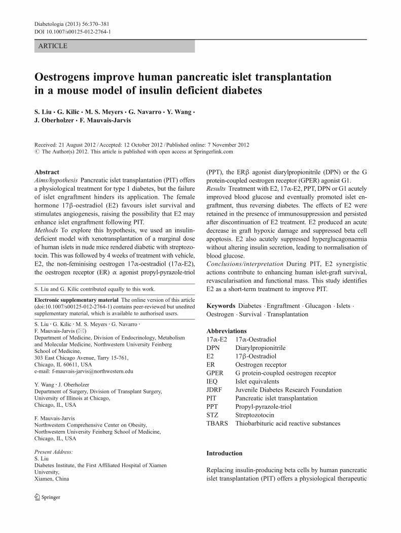
Oestrogens ameliorate diabetes We investigated the thera-peutic action of oestrogens on islet transplantation out-comes. We used male immunodeficient nude micerendered totally insulin deficient by injection of a singlehigh dose of STZ (200 mg/kg). In these mice, we performedeither sham transplantation or xenotransplantation of a mar-ginal dose of human islets (1,000 IEQ) under the renalcapsule (PIT). This was followed by treatment with vehicleor E2, leading to serum concentrations within the physio-logical range (E2 levels: vehicle 68.49±25.5 pg/ml; E2injection 321.93±77.15 pg/ml), the non-feminising E2 ste-reoisomer 17α-E2 [29], the ERα-selective agonist PPT [30],the ERβ-selective agonist DPN [31] and the G protein-coupled oestrogen receptor (GPER) agonist G1 [32]. Fol-lowing PIT or sham transplantation, blood glucose wasmonitored for 3 weeks. PIT mice treated with vehicleremained hyperglycaemic, but conversely, PIT mice treatedwith E2, 17α-E2, PPT, DPN or G1 showed a dramaticimprovement in blood glucose (Fig. 1a–f) starting 1 dayafter transplantation. In sham-operated diabetic mice, E2,17α-E2, PPT, DPN and G1 treatment had no effect on bloodglucose (Fig. 1a–f) or body weight (electronic supplemen-tary material [ESM] Fig. 1) compared with vehicle. Thisfinding demonstrated that the improvement in blood glucoseby ER agonists required the presence of transplanted humanislets. A similar protection of human PIT was observed infemale nude mice treated with E2 (ESM Fig. 2), demon-strating that E2 protection is not sex specific. Thus, weperformed the rest of the study in male mice.
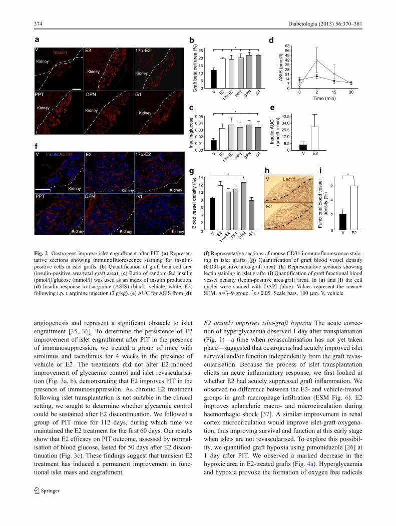
Oestrogens enhance islet engraftment At 4 weeks after PIT,mice treated with E2, 17α-E2, PPT, DPN or G1 showedprotection of islet-graft beta cell mass (Fig. 2a, b) associatedwith a rise in human serum insulin concentrations comparedwith vehicle-treated PIT mice (Fig. 2c). Insulin response toi.p. arginine was also increased in the E2- compared with thevehicle-treated mice (Fig. 2d, e). Additionally, the endoge-nous pancreatic beta cell mass of STZ-treated PIT recipientmice was efficiently reduced and unaffected by ER agonist
372 Diabetologia (2013) 56:370–381
treatments (ESM Fig. 3). Thus, activation of ERα, ERβ andGPER with oestrogens preserves functional islet mass andreverses diabetes after PIT.
We next quantified the endothelial cell area in islet graftsusing the mouse endothelial cell marker CD31. At 4 weeksafter PIT, mice treated with E2, 17α-E2, PPT or DPNshowed an improvement in islet revascularisation comparedwith vehicle-treated mice (Fig. 2f, g). Conversely, the micetreated with G1 did not show any improvement comparedwith controls (Fig. 2f, g). To determine the functionality ofthe E2-induced reconstructed islet vasculature network, weapplied in vivo staining for lectin—a commonly used mark-er of vessel functionality [33]—to a group of mice treatedwith vehicle or E2. We detected increased lectin staining inE2-treated mice compared with vehicle-treated mice(Fig. 2h, i), demonstrating that E2 increased functionalvessel density in the transplanted islets. While the contribu-tion of endothelial cells from freshly isolated donor islets tothe formation of functional vessels within islet grafts hasbeen demonstrated [33, 34], cultured islets lose most of theirendothelial cell populations within 3 days of culture [34].
We thus examined whether E2 treatment could preserve theintra-islet endothelial cell population in freshly isolatedmouse islets. Consistent with a previous report [34], weobserved that the islet endothelial cell number decreasedwithin 1 day of culture and totally disappeared after 3 days.However, we observed no beneficial effect of E2 treatmenton the maintenance of intra-islet endothelial cell population(ESM Fig. 4). As E2 favours islet survival, we tested wheth-er in vitro E2 treatment of human islets prior to PIT couldimprove PIT outcome using the same model system. Weobserved that E2 treatment of cultured human islets prior toPIT had no significant effect on subsequent glycaemic con-trol either alone or in association with in vivo E2 treatment(ESM Fig. 5).
Improvement of islet engraftment persists in the presenceof immunosuppression and after discontinuation of E2treatment The Edmonton protocol has facilitated humanislet transplantation by using non-steroidal immunosuppres-sive treatment such as sirolimus (rapamycin) and tacrolimus(FK-506) [1, 3]. However, these regimens undermine
c
BS AS 1 3 5 7 11 14 18 21
Blo
od g
luco
se (
mm
ol/l)
0
5.5
11.0
16. 5
22.0
27.5
d
Blo
od g
luco
se (
mm
ol/l)
BS AS 1 3 5 7 11 14 18 210
5.5
11.0
16.5
22.0
27.5
Time after transplantation (days)
22.0
BS AS 1 3 5 7 11 14 18 21
Blo
od g
luco
se (
mm
ol/l)
0
5.5
11.0
16.5
27.5
b
Time after transplantation (days)
22.0
a
BS AS 1 3 5 7 11 14 18 21
Blo
od g
luco
se (
mm
ol/l)
0
5.5
11.0
16.5
27.5
Time after transplantation (days)
Blo
od g
luco
se (
mm
ol/l)
e
Time after transplantation (days)BS AS 1 3 5 7 11 14 18 21
0
5. 5
11.0
16.5
22.0
27.5
Time after transplantation (days)
f
-11
0
V V E2PPT
DPN V E2G1PPT
DPN G1
17α-E
2
17α-E
2
11
22
33
44
***
Diabetes
PIT
Glu
cose
AU
C(m
mol
/l ×
day
s ×
105
)
Diabetes+PIT+E2
Control+sham+VDiabetes+PIT+V
Diabetes+sham+E2Diabetes+sham+V
Diabetes+PIT+17α-E2
Control+sham+VDiabetes+PIT+V
Diabetes+sham+17α-E2Diabetes+sham+V
Diabetes+PIT+PPT
Control+sham+VDiabetes+PIT+V
Diabetes+sham+PPTDiabetes+sham+V
Diabetes+PIT+DPN
Control+sham+VDiabetes+PIT+V
Diabetes+sham+DPNDiabetes+sham+V
Diabetes+PIT+G1
Control+sham+VDiabetes+PIT+V
Diabetes+sham+G1Diabetes+sham+V
Fig. 1 Oestrogens amelioratediabetes after PIT. Effect of (a)E2, (b) 17α-E2, (c) PPT, (d)DPN and (e) G1 on blood glu-cose after PIT. (f) Blood glu-cose AUC was calculated from(a–e). Values represent themean±SEM, n04–18/group.*p<0.05, **p<0.01. AS, afterSTZ; BS, before STZ; V,vehicle
Diabetologia (2013) 56:370–381 373
angiogenesis and represent a significant obstacle to isletengraftment [35, 36]. To determine the persistence of E2improvement of islet engraftment after PIT in the presenceof immunosuppression, we treated a group of mice withsirolimus and tacrolimus for 4 weeks in the presence ofvehicle or E2. The treatments did not alter E2-inducedimprovement of glycaemic control and islet revascularisa-tion (Fig. 3a, b), demonstrating that E2 improves PIT in thepresence of immunosuppression. As chronic E2 treatmentfollowing islet transplantation is not suitable in the clinicalsetting, we sought to determine whether glycaemic controlcould be sustained after E2 discontinuation. We followed agroup of PIT mice for 112 days, during which time wemaintained the E2 treatment for the first 60 days. Our resultsshow that E2 efficacy on PIT outcome, assessed by normal-isation of blood glucose, lasted for 50 days after E2 discon-tinuation (Fig. 3c). These findings suggest that transient E2treatment has induced a permanent improvement in func-tional islet mass and engraftment.
E2 acutely improves islet-graft hypoxia The acute correc-tion of hyperglycaemia observed 1 day after transplantation(Fig. 1)—a time when revascularisation has not yet takenplace—suggested that oestrogens had acutely improved isletsurvival and/or function independently from the graft revas-cularisation. Because the process of islet transplantationelicits an acute inflammatory response, we first looked atwhether E2 had acutely suppressed graft inflammation. Weobserved no difference between the E2- and vehicle-treatedgroups in graft macrophage infiltration (ESM Fig. 6). E2improves splanchnic macro- and microcirculation duringhaemorrhagic shock [37]. A similar improvement in renalcortex microcirculation would improve islet-graft oxygena-tion, thus improving survival and function at this early stagewhen islets are not revascularised. To explore this possibil-ity, we quantified graft hypoxia using pimonidazole [26] at1 day after PIT. We observed a marked decrease in thehypoxic area in E2-treated grafts (Fig. 4a). Hyperglycaemiaand hypoxia provoke the formation of oxygen free radicals
b
Gra
ft be
ta c
ell a
rea
(%)
0
5
10
15
20
25 *
*
Insu
lin/g
luco
se
0.00
0.01
0.02
0.03
0.04
0.05
cKidney
DPN
Kidney
G1
Kidney
PPT
Kidney
17α-E2
Kidney
E2
a
V E20
8.5
17.0
25.5
34.0
42.5
Insu
lin A
UC
(pm
ol/l
× m
in)
d
AS
IS (
pmol
/l)
07
1421283542495663
0 2 15 30
Time (min)
DPN
Kidney
Kidney
E2
Kidney
17α-E2
Kidney
PPT
Kidney
G1
V Insulin/CD31
Kidney
0
2
4
6
8
10
12
14
Blo
od v
esse
l den
sity
(%
) *
V E2
Fun
ctio
nal b
lood
ves
sel
dens
ity (
%)
0
2
4
6
*
f
e
gV Lectin
E2
h i
InsulinV
Kidney
V E2PPT
DPN G1
17α-E
2
V E2PPT
DPN G1
17α-E
2
V E2PPT
DPN G1
17α-E
2
Fig. 2 Oestrogens improve islet engraftment after PIT. (a) Represen-tative sections showing immunofluorescence staining for insulin-positive cells in islet grafts. (b) Quantification of graft beta cell area(insulin-positive area/total graft area). (c) Ratio of random-fed insulin(pmol/l)/glucose (mmol/l) was used as an index of insulin production.(d) Insulin response to L-arginine (ASIS) (black, vehicle; white, E2)following i.p. L-arginine injection (3 g/kg). (e) AUC for ASIS from (d).
(f) Representative sections of mouse CD31 immunofluorescence stain-ing in islet grafts. (g) Quantification of graft blood vessel density(CD31-positive area/graft area). (h) Representative sections showinglectin staining in islet grafts. (i) Quantification of graft functional bloodvessel density (lectin-positive area/graft area). In (a) and (f) the cellnuclei were stained with DAPI (blue). Values represent the mean±SEM, n03–9/group. *p<0.05. Scale bars, 100 μm. V, vehicle
374 Diabetologia (2013) 56:370–381
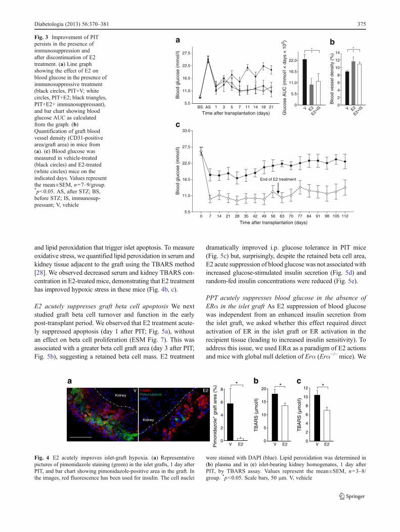
and lipid peroxidation that trigger islet apoptosis. To measureoxidative stress, we quantified lipid peroxidation in serum andkidney tissue adjacent to the graft using the TBARS method[28]. We observed decreased serum and kidney TBARS con-centration in E2-treated mice, demonstrating that E2 treatmenthas improved hypoxic stress in these mice (Fig. 4b, c).
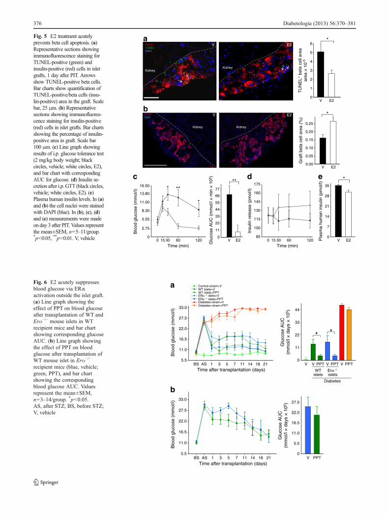
E2 acutely suppresses graft beta cell apoptosis We nextstudied graft beta cell turnover and function in the earlypost-transplant period. We observed that E2 treatment acute-ly suppressed apoptosis (day 1 after PIT; Fig. 5a), withoutan effect on beta cell proliferation (ESM Fig. 7). This wasassociated with a greater beta cell graft area (day 3 after PIT;Fig. 5b), suggesting a retained beta cell mass. E2 treatment
dramatically improved i.p. glucose tolerance in PIT mice(Fig. 5c) but, surprisingly, despite the retained beta cell area,E2 acute suppression of blood glucose was not associatedwithincreased glucose-stimulated insulin secretion (Fig. 5d) andrandom-fed insulin concentrations were reduced (Fig. 5e).
PPT acutely suppresses blood glucose in the absence ofERα in the islet graft As E2 suppression of blood glucosewas independent from an enhanced insulin secretion fromthe islet graft, we asked whether this effect required directactivation of ER in the islet graft or ER activation in therecipient tissue (leading to increased insulin sensitivity). Toaddress this issue, we used ERα as a paradigm of E2 actionsand mice with global null deletion of Erα (Erα−/− mice). We
0
5.5
11.0
16.5
22.0
*
Glu
cose
AU
C (
mm
ol/l
× d
ays
× 1
05 )
*
0
2
4
6
8
10
12
14
Blo
od v
esse
l den
sity
(%
)
b
Blo
od g
luco
se (
mm
ol/l)
Time after transplantation (days)0 7 14 21 28 35 42 49 56 63 70 77 84 91 98 105 112
5.5
11.0
16.5
22.0
27.5
33.0
End of E2 treatment
c
21 V E2
E2+IS V E2
E2+IS
Time after transplantation (days)BS AS 1 3 5 7 11 14 18
Blo
od g
luco
se (
mm
ol/l)
5.5
11.0
16.5
22.0
27.5
aFig. 3 Improvement of PITpersists in the presence ofimmunosuppression andafter discontinuation of E2treatment. (a) Line graphshowing the effect of E2 onblood glucose in the presence ofimmunosuppressive treatment(black circles, PIT+V; whitecircles, PIT+E2; black triangles,PIT+E2+ immunosuppressant),and bar chart showing bloodglucose AUC as calculatedfrom the graph. (b)Quantification of graft bloodvessel density (CD31-positivearea/graft area) in mice from(a). (c) Blood glucose wasmeasured in vehicle-treated(black circles) and E2-treated(white circles) mice on theindicated days. Values representthe mean±SEM, n07–9/group.*p<0.05. AS, after STZ; BS,before STZ; IS, immunosup-pressant; V, vehicle
V E20
2
4
6
8
Pim
onid
azol
e+ g
raft
area
(%
) *
V E2
TB
AR
S (
µmol
/l)
0
5
10
15
20 *b
V InsulinPimonidazoleDAPI
Kidney
Kidney
aE2
V E20
2
4
6
8
10
12 *c
TB
AR
S (
µmol
/l)
Fig. 4 E2 acutely improves islet-graft hypoxia. (a) Representativepictures of pimonidazole staining (green) in the islet grafts, 1 day afterPIT, and bar chart showing pimonidazole-positive area in the graft. Inthe images, red fluorescence has been used for insulin. The cell nuclei
were stained with DAPI (blue). Lipid peroxidation was determined in(b) plasma and in (c) islet-bearing kidney homogenates, 1 day afterPIT, by TBARS assay. Values represent the mean±SEM, n03–8/group. *p<0.05. Scale bars, 50 μm. V, vehicle
Diabetologia (2013) 56:370–381 375
Time after transplantation (days)BS AS 1 3 5 7 11 14 18 21
Blo
od g
luco
se (
mm
ol/l)
5.5
11.0
16.5
22.0
27.5
33.0
a
Time after transplantation (days)BS AS 1 3 5 7 11 14 18 21
Blo
od g
luco
se (
mm
ol/l)
5.5
11.0
16.5
22.0
27.5
33.0
Glu
cose
AU
C(m
mol
/l ×
day
s ×
105
)
V PPT0
5.5
11.0
16.5
22.0
27.5
b
Glu
cose
AU
C(m
mol
/l ×
day
s ×
105
)
0
11
22
33
44
* *
V V PPTV PPT PPT
Diabetes
WTislets
Erα -/-
islets
V
Control+sham+VWT islets+VWT islets+PPTERα -/- islets+VERα -/- islets+PPTDiabetes+sham+VDiabetes+sham+PPT
Fig. 6 E2 acutely suppressesblood glucose via ERαactivation outside the islet graft.(a) Line graph showing theeffect of PPT on blood glucoseafter transplantation of WT andErα−/− mouse islets in WTrecipient mice and bar chartshowing corresponding glucoseAUC. (b) Line graph showingthe effect of PPT on bloodglucose after transplantation ofWT mouse islet in Erα−/−
recipient mice (blue, vehicle;green, PPT), and bar chartshowing the correspondingblood glucose AUC. Valuesrepresent the mean±SEM,n03–14/group. *p<0.05.AS, after STZ; BS, before STZ;V, vehicle
c
0 15 30 60 1200
2.75
5.55
8.30
11.00
13.80
16.50
Blo
od g
luco
se (
mm
ol/l)
Time (min)
*
*
**
0 15 30 60 12085
100
115
130
145
160
175
Insu
lin r
elea
se (
pmol
/l)Time (min)
V E20
11
22
33
44
55
66
77
Glu
cose
AU
C (
mm
ol/l
× m
in ×
105
)
**
E20
7
14
21
28
35
Pla
sma
hum
an in
sulin
(pm
ol/l)
V
*d e
b
V E20.00
0.05
0.10
0.15
0.20
0.25
Gra
ft be
ta c
ell a
rea
(%)
*
Kidney Kidney
InsulinDAPI
V E2
*
V E20
1
2
3
4
5
6
TU
NE
L+ b
eta
cell
area
area
× 1
0-5
V
KidneyKidney
E2InsulinTUNELDAPI
a
Kidney
E2
Fig. 5 E2 treatment acutelyprevents beta cell apoptosis. (a)Representative sections showingimmunofluorescence staining forTUNEL-positive (green) andinsulin-positive (red) cells in isletgrafts, 1 day after PIT. Arrowsshow TUNEL-positive beta cells.Bar charts show quantification ofTUNEL-positive/beta cells (insu-lin-positive) area in the graft. Scalebar, 25 μm. (b) Representativesections showing immunofluores-cence staining for insulin-positive(red) cells in islet grafts. Bar chartsshowing the percentage of insulin-positive area in graft. Scale bar100 μm. (c) Line graph showingresults of i.p. glucose tolerance test(2 mg/kg body weight; blackcircles, vehicle; white circles, E2),and bar chart with correspondingAUC for glucose. (d) Insulin se-cretion after i.p.GTT (black circles,vehicle; white circles, E2). (e)Plasma human insulin levels. In (a)and (b) the cell nuclei were stainedwith DAPI (blue). In (b), (c), (d)and (e) measurements were madeon day 3 after PIT.Values representthe mean±SEM, n05–11/group.*p<0.05, **p<0.01. V, vehicle
376 Diabetologia (2013) 56:370–381
first performed an allotransplantation of a marginal dose ofislets (150) isolated from donor littermate wild-type (WT) orErα−/− mice under the kidney capsule of recipient WT mice.These recipient mice were treated with vehicle or the ERα-selective agonist PPT. As expected, WT mice transplantedwith WT or Erα−/− islets and treated with vehicle remainedhyperglycaemic. However, PPT treatment led to a similarearly decrease in blood glucose in WT mice transplanted withWT or Erα−/− islets, demonstrating that PPT improves PIT inthe absence of islet ERα (Fig. 6a). Next, we performed anallotransplantation of a marginal dose ofWT islets in recipientErα−/− mice followed by treatment with vehicle or PPT.Vehicle- and PPT-treated mice showed a similar and minorblood-glucose-lowering effect due to the transplantation ofWT islets in Erα−/− mice. However, PPT treatment did notproduce a stronger hypoglycaemic effect than vehicle in theseERα-deficient mice. Together, these data demonstrate therequirement of ERα activation in the recipient mouse tissuesto acutely suppress blood glucose (Fig. 6b).
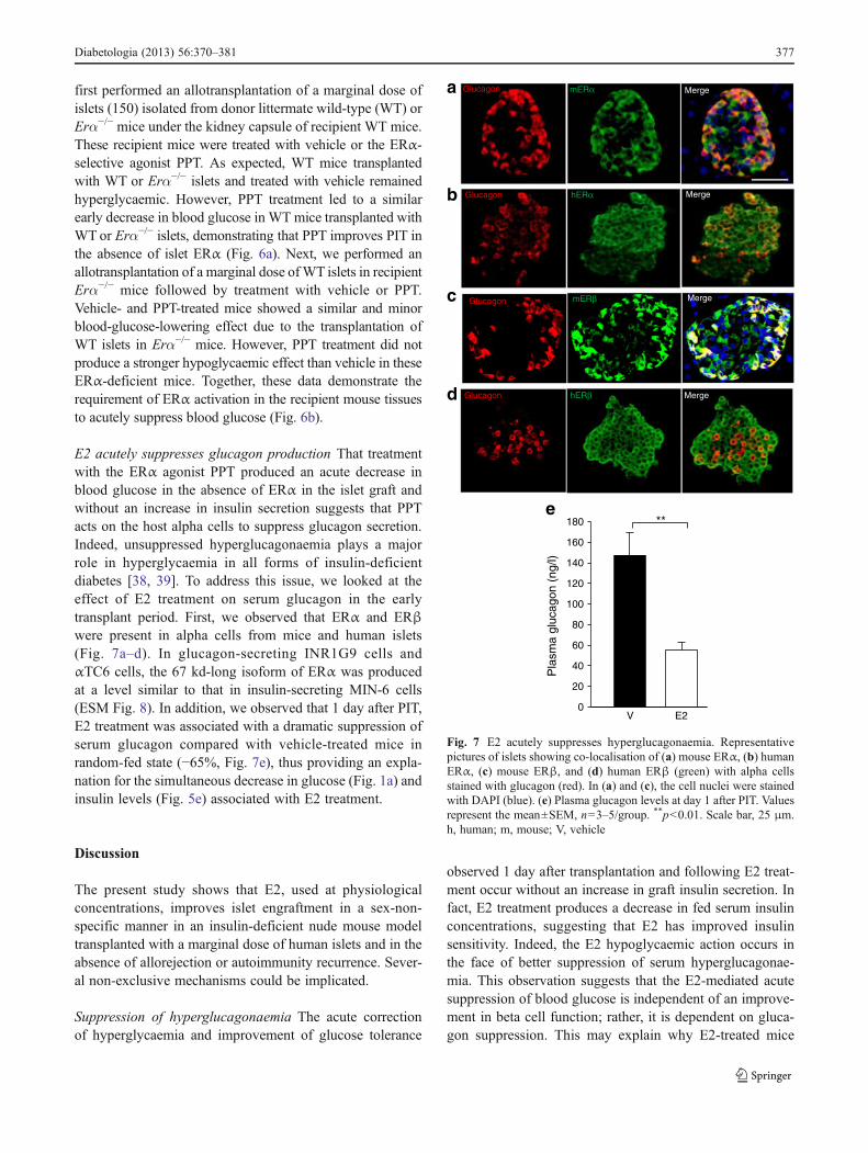
E2 acutely suppresses glucagon production That treatmentwith the ERα agonist PPT produced an acute decrease inblood glucose in the absence of ERα in the islet graft andwithout an increase in insulin secretion suggests that PPTacts on the host alpha cells to suppress glucagon secretion.Indeed, unsuppressed hyperglucagonaemia plays a majorrole in hyperglycaemia in all forms of insulin-deficientdiabetes [38, 39]. To address this issue, we looked at theeffect of E2 treatment on serum glucagon in the earlytransplant period. First, we observed that ERα and ERβwere present in alpha cells from mice and human islets(Fig. 7a–d). In glucagon-secreting INR1G9 cells andαTC6 cells, the 67 kd-long isoform of ERα was producedat a level similar to that in insulin-secreting MIN-6 cells(ESM Fig. 8). In addition, we observed that 1 day after PIT,E2 treatment was associated with a dramatic suppression ofserum glucagon compared with vehicle-treated mice inrandom-fed state (−65%, Fig. 7e), thus providing an expla-nation for the simultaneous decrease in glucose (Fig. 1a) andinsulin levels (Fig. 5e) associated with E2 treatment.
Discussion
The present study shows that E2, used at physiologicalconcentrations, improves islet engraftment in a sex-non-specific manner in an insulin-deficient nude mouse modeltransplanted with a marginal dose of human islets and in theabsence of allorejection or autoimmunity recurrence. Sever-al non-exclusive mechanisms could be implicated.
Suppression of hyperglucagonaemia The acute correctionof hyperglycaemia and improvement of glucose tolerance
observed 1 day after transplantation and following E2 treat-ment occur without an increase in graft insulin secretion. Infact, E2 treatment produces a decrease in fed serum insulinconcentrations, suggesting that E2 has improved insulinsensitivity. Indeed, the E2 hypoglycaemic action occurs inthe face of better suppression of serum hyperglucagonae-mia. This observation suggests that the E2-mediated acutesuppression of blood glucose is independent of an improve-ment in beta cell function; rather, it is dependent on gluca-gon suppression. This may explain why E2-treated mice
V E20
20
40
60
80
100
120
140
160
180
Pla
sma
gluc
agon
(ng
/l)
**e
Glucagon mERα Mergea
b Glucagon hERα Merge
d hERβGlucagon Merge
CGlucagon mERβ Mergec
Fig. 7 E2 acutely suppresses hyperglucagonaemia. Representativepictures of islets showing co-localisation of (a) mouse ERα, (b) humanERα, (c) mouse ERβ, and (d) human ERβ (green) with alpha cellsstained with glucagon (red). In (a) and (c), the cell nuclei were stainedwith DAPI (blue). (e) Plasma glucagon levels at day 1 after PIT. Valuesrepresent the mean±SEM, n03–5/group. **p<0.01. Scale bar, 25 μm.h, human; m, mouse; V, vehicle
Diabetologia (2013) 56:370–381 377
have suppressed hyperglucagonaemia and lower insulinconcentrations, as a decrease in hyperglucagonaemiaimproves glucagon-induced hepatic insulin resistance. Thisobservation is also consistent with the ‘glucagonocentric’vision of diabetes pathophysiology in which glucagon ex-cess, rather than insulin deficiency, is the sine qua non ofhyperglycaemia in all forms of diabetes, at least in rodents[40]. Indeed, total insulin deficiency by beta cell destructionin glucagon-receptor-null mice, which are unresponsive toglucagon, does not cause diabetic abnormalities [41]. Weobserve that ERα and ERβ are produced in mouse andhuman alpha cells, further indicating that alpha cells aredirect targets of E2 actions. In addition, our experimentssuggest that ERα needs to be activated in alpha cells of thehost pancreas as the hypoglycaemic effect of an ERα-selective agonist is lost when islets from WT mice aretransplanted in Erα null mice (lacking ERα in alpha cells).Accordingly, the ERα agonist can still acutely suppressblood glucose and permanently improve diabetes whenERα-deficient islets are transplanted in WT mice. Thisdemonstrates that ERα action in graft alpha cells is notnecessary to suppress blood glucose. Recently, leptin hasbeen shown to suppress glucagon and correct diabetes inmice, in the absence of insulin [42]. Our results suggest thatE2 requires the presence of insulin—or at least functionalbeta cells—to suppress glucagon as ER agonists have nohypoglycaemic effect in diabetic mice in the absence oftransplanted islets. This observation is consistent with theconcept that insulin suppresses glucagon secretion from
alpha cells via paracrine mechanisms [43] and that insulinsignalling in alpha cells is required for this process [44].Thus, in the alpha cells of diabetic mice E2 may act as aninsulin sensitiser in suppressing glucagon production. Ad-ditional studies in mice lacking ERs in alpha cells areneeded to address this issue.
Protection of functional beta cell mass Oestrogens haveacutely improved islet survival following transplantation,as demonstrated by E2-mediated acute suppression of graftapoptotic beta cells. This is the first evidence that E2, usedat therapeutic doses, protects human islets from apoptosis inan in vivo diabetic environment. We and others have shownthat E2 promotes islet survival in conditions of oxidativestress and pro-inflammatory cytokine injury in culture andin vivo [11–13, 17]. Higher islet functionality and decreasedapoptosis are also observed after transplantation of rat isletsrecovered from E2-treated brain-dead donors [45]. This anti-apoptotic protection probably accounts for the early reten-tion of graft beta cell mass, which persists after 3 weeks ofER agonist treatment. In addition, we believe that E2 anti-apoptotic protection is also mediated via activation of ERsin recipient endothelial cells. E2 dilates the mesenteric ar-teries and increases mesenteric blood flow [46, 47]. E2 alsoimproves splanchnic circulation during haemorrhagic shock,which increases oxygenation [37]. We observed that E2acutely and dramatically improved islet-graft hypoxia,which is associated with decreased systemic and kidneyoxidative damage. This protection of islet oxygenation is
Pancreas
Acute effect
Kidney
Late effect
Beta cell mass
E2Oxygenation
Alpha cellsApoptosis Endothelial
cells
KidneyGraft
Functional beta cell mass
Hyperglycaemia
Glucagon
Revascularisation
Endothelialcells
Graft
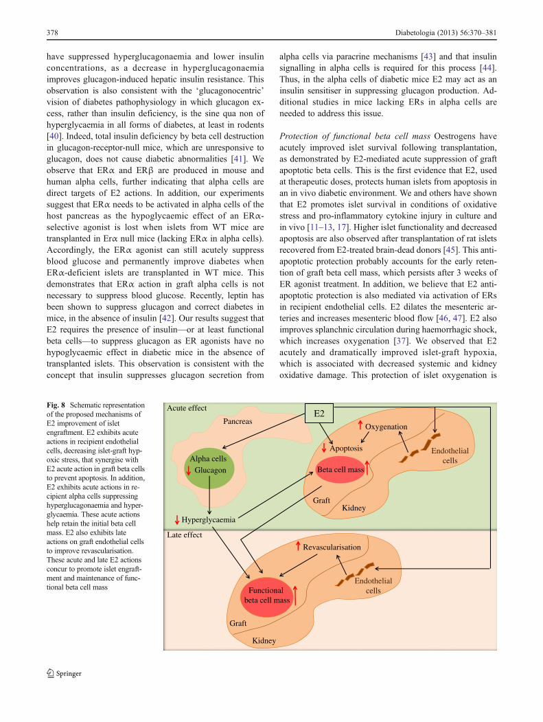
Fig. 8 Schematic representationof the proposed mechanisms ofE2 improvement of isletengraftment. E2 exhibits acuteactions in recipient endothelialcells, decreasing islet-graft hyp-oxic stress, that synergise withE2 acute action in graft beta cellsto prevent apoptosis. In addition,E2 exhibits acute actions in re-cipient alpha cells suppressinghyperglucagonaemia and hyper-glycaemia. These acute actionshelp retain the initial beta cellmass. E2 also exhibits lateactions on graft endothelial cellsto improve revascularisation.These acute and late E2 actionsconcur to promote islet engraft-ment and maintenance of func-tional beta cell mass
378 Diabetologia (2013) 56:370–381
probably instrumental in E2 acute anti-apoptotic protectionas, following PIT, hypoxic stress is a critical factor in theearly loss of islet mass [8–10]. Initially, E2-treated graft betacells are non-functional and show no insulin-secretory re-sponse to the glucose challenge, probably as a result ofhyperglycaemia-induced glucose desensitisation and/or lackof islet vascularisation. However, following 3 weeks ofnormoglycaemia, PIT mice treated with ER agonists showa restoration of beta cell function. Thus, E2 acute normal-isation of blood glucose, via suppression of hyperglucago-naemia, has favoured the subsequent restoration of beta cellfunction following correction of glucose desensitisation.
Action on recipient endothelial cells Apart from E2-mediated acute improvement of islet-graft oxygenation, thelikely action of oestrogens involves the enhancement of isletrevascularisation through action on recipient endothelialcells, as E2 is a potent angiogenic factor in the endometriumand following vascular injury [22, 23]. Accordingly, E2increases the functional blood vessel density of the humanislet graft. More importantly, immunosuppression with siroli-mus and tacrolimus typically inhibits islet revascularisationand engraftment. Yet E2 stimulation of revascularisation per-sists in the presence of this immunosuppression regimen.Furthermore, after discontinuation of E2 treatment the correc-tion of diabetes endures, demonstrating that E2 has induced astable engraftment. E2 treatment promotes revascularisationof the transplanted human islets through ERs on the endothe-lial cell from the recipient kidney cortex and without theparticipation of endothelial cells from the human graft, asthese cells had disappeared at the time of transplantation.However, G1, which exhibits potent anti-apoptotic action inhuman islets [13], improves PIT outcome without enhancinggraft revascularisation, suggesting that the acute hypoglycae-mic and anti-apoptotic effects of oestrogens are more impor-tant to the engraftment process than the moderate increase inrevascularisation.
Thus, the synergistic actions of E2 on ERs in recipientalpha cells, endothelial cells and graft beta cells leading,respectively, to the suppression of hyperglucagonaemia andhyperglycaemia, the protection from islet-graft hypoxia,oxidative stress and apoptosis and ultimately to revascular-isation of the graft, have concurred to promote islet engraft-ment and maintenance of beta cell functional mass. Asummary of the proposed mechanisms of E2 action is shownin Fig. 8.
Our findings have direct therapeutic implications for PITin type 1 diabetes. Although long-term E2 treatment follow-ing PIT is not suitable because of the risk of cancer, con-versely, transient E2 treatment represents a safe andimmediately available alternative to improve PIT in women.Indeed, fertile women with type 1 diabetes show E2 defi-ciency compared with healthy women [48]. Therefore,
women with type 1 diabetes undergoing islet transplantationhave lost their endogenous E2 protection and could benefitfrom short-term oestrogen supplementation using therapeu-tic doses. Furthermore, 17α-E2, which is endogenous inhumans, has few of the biological effects associated withthe female hormone activity and may be a candidate for sex-neutral therapy in PIT.
In conclusion, E2 enhances islet engraftment after PIT.As oestrogens are approved by the Food and Drug Admin-istration, further testing in women with the addition ofoestrogens to the Edmonton protocol should be considered.This could provide an immediate therapeutic alternative toimprove PIT and achieve insulin independence with fewerislets, long before other surrogate islet beta cell sources orbeta cell regeneration therapy can be developed.
Acknowledgements We are grateful to B. Hering from the Univer-sity of Minnesota for helpful discussion and to M. Brissova andA. Powers from Vanderbilt University for providing protocol andadvice on in vivo lectin infusion. We acknowledge the Integrated IsletDistribution Program (IIDP) funded by the National Institute of Diabetesand Digestive and Kidney Diseases (NIDDK) and the Juvenile DiabetesResearch Foundation (JDRF) for providing human islet for research.
Funding This work was supported by grants from the NationalInstitutes of Health (RO1 DK074970, P50 HD044405), the JDRF(1-2006-837), the March of Dimes Birth Defects Foundation(6-FY07-312) and the American Heart Association (11IRG5570010) toF. Mauvais-Jarvis.
Contribution statement SL and GK performed experiments, ana-lysed data and wrote the manuscript. MSM and GN performed experi-ments, analysed data and revised the manuscript; YW and JOparticipated in design of the study and manuscript revision. FMJconceptualised and designed the study, analysed data and wrote themanuscript. All authors gave approval of the final version of thismanuscript for publication.
Duality of interest F. Mauvais-Jarvis has received research supportfrom Pfizer. The remaining authors declare that there is no duality ofinterest associated with this manuscript.
Open Access This article is distributed under the terms of the CreativeCommons Attribution License which permits any use, distribution, andreproduction in any medium, provided the original author(s) and thesource are credited.
References
1. Shapiro AM, Lakey JR, Ryan EA et al (2000) Islet transplantationin seven patients with type 1 diabetes mellitus using aglucocorticoid-free immunosuppressive regimen. N Engl J Med343:230–238
Diabetologia (2013) 56:370–381 379
2. Hering BJ, Kandaswamy R, Ansite JD et al (2005) Single-donor,marginal-dose islet transplantation in patients with type 1 diabetes.JAMA 293:830–835
3. Shapiro AM, Ricordi C, Hering BJ et al (2006) International trialof the Edmonton protocol for islet transplantation. N Engl J Med355:1318–1330
4. Robertson RP (2010) Islet transplantation a decade later and strat-egies for filling a half-full glass. Diabetes 59:1285–1291
5. Weir GC, Bonner-Weir S (2004) Five stages of evolving beta-celldysfunction during progression to diabetes. Diabetes 53(Suppl 3):S16–S21
6. Davalli AM, Ogawa Y, Ricordi C, Scharp DW, Bonner-Weir S,Weir GC (1995) A selective decrease in the beta cell mass ofhuman islets transplanted into diabetic nude mice. Transplantation59:817–820
7. Davalli AM, Scaglia L, Zangen DH, Hollister J, Bonner-Weir S,Weir GC (1996) Vulnerability of islets in the immediate posttrans-plantation period. Dynamic changes in structure and function.Diabetes 45:1161–1167
8. Emamaullee JA, Shapiro AM (2006) Interventional strategies toprevent beta-cell apoptosis in islet transplantation. Diabetes55:1907–1914
9. Carlsson PO, Palm F, Andersson A, Liss P (2001) Markedlydecreased oxygen tension in transplanted rat pancreatic islets irre-spective of the implantation site. Diabetes 50:489–495
10. Mattsson G, Jansson L, Carlsson PO (2002) Decreased vasculardensity in mouse pancreatic islets after transplantation. Diabetes51:1362–1366
11. Contreras JL, Smyth CA, Bilbao G, Young CJ, Thompson JA,Eckhoff DE (2002) 17beta-Estradiol protects isolated human pan-creatic islets against proinflammatory cytokine-induced cell death:molecular mechanisms and islet functionality. Transplantation74:1252–1259
12. Le May C, Chu K, Hu M et al (2006) Estrogens protect pancreaticbeta-cells from apoptosis and prevent insulin-deficient diabetesmellitus in mice. Proc Natl Acad Sci USA 103:9232–9237
13. Liu S, Le May C, Wong WP et al (2009) Importance of extranuclearestrogen receptor-alpha and membrane G protein-coupled estrogenreceptor in pancreatic islet survival. Diabetes 58:2292–2302
14. Wong WP, Tiano JP, Liu S et al (2010) Extranuclear estrogenreceptor-alpha stimulates NeuroD1 binding to the insulin promoterand favors insulin synthesis. Proc Natl Acad Sci USA 107:13057–13062
15. Liu S, Mauvais-Jarvis F (2010) Minireview: estrogenic protectionof beta-cell failure in metabolic diseases. Endocrinology 151:859–864
16. Alonso-Magdalena P, Ropero AB, Carrera MP et al (2008) Pan-creatic insulin content regulation by the estrogen receptor ERalpha. PLoS One 3:e2069
17. Tiano JP, Delghingaro-Augusto V, Le May C et al (2011) Estrogenreceptor activation reduces lipid synthesis in pancreatic islets andprevents beta cell failure in rodent models of type 2 diabetes. J ClinInvest 121:3331–3342
18. Tiano JP, Mauvais-Jarvis F (2012) Importance of oestrogen recep-tors to preserve functional beta-cell mass in diabetes. Nat RevEndocrinol 8:342–351
19. Shifren JL, Tseng JF, Zaloudek CJ et al (1996) Ovarian steroidregulation of vascular endothelial growth factor in the humanendometrium: implications for angiogenesis during the menstrualcycle and in the pathogenesis of endometriosis. J Clin EndocrinolMetab 81:3112–3118
20. Mueller MD, Vigne JL, Minchenko A, Lebovic DI, Leitman DC,Taylor RN (2000) Regulation of vascular endothelial growth factor(VEGF) gene transcription by estrogen receptors alpha and beta.Proc Natl Acad Sci USA 97:10972–10977
21. Losordo DW, Isner JM (2001) Estrogen and angiogenesis: a re-view. Arterioscler Thromb Vasc Biol 21:6–12
22. Morales DE, McGowan KA, Grant DS et al (1995) Estrogenpromotes angiogenic activity in human umbilical vein endothelialcells in vitro and in a murine model. Circulation 91:755–763
23. MendelsohnME, Karas RH (1999) The protective effects of estrogenon the cardiovascular system. N Engl J Med 340:1801–1811
24. Dupont S, Krust A, Gansmuller A, Dierich A, Chambon P, MarkM (2000) Effect of single and compound knockouts of estrogenreceptors alpha (ERalpha) and beta (ERbeta) on mouse reproduc-tive phenotypes. Development 127:4277–4291
25. Molano RD, Pileggi A, Berney T et al (2003) Long-term isletallograft survival in nonobese diabetic mice treated with tacroli-mus, rapamycin, and anti-interleukin-2 antibody. Transplantation75:1812–1819
26. Olsson R, Olerud J, Pettersson U, Carlsson PO (2011) Increasednumbers of low-oxygenated pancreatic islets after intraportal islettransplantation. Diabetes 60:2350–2353
27. Chen C, Kuehn C, Bretzel RG, Linn T (2009) Anti-inflammatorythalidomide improves islet grafts survival and functions in a xeno-genic environment. PLoS One 4:e6312
28. Armstrong D, Browne R (1994) The analysis of free radicals, lipidperoxides, antioxidant enzymes and compounds related to oxida-tive stress as applied to the clinical chemistry laboratory. Adv ExpMed Biol 366:43–58
29. Moos WH, Dykens JA, Howell N (2008) 17α-Estradiol: a less-feminizing estrogen. Drug Dev Res 69:177–184
30. Stauffer SR, Coletta CJ, Tedesco R et al (2000) Pyrazoleligands: structure-affinity/activity relationships and estrogenreceptor-alpha-selective agonists. J Med Chem 43:4934–4947
31. Meyers MJ, Sun J, Carlson KE, Marriner GA, KatzenellenbogenBS, Katzenellenbogen JA (2001) Estrogen receptor-beta potency-selective ligands: structure-activity relationship studies of diaryl-propionitriles and their acetylene and polar analogues. J MedChem 44:4230–4251
32. Bologa CG, Revankar CM, Young SM et al (2006) Virtual andbiomolecular screening converge on a selective agonist forGPR30. Nat Chem Biol 2:207–212
33. Brissova M, Fowler M, Wiebe P et al (2004) Intraislet endothelialcells contribute to revascularization of transplanted pancreaticislets. Diabetes 53:1318–1325
34. Nyqvist D, Kohler M, Wahlstedt H, Berggren PO (2005)Donor islet endothelial cells participate in formation of functionalvessels within pancreatic islet grafts. Diabetes 54:2287–2293
35. Zhang N, Su D, Qu S et al (2006) Sirolimus is associated withreduced islet engraftment and impaired beta-cell function. Diabetes55:2429–2436
36. Kang L, Zhang X, Xie Y et al (2010) Involvement of estrogenreceptor variant ER-alpha36, not GPR30, in nongenomic estrogensignaling. Mol Endocrinol 24:709–721
37. Kuebler JF, Jarrar D, Toth B et al (2002) Estradiol administrationimproves splanchnic perfusion following trauma-hemorrhage andsepsis. Arch Surg 137:74–79
38. Dobbs R, Sakurai H, Sasaki H et al (1975) Glucagon: role in thehyperglycemia of diabetes mellitus. Science 187:544–547
39. Raskin P, Unger RH (1978) Hyperglucagonemia and its suppres-sion. Importance in the metabolic control of diabetes. N Engl JMed 299:433–436
40. Unger RH, Cherrington AD (2012) Glucagonocentric restructuringof diabetes: a pathophysiologic and therapeutic makeover. J ClinInvest 122:4–12
41. Lee Y, Wang MY, Du XQ, Charron MJ, Unger RH (2011) Gluca-gon receptor knockout prevents insulin-deficient type 1 diabetes inmice. Diabetes 60:391–397
42. Yu X, Park BH, Wang MY, Wang ZV, Unger RH (2008) Makinginsulin-deficient type 1 diabetic rodents thrive without insulin.Proc Natl Acad Sci USA 105:14070–14075
380 Diabetologia (2013) 56:370–381

43. Maruyama H, Hisatomi A, Orci L, Grodsky GM, Unger RH (1984)Insulin within islets is a physiologic glucagon release inhibitor.J Clin Invest 74:2296–2299
44. Kawamori D, Kurpad AJ, Hu J et al (2009) Insulin signaling inalpha cells modulates glucagon secretion in vivo. Cell Metabolism9:350–361
45. Eckhoff DE, Eckstein C, Smyth CA et al (2004) Enhanced isolatedpancreatic islet recovery and functionality in rats by 17β-estradioltreatment of brain death donors. Surgery 136:336–345
46. Ma XL, Gao F, Chen J et al (2001) Endothelial protective andantishock effects of a selective estrogen receptor modulator in rats.Am J Physiol Heart Circ Physiol 280:H876–H884
47. Vacca G, Battaglia A, Grossini E, Mary DA, Molinari C, Surico N(1999) The effect of 17beta-oestradiol on regional blood flow inanaesthetized pigs. J Physiol 514:875–884
48. Salonia A, Lanzi R, Scavini M et al (2006) Sexual function andendocrine profile in fertile women with type 1 diabetes. DiabetesCare 29:312–316
Diabetologia (2013) 56:370–381 381