odontogenesis i
TRANSCRIPT
1
ODONTOGENESIS
Presented byDr.G.Umamaheswari
I year PG
2
INTRODUCTION
Teeth development –interaction between the epithelium and underlying ectomesenchymal tissue.
Molecules and signaling pathways are responsible fo intiation , differentiation and morphogenesis.
Interaction may be short, mid, long term cell to cell or cell to tissue interactions.•
3
Ectoderm:Enamel NEURAL CREST CELLS: constitute much
mesenchyme of head and neck Derived from ectodermal germ layer-
ectomesenchyme or neuroectoderm CT of face, dental structures
Origin of dental tissue:
Molecule involved: wnt has been implicated in neural tube formation and neural crest migration
4
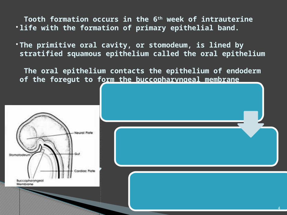
Tooth formation occurs in the 6th week of intrauterine life with the formation of primary epithelial band.
The primitive oral cavity, or stomodeum, is lined by stratified squamous epithelium called the oral epithelium
The oral epithelium contacts the epithelium of endoderm of the foregut to form the buccopharyngeal membrane
•
•
Buccopharngygeal membrane ruptures at about 27th day of gestation and the primitive oral cavity establishes a connection with the foregut
Most of the connective tissue cells underlying the oral epithelium are of neural crest or ectomesenchyme in origin
These cells instruct the overlying ectoderm to start the tooth development, which begins in the anterior portion of the future maxilla & mandible and proceeds posteriorly
5
PRIMARY EPITHELIAL BAND
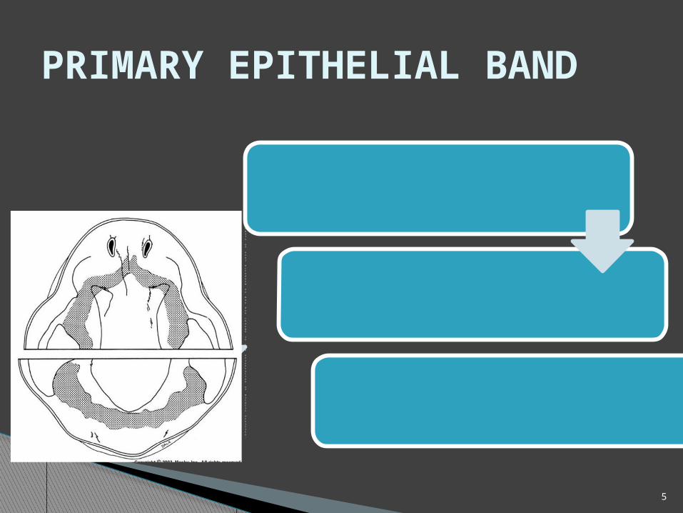
After rupture of Buccopharnygeal membrane ,about 37 days of development, the continuous band of thickened epithelium forms around the mouth
Horse shoe shaped and correspond to the ectoderm of future dental arches [U/L jaws]
Rapid proliferation of basal cells of oral ectoderm or may due change in the orientation of mitotic activity.
6
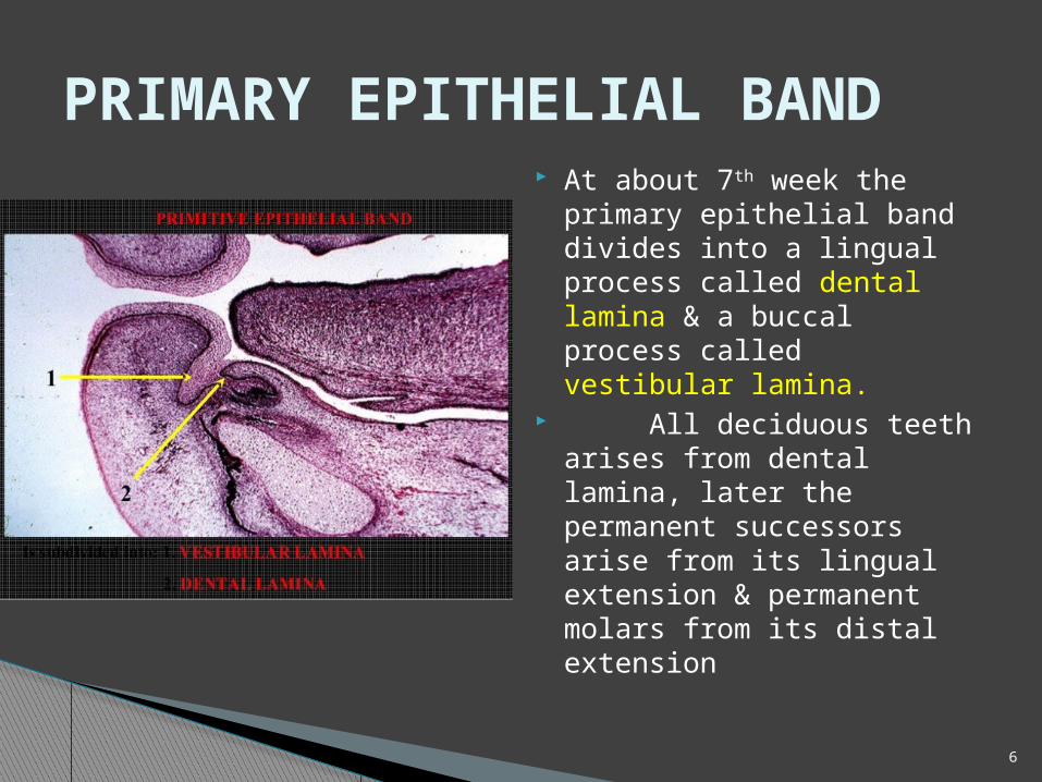
At about 7th week the primary epithelial band divides into a lingual process called dental lamina & a buccal process called vestibular lamina.
All deciduous teeth arises from dental lamina, later the permanent successors arise from its lingual extension & permanent molars from its distal extension
PRIMARY EPITHELIAL BAND
7
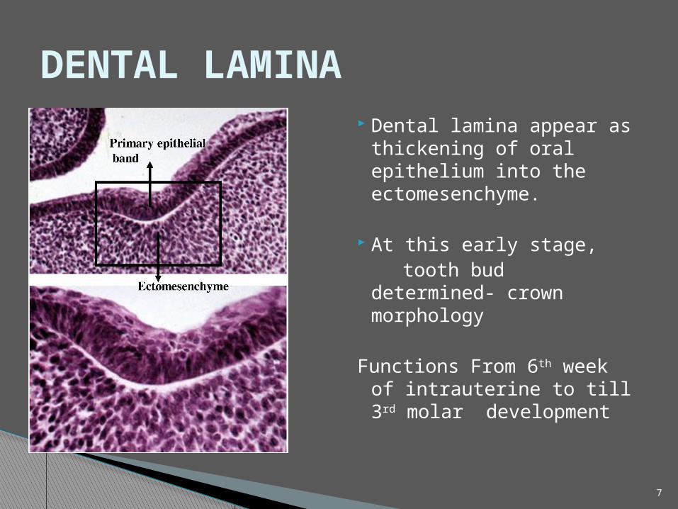
Dental lamina appear as thickening of oral epithelium into the ectomesenchyme.
At this early stage, tooth bud determined-
crown morphology
Functions From 6th week of intrauterine to till 3rd molar development
DENTAL LAMINA
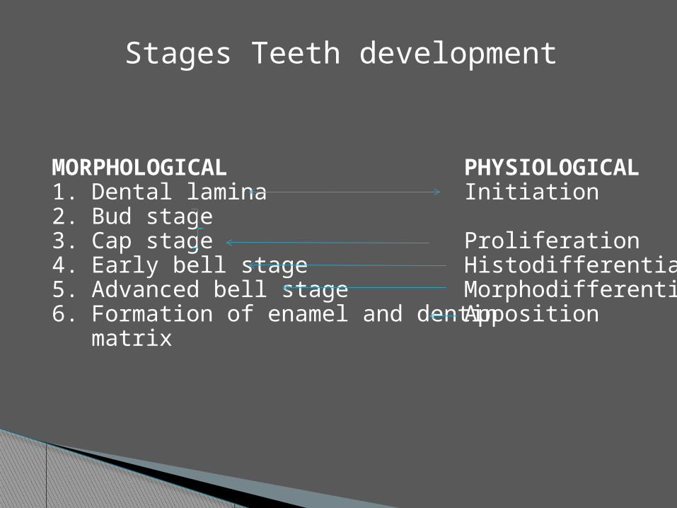
MORPHOLOGICAL1. Dental lamina2. Bud stage3. Cap stage4. Early bell stage5. Advanced bell stage6. Formation of enamel and dentin matrix
PHYSIOLOGICALInitiation
ProliferationHistodifferentiationMorphodifferentiationApposition
Stages Teeth development
9
BUD STAGE
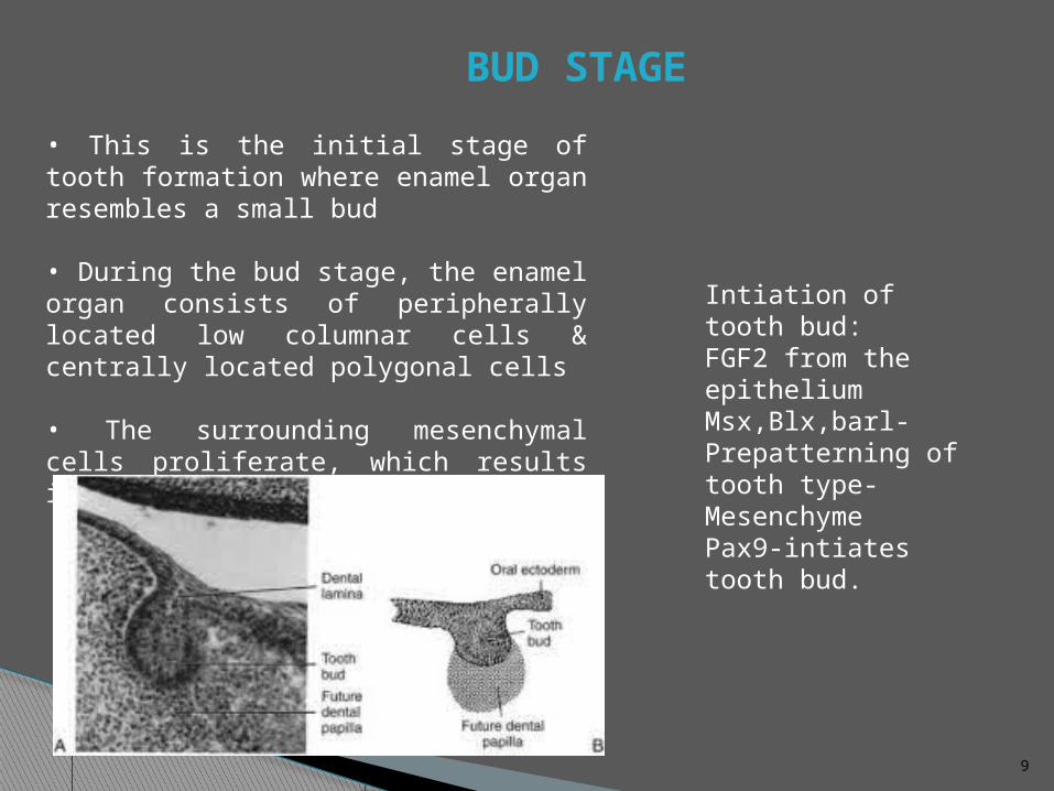
• This is the initial stage of tooth formation where enamel organ resembles a small bud
• During the bud stage, the enamel organ consists of peripherally located low columnar cells & centrally located polygonal cells
• The surrounding mesenchymal cells proliferate, which results in their condensation in two areas
Intiation of tooth bud:FGF2 from the epitheliumMsx,Blx,barl-Prepatterning of tooth type-MesenchymePax9-intiates tooth bud.
10
CAP STAGE
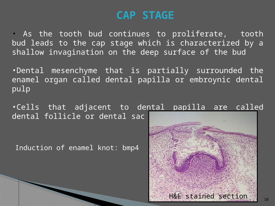
• As the tooth bud continues to proliferate, tooth bud leads to the cap stage which is characterized by a shallow invagination on the deep surface of the bud
•Dental mesenchyme that is partially surrounded the enamel organ called dental papilla or embroynic dental pulp
•Cells that adjacent to dental papilla are called dental follicle or dental sac
H&E stained section
Induction of enamel knot: bmp4
11
OUTER & INNER ENAMEL EPITHELIUM
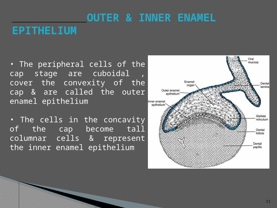
• The peripheral cells of the cap stage are cuboidal , cover the convexity of the cap & are called the outer enamel epithelium
• The cells in the concavity of the cap become tall columnar cells & represent the inner enamel epithelium
12
STELLATE RETICULUM•Polygonal cells located between the outer and the inner enamel
epithelium, begin to separate due to water being drawn into the enamel organ from the surrounding dental papilla
• As a result the polygonal cells become star shaped but maintain contact with each other by their cytoplasmic process
• As the star shaped cells form a cellular network, they are called the stellate reticulum
13
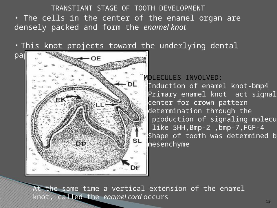
• The cells in the center of the enamel organ are densely packed and form the enamel knot
• This knot projects toward the underlying dental papilla
MOLECULES INVOLVED:•Induction of enamel knot-bmp4 Primary enamel knot act signaling center for crown pattern determination through the production of signaling molecules like SHH,Bmp-2 ,bmp-7,FGF-4•Shape of tooth was determined by mesenchyme
TRANSTIANT STAGE OF TOOTH DEVELOPMENT
At the same time a vertical extension of the enamel knot, called the enamel cord occurs
14
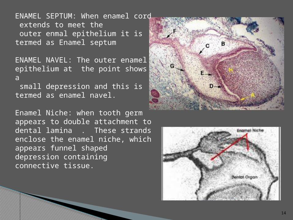
ENAMEL SEPTUM: When enamel cord extends to meet the outer enmal epithelium it is termed as Enamel septum
ENAMEL NAVEL: The outer enamel epithelium at the point shows a small depression and this is termed as enamel navel.
Enamel Niche: when tooth germ appears to double attachment to dental lamina . These strands enclose the enamel niche, which appears funnel shaped depression containing connective tissue.
15
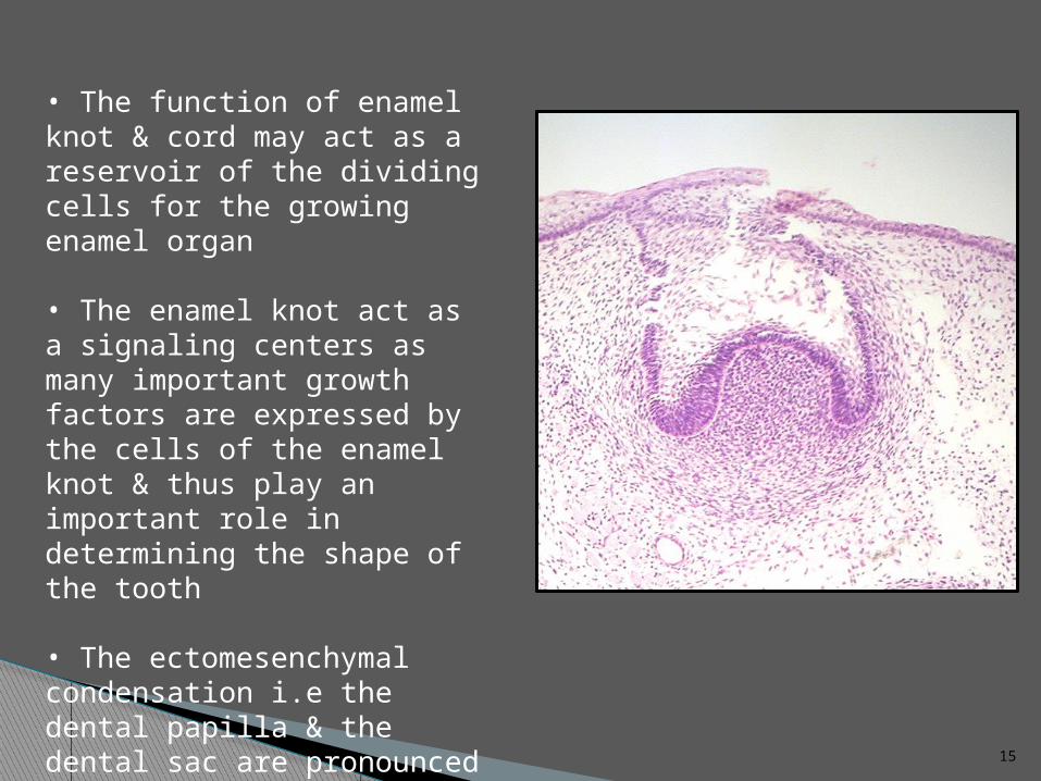
• The function of enamel knot & cord may act as a reservoir of the dividing cells for the growing enamel organ
• The enamel knot act as a signaling centers as many important growth factors are expressed by the cells of the enamel knot & thus play an important role in determining the shape of the tooth
• The ectomesenchymal condensation i.e the dental papilla & the dental sac are pronounced during this stage of dental development
16
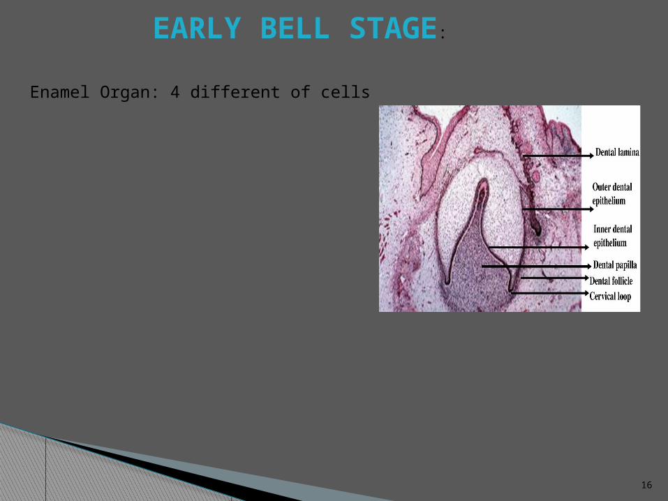
EARLY BELL STAGE:
Enamel Organ: 4 different of cells
17
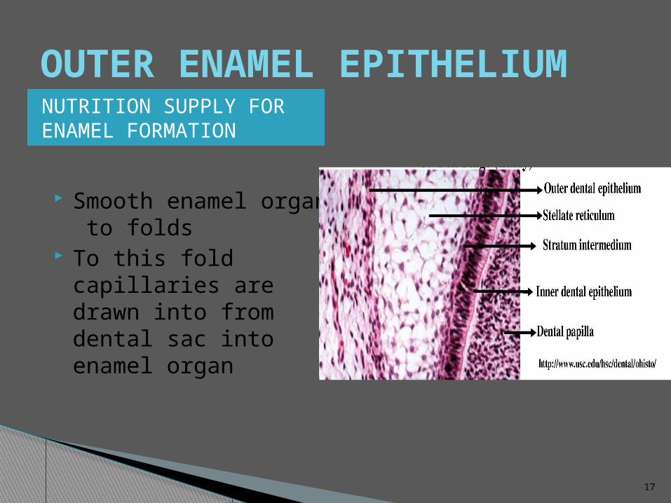
OUTER ENAMEL EPITHELIUMNUTRITION SUPPLY FOR ENAMEL FORMATION
Smooth enamel organ to folds
To this fold capillaries are drawn into from dental sac into enamel organ
18
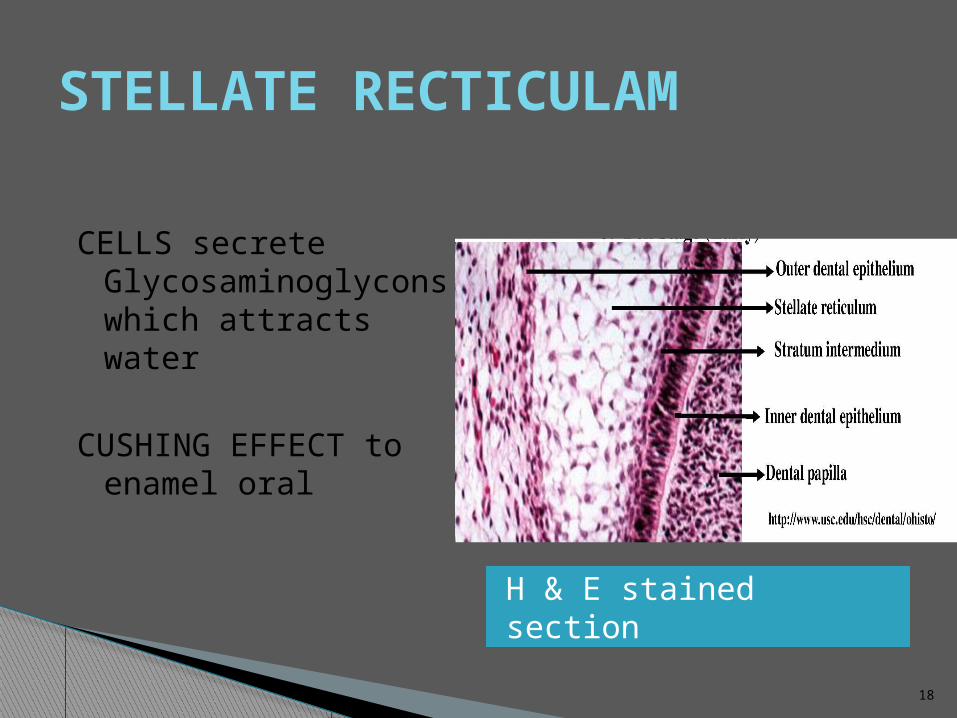
STELLATE RECTICULAM
H & E stained section
CELLS secrete Glycosaminoglycons which attracts water
CUSHING EFFECT to enamel oral
19
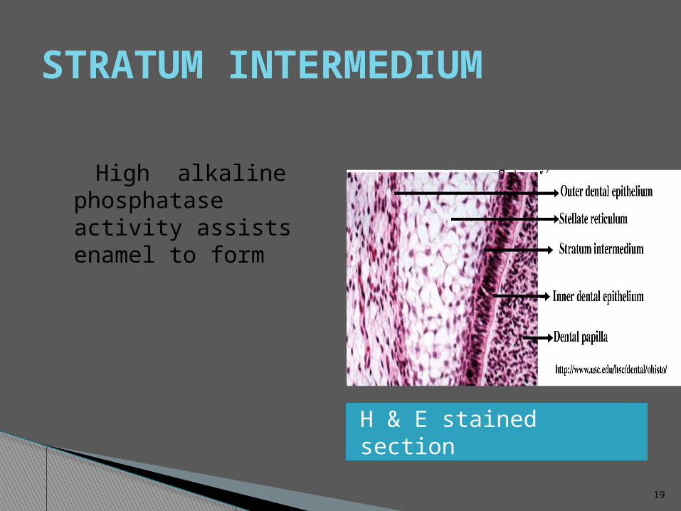
STRATUM INTERMEDIUM
H & E stained section
High alkaline phosphatase activity assists enamel to form
20
INNER ENAMEL EPITHELIUM
Short columanar to tall columnar Differentiated into pre-
ameloblast Pre-ameloblast cells provide
signal to adjacent dental papilla to differentiate into odontoblast
21
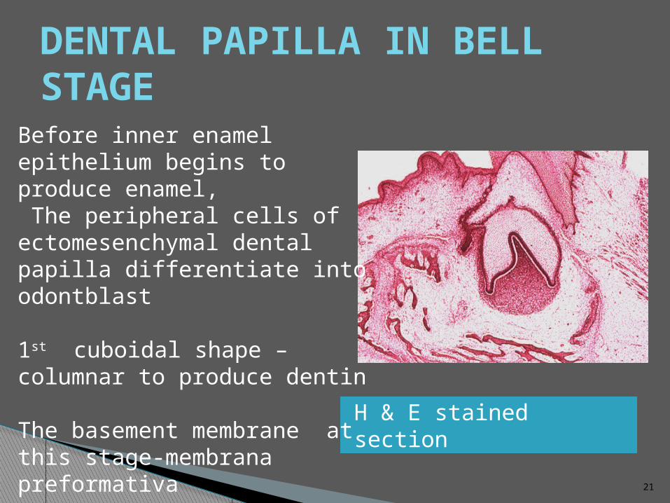
DENTAL PAPILLA IN BELL STAGE
H & E stained section
Before inner enamel epithelium begins to produce enamel, The peripheral cells of ectomesenchymal dental papilla differentiate into odontblast
1st cuboidal shape –columnar to produce dentin
The basement membrane at this stage-membrana preformativa
Cell free zone: 1-2 µm wide
22
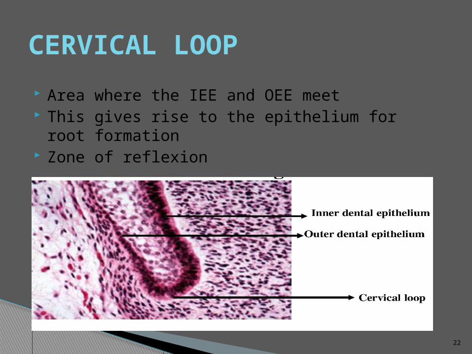
CERVICAL LOOP
Area where the IEE and OEE meet This gives rise to the epithelium for root
formation Zone of reflexion
23
ADVANCED BELL STAGE/MORPHODIFFERENTIATION
Characterized by the commencement of mineralization & root formation
For dentinogenesis and amelogenesis to takes place normally differentiating odontoblast and ameloblast will receive signals from each other called Reciprocal Induction
The boundary between the inner enamel epithelium & odontoblasts outline the future dentinoenamel junction
Formation of dentin occurs first after received signals from preameloblast.
After the first layer of dentin is formed, the ameloblasts lay down enamel over the dentin in the future incisal & cuspal areas
24
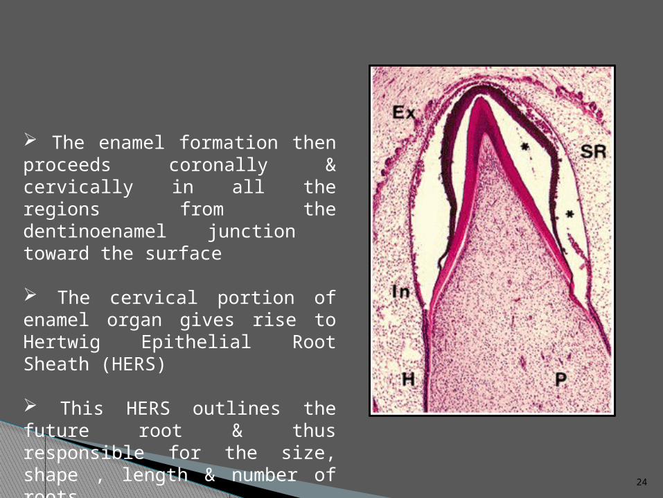
The enamel formation then proceeds coronally & cervically in all the regions from the dentinoenamel junction toward the surface
The cervical portion of enamel organ gives rise to Hertwig Epithelial Root Sheath (HERS)
This HERS outlines the future root & thus responsible for the size, shape , length & number of roots
25
FORMATION OF ENAMEL & DENTIN MATIX ( APPOSITION)
• Apposition is the deposition of the matrix of the hard enamel structures
• Appositional growth is characterised by regular & rhythmic deposition of the extracellular matrix, which is of itself incapable of further growth4 stages: Elongation of IEE Differentiation of odontoblast Formation of dentin Formation of enamel
26
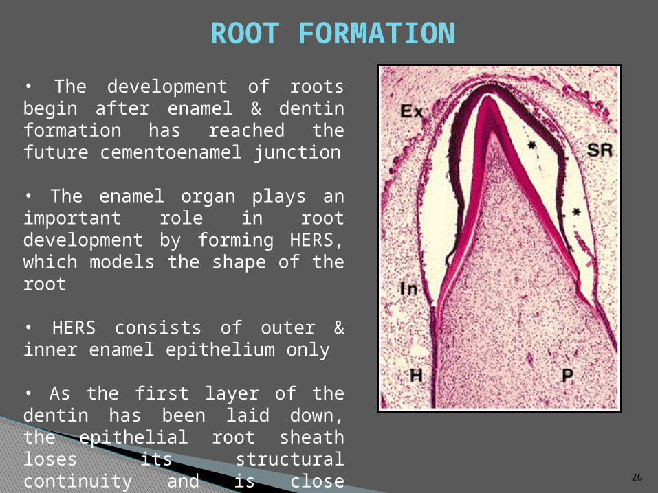
• The development of roots begin after enamel & dentin formation has reached the future cementoenamel junction
• The enamel organ plays an important role in root development by forming HERS, which models the shape of the root
• HERS consists of outer & inner enamel epithelium only
• As the first layer of the dentin has been laid down, the epithelial root sheath loses its structural continuity and is close relation to the surface of the root
ROOT FORMATION
27
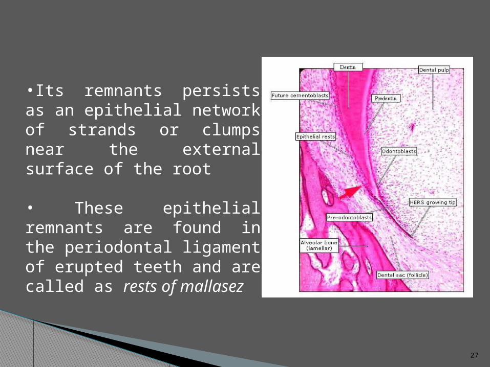
•Its remnants persists as an epithelial network of strands or clumps near the external surface of the root
• These epithelial remnants are found in the periodontal ligament of erupted teeth and are called as rests of mallasez
28
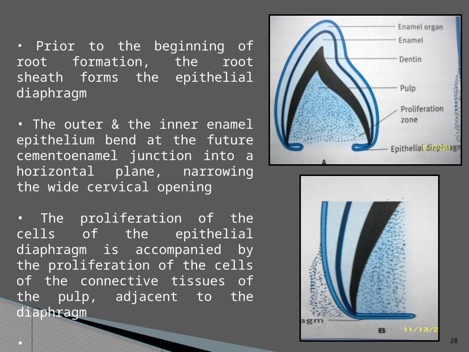
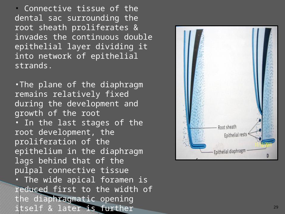
• Prior to the beginning of root formation, the root sheath forms the epithelial diaphragm
• The outer & the inner enamel epithelium bend at the future cementoenamel junction into a horizontal plane, narrowing the wide cervical opening
• The proliferation of the cells of the epithelial diaphragm is accompanied by the proliferation of the cells of the connective tissues of the pulp, adjacent to the diaphragm
•
29
• Connective tissue of the dental sac surrounding the root sheath proliferates & invades the continuous double epithelial layer dividing it into network of epithelial strands.
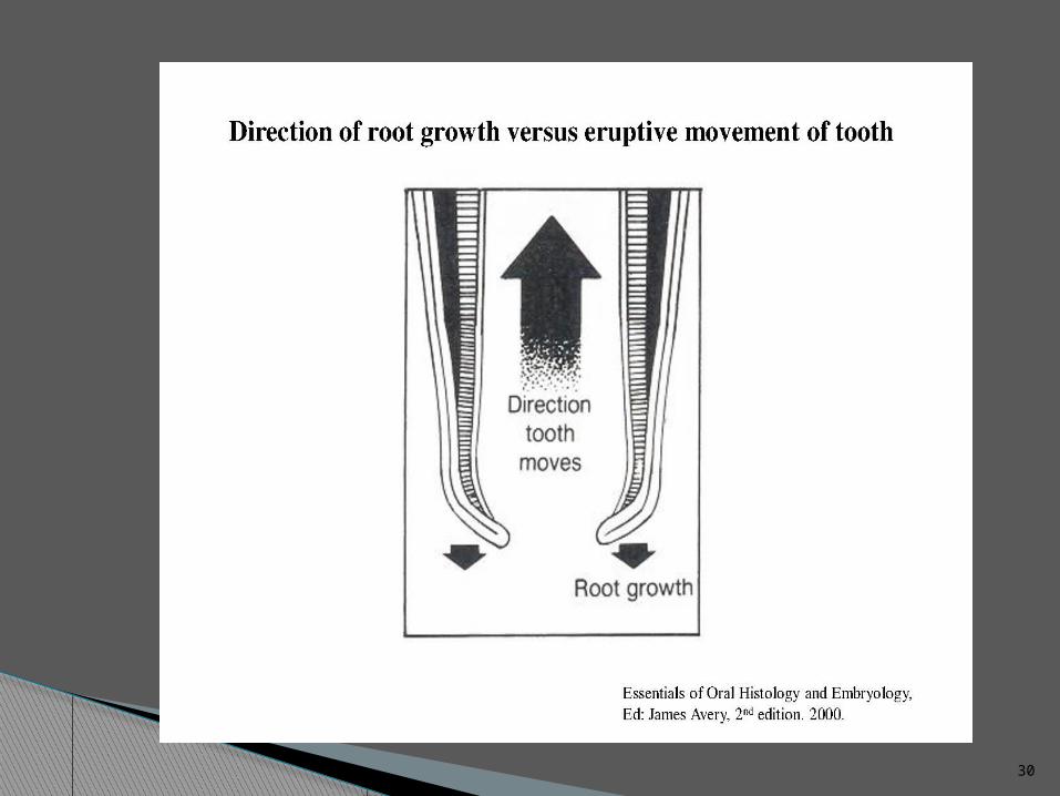
•The plane of the diaphragm remains relatively fixed during the development and growth of the root• In the last stages of the root development, the proliferation of the epithelium in the diaphragm lags behind that of the pulpal connective tissue• The wide apical foramen is reduced first to the width of the diaphragmatic opening itself & later is further narrowed by opposition of dentin & cementum to the apex of the root
30
31
32
REFERENCES
• Orban’s, Textbook of oral histology & embryology
• Ten cates, Textbook of oral histology
• Avery JK:Text book of oral Histology

33