ocular manifestations of tb infection
TRANSCRIPT
OCULAR MANIFESTATIONS OF TB
INFECTION
MD SHAHID MANZOOR

INTRODUCTION
Tuberculosis is a chronic infection caused by Mycobacterium
tuberculosis that is characterized by the formation of
necrotizing granulomas.
Tuberculos is primarily involves the lung. Other organs
including the eye may be involved secondarily. Tuberculosis
may affect any part of the eye with variable clinical features.
Ocular TB incidence ranges from 1.4 - 5.74%.

INTRODUCTION
Ocular tuberculosis is an extrapulmonary form.
Primary infection of the eye is rare.
Secondary ocular tuberculosis is the ocular involvement
as a result of haematogenous spread from a distant site or
a direct invasion from adjacent areas like skin, sinus
or cranial cavity or, as hypersensitivity response to
distant infection.
The disease is usually chronic and insidious with
exacerbations & remissions.

INTRODUCTION
Infection
1. The number and virulence of the organisms,
2. Immunity of the host.
The clinical presentation.
Direct infection by TB.
Hypersensitivity reactions to tubercular proteins.

PATHOPHYSIOLOGY
Tuberculosis is caused by M. tuberculosis, which is an
obligate aerobic, slow growing, nonspore forming,
nonmotile bacterium.
Humans are the only natural host.
It is primarily spread as an airborne aerosol which gains
access to susceptible hosts through the lung and results in
a latent or dormant infection in hosts with normally
functioning immune systems.
Usually, between 5 and 200 inhaled bacilli are needed to
cause infection in humans.
PATHOPHYSIOLOGY
Infect end organs typically having high regional oxygen
tension (apices of the lungs, kidneys, bones, meninges,
eye, and choroid).
M.tuberculosis tends to grow successfully in the choroid
and ciliary body where the oxygen tension is high
compared with other ocular structures.
The hallmark of extra-pulmonary TB is caseating
granuloma and necrosis

CLINICAL PRESENTATION
A ) ADNEXAL MANIFESTATIONS
B) ANTERIOR SEGMENT MANIFESTATION
C) POSTERIOR SEGMENT MANIFESTATIONS
D) NEURO-OPHTHALMIC MANIFESTATIONS
E) DRUG-RELATED OCULAR TOXICITY IN TB-
INFECTED PATIENTS

A) ADNEXAL MANIFESTATIONS
1) Lupus Vulgaris
2) Eyelid Tuberculous Granuloma
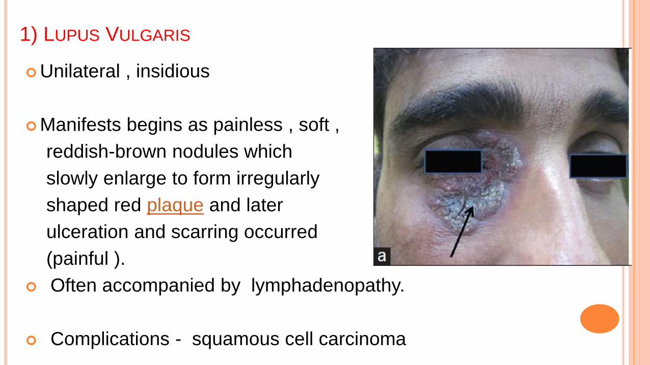
1) LUPUS VULGARIS
Unilateral , insidious
Manifests begins as painless , soft ,
reddish-brown nodules which
slowly enlarge to form irregularly
shaped red plaque and later
ulceration and scarring occurred
(painful ).
Often accompanied by lymphadenopathy.
Complications - squamous cell carcinoma

1) LUPUS VULGARIS
1) LUPUS VULGARIS
On diascopy, it shows characteristic "apple-jelly" color.
Biopsy will reveal tuberculoid granuloma with few
AF bacilli.
Mantoux test is positive.
Treatment includes ATT.

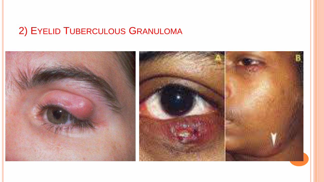
2) EYELID TUBERCULOUS GRANULOMA
Unilateral , insidious
Manifests with a violet-brown ,
non-tender, mobile nodule.
Often accompanied by
lymphadenopathy.
The nodule may ulcerate after some time and spread locally
in an irregular fashion and it is often accompanied by pain
and discharge.
Complications include trichiasis and entropion formation. .
2) EYELID TUBERCULOUS GRANULOMA

2) EYELID TUBERCULOUS GRANULOMA
The diagnosis can be difficult and it may require
biopsy in which acid fast bacilli (AFB) and the
characteristic histopathology may be seen.
The growth of Mycobacterium tuberculosis from such a
specimen remains the gold standard for the diagnosis of
TB. PCR based tests of the pathological specimens have
been proven to be useful.

B) ANTERIOR SEGMENT INVOLVEMENT
1) Tuberculous conjunctivitis.
2) Conjunctival granuloma.
3) Phlyctenular keratoconjunctivitis.
4) Tuberculous Scleritis
5) Interstitial keratitis
6) Iridocyclitis
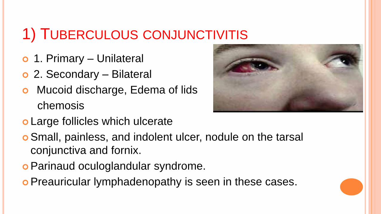
1) TUBERCULOUS CONJUNCTIVITIS
1. Primary – Unilateral
2. Secondary – Bilateral
Mucoid discharge, Edema of lids
chemosis
Large follicles which ulcerate
Small, painless, and indolent ulcer, nodule on the tarsal
conjunctiva and fornix.
Parinaud oculoglandular syndrome.
Preauricular lymphadenopathy is seen in these cases.


1) TUBERCULOUS CONJUNCTIVITIS
Diagnosis
Isolation of organism from conjunctival secretions or scrapings.
Treatment
Primary focus – Excision of conjunctiva
Topical antibiotics
Topical steroid
ATT

2) CONJUNCTIVAL GRANULOMAS
It is a Type IV Hypersensitivity reaction,
presents as an inflammatory mass on
the conjunctiva.
It is usually occurs due to tuberculosis
but can be associated with Staphylococcus aureus .
Complications include keratitis ,scleritis, corneal ulcer
Subsequent calcification of granulomas can impede vision,
and inflammation can cause irreversible damage to ocular
tissue.

3) PHYLYCTENULAR KERATOCONJUNCTIVITIS
Presents with photophobia, tearing and
blepharospasm .
Mainifests as slightly raised,
small, pinkish white or yellow nodules
surrounded by dilated vessels located
on conjunctiva near the limbus or
on peripheral cornea.
Classically, there is no clear zone between the limbus and the
lesion.

3) PHYLYCTENULAR KERATOCONJUNCTIVITIS

3) PHYLYCTENULAR KERATOCONJUNCTIVITIS
After a few days, the superficial part of the raised nodule
becomes gray and soft; the center of the lesion then
ulcerates, sloughs, and clears without scarring.
Delayed hypersensitivity reaction to mycobacterial antigens
More commonly in children, bilateral.
Responds promptly to topical application of
corticosteroids.
These ulcers are associated with neovascularization and
can go on to perforate.

4) TUBERCULOUS SCLERITIS
Mostly it presents as an anterior
scleritis while posterior scleritis
is rare .
Localized focal elevated nodules
of the sclera or Necrotizing.
The sclera may be infected by direct spread from a local
conjunctival or choroidal lesion, or more commonly by
haematogenous spread.

4) TUBERCULOUS SCLERITIS
This may undergo necrosis and may lead to scleromalacia
It does not respond to topical steroids and requires
antituberculous therapy

5) INTERSTITIAL KERATITIS
an inflammation of the corneal stroma
without primary involvement of the
epithelium or endothelium.
In most cases, the inflammation is
an immune-mediated process
triggered by an appropriate antigen.
Treatment- systemic antitubercular drugs, topical steroids
and cycloplegics


6) ANTERIOR UVEITIS
unilateral or bilateral chronic granulomatous disease
which presents with large, mutton
fat keratic precipitates,
posterior synechiae, and iris
or angle granulomas..
In severe cases, hypopyon
Iris nodules may be present near the pupillary border
(Koeppe) or on the iris surface (Bussaca).

6) ANTERIOR UVEITIS

6) ANTERIOR UVEITIS
Anterior uveitis is often accompanied by vitritis
an insidious onset and runs a chroniccourse, inevitably complicated by the development of cataract and posterior synechiae.
Administration of antituberculosis treatment can help in reducing the number of recurrences in these eyes.

INTERMEDIATE UVEITIS
Chronic, low-grade, vitritis with
snowball opacities, snow banking,
peripheral vascular sheathing,
and peripheral granuloma.
In a North Indian population, TB was found to be an
unusually common etiology of intermediate uveitis
(46.7%).


The most frequent complications related to Tb uveitis
included cystoid macular edema (40%) and cataract
(38.9%).
Other less common complications - epiretinal
membrane, raised intraocular pressure, optic disc pallor,
peripheral neovascularization, retinal
detachment and vitreous hemorrhage.
In eyes with media opacity, ultrasound biomicroscopy can
assist in detecting the presence of a granuloma in the ciliary
body region.

C) POSTERIOR SEGMENT MANIFESTATIONS
The ocular changes can be divided into four groups:
Choroidal tubercles,
Choroidal tuberculoma,
Serpiginous like choroiditis and
Subretinal abscess.

1) CHOROIDAL TUBERCLES
Most common manifestation
of intra-ocular tuberculosis and
result from hematogenous spread.
less than 5, upto 50 in number,
Unilateral or bilateral,
grayish white to yellow in color
with indistinct borders,
are located mostly in the posterior pole.

1) CHOROIDAL TUBERCLES

1) CHOROIDAL TUBERCLES
are seen in military tuberculosis and central nervous
system tuberculosis (meningitis)
On fluorescein angiography, they are hypofluorescent
during dye transit with late hyperfluorescence.
Active Choroidal tubercles usually respond well to ATT
and generally take up to 3 to 4 months to heal. On
healing, the tubercles result in pigmented and atrophic
scars.

2) CHOROIDAL TUBERCULOMA
Choroidal tubercle continues to
grow, it forms a solitary mass known as
tuberculoma
Present as a solitary, yellowish,
subretinal mass with surrounding
exudative retinal detachment ,
mimicking a choroidal tumor.
May be located anywhere
Measure from 4 to 14 mm in size and generally


2) CHOROIDAL TUBERCULOMA
May occur in immunocompetent patients and in
patients with disseminated tuberculosis, may have
overlying hemorrhages, retinal folds, and surrounding
exudative retinal detachment
On ultrasonography, these lesions are solid, elevated
masses with moderate to low internal reflectivity.
respond well to antituberculosis treatment

3) SERPIGINOUS LIKE CHOROIDITIS
It is a bilateral, chronic, progressive
and recurrent inflammation that primarily
involves the choroid and choriocapillaris
and progresses to involve the retina .
These lesions begin in the peri papillary area and spread
centrifugally.
Multifocal form where the lesions are discrete and noncontiguous
initially but later in the course may form a diffuse, contiguous
pattern.
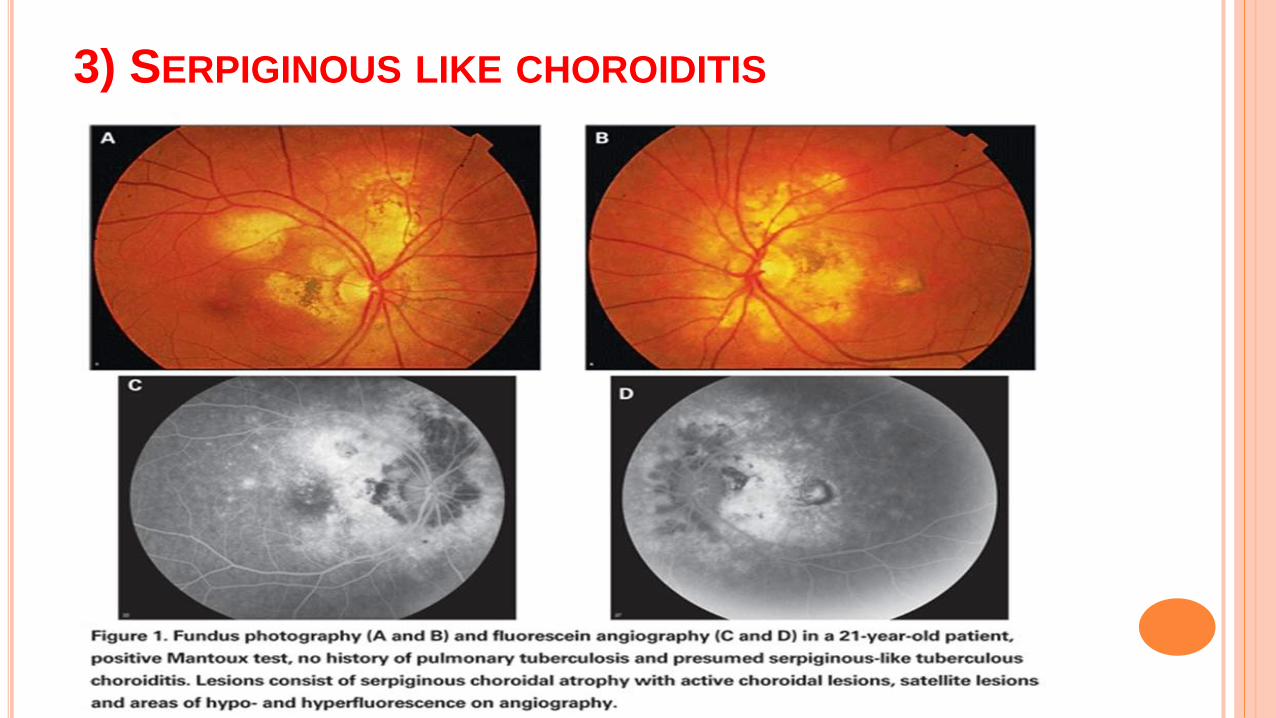
3) SERPIGINOUS LIKE CHOROIDITIS

3) SERPIGINOUS LIKE CHOROIDITIS
It may represent an immune-mediated hypersensitivity reaction
with relentless progression despite administration of systemic
corticosteroids and immunosuppressive agents.
Antituberculosis treatment in conjunction with oral
corticosteroids/immunosuppressive agents may reduce the number
of recurrences.
The healing of such lesions may lead to peripapillary
retinochoroiditis scar.
The retinochoroiditis may become extensive and may involve the
ciliary body causing cyclitis with hypotony and phthisis bulbi.
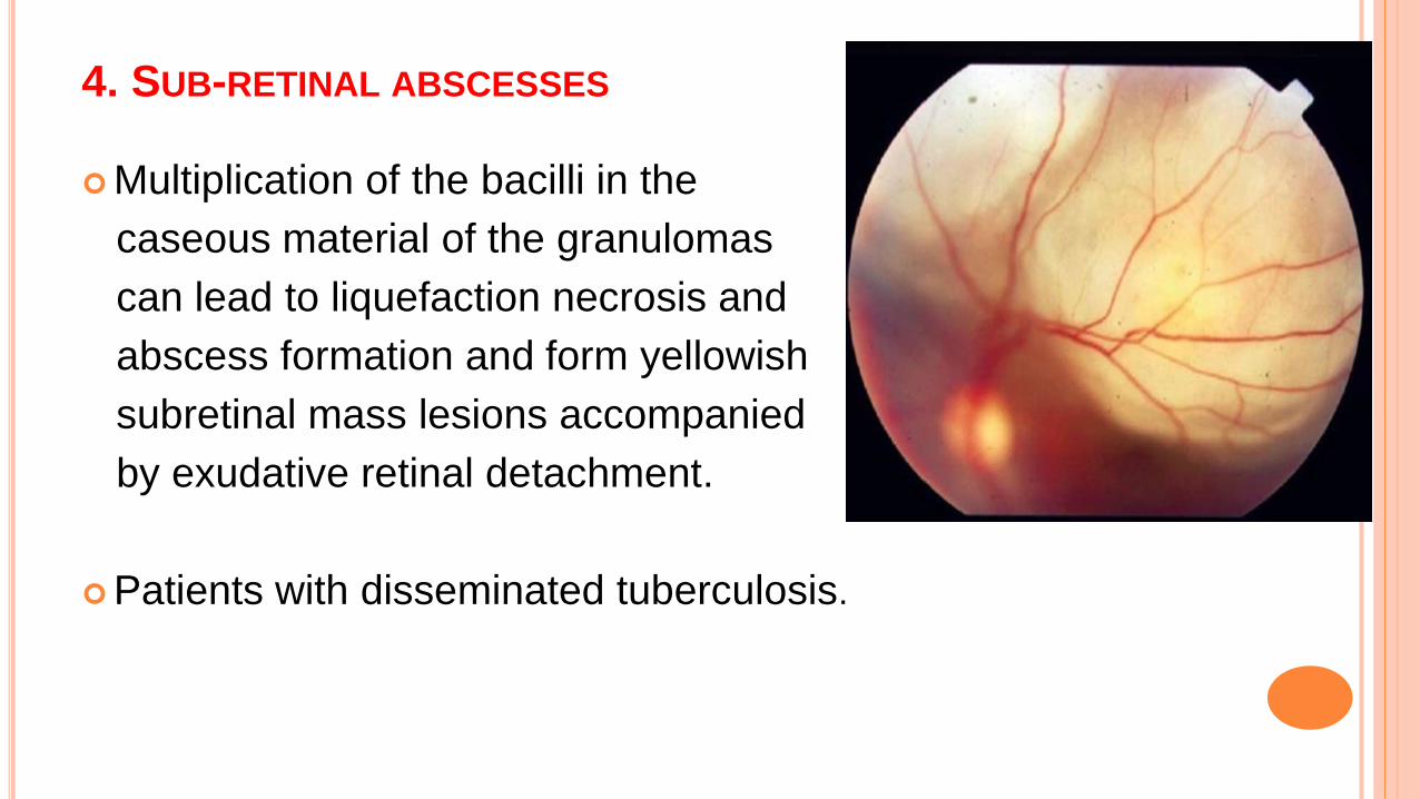
4. SUB-RETINAL ABSCESSES
Multiplication of the bacilli in the
caseous material of the granulomas
can lead to liquefaction necrosis and
abscess formation and form yellowish
subretinal mass lesions accompanied
by exudative retinal detachment.
Patients with disseminated tuberculosis.

4. SUB-RETINAL ABSCESSES

4. SUB-RETINAL ABSCESSES
Rarely, these lesions can rupture into the vitreous cavity
and may lead to endophthalmitis or panophthalmitis
usually heal with ATT and healed lesions may show
pigmentation and atrophy with chances of good visual
recovery .
Sub-retinal neo-vascularization may develop within the
scar

RETINAL VASCULITIS
in patients with tuberculosis involve
the veins or, rarely, the arteries.
The characteristic features include
bilateral, vitreous infiltrates
(vitritis), perivascular cuffing,
infiltrates, retinal haemorrhages,
perivascular choroiditis scars, neo-
vascularization and neuro-retinitis.

On FFA, extensive peripheral
capillary nonperfusion areas that lead to retinal/optic
disc neovascularization characterize
TB retinal vasculitis.
It has been long speculated that
patients of idiopathic retinal vasculitis, the so called Eales'
disease, may indeed be tuberculous retinal vasculitis.
presence of MTB DNA was demonstrated in
epiretinal membranes of patients with Eales' disease who
underwent vitreous surgery.

EALES’ DISEASE
Bilateral, idiopathic, occlusive, peripheral periphlebitis and
neovascularization.
Recurrent attacks of diminution of vision in young males –
recurrent vitreous hemorrhage
The disease is characterized by three overlapping stages:
(a) periphlebitis,
(b) occlusion and
(c) retinal neovascularization.
Mild uveitis is common

EALES’ DISEASE
(A) Peripheral vascular sheathing
and occlusion in the
superotemporal quadrant.
(A) (B) peripheral neovascularization;
(B) (C) haemorrhage from new vessels

EALES’ DISEASE
Complications include tractional retinal detachment,
rubeosis iridis, glaucoma and cataract.
.The treatment includes systemic corticosteroids,
antituberculosis treatment, laser photocoagulation of the
ischemic retina, and pars plana vitrectomy in cases of no
resolving vitreous hemorrhage or tractional retinal
detachment.
The visual prognosis is good in the majority of cases.
D) NEURO-OPHTHALMIC MANIFESTATIONS
The optic neuropathy develops either from direct
infection induced by the mycobacteria or from a
hypersensitivity to the infectious agent.
The involvement may manifest as an optic nerve
tubercle, papillitis, papilledema, optic neuritis,
retrobulbar neuritis, neuroretinitis.

• Clinical features
1. Sudden painful loss of vision
2. Vitreous haze
3. Hyperemia of the disc
4. blurring of disc margins
5. optic atrophy
• Treatment
– Systemic steroids: methyl prednisolone 1g IV
– ATT

E) DRUG-RELATED OCULAR TOXICITY IN TB-
INFECTED PATIENTS
1 )Ethambutol.
Ocular toxic effects include optic neuritis, colour vision
abnormalities and visual field defects.
Toxicity is dose- and duration-dependent; the incidence is
up to 6% at a daily dose of 25 mg/kg and rare with a daily
dose not exceeding 15 mg/kg.
Toxicity typically occurs within 3–6 months of starting
treatment.

2) Rifampicin - orange-red discoloration of tears
3) Isoniazid
Optic neuritis
Steven Johnson syndrome involving lids and
conjunctiva
4) Rifabutin used for the treatment of pulmonary
tuberculosis may in itself cause anterior uveitis .

COMPLICATIONS OF OCULAR TB
Cataract
Glaucoma
Cystoid
Macular edema
Retinal detachment
Corneal Scarring

DIFFERENTIAL DIAGNOSIS OF OCULAR TB
Infectious Disorders Noninfectious Disorders
Syphilis
Toxoplasmosis
Toxocariasis
Candidiasis
Brucellosis
Leprosy
Nocardiasis
Coccidiomycosis
Leptospirosis
Cat scratch disease
Lyme disease
Sarcoidosis
Behçet's disease
Metastasis
Tumors
Autoimmune vasculitis

DIAGNOSIS
Confirmation of the diagnosis is a challenge since
intraocular tissue or fluids are examined rarely. The
diagnosis of ocular tuberculosis has thus remained largely
presumptive and dependent on associated systemic
infection. Owing to large variations in the clinical spectrum,
it is difficult to diagnose the disease based on clinical
findings alone.
The diagnosis is typically made based on the clinical
presentation in conjunction with corroborative evidence,
direct evidence, and therapeutic response
DIAGNOSIS
I. Clinical signs
II. Ocular investigations
III. Systemic investigations
IV. Exclusion of other uveitis entities
V. Therapeutic test
VI. New diagnostic assays
I. CLINICAL SIGNS
Presence of features of any one of the following: uveitis,
cyclitis ,choroiditis retinitis ,retinal vasculitis, neuro-
retinitis ,optic neuropathy, endophthalmitis and pan-
ophthalmitis
An intractable disease course with multiple recurrences
on nonspecific treatment (corticosteroids) is a clue
suggesting a possible tubercular etiology.

II. OCULAR INVESTIGATIONS
a. Demonstration of AFB by microscope or culture
of M tuberculosis from the intraocular
fluid/tissue-media.
b. Positive polymerase chain reaction from
intraocular fluids for IS 6110 or other
conserved sequences in M.tuberculosis genome.

III. SYSTEMIC INVESTIGATIONS
a. Positive Mantoux reaction
b. Evidence of healed or active tubercular lesion on
radiography of the chest.
c. Evidence of confirmed active extrapulmonary
tuberculosis (either by microscopic examination or by
culture of the affected tissue for M tuberculosis).

IV. EXCLUSION OF OTHER UVEITIS ENTITIES
Other causes of uveitis must be excluded by various
laboratory investigations including serology for syphilis,
toxoplasmosis, sarcoidosis, and others.

V. THERAPEUTIC TEST
A positive response to 4-drug ATT (isoniazid, rifampicin,
ethambutol, and pyrazinamide) over a period of 4 to 6
weeks can be diagnostic.
Therapeutic trial with single drug isoniazid should be
avoided due to risk of development of resistance.

NEW DIAGNOSTIC ASSAYS
Interferon-g release assays (IGRA)
It based on the in vitro assays that measure interferon-g released
by sensitized T cells after stimulation by Mycobacterium
tuberculosis antigens.
Two kits are available commercially:
TSPOT. TB test (Oxford Immunotec Ltd, Oxford, UK) and the
QuantiFERON —TB GOLD (QFTG: Cellestis Ltd, Carnegie, Australia)

TREATMENT
The treatment of tuberculosis is complex ,high levels of
patient adherence are required.
Inappropriate management can result in life-threatening
consequences as well as drug resistance.
A multiple drug regimen is recommended to avoid
resistance.

Addition of ATT to corticosteroids in uveitis patients with
latent/manifest TB also leads to significant reduction in
recurrences of uveitis (Bansal et al 2008).
Systemic corticosteroids used for the first 4–6 weeks,
together with ATT, may limit damage to ocular tissues caused
from delayed type hypersensitivity.
However, one should avoid using corticosteroids alone
without concomitant ATT as the corticosteroids may promote
multiplication of bacilli, leading to panophthalmitis or they
may cause a flare-up of systemic tuberculosis by activating a
latent infection.

SYSTEMIC SIDE EFFECTS OF ATT
Close follow-up and monitoring of the liver function tests
and renal function are also mandatory.

ISONIAZID
Hepatotoxicity
Elderly, slow acetylators more prone
Polyneuropathy
Prevented by concurrent pyridoxine
Rashes, acne
Heamatological –haemolytic anaemia in G6PD deficiency
RIFAMPICIN
Mild elevation of liver enzymes – common
Rashes, hepatotoxicity, thrombocytopenia
Orange discoloration of urine, sweat, tears
Potent CYP-P450 inducer- reduce the serum level of
drugs warfarin, oestrogen but can potentiate the action
of neuromuscular blocking agents

PYRAZINAMIDE
GI disturbances
Hepatotoxicity
Hyperuricaemia – gout
Arthralgia

SUMMARY
Incidence of ocular tuberculosis – 1.4%
• Involves all the ocular structures except lens
• Most common manifestation
Chroiditis, Anterior Uveitis, sclerokeratitis.
• Consider the diagnosis of TB especially in patients presenting with
occlusive retinal vasculitis & choroiditis.
• All patients on ATT should be screened for ocular toxicity