occurrence and characteristics of group 1 introns found at three
TRANSCRIPT
RESEARCH ARTICLE Open Access
Occurrence and characteristics of group 1 intronsfound at three different positions within the 28Sribosomal RNA gene of the dematiaceousPhialophora verrucosa: phylogenetic andsecondary structural implicationsKayoko Takizawa1*, Toko Hashizume2 and Katsuhiko Kamei1
Abstract
Background: Group 1 introns (ribozymes) are among the most ancient and have the broadest phylogeneticdistribution among the known self-splicing ribozymes. Fungi are known to be rich in rDNA group 1 introns. In thepresent study, five sequences of the 28S ribosomal RNA gene (rDNA) regions of pathogenic dematiaceousPhialophora verrucosa were analyzed using PCR by site-specific primers and were found to have three insertions,termed intron-F, G and H, at three positions of the gene. We investigated the distribution of group 1 introns in thisfungus by surveying 34 strains of P. verrucosa and seven strains of Phialophora americana as the allied species.
Results: Intron-F’s (inserted at L798 position) were found in 88% of P. verrucosa strains, while intron-G’s (inserted atL1921) at 12% and intron-H’s (inserted at L2563) at 18%. There was some correlation between intron distributionand geographic location. In addition, we confirmed that the three kinds of introns are group 1 introns from resultsof BLAST search, alignment analysis and Reverse Transcriptase-Polymerase Chain Reaction (RT-PCR). Prediction ofsecondary structures and phylogenetic analysis of intron sequences identified introns-F and G as belonging tosubgroup IC1. In addition, intron-H was identified as IE.
Conclusion: The three intron insertions and their insertion position in the 28S rDNA allowed the characterizationof the clinical and environmental isolates of P. verrucosa and P. americana into five genotypes. All subgroups ofintrons-F and G and intron-H were characterized and observed for the first time in both species.
BackgroundThe type species Phialophora verrucosa was described byMedlar in 1915 [1] when he isolated the fungus from ahuman skin disease. The species is ubiquitous and cos-mopolitan, and are important plant saprobes as well ashuman pathogens. Identification is based on conidialontogeny and molecular systematics. Few studies invol-ving molecular genotyping techniques have beenreported for P. verrucosa. A study analyzed restrictionfragment length polymorphisms (RFLP) of mitochondrial
DNA to determine genetic variations and phylogeneticrelationships among P. verrucosa strains [2].Different molecular typing tools, such as random
amplification of polymorphic DNA (RAPD), RFLP,pulsed-field gel electrophoresis (PFGE), multilocusenzyme electrophoresis (MLEE) and multilocussequence typing (MLST), have been developed to pro-vide a better understanding of the molecular epidemiol-ogy of fungal pathogens, e.g., Candida albicans [3-5]and Aspergillus fumigatus [6,7] and medically importantfilamentous fungi [8]. However, although the majority ofthe reported group 1 intron sequences have been foundin a wide range of fungi (Comparative RNA Web[CRW] site: http://www.rna.ccbb.utexas.edu/[9], few stu-dies about sequence and structure variation, distribution
* Correspondence: [email protected] Mycology Research Center, Chiba University, 1-8-1 Inohana, Chuo-ku, Chiba, 260-8673, JapanFull list of author information is available at the end of the article
Takizawa et al. BMC Microbiology 2011, 11:94http://www.biomedcentral.com/1471-2180/11/94
© 2011 Takizawa et al; licensee BioMed Central Ltd. This is an Open Access article distributed under the terms of the CreativeCommons Attribution License (http://creativecommons.org/licenses/by/2.0), which permits unrestricted use, distribution, andreproduction in any medium, provided the original work is properly cited.

and phylogenetic relationships of introns from a singlespecies have been performed in detail. We focused ongroup 1 introns within 28S rDNA from P. verrucosa toevaluate the prevalence of intron polymorphism at thestrain level.As the first step to determine intron sequence diver-
gence, sequences of 28S rDNA of five representativestrains of P. verrucosa were analyzed to find insertions.Based on these five sequences, site-specific primers weredesigned for use in PCR to detect insertions on other P.verrucosa and P. americana strains studied, in order toinvestigate incidence and distribution of insertions.Moreover, to characterize the insertions, we analyzedthe phylogeny of the introns found in this study andpredicted their secondary structures.
ResultsNucleotide structure of P. verrucosa 28S sequences andthe characterization intronic insertionSince the sequence information of the 28S region wasnot available in public databases, we sequenced the28S, including ITS, regions of five representativestrains (Table 1) of P. verrucosa. Alignment of the fiveP. verrucosa rDNA sequences revealed the nucleotidesizes of 18S, ITS and 28S and the location of intron-Fand G insertions (see Table 2 and Additional file 1). Itwas found that the sequences were composed of 29nucleotides of partial 18S, 534-535 nucleotides of ITSand from 3349 to 4133 nucleotides of 28S regions.These genomic sequences were deposited at DDBJ andaccession numbers are listed in Table 2. Twenty-fiveand two nucleotide substitutions were found withinthe ITS and D1D2 regions, respectively. The two bpsubstitutions in D1D2 were located at 1036 and 1042nucleotide positions in the 28S region. These poly-morphisms were confirmed in the five strains of P. ver-rucosa strains except for the two insertions in intron-Fand G at 924 and 2239 positions of the 28S, respec-tively. None of the insertions were found in the Yaostrain. BLAST comparisons showed with relatively highhomology values that the sequences of both insertionswere individually homogeneous to group 1 introns inthe database. The positions of insertion for intron-Fand G correspond to nucleotide positions 798 and1921 of Escherichia coli 23S nucleotide sequenceaccession number J01695 [10]. In addition, althoughthese five strains of P. verrucosa did not possessintron-H, this region of PV28 strain was amplifiedwith the site-specific primer pair (in Table 3) designedfrom the sequences obtained from another experiment(data not shown) and a 403-nucleotide insert repre-senting intron-H was sequenced. The insertion posi-tions of the intron-H correspond to nucleotidepositions 2563 of E. coli 23S nucleotide sequence.
Survey of insertions of P. verrucosa and P. americanaWe amplified intron insertion regions using site-specificprimer pairs we have designed for intron-F (inF-F andinF-R), intron-G (inG-F and inG-R) and intron-H(L2563F and L2563R), within the 28S region (Table 3).These primer pairs were used to screen and detect PCRamplicons for insertion regions within 34 P. verrucosaand seven P. americana strains. Amplicons were elutedin agarose gel to gain information regarding the introninsertions. No-insertion amplicons for intron-F andintron-G primers were in the size 142 and185 bps,respectively. When insertions were present, intron-F pri-mer pair yielded amplicons in the size range from 531to 533 bps, and intron-Gs in the size 575 or 578 bps.Moreover, amplicons of about 643 bps for intron-Hswere also eluted. It was revealed that there were 30intron-F’s, four intron-G’s and six intron-H’s within P.verrucosa and only two intron-Fs within P. americanaas shown in Table 1. There was some correlationbetween intron distribution of P. verrucosa and geo-graphic location, i.e., intron-Fs were found to have pre-valence of 88% in P. verrucosa and intron-Hs werefound specifically in the South American Continent. Nointrons were found except for two intron-Fs in P. ameri-cana. In addition, the agarose gel profiles allowed us tocharacterize genotypes and distribution frequencies ofinsertions from P. verrucosa including no-insertion asshown in Table 1. It was found that occurrence of geno-types F, FG, FH, FGH and N were at 64, 6, 12, 6 and12%, respectively.
Characterization of the P. verrucosa intronic insertionRT-PCR was carried out to identify the property ofthese insertions, namely, whether they are introns orunusual extensions incorporated into mature rRNA.Four representative strains were selected among the 41strains surveyed. And it was found that two strains (PV1and PV3) had two introns individually, while the othertwo strains (PV2 and PV41) had only one intron asshown in Figure 1. Insertions of strain PV1 and PV3were eluted at 142 bps on lane 2 and 3 with intron-Fprimer pair, and 185 bps on lane 4 and 5 with intron-Gprimer pair, respectively. PV2 and PV41 exhibited 142bps amplicons with intron-F primer pair as shown onlane 15 and 16, respectively. An intron-lacking Yaostrain gave 142 and 192 bps amplicons with intron-Fand G primer pairs on lane 10 and 11, respectively. Theother lanes; namely, 6, 7, 8, 9, 13 and 14 show PCR pro-ducts of genomic DNA as templates and lane 12 isnegative control. These shows that all the insertionswere excised after cDNA was transcribed, indicatingthat they were, as predicted, actively spliced introns.These results point to the possibility that these inser-tions are group 1 introns.
Takizawa et al. BMC Microbiology 2011, 11:94http://www.biomedcentral.com/1471-2180/11/94
Page 2 of 12

Moreover, we analyzed sequences of the splicedintrons to confirm the boundaries of exon and intronsequences. The last nucleotide of the upstream exonwas confirmed to be a T (U in RNA) and the lastnucleotide of the intron was a G, consistent with group1 introns [11,12].
Phylogenetic relationships of introns F and G of P.verrucosaSequences of intron-F and G of ten P. verrucosa strainswere sequenced and it was found that DNA sequencepolymorphisms exist among the two introns, i.e., theintron-Fs ranged in the size from 389 to 391 bps and
Table 1 Thirty-four strains of P. verrucosa and seven strains of P. americana examined in this study
Strain Isolation source Locality Insertion Genotype Sample ID
Intron-F Intron-G Intron-H
IFM 4928 Human Japan + + - FG PV1
IFM 5089 Human Japan + - - F PV2
IFM 51934 Human China - - - N Yao
IFM 41779 Human China + + - FG PV3
IFM 41750 Bark China + - - F TH9
IFM41710 Corn China + - na F (na)
IFM 41780 Human China + - - F
IFM 41721 Rotten wood China + - - F
IFM 41724 Rotten wood China + - - F
IFM 41725 Straw China + - - F
IFM 41739 Bark China + - - F
IFM 41740 Bark China + - - F
IFM 41746 Straw China + - - F
IFM 41749 Bark China + - - F
IFM 41752 Bark China + - - F
IFM 41764 Soil China + - - F
IFM 41755 Unknown China + - - F
IFM 41765 Soil China + - - F
IFM 41778 Straw China + - - F
IFM 41872 Soil Colombia - - - N
IFM 41871 Soil Colombia + - - F PV41
IFM 41879 Soil Colombia + - + FH
IFM 41881 Soil Colombia + - - F
IFM 41885 Soil Colombia + - - F
IFM 41884 Cactus Venezuela - - - N
IFM 41888 Rotten wood Venezuela - - - N
IFM 41887 Rotten wood Venezuela + - + FH PV28
IFM 41886 Cactus Venezuela + - + FH TH31
IFM 41892 Soil Venezuela + - + FH TH35
IFM 41883 Soil Venezuela + - - F
IFM 41893 Soil Venezuela + - - F
IFM 41897 Soil Brazil + + + FGH PV33
IFM 41898 Soil Brazil + + + FGH PV34
IFM 41899 Soil Brazil + - - F
*CBS 273.37 Human Brazil - - - N
*CBS 400.67 Soil Brazil - - - N
*CBS 281.35 Human USA - - - N
*CBS 220.97 Linden tree USA - - - N
*CBS 840.69 Decaying timber Finland - - - N
*CBS 221.97 Unknown Uruguay + - - F
*CBS 223.97 Human USA + - - F
*: P. americana, +: with insertion, -: no insertion, na: not analized.
Takizawa et al. BMC Microbiology 2011, 11:94http://www.biomedcentral.com/1471-2180/11/94
Page 3 of 12
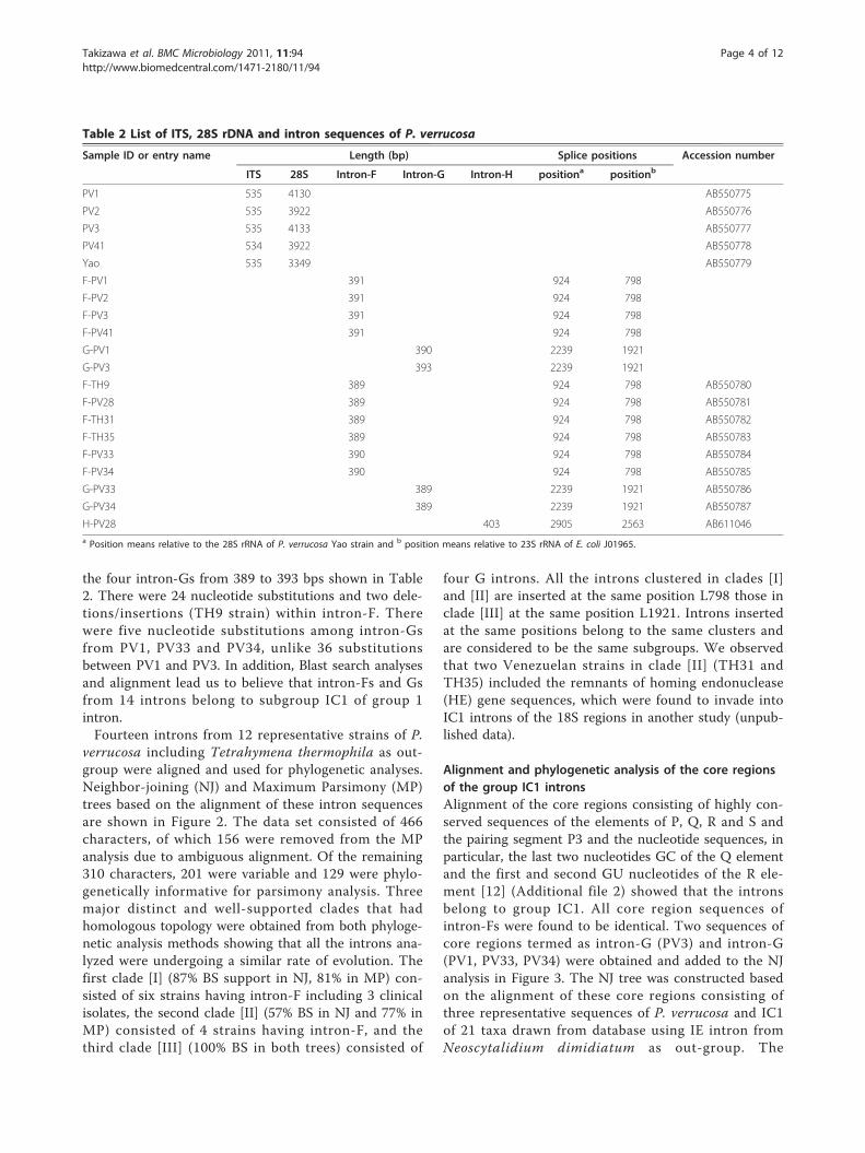
the four intron-Gs from 389 to 393 bps shown in Table2. There were 24 nucleotide substitutions and two dele-tions/insertions (TH9 strain) within intron-F. Therewere five nucleotide substitutions among intron-Gsfrom PV1, PV33 and PV34, unlike 36 substitutionsbetween PV1 and PV3. In addition, Blast search analysesand alignment lead us to believe that intron-Fs and Gsfrom 14 introns belong to subgroup IC1 of group 1intron.Fourteen introns from 12 representative strains of P.
verrucosa including Tetrahymena thermophila as out-group were aligned and used for phylogenetic analyses.Neighbor-joining (NJ) and Maximum Parsimony (MP)trees based on the alignment of these intron sequencesare shown in Figure 2. The data set consisted of 466characters, of which 156 were removed from the MPanalysis due to ambiguous alignment. Of the remaining310 characters, 201 were variable and 129 were phylo-genetically informative for parsimony analysis. Threemajor distinct and well-supported clades that hadhomologous topology were obtained from both phyloge-netic analysis methods showing that all the introns ana-lyzed were undergoing a similar rate of evolution. Thefirst clade [I] (87% BS support in NJ, 81% in MP) con-sisted of six strains having intron-F including 3 clinicalisolates, the second clade [II] (57% BS in NJ and 77% inMP) consisted of 4 strains having intron-F, and thethird clade [III] (100% BS in both trees) consisted of
four G introns. All the introns clustered in clades [I]and [II] are inserted at the same position L798 those inclade [III] at the same position L1921. Introns insertedat the same positions belong to the same clusters andare considered to be the same subgroups. We observedthat two Venezuelan strains in clade [II] (TH31 andTH35) included the remnants of homing endonuclease(HE) gene sequences, which were found to invade intoIC1 introns of the 18S regions in another study (unpub-lished data).
Alignment and phylogenetic analysis of the core regionsof the group IC1 intronsAlignment of the core regions consisting of highly con-served sequences of the elements of P, Q, R and S andthe pairing segment P3 and the nucleotide sequences, inparticular, the last two nucleotides GC of the Q elementand the first and second GU nucleotides of the R ele-ment [12] (Additional file 2) showed that the intronsbelong to group IC1. All core region sequences ofintron-Fs were found to be identical. Two sequences ofcore regions termed as intron-G (PV3) and intron-G(PV1, PV33, PV34) were obtained and added to the NJanalysis in Figure 3. The NJ tree was constructed basedon the alignment of these core regions consisting ofthree representative sequences of P. verrucosa and IC1of 21 taxa drawn from database using IE intron fromNeoscytalidium dimidiatum as out-group. The
Table 2 List of ITS, 28S rDNA and intron sequences of P. verrucosa
Sample ID or entry name Length (bp) Splice positions Accession number
ITS 28S Intron-F Intron-G Intron-H positiona positionb
PV1 535 4130 AB550775
PV2 535 3922 AB550776
PV3 535 4133 AB550777
PV41 534 3922 AB550778
Yao 535 3349 AB550779
F-PV1 391 924 798
F-PV2 391 924 798
F-PV3 391 924 798
F-PV41 391 924 798
G-PV1 390 2239 1921
G-PV3 393 2239 1921
F-TH9 389 924 798 AB550780
F-PV28 389 924 798 AB550781
F-TH31 389 924 798 AB550782
F-TH35 389 924 798 AB550783
F-PV33 390 924 798 AB550784
F-PV34 390 924 798 AB550785
G-PV33 389 2239 1921 AB550786
G-PV34 389 2239 1921 AB550787
H-PV28 403 2905 2563 AB611046a Position means relative to the 28S rRNA of P. verrucosa Yao strain and b position means relative to 23S rRNA of E. coli J01965.
Takizawa et al. BMC Microbiology 2011, 11:94http://www.biomedcentral.com/1471-2180/11/94
Page 4 of 12

phylogeny of intron-F and G formed separate clades asshown in Figure 3, and indicated that both introns werelikely acquired independently. Indeed, all intron-Fs werefound to be closely related to Myriosclerotinia ciboriumand Sclerotinia tetraspora introns which are located atL798. Two sequences of intron-G located at L1921 weregrouped together with 85% BS value and found to be onthe neighboring clade with Cordyceps prolifica intronlocated at L1921. The phylogenetic tree suggests thatboth introns may be inserted prior to the divergence ofthe species formerly belonging to clade [IV] and [V].Collectively, this tree displays that all introns of P. ver-rucosa generated by the core regions are members ofsubgroup IC1s.
Modeling of the P. verrucosa insertions revealed that theywere group IC1 intronsThe predicted secondary structure of the intron-F and Gwere constructed as follows. The conserved P, Q, R and Sregions of intron-F (L798) from PV1 were initially alignedwith the same regions from other taxa, and then regions
of P1 through P10 were constructed and added on thebasis of the secondary structure model as shown in Figure4[A][13]. The P10 region was formed using the internalguide sequence (IGS). The catalytic core was defined by aset of structurally conserved elements, including elementsP3 to P8. A G-C pair within P7, i.e. G391-C277 of intron-F was assumed to be G-binding positions [14]. ExtendedP5 and P9 stems were displayed in the putative structureof intron-F from PV1. Nine intron-Fs from nine strains(PV2, 3, 28, 33, 34 and 41 and TH9, 31 and 35) of P. verru-cosa were predicted to be the same structures as the puta-tive structure of intron-F derived from PV1 drawn inFigure 4[A], alternatively, shown in Additional file 3.These nucleotide variations among intron-F were observedmainly in the loop and at four positions where one nucleo-tide of P5a, two of P5.1a and one of P5.2 stem were posi-tioned. The base pairs GU and CG within P6 were formedin the core region of intron-F [12]. The nucleotides A71,A72, U73 were located in segments J3/4 of PV1 intron-F[15-18]. These predictions of secondary structure revealedthat all intron-Fs were IC1 group 1 introns.
Table 3 Primers used for the amplification and sequencing of P. verrucosa
Primer Sequence (5’-3’) 5’ position* Source 5’ position including ITS
ITS1 TCCGTAGGTGAACCTGCGG -563 White TJ, et al. [48] 1
ITS3 GCATCGATGAAGAACGCAGC -309 White TJ, et al. [48] 255
NL1 GCATATCAATAAGCGGAGGAAA 39 O’Donnell K [49] 603
3PV26 CCGTCTTGAAACACGGACC 633 This work 1197
inFG-F CCGAAAGATGGTGAACTATGCC 795 This work 1359
inF-F ACGTGCAAATCGATCGTCAA 868 This work 1432
inF-R CAAGGCCTCTAATCATTCGCT 1009 This work 1573
8PV26 GAACCTTTCCCCACTTCAG 1487 This work 2051
11PV26 AAGCCATAGGGAAGTTCCGT 1525 This work 2089
9PV26 GTCGTACTCATAACCGCAG 1818 This work 2382
CA-INT-L ATAAGGGAAGTCGGCAAAATAGATCCGTAA 1881 McCullough MJ, et al. [50] 2445
2PV26 TCCCGAAGTTACGGATCTA 1918 This work 2482
16PV26 CCCAACCCTTAGAGCCAATC 1942 This work 2506
10PV26 CCGTACCAGTTCTAAGTTG 2089 This work 2653
inG-F GATGGCCAGAAAGTGGTGTTG 2130 This work 2694
inG-R TAGGGACAGTGGGAATCTCGT 2314 This work 2878
26S-INT3 CTAGCGAAACCACAGCCAAG 2323 This work 2887
CA-INT-R CCTTGGCTGTGGTTTCGCTAGATAGTAGAT 2343 McCullough MJ, et al. [50] 2907
inFG-R GCTCTCCCACCTATTCTACACC 2445 This work 3009
12PV26 TGGTATTTCACCGGCGATTG 2464 This work 3028
IGS1-M-R CTGCCACAAGCCAGTTATC 2805 This work 3369
IGS3-M CTTCGATGTCGGCTCTTCCT 2838 This work 3402
IGS-L TAGTACGAGAGGAACCGT 2991 Williamson ECM et al. [51] 3555
3IGS-PV TCTAAGTCAGAATCCGTGCCG 3090 This work 3654
5IGS1-PV ACGAGCTACTGAGCGTAAG 3318 This work 3882
6IGS-PV GACCACAGTCAGGCTTACG 3349 This work 3913
L2563 F CACAGGGATAACTGGCTTGTGG 2781 This work 3345
L2563R ATCTGAATCAACGGTTCCTCTCG 3018 This work 3582
* The 5’ position is relative to the 28S rDNA sequence of the P. verrucosa Yao strain.
Takizawa et al. BMC Microbiology 2011, 11:94http://www.biomedcentral.com/1471-2180/11/94
Page 5 of 12

The structure of intron-G (L1921) from PV1 wasdrawn just as was done for intron-Fs (Figure 4[B]). A G-C pair within P7, i.e. G390-C360, was assumed to be theG-binding positions. The GU-CG pair of P6 and theAAU in J3/4 was the same as in the intron-F coreregion of PV1. This putative intron-G exhibitedexpanded regions of P1 and P5. The three intron-Gs ofPV1, PV33 and PV34 were found to be similar amongthe three strains. Different features were found in PV3as shown in Figure 4[C] wherein the sequence of PV3differed in P1 region among four trains; namely, shortstems in P1b and P1c and small bulge loops of L1 andL1a (Additional file 4). Moreover, PV3 added P2.0 andP8c, although the other intron-Gs did not. Predictionstructures in the remaining two introns of PV33 andPV34 are not shown. Nevertheless, all subgroups ofintron-G were also identified as IC1, based on
comparison of tertiary structures across segments P3-7of the four strains.In conclusion, we have identified that the ten intron-
Fs and four intron-Gs of P. verrucosa belong to IC1group 1 introns.
Characterization of intron-HLoss of P5abcd domain in derived S788 introns was cor-related with inability to self-splice in vitro in a previousreport [19]. Accordingly, we have not confirmed inser-tion positions of intron-H by RT-PCR. However, weexamined PV-28 strain as the representative strain ofintron-H by analyzing the sequence alignment of thecore region of subgroup IE from other organisms in thedatabase. Moreover, we predicted the secondary struc-ture of this intron-H as shown in Figure 5. The second-ary structure modeling revealed distinctive IE sequences,namely, the non-expanding P5 and the expanding P9regions.
DiscussionTo date, although a variety of introns from eukaryoteshave been described in the rRNA gene loci of fungi [9],few introns in Phialophora species have been reported.An unusually small group 1 intron of 67 bps from thenuclear 18S rDNA has been described in a splicingstudy of Capronia semiimmersa, a teleomorph of P.americana which is known to be similar to P. verrucosa[20-22]. These small introns contain only P1, P7 and
Neoscytalidium dimidiatum AF258604, S1199 [A] --- IE --- outgroup
Acanthamoeba griffini AGU02540, S516 [Ac]
Exophiala calicioides AB007685, S1767* [A]
Physarum polycephalum L03183, L1925 [M]
Tetrahymena thermophila J01235=V01416, L1925 [C]
Dunaliella parva M62998, S1512 [C]
Naegleria sp. AJ001314, L1949 [H]
Cordyceps prolifica AB044640, L1921 [A]
Intron-G (PV1, PV33, PV34), L1921 [A]
Intron-G (PV3), L1921 [A]
Myriosclerotinia scirpicola AJ226075, S788 [A]
Myriosclerotinia ciborium AJ2260084, L798 [A]
Sclerotinia tetraspora AJ226090, L798 [A]
Plasmodiophora brassicae PBU18981, S943 [P]
Fusarium solani AF150487, S943 [A]
Isaria japonica AB016607, S943 [A]
Neoscytalidium dimidiatum AY307355, S943 [A]
Exophiala dermatitidis Z75304, S1165* [A]
Ajellomyces capsulatus Z75305, S943 [A]
Cadophora gregata AF056487, S1506 [A]
Cenococcum geophilum Z48534, S1506 [A]
Penicillium pulvillorum AF178517, S1511 [A]
Porphyra tenera AB029880, S516 [R]
Intron-F, L798 [A]
0.1 substitutions/site
73
6551
8560
80
[IV]
[V]
Figure 3 Phylogenetic tree of IC1 intron based on elements P,Q, R, S and a segment of P3. Numerals at each node arebootstrap probabilities from NJ analysis. Insertion positions are givenafter the sample ID or accession number. * indicates the insertionposition relative to the 18S rDNA of the S. cerevisiae sequence.
1 2 3 4 5 6 7 8 9 10 11 12 13 14 15 16 17
Mar
ker
PV1
PV3
PV1
PV3
PV1
PV3
PV1
PV3
Yao
Yao
PV2
PV41
PV2
PV41
Mar
ker
1,000bp
500bp
100bp
Figure 1 Amplification pattern by RT-PCR with the site-specificprimer pairs for intron-F and G. PCR products of from cDNAamplified with the primers inF-F and inF-R are eluted in lanes 2, 3,15 and 16, and with primers inG-F and inG-R in lanes 4 and 5. PCRproducts from genomic DNA amplified with primer pair for intron-Fare eluted in lanes 6, 7, 10, 13 and 14, and with primer pair forintron-G in lanes 8, 9 and 11. Lane 12 is the negative control.
10 changes
Tetrahymena thermophila
inG_PV3 [China*]
inG_PV1 [Japan*]
inG_PV34 [Brazil]
inG_PV33 [Brazil]
inF_TH35 [Venezuela]
inF_TH31 [Venezuela]
inF_PV28 [Venezuela]
inF_TH9 [China]
inF_PV34 [Brazil]
inF_PV33 [Brazil]
inF_PV2 [Japan*]
inF_PV41 [Colombia]
inF_PV3 [China*]
inF_PV1 [Japan*]
10097
86
99
77
100
81
55
55
A
0.05 substitutions/site
87
87
89 100
57
100
100
65
78
B
[I]
[II]
[III]
Figure 2 Phylogenetic relationship of intron-F and G within28S of P. verrucosa. The trees were generated using MP (A) andNJ (B). One of three equally MP trees (tree length = 353,consistency index (CI) = 0.9575, homoplasy index (HI) = 0.0425, CIexcluding uninformative characters = 0.9268, HI uninformativecharacters = 0.0732, retention index = 0.9679, rescaled consistencyindex = 0.9268). * indicates a clinical isolate of P. verrucosa.
Takizawa et al. BMC Microbiology 2011, 11:94http://www.biomedcentral.com/1471-2180/11/94
Page 6 of 12

[C]
A -U U C -G
G -U -G -G -
CC -C -U -C -
G -C -G -G -
P7 P3
P8
CAA C
GC
UUC
C
GG
P8a
P8b
UGGGU
CACCUGGAG
CGCC
P8c5'
P1c
U - a
C - gU - aG - cA - u
A - uP1
3'a -u -
c -u
G - cU G
P10
P1a
P1b
G - C
G G
A
A
A A
G
C A
C - GU - AG - CACGG
CAA
u
G UG - C
G - CG - C
G A
U AA U
P9.0
[L1b]
[L1]
[L1a]
[L1c]
CUGUCCCCCCUUUG
GGGAGAGU
UC
UC
P2.0
P2a P2 P2.1
48
62
A
A
A
[A]
P2 P2.1
A
UAG
AUGC GGCGA
GGCG CCGCU AAA
CGUUGGACA ACU
AA
GUGACCUGUUGGACU
P2a
CU -A -C -U A -C -
A -U U C -G
G -U -G -G -A -C -C -U -C -
G -C -G -G -
R
A
U
A
G
A
P3
P8
CAA C
S
GC
UUU
C
GG
G
P8a
P8b
ACGACUAA
C -U -G -A -U -
P7
AUGGUG
UGGGU
CACCUGGAG
CGCC
UCAUA A A
AAUCCU U
AGGGCGUUGA
GUAGG
CA
UCGGGGG
G -G -A -
AU C -C -G -C -A -G C -C
AG U -C -C -U
GG G -C -C -C -U U
C
Q P
P4
P6
P6a
P6b
A
A
A
CU CG
AGGU
P5
U GC - GG UG - CG - CU GG - CC - G
GGU
G AA A
P9
5'
P1c
U - a
C - gU - aG - cG - u
A - uP1
3'u -u -c -u -
P10
A
P1a
UC-GC
UUU A
CG - CU
U-A GGUGACUUC-GC-G
CCACUGGAAA
U
P1b
G - CAA
U - AA - UCAAGGAGA
CU
A A
P5a
AGC -U -C -A -A -G -C -U -A -C -C -A -
A
G C -G -U C -C -G -A -U
GAGUUCGC AA A CUGGUAUGG UCC AG
GCC GGGU A
GUGCGGGCUG
GA
G
UA
AG
P5b
P5.1
P5.2aP5.2
A
A
AU-AAUG-CC A
P5c
A UU AG Uc - G P9.0
*360
390
[B]
U - a
G - cG - cU - aG uG U
A
AA
GCC
ACUC
U - a
5'
P1
UCA
UAA
GAGGUCUUCAUA AGGCA
CGGGC
GGAGGC
AGCUCCAGGGACC
GCCCACUCCA
------
---
U
UGG UCCAGACC AGGUU
GC CAA-
---
AC
A A
----
C G
P5a
P5b
P5.1a
P5.1 P5.2 P5.2a
G A
Q
P4
P6
A
AAUCCCU
GGGU
---
CA
P5
GGGGCGUUG
GUAGGCAUCCGC
AUCUGCAGC
AGUCCU
GGGCG
-----
-CA
AC
---
---
AC
P
P6a
P6b
A U
A AGUGG
GGUCA A
G CCUACGC
G UCACC
CCA
C C G
A A CGGUGC
GG U
A
P9.1a
P9.1b
P9.1
C A
P9.1c-----
----
--
----
CCCCGUCUUA
CGGGCAC GG
G CCC A CGG G P9.2
GGACCCAA
GCCUGGGG
P9
UGGUGCUUG
G -CG -CA - U
GGUAAACG
P9.03'auggu c
CAGACUAGA
UGGCAG
UGGGCUC
A -C -UG -U -C -
A -UUC -G -A -G -
CUGAU
R
A
U
A
G
A
P3
P8
CA
A GC
C
UU
-----
G
S
GCCC A GUG U CGCGGUCUAGGG UCGC A A GUGCCGGA
UAA
C
P2 P2.1
AAG
ACAG
P10
P7
A
AU-AAUG-CU A
P5c
P2a
*391277
Figure 4 A-C. - Diagrams for predicted secondary structure of P. verrucosa. [A]: intron-F from rDNA of PV1, [B]: intron-G from PV1 and [C]:intron-G from PV3. Capital letters indicate intron sequences and lowercase letters indicate flanking exon sequences. Arrows point to the 5’ and3’ splice sites. The guanosin cofactor-binding sites are marked with *.
Takizawa et al. BMC Microbiology 2011, 11:94http://www.biomedcentral.com/1471-2180/11/94
Page 7 of 12

P10 elements, because most of the core regions com-mon in almost all other group 1 introns are missing.Four intron sequences have been reported or registeredin dematiaceous fungi; namely, 283 bps within the smallsubunit (SSU) rDNA from Cadophora gregata f. sp.adzukicola [23], 339 bps within SSU from Cadophorafinlandica (accession number: AF486119), 456 bpswithin the large subunit (LSU) rDNA from C. semiim-mersa [24] and 397 bps within LSU from Cladophialo-phora carrionii [24]. These introns have not beensubjected to secondary structure analysis. Therefore, weaimed to identify the introns that we found in this studyand to investigate the prevalence and phylogenetic rela-tionships of 28S group 1 intron at the intra-species level.The intron-F, G and H in the 28S rDNA of both spe-
cies were found to belong to two subgroups, IC1 and IE,of group 1 intron. IC1 at L798 is the most commoninsertion position as shown in Table 1 and in the CRWwebsite, and insertions at L1921 and L2563 were foundcomparatively in the database. The loss of most of P5 inthe secondary structure of intron-H is believed to be arelatively recent evolutionary event [19]. The threeinsertions possessed all the ten elements (P1-P10) com-mon in group 1 introns. Enzymatic core regions areespecially well conserved in primary and secondary
structures, as described in previous reports [12,25], sug-gesting that they were derived from a common origin.Peripheral elements of the core have various forms andthese variations have been used to subdivide intronsinto five major subgroups [17,26]. In this study, the phy-logeny obtained in Figure 2 and 3 showed that all IC1introns inserted into P. verrucosa have been survivingwith base substitution/insertion/deletion, especiallyamong peripheral elements as a consequence of someevents after the individual insertion of IC1 at L798 andL1921, and may have spread by homing (e.g., [27-29]) orreverse splicing [30-32].Comparisons of intron-F and G indicate comparative
high sequence divergence within P. verrucosa whereinthe sequence similarity among intron-F’s was 94%, and99% among intron-G’s with the exception of PV3 and90% among all the four intron-G’s. The phylogeneticrelationships among both introns were very similar toeach other (Figure 2). These results suggest that theyhave diverged from a common origin.Thirty isolates of P. verrucosa and two isolates of P.
americana possessed intron-F, G and H either individu-ally or as a combination of these introns. Genotypesbased on the combinations of presence or absence ofintrons, type and position of insertions were establishedto discriminate among the isolates surveyed. As a result,five genotypes; namely, F, FG, FH, FGH and N wereidentified, as shown in Table 1. Type-F was isolated inall the countries where the strains used in the study.Intron distribution was found to have some correlationwith geographic location, albeit the number of isolatesused was small. For example, most of the Chinese iso-lates except for Yao-strain had only type-F. Isolatesfrom South American continent had slight tendency tohave an intron-H, wherein they were either type-FH orFGH. Intron-G’s occurred as type-FG in the clinical iso-lates of Japan and China and as two type-FGH’s in soilisolates of Brazil. In addition, according to an interpreta-tion from a different viewpoint, insight into possiblecorrelation of geographic origin among introns from P.verrucosa strains have emerged from these insertionposition results, namely, the spread of L798 among alarge number of P. verrucosa isolates and the existenceof L1921 and L2563 that coexist with the other introninsertions among the species and strains that have lostintrons. L1921 positions are only seen in two clinicalisolates from Japan and China and two isolates fromBrazilian soil. The L2563 position seems to be specificto the South American continent in six environmentalisolates. The possible correlation tendencies shown inour results have also been reported in previous studiesdescribed below. For example, group 1 intron CgSSUwas found in the SSU rDNA of deuteromycetes mycor-rhizal fungus Cenococcum geophilum, and the intron-
A
A Uu Gc Ga Uc Gc Gcacuuguu5’
C
A G
C GU AC G
C A
C GG C
U AC GC G
AU GU U
U C C
GUA G AA
P3
AA A
A CG
G G
AA
A GU GC GC GC GG CU AG U
G
U UG CG C
A CA GC GC GU G
P4
P5
P5a
A
CG
AGCUGGUC
UAGUG A
A G
UGC
CGACUAGU
UCA
P6
P6a
P6b
P6c
GUC
G GA U
A G
UCGUCUCG
UCGUGCAGGGAAU
C G C C G G G G G G C U U A C U G G G A G G A U G
G C G G C U C C C C G A G U G A C C C U C C UAG G
A CU G
A C
C A
A G
A
U
P9.2d P9.2c P9.2b P9.2a
G A A U U G G G G
C U U A A C U C U
C A
C A
U U
A A
A
P9.3
A
CCCUCA
AA
U G G G A
A C C C G
P9
AA
A UC CA AC UG CG CC GG UC G
C GG CG UU G
G GG CG CG CG CC G
A
U
P9.1d
P9.1c
P9.1b
P9.1a
U C C G A C U
A G G
P9.4
G A
G CU
UA
aauaggga3’
[ P10 ]
P1c
P1b
P1a
P1
CCAGC
CU
CC
A U
U G G G G A
A C C C C U A
C G A G G A C G U C U G U G A U
AG G U U C C U G C A G G U A C G
G A
G
P2.1 P2.1a P2.1bP2
AACGCGCGCAAAGG
CAUCGGUAGAGC
P8
P7
Figure 5 Diagrams for predicted secondary structure of intron-H from PV28 strain. Capital letters indicate intron sequences andlowercase letters indicate flanking exon sequences. Arrows point tothe 5’ and 3’ splice sites.
Takizawa et al. BMC Microbiology 2011, 11:94http://www.biomedcentral.com/1471-2180/11/94
Page 8 of 12
positive isolates occurred mainly in North America andEurope and negative isolates in Western, Midwesternand Southern North America [33]. In other studies onintron distribution from a single species, four differentgroup 1 intron combinations within LSU rDNA fromentomopathogenic hyphomycete Beauveria bassianawere divided into 13 genotypes to investigate distribu-tion frequencies in the population and it was found thatthere was a tenuous correlation with geographic originor insect host species [34]. Moreover, M. Márquez, et al.have found three intron insertion positions within LSUrDNA and established seven genotypes among 26 bio-control isolates for entomopathogenic anamorphicMetarhizum anisopliae [35].Meanwhile, we found that five isolates of P. americana
had no introns, even though two isolates were detectedas type-F. Intron-loss strains might have been lost fromtaxa possessing intron-F in the common ancestor of thespecies during its evolution [36]. Further, the lateraltransfers appear to have been rare events in P. ameri-cana. The fact that intron-F was found in almost all iso-lates of P. verrucosa, it is believed that intron-F may bespecific to P. verrucosa. To confirm this hypothesis,more isolates are needed in the survey and the relation-ships of the clinical background of the individualpatients and the ecological niches of saprobic isolatesmust be investigated. Further analysis of genotypeswithin the complete nuclear rDNA gene must be doneand the presence of HE gene sequences must be ana-lyzed since they provide key information on intron phy-logeny and origin. This study is a first step in the studyof introns in P. verrucosa and P. americana.
ConclusionThe three insertions within 28S rDNA of clinical andenvironmental isolates of P. verrucosa and P. americanaallowed us to characterize them into five genotypesusing agarose gel electrophoresis patterns. The twoinsertions, namely, intron-F and G, were characterizedas subgroup IC1 by subjecting them to RT-PCR, sec-ondary structure and phylogenetic analysis to determinewhether they are true introns, to characterize subgroupand to infer evolutionary relationships, respectively.Another insertion, intron-H, was characterized as an IEintron using BLAST search and by prediction of second-ary structure. Furthermore, we also developed a systemto classify genotypes based on the presence and distribu-tion of group 1 introns and the distributions as DNApolymorphism among the two species.
MethodsFungal strains and culture conditionsWe studied 34 P. verrucosa strains including of five clin-ical isolates as shown in Table 1. Seven P. americana
strains including of three clinical isolates were used asallied species. All the isolates were preserved by usingL-drying method and were sub-cultured on potato dex-trose ager (Difco) slant before extraction of genomicDNA. For an extraction of total RNA, liquid cultivationwas performed in 50-ml Erlenmyer flask containing 20ml of potato dextrose medium at 30°C for seven dayson a rotary shaker at 120 rpm.
Extraction of genomic DNA and total RNADNA extraction was performed using an InstaGeneMatrix extraction kit (BioRad, Hercules, CA, USA)according to the manufacturer’s instructions with minorrevisions. Particularly, cells were ground with micro pes-tle before incubation at 56°C. The extracted DNA wasthen diluted 1:10 and used as template DNA for PCRamplification.Total RNA was extracted by using the Nucleic Acid
Purification Kit MagExtractor (TM -RNA- TOYOBO,Osaka, Japan). The following procedures were donebefore carrying out the manufacturer’s instructions.Approximately 20 mg (wet weight) of mycelia werewashed with water and then rinsed with Schizosaccharo-myces pombe spheroplast buffer (20 mM citrate-phos-phate buffer (pH 5.6), 50 mM EDTA and 0.9 Msorbitol). This was followed by addition of 100 μl of buf-fer plus 20 units of Lyticase (L-5263; SIGMA, MO,USA) and 0.01 units of Chitinase (C-7809; SIGMA, MO,USA). The suspension was mixed by vortexing, incu-bated at 37°C for 30 min and added with 700 μl of alysis-adsorption solution including 1% 2-mercaptoetha-nol and pricked with a pipette tip until the solutionbecame viscous. The extracted RNA was treated withRNase-Free DNase Set (QIAGEN). Approximately morethan 20 ng/μl RNA was obtained.
PCR amplification and sequencing analysisA primer walking method was performed to obtain thesequences of the entire 28S rDNA region including ITS.PCR Master Mix (Promega, Madison, WI, USA) andTaKaRa LA Taq (TAKARA Bio Inc, Sigma, Japan) wereused depending on the amplification sizes. The PCRconditions for PCR Master Mix consisted of denatura-tion for 4 min at 95°C, followed by 30 amplificationcycles of denaturation at 94°C for 1 min, annealing atprimer-dependent temperatures based on Tm values for1 min and extension at 72°C for 1.5 min, and then 1cycle of 5 min at 72°C. For TaKaRa LA Taq consisted ofdenaturation for 1 min at 94°C, followed by 30 cycles ofdenaturation at 98°C for 5 sec, annealing at primer-dependent temperatures for 30 sec and extension at 72°C for 2 min, and then 1 cycle of 72°C for 10 min. PCRproducts were purified with SAP-IT (USB Corporation,Cleveland, OH, USA) and then sequenced with primers
Takizawa et al. BMC Microbiology 2011, 11:94http://www.biomedcentral.com/1471-2180/11/94
Page 9 of 12

listed in Table 2 and the BigDye Terminator v3 CycleSequencing Kit (Applied Biosystems, Foster City, CA,USA) on an ABI Prism 3130 × l Sequencer (AppliedBiosystems, Hitachi). The nucleotide sequences weredetermined from both strands. To determine base sub-stitutions and intron insertion positions, sequences werealigned by using the alignment function of GENETYXver. 9.1.1 (GENETYX COOPERATION, Tokyo, Japan).
Determining incidence of introns by agarose gelThe extracted DNA was used as template DNA for theamplification of the insertion regions (intron-F, G andH). PCR was performed individually using PCR MasterMix and the primer pair inF-F and inF-R for intron-Fand inG-F and inG-R for intron-G which we newlydesigned. Primer pair L2563F and L2563R for intron-Hwas designed based on sequences of exon and group 1intron on CRW website, because the intron was notinserted in the five representative strains used. PCR con-ditions were the same as described above and the result-ing DNA fragments were resolved by electrophoresis ona 2% agarose gel (NuSieve® 3:1 Agarose, TAKARA BioInc, Sigma, Japan) in Tris-borate-EDTA buffer. Presenceor absence of individual intron was listed as positive/negative in Table 1. In addition, the strains were cate-gorized into five intron types; namely, F, FG, FH, FGHand N on the basis of the intron insertions.
RT-PCR and colony sequencingThe RT-PCR from total RNA was performed using aSuperScript ™ III One Step RT-PCR System with Plati-num Taq DNA Polymerase (Invitrogen, CA, USA)according to the manufacturer’s instructions. Tenmicroliters of RNA was used for a 50 μl reaction mix-ture consisting of 25 μl of ReactionMix, 2 μl of Super-Script™ III RT/Platinum Taq Mix, 1 ul of each primerinF-F (10 μM) and inF-R (10 μM) for RT-PCR ofintron-F and 11 μl of autoclaved distilled water. For RT-PCR of intron-G, primer pair inG-F and inG-R wasused. RT-PCR was carried out in the following condi-tions: cDNA synthesis at 55°C for 30 min, denaturationat 94°C for 2 min, and PCR amplification at 40 cycles of94°C for 15 sec, 55°C for 30 sec and 68°C for 1.5 minand final extension at 68°C for 5 min. Amplificationproducts were eluted in 3.5% polyacrylamide gel in tris-acetate-EDTA buffer on an electrophoresis run condi-tion of 100 V for 30 min and followed by 75 V for 25min, together with genomic DNAs amplified with thesame primer pairs as control (shown in Figure 1).The RT-PCR products were purified with the SUPREC-
PCR (TAKARA Bio Inc, Sigma, Japan) and ligated intothe pGEM-T Easy Vector System (Promega, Madison,WI, USA). Plasmids were transformed into E. coli compe-tent cells (ECOS TM Competent E. coli, JM109, NIPPON
GENE Co., LTD., Japan). Transconjugants were selectedon LB agar plates containing 50 μg/ml ampicilin and 40μg/ml of 5-bromo-4-chloro-3-indoyl-b-D-galactopyrano-side (X-Gal). The presence of the expected insert wasconfirmed by PCR and agarose gel electrophoresis. Theinserts were sequenced with T7 (5’-TAATACGACT-CACTATAGGG-3’) and M13 reverse primers (5’-AGGAAACAGCTATGACCATGA-3’).
Phylogenetic analysis of introns from P. verrucosaNucleotide sequences were aligned using the BioEditprogram version 7.0.9.0 [37]. For phylogenetic analysis,alignment gaps were treated as missing data and ambig-uous positions were excluded from the analysis. NJ ana-lysis [38] as distance matrix method and MP analysis ascharacter state method were carried out using PAUP4.0b10 [39]. For NJ analysis, the distances betweensequences were calculated using Kimura’s two-para-meter model [40]. MP analysis was undertaken with theheuristic search option using the tree-bisection-recon-struction (TBR) algorithm with 1000 random sequenceadditions to find the global optimum tree. All positionswere treated as unordered and unweighted. The maxi-mum tree number was set at 104. To estimate cladesupport, the bootstrap procedure of Felsenstein [41] wasemployed with 1000 replicates in both MP and NJ ana-lyses. Bootstrap (BS) values higher than 50% areindicated.
Alignment and phylogenetic analysis of core sequencesFor the comparison with highly conserved sequences ofsubgroup IC1 from 20 taxa, sequences of elements of P,Q, R and S and the pairing segment P3 were obtainedfrom DDBJ database (accession numbers shown aftersample name in Figure 3). These regions do not includeIGS, because the sequences in the upstream region ofintron insertion positions do not share a common IGS[42]. The NJ tree was constructed after alignment of allthe sequences, which ranged from 57 to 60 bps (Addi-tional File 2). Insertion positions are shown after thesample ID or accession number. The insertion positionnumbering of the taxa refers to the 23S nucleotidesequence of E. coli [10] except for the Exophiala cali-cioides which is based on the 18S of Saccharomyces cer-evisiae. Taxonomic affiliation was indicated by letters inparentheses; namely, [A], Fungi/Ascomycota; [Ac],Acanthamoebidae; [C], Chlorophyta; [H], Heterolobosea;[M], Mycetozoa and [R], Rhodophyta.
Secondary structure modelingThe secondary structures are proposed from modeling byMichel et al. [14,26,43] and computational analysis wasdone using the Mfold web server available at http://mfold.rna.albany.edu/[44] and GENETYX Ver.9 software,
Takizawa et al. BMC Microbiology 2011, 11:94http://www.biomedcentral.com/1471-2180/11/94
Page 10 of 12

with manual adjustments. The pairing segments of P1-P10 locations are indicated in Figure 4 and 5. Moreover,the model was manually optimized based on previousstudies of group 1 introns [17,45-47].
Additional material
Additional file 1: Schematic representation of the large ribosomalsubunit 28S gene. The hatched and dotted boxes correspond to thegroup 1 intron of P. verrucosa inserted at positions 798, 1921 and 2563relative to the 23S rDNA of the E. coli J01965 sequence. The numberingin the parentheses is relative to the ITS and 28S rDNA sequence of P.verrucosa.
Additional file 2: Partial alignment of IC1 introns of P. verrucosaand selected introns from the database. Highly conserved sequencesof the elements of P, Q, R and S and the pairing segment P3 are alsoshown. Intron insertion positions relative to E. coli are given after thesample ID or taxon name. * indicates the insertion position relative tothe 18S rDNA of the S. cerevisiae sequence. Letters in parenthesesindicate taxonomic affiliation: [A], Fungi/Ascomycota; [Ac],Acanthamoebidae; [C], Chlorophyta; [H], Heterolobosea; [M], Mycetozoa;[R], Rhodophyta.
Additional file 3: Alignment of intron-F used for the phylogeneticanalysis and the modeling of secondary structure. The gaps weremarked with dashes. The highly conserved (ribozymatic core) regions ofthe P, Q, R and S were marked with dotted lines. Boxed nucleotidesparticipate in the pairing segments of P1-P10 of the secondary structuremodel.
Additional file 4: Alignment of intron-G used for the phylogeneticanalysis and the modeling of secondary structure. The gaps weremarked with dashes. The highly conserved (ribozymatic core) regions ofthe P, Q, R and S were marked with dotted lines. Boxed nucleotidesparticipate in the pairing segments of P1-P10 of the secondary structuremodel.
AcknowledgementsThis study was supported in part by the National BioResource Project of theMinistry of Education, Culture, Sports, Science and Technology, Japan.
Author details1Medical Mycology Research Center, Chiba University, 1-8-1 Inohana, Chuo-ku, Chiba, 260-8673, Japan. 2Advanced Engineering Services Co. Ltd., TsukubaMitsui Building, 1-6-1 Takezono, Tsukuba, Ibaraki, 305-0032, Japan.
Authors’ contributionsKT: conceived the study, designed the experimental plan, performed theexperiments, wrote and revised the manuscript. TH: performed theexperiments. KK: participated in the coordination of the study, helped draftand revise the manuscript. All authors read and approved the finalmanuscript.
Received: 30 November 2010 Accepted: 8 May 2011Published: 8 May 2011
References1. Medlar EM: A new fungus, Phialophora verrucosa, pathogenic for men.
Mycologia 1915, 7:200-203.2. Yamagishi Y, Kawasaki K, Ishizaki H: Mitochondrial DNA analysis of
Phialophora verrucosa. Mycoses 1997, 40(9-10):329-334.3. Botterel F, Desterke C, Costa C, Bretagne S: Analysis of microsatellite
markers of Candida albicans used for rapid typing. J Clin Microbiol 2001,39(11):4076-4081.
4. Gil-Lamaignere C, Roilides E, Hacker J, Müller FMC: Molecular typing forfungi - A critical review of the possibilities and limitations of currentlyand future methods. Clin Microbiol Infect 2003, 9(3):172-185.
5. Pujol C, Joly S, Lockhart SR, Noel S, Tibayrenc M, Soll DR: Parity among therandomly amplified polymorphic DNA method, multilocus enzymeelectrophoresis, and Southern blot hybridization with the moderatelyrepetitive DNA probe Ca3 for fingerprinting Candida albicans. J ClinMicrobiol 1997, 35(9):2348-2358.
6. Vanhee LME, Symoens F, Jacobsen MD, Nelis HJ, Coenye T: Comparison ofmultiple typing methods for Aspergillus fumigatus. Clin Microbiol Infect2009, 15(7):643-650.
7. Alvarez-Perez S, Garcia ME, Bouza E, Pelaez T, Blanco JL: Characterization ofmultiple isolates of Aspergillus fumigatus from patients: Genotype,mating type and invasiveness. Med Mycol 2009, 47(6):601-608.
8. Nagy E, Kredics L, Antal Z, Papp T: Molecular diagnosis, epidemiology andtaxonomy of emerging medically important filamentous fungi. Rev MedMicrobiol 2004, 15:153-162.
9. Cannone JJ, Subramanian S, Schnare MN, Collett JR, D’Souza LM, Du Y,Feng B, Lin N, Madabusi LV, Müller KM, Pande N, Shang Z, Yu N, Gutell RR:The Comparative RNA Web (CRW) Site: an online database ofcomparative sequence and structure information for ribosomal, intron,and other RNAs. BMC Bioinformatics 2002, 3:2.
10. Brosius J, Dull TJ, Noller HF: Complete nucleotide sequence of a 23Sribosomal RNA gene from Escherichia coli. Proc Natl Acad Sci USA 1980,77(1):201-204.
11. Cech TR: The generality of self-splicing RNA: Relationship to nuclearmRNA splicing. Cell 1986, 44(2):207-210.
12. Cech T: Conserved sequences and structures of group 1 introns: buildingan active site for RNA catalysis – a review. Gene 1988, 73(2):259-271.
13. Cech TR, Damberger SH, Gutell RR: Representation of the secondary andtertiary structure of group 1 introns. Nat Struct Biol 1994, 1(5):273-280.
14. Michel F, Hanna M, Green R, Bartel DP, Szostak JW: The guanosine bindingsite of the Tetrahymena ribozyme. Nature 1989, 342:391-395.
15. Lehnert V, Jaeger L, Michel F, Westhof E: New loop-loop tertiaryinteractions in self-splicing introns of subgroup IC and ID: a complete3D model of the Tetrahymena thermophila ribozyme. Chem Biol 1996,3(12):993-1009.
16. Holst-Jensen A, Vaage M, Schumacher T, Johansen S: Structural characteristicsand possible horizontal transfer of group 1 introns between closely relatedplant pathogenic fungi. Mol Biol Evol 1999, 16(1):114-126.
17. Suh S, Jones KG, Blackwell M: A group 1 intron in the nuclear smallsubunit rRNA gene of Cryptendoxyla hypophloia, an ascomycetousfungus: Evidence for a new major class of group 1 introns. J Mol Evol1999, 48(5):493-500.
18. Bhattacharya D, Cannone J, Gutell R: Group 1 intron lateral transferbetween red and brown algal ribosomal RNA. Curr Genet 2001, 40(1):82-90.
19. Haugen P: Long-term evolution of the S788 fungal nuclear small subunitrRNA group I introns. RNA 2004, 10(7):1084-1096.
20. Scott OR, Zhong HY, Shinohara M, LoBuglio KL, Wang CJK: Messenger RNAintron in the nuclear 18S ribosomal RNA gene of deuteromycetes. CurrGenet 1993, 23(4):338-342.
21. Yan Z, Rogers SO, Wang CJK: Assessment of Phialophora species basedon ribosomal DNA internal transcribed spacers and morphology.Mycologia 1995, 87(1):72-83.
22. Harris L, Rogers SO: Splicing and evolution of an unusually small group 1intron. Curr Genet 2008, 54(4):213-222.
23. Chen W: Characterization of a group 1 intron in the nuclear rDNAdifferentiating Phialophora gregata f. sp. adzukicola from P. gregata f. sp.sojae. Mycoscience 1998, 39(3):279-283.
24. Gueidan C, Villasenor CR, de Hoog GS, Gorbushina AA, Untereiner WA,Lutzoni F: A rock-inhabiting ancestor for mutualistic and pathogen-richfungal lineages. Stud Mycol 2008, 61:111-119.
25. Burke JM: Molecular genetics of group 1 introns: RNA structures andprotein factors required for splicing–a review. Gene 1988, 73(2):273-294.
26. Michel F, Westhof E: Modelling of the three-dimensional architecture ofgroup 1 catalytic introns based on comparative sequence analysis. J MolBiol 1990, 216:585-610.
27. Dujon B: Group 1 introns as mobile genetic elements: Facts andmechanistic speculations – a review*. Gene 1989, 82(1):91-114.
28. Jurica MS, Stoddard BL: Homing endonucleases: structure, function andevolution. Cell Mol Life Sci 1999, 55(10):1304-1326.
29. Brett SC, Barry LS: Homing endonucleases: structural and functionalinsight into the catalysts of intron/intein mobility. Nucleic Acids Res 2001,29(18):3757-3774.
Takizawa et al. BMC Microbiology 2011, 11:94http://www.biomedcentral.com/1471-2180/11/94
Page 11 of 12
30. Woodson SA, Cech TR: Reverse self-splicing of the Tetrahymena group 1intron: Implication for the directionality of splicing and for introntransposition. Cell 1989, 57(2):335-345.
31. Roman J, Woodson SA: Reverse splicing of the Tetrahymena IVS: evidencefor multiple reaction sites in the 23S rRNA. RNA 1995, 1:478-490.
32. Roman J, Woodson SA: Integration of the Tetrahymena group 1 introninto bacterial rRNA by reverse splicing in vivo. Proc Natl Acad Sci USA1998, 95:2134-2139.
33. Shinohara ML, LoBuglio KF, Rogers SO: Group-1 intron family in thenuclear ribosomal RNA small subunit genes of Cenococcum geophilumisolates. Curr Genet 1996, 29(4):377-387.
34. Wang C, Li Z, Typas MA, Butt TM: Nuclear large subunit rDNA group 1intron distribution in a population of Beauveria bassiana strains:phylogenetic implications. Mycol Res 2003, 107(10):1189-1200.
35. Márquez M, Iturriaga EA, Quesada-Moraga E, Santiago-Álvarez C, Monte E,Hermosa R: Detection of potentially valuable polymorphisms in fourgroup 1 intron insertion sites at the 3’-end of the LSU rDNA genes inbiocontrol isolates of Metarhizium anisopliae. BMC Microbiol 2006, 6:77-84.
36. Haugen P, Simon DM, Bhattacharya D: The natural history of group Iintrons. Trends Genet 2005, 21(2):111-119.
37. Hall TA: BioEdit: a user-friendly biological sequence alignment editor andanalysis program for Windows 95/98/NT. Nucleic Acids Symp Ser 1999,41:95-98.
38. Saitou N, Nei M: The neighbor-joining method: a new method forreconstructing phylogenetic trees. Mol Biol Evol 1987, 4:406-425.
39. Swofford DL: PAUP: Phylogenetic analysis using parsimony [and othermethods]. Sinauer, Sunderland, MA; 2003, Version 4.
40. Kimura M: A simple method for estimating evolutionary rates of basesubstitutions through comparative studies of nucleotide sequences. JMol Evol 1980, 16(2):111-120.
41. Felsenstein J: Confidence limits on phylogenies: An approach using thebootstrap. Evolution 1985, 39(4):783-791.
42. Hoshina R, Imamura N: Phylogenetically close group 1 introns withdifferent positions among Paramecium bursaria photobionts imply aprimitive stage of intron diversification. Mol Biol Evol 2009,26(6):1309-1319.
43. Cech TR, Damberger SH, Gutell RR: Representation of the secondary andtertiary structure of group 1 introns. Nat Struct Biol 1994, 1(5):273-280.
44. Zuker M: Mfold web server for nucleic acid folding and hybridizationprediction. Nucleic Acids Res 2003, 31(13):3406-3415.
45. Johansen S, Johansen T, Haugli F: Structure and evolution of myxomycetenuclear group 1 introns: a model for horizontal transfer by intronhoming. Curr Genet 1992, 22:297-304.
46. Egger K: Sequence and putative secondary structure of group 1 intronsin the nuclear-encoded ribosomal RNA genes of the fungusHymenoscyphus ericae. Biochimica et Biophysica Acta (BBA) - Gene Structureand Expression 1995, 1261(2):275-278.
47. Perotto S, Nepote-Fus P, Saletta L, Bandi C, Young JPW: A diversepopulation of introns in the nuclear ribosomal genes of ericoidmycorrhizal fungi includes elements with sequence similarity toendonuclease-coding genes. Mol Biol Evol 2000, 17(1):44-59.
48. White TJ, Bruns TD, Lee SB, Taylor JW: Amplification and directsequencing of fungal ribosomal RNA genes for phylogenetics. In PCRProtocols: a Guide to Methods and Applications. Edited by: Innis MA, GelfandDH, Sninsky JJ, White TJ. London: Academic Press; 1990:315-322.
49. O’Donnell K: Fusarium and its near relatives. In The fungal holomorph:mitotic, meiotic and pleomorphic speciation in fungal systenatics Edited by:Reynolds DR, Taylor JW 1993, 225-233.
50. McCullough MJ, Clemons KV, Stevens DA: Molecular and phenotypiccharacterization of genotypic Candida albicans subgroups andcomparison with Candida dubliniensis and Candida stellatoidea. J ClinMicrobiol 1999, 37(2):417-421.
51. Williamson ECM, Speers D, Arthur IH, Harnett G, Ryan G, Inglis TJJ:Molecular epidemiology of Scedosporium apiospermum infectiondetermined by PCR amplification of ribosomal intergenic spacersequences in patients with chronic lung disease. J Clin Microbiol 2001,39(1):47-50.
doi:10.1186/1471-2180-11-94Cite this article as: Takizawa et al.: Occurrence and characteristics ofgroup 1 introns found at three different positions within the 28Sribosomal RNA gene of the dematiaceous Phialophora verrucosa:phylogenetic and secondary structural implications. BMC Microbiology2011 11:94.
Submit your next manuscript to BioMed Centraland take full advantage of:
• Convenient online submission
• Thorough peer review
• No space constraints or color figure charges
• Immediate publication on acceptance
• Inclusion in PubMed, CAS, Scopus and Google Scholar
• Research which is freely available for redistribution
Submit your manuscript at www.biomedcentral.com/submit
Takizawa et al. BMC Microbiology 2011, 11:94http://www.biomedcentral.com/1471-2180/11/94
Page 12 of 12