obesity induced dysfunction of gastric vagal …1 obesity induced dysfunction of gastric vagal...
TRANSCRIPT

1
Obesity Induced Dysfunction of Gastric
Vagal Afferent Signalling
Stephen James Kentish
Discipline of Medicine
School of Medicine
The University of Adelaide
July 2013

2
This work contains no material which has been accepted for the award of any
other degree or diploma in any university or other tertiary institution to
Stephen James Kentish and, to the best of my knowledge and belief, contains
no material previously published or written by another person, except where
due reference has been made in the text.
I give consent to this copy of my thesis when deposited in the University
Library, being made available for loan and photocopying, subject to the
provisions of the Copyright Act 1968.
The author acknowledges that copyright of published works contained within
this thesis (as listed below*) resides with the copyright holder(s) of those
works.
I also give permission for the digital version of my thesis to be made available
on the web, via the University‘s digital research repository, the Library
catalogue, the Australasian Digital Theses Program (ADTP) and also through
web search engines, unless permission has been granted by the University to
restrict access for a period of time.
Stephen James Kentish
BSc. (Biomed) (Honours, First Class)
July 11th 2013

3
TABLE OF CONTENTS
Acknowledgements 10
Conference Proceedings 12
Abbreviations 14
Abstract 17
CHAPTER 1: GENERAL INTRODUCTION 20
1.1 OBESITY 21
1.1.1 Causes 25
1.1.1.1 Environmental 25
1.1.1.2 Genetic 29
1.2 REGULATION OF FOOD INTAKE 32
1.2.1 Central mechanisms 37
1.2.1.1 Hypothalamic control of feeding 37
1.2.1.2 Reward pathway involvement in feeding 44
1.2.2 Peripheral regulation of food intake 48
1.2.2.1 Adipose tissue 51
1.2.2.2 Proximal gastrointestinal tract 56
1.2.2.2.1 Nutrient sensing 57
1.2.2.2.1.1 Small intestine 57
1.2.2.2.1.2 Stomach 58
1.2.2.2.2 Innervation of the gastrointestinal tract 60
1.2.2.2.2.1 Enteric nervous system 63
1.2.2.2.2.2 Spinal afferents 65
1.2.2.2.2.3 Vagal afferents 67
1.2.2.2.2.3.1 Tension receptors 69

4
1.2.2.2.2.3.2 Mucosal receptors 70
1.2.2.2.2.4 Vagal afferent mediators 78
1.2.2.2.2.4.1 Gastric mediators 78
Leptin 78
Neuropeptide W 84
Ghrelin 85
1.2.2.2.2.4.2 Intestinal mediators 87
CCK 87
GLP-1 89
PYY 91
1.2.2.2.2.5 The vagus in obesity 93
1.3 HYPOTHESES 95
CHAPTER 2: DIET INDUCED ADAPTATION OF VAGAL AFFERENT 96
FUNCTION
2.1 ABSTRACT 101
2.2 INTRODUCTION 102
2.3 MATERIALS AND METHODS 105
2.3.1 Ethical approval 105
2.3.2 Short term restriction of food intake 105
2.3.3 High fat diet model 105
2.3.4 In vitro mouse gastro-oesophageal afferent preparation 106
2.3.5 Characterisation of gastro-oesophageal vagal afferent 106
properties
2.3.6 Effect of ghrelin on the mechanosensitivity of vagal afferents 108

5
2.3.7 Drugs 109
2.3.8 Quantitative reverse-transcription polymerase chain 109
reaction
2.3.9 Tracing studies 111
2.4 RESULTS 113
2.4.1 Short term restriction of food intake reduces 113
mechanosensitivity of vagal afferents
2.4.2 Effects of long term alterations in diet 113
2.4.3 Anatomy of vagal afferent endings and ghrelin containing 114
cells in the gastric mucosa
2.4.4 Ghrelin receptor expression in vagal afferent pathways 115
2.4.5 Vagal afferent responses to ghrelin are altered by changes 115
in food intake
2.5 DISCUSSION 118
Figure 2.1 123
Figure 2.2 125
Figure 2.3 127
Figure 2.4 129
Figure 2.5 131
Figure 2.6 133
Figure 2.7 135
CHAPTER 3: GASTRIC VAGAL AFFERENT MODULATION BY 137
LEPTIN IS INFLUENCED BY FOOD INTAKE STATUS
3.1 ABSTRACT 142
3.2 INTRODUCTION 144

6
3.3 MATERIALS AND METHODS 146
3.3.1 Ethical approval 146
3.3.2 Mice 146
3.3.3 In vitro mouse gastro-oesophageal afferent preparation 147
3.3.4 Characterization of gastric vagal afferent properties 147
3.3.5 Single unit vagal afferent recordings 148
3.3.6 Drugs 149
3.3.7 Nodose ganglia quantitative RT-PCR 149
3.3.8 Anterograde tracing 150
3.3.9 Retrograde tracing 151
3.3.10 Laser capture microdissection 152
3.3.11 Immunohistochemistry 153
3.3.12 Statistical analysis 154
3.4 RESULTS 155
3.4.1 Effect of leptin on gastric vagal afferent 155
mechanosensitivity in fed and fasted mice
3.4.2 Effect of leptin in diet-induced obese mice 156
3.4.3 Leptin receptor expression 157
3.4.4 Leptin localization 157
3.4.5 The second messaging systems utilized by leptin 158
3.5 DISCUSSION 160
Figure 3.1 166
Figure 3.2 168
Figure 3.3 170
Figure 3.4 172

7
Figure 3.5 174
Figure 3.6 176
Figure 3.7 178
Supplementary Figure 3.1 180
Supplementary Figure 3.2 182
Supplementary Figure 3.3 184
CHAPTER 4: A CHRONIC HIGH FAT DIET ALTERS THE 186
HOMOLOGOUS AND HETEROLOGOUS CONTROL OF
SATIETY PEPTIDE RECEPTOR EXPRESSION
4.1 ABSTRACT 189
4.2 INTRODUCTION 190
4.3 MATERIALS AND METHODS 192
4.3.1 Ethical approval 192
4.3.2 High fat diet model 192
4.3.3 Cell culture 192
4.3.4 RNA isolation 194
4.3.5 Quantitative RT-PCR 194
4.3.6 Quantification of plasma concentrations of peptides 195
4.3.7 Data analysis 196
4.4 RESULTS 197
4.4.1 Effect of high fat diet on body weight and plasma 197
hormones
4.4.2 Effect of leptin on expression of Ob-R, GHS-R, GPR7 197
and CCK1R in SLD and HFD vagal cell bodies
8
4.4.3 Effect of ghrelin on expression of Ob-R, GHS-R, GPR7 198
and CCK1R in SLD and HFD vagal cell bodies
4.4.4 Effect of NPW on expression of Ob-R, GHS-R, GPR7 199
and CCK1R in SLD and HFD vagal cell bodies
4.5 DISCUSSION 201
Figure 4.1 206
Figure 4.2 208
Figure 4.3 210
Figure 4.4 212
CHAPTER 5: ALTERED GASTRIC VAGAL MECHANOSENSITIVITY 214
IN DIET INDUCED OBESITY PERSISTS ON RETURN TO NORMAL
CHOW AND IS ACCOMPANIED BY INCREASED FOOD INTAKE
5.1 ABSTRACT 217
5.2 INTRODUCTION 219
5.3 MATERIALS AND METHODS 222
5.3.1 Ethical approval 222
5.3.2 Animals 222
5.3.3 In vitro mouse gastro-oesophageal afferent 222
preparation
5.3.4 Characterization of gastric vagal afferent properties 223
5.3.5 Effect of leptin on the mechanosensitivity of vagal 224
afferents
5.3.6 Data recording and analysis 224

9
5.3.7 Nodose ganglia quantitative RT-PCR 224
5.3.8 Retrograde tracing 225
5.3.9 Laser capture microdissection 226
5.3.10 Plasma hormone measurements 226
5.3.11 Statistical analysis 227
5.4 RESULTS 228
5.4.1 Diet induced changes to mouse weight, fat mass, 228
food consumption and plasma peptide levels
5.4.2 High fat diet induced changes in gastric mechanosensitivity 229
are not altered by reverting to ‗normal‘ chow feed
5.4.3 Leptin‘s effects on gastric vagal afferent 229
mechanosensitivity are dependent on diet
5.4.4 Diet induced changes in the expression of leptin 230
receptor in vagal afferents
5.5 DISCUSSION 231
Figure 5.1 235
Figure 5.2 237
Figure 5.3 239
Figure 5.4 241
Figure 5.5 243
CHAPTER 6: CONCLUSIONS 245
REFERENCES 253

10
ACKNOWLEDGEMENTS
I would like to sincerely thank my primary supervisor Associate Professor
Amanda Page for the tireless supervision she has provided me over the past
three years. Without the support she provided I doubt that I would have been
able to complete this thesis. I am truly thankful for her guidance and support
in allowing me to develop research ideas and implement them. I am
furthermore grateful for the opportunities provided to me to attend a number of
local and international scientific meetings.
I am also grateful to my co-supervisor, Professor Ashley Blackshaw. Whilst for
the majority of my candidature he was not in the country; the advice and
guidance he provided both remotely and in person were well appreciated.
I would also like to offer my thanks to a Tracey O‘Donnell for her contributions
in caring for the obese mice and Gary Wittert for his excellent advice at
various stages of my candidature as well as acting as a pseudo supervisor. I
would also like to thank every member of the Nerve Gut Laboratory for their
support. My time in this lab has made me realise just how specialised the
work is and how much effort is put in. I wish every member of the lab every
success imaginable.

11
On a more personal note I wish to thank my parents for seeing me through
the initial stages of my education and encourage me to take my desire to
learn as far as I have (Pete I think I may have beaten your PhD completion
record by just a tad).
I would also like to give very special endless heartfelt thanks to my partner of
6 years, Alice. She who has had to endure the mountains of paper, the
ridiculous hours, the stress that shaves years off your life and all with a smile
on her face and a kind word to be said.
“Far and away the best prize that life offers is the
chance to work hard at work worth doing.”- Thomas
Jefferson

12
CONFERENCE PROCEEDINGS
Kentish S.J., O‘Donnell T., Wittert G., Page A.J. (2012) Obesity induced
suppression of gastric satiety signals are only partially reversed by dietary
change. Presented at NGM 2012, Bologna, Italy.
Kentish S.J., O‘Donnell T., Wittert G., Page A.J. (2012) Obesity induced
suppression of gastric satiety signals are not reversed by dietary change.
Presented at SSIB 2012, Zurich, Switzerland.
Kentish S.J., Wittert G., Blackshaw L.A., Page A.J. (2012) A chronic high fat
diet alters the homologous and heterologous control of satiety peptide
receptor expression. Presented at DDW 2012, San Diego, United States of
America.
Kentish S.J., O'Donnell T.A., Wittert G., Blackshaw L.A., Page A.J. (2011)
High fat diet feeding switches the second messenger system activated by
leptin on vagal afferents. Presented at ANZOS 2011, Adelaide, Australia.
Kentish S., O‘Donnell T., Wittert G., Blackshaw L.A. & Page A. (2011) The
―leptin switch‖ mechanism in gastric vagal afferents. Presented at Digestive
Disease Week 2011, Chicago, United States of America.
13
Kentish S., O‘Donnell T., Li H., Wittert G., Blackshaw L.A. & Page A. (2011)
Obesity switches the effect of leptin on vagal afferent mechanosensitivity.
Presented at Digestive Disease Week 2011, Chicago, United States of
America .
Kentish S., T., Wittert G., Blackshaw L.A. & Page A. (2010) Increased Ghrelin
Inhibition of Vagal Afferents in an Obese Mouse Model. Presented at
Digestive Disease Week 2010, New Orleans, United States of America.
14
ABBREVIATIONS
ACh; Acetylcholine
AgRP; Agouti-related peptide
α-MSH; α-Melanocyte-stimulating hormone
ANOVA; Analysis of variance
AP; Area postrema
ARC; Arcuate nucleus
AT; Adaptive thermogenesis
BBB; Blood brain barrier
BKCa; Large conductance calcium activated potassium channel
CART; Cocaine- and amphetamine-regulated transcript
CB1; Cannabinoid receptor 1
CCK; Cholecystokinin
CCK1R; CCK receptor 1
ChAT; Choline acetyltransferase
CNS; Central nervous system
CT; Cycle threshold
db/db; Leptin receptor knockout mouse
DMH; Dorsal medial hypothalamus
DMV; Dorsal motor nucleus of the vagus
DRG; Dorsal root ganglion
ENS; Enteric nervous system
GABA; γ-Amino butyric acid
GHS-R; Growth hormone secretagogue receptor

15
GPCRs; G-protein coupled receptors
GPR7; G-Protein coupled receptor 7 (endogenous receptor for neuropeptide
W)
IGLE; Intraganglionic laminar endings
IL-; Interleukin
IMA; Intramuscular array
IP; Intraperitoneal
IRS; Insulin receptor substrate
JAK; Janus kinase
LDL; low density lipoprotein
LepR; Leptin receptor
LH; Lateral hypothalamus
MCR; Melanocortin receptor
mRNA; Messenger RNA
NANC; Non-adrenergic non-cholinergic
NPW; Neuropeptide W
NPY; Neuropeptide Y
NTS; Nucleus tractus solitarii
Ob/Ob; Leptin knockout mouse
PDE; Phosphodiesterase
PI3K; Phosphatidylinositide 3-kinases
PLC; Phospholipase C
POMC; Pro-opiomelanocortin
PYY; Peptide YY
PVN; Paraventricular nucleus

16
QRT-PCR; Quantative reverse transcription polymerase chain reaction
RMR; Resting metabolic rate
RNA; Ribonucleic acid
RT; Reverse transcription
RYGB; Roux-en-Y gastric bypass
5-HT; 5-hydroxytryptamine
SEM; Standard error of the mean
SGLT1; Sodium-glucose transporter 1
SOCS; Suppressor of cytokine signalling
STAT; Signal transducer and activator of transcription
TRPC; Transient receptor potential: Canocial subtype
TRPV; Transient receptor potential: Vannilloid subtype
UCP; Uncoupling protein
VTA; Ventral tegmental area

17
ABSTRACT
Background: The stomach has the ability to respond to chemical and
mechanical stimuli to mediate satiety through vagal pathways. Within the
stomach specialised endocrine and epithelial cells synthesise and secrete
leptin and ghrelin, which influence food intake through vagal afferent
pathways. However, it remains to be determined if mechanosensitive gastric
vagal afferent signalling is disrupted in obesity and whether this may play a
role in the overconsumption of energy required for the maintenance of diet-
induced obesity. Furthermore, whether leptin can modulate mechanically
sensitive gastric vagal afferents and whether any ability of leptin and ghrelin to
modulate mechanically sensitive endings is altered in obesity has not been
conclusively determined.
Aims: To determine in lean mice and in high fat diet induced obese mice:
1) The effect of gastric peptides ghrelin and leptin on gastric vagal afferent
mechanosensitivity.
2) The effect of gastric peptides on the expression of their own and other
peptide receptors.
3) The reversibility of diet-induced obesity.
Methods: Lean and diet-induced obese mice were created by feeding 8 week
old female C57BL/6 mice a standard chow diet (N=4-20; 7% energy from fat)
or a high-fat diet (N=4-20; 60% of energy from fat) respectively. An in vitro
gastro-oesophageal vagal flat sheet preparation was utilised to determine the

18
mechanosensitivity of vagal afferent endings and the effect of leptin, ghrelin
and diet-induced obesity on this mechanosensitivity. Messenger RNA (mRNA)
content in nodose ganglia was measured by QRT-PCR. Specific gastric vagal
afferent cell bodies were identified by retrograde labelling and this technique
was combined with QRT-PCR to determine mRNA content in specific gastric
cell bodies. Anterograde tracing by injection of tracer into the nodose ganglia
allowed visualisation of the distribution of gastric vagal afferents in relation to
leptin and ghrelin positive cells. Nodose ganglia were cultured overnight in
medium containing leptin, ghrelin or neuropeptide W (NPW) followed by QRT-
PCR to determine any homologous or heterologous receptor expression
regulation.
Results: Diet-induced obesity caused a reduction in the mechanosensitivity of
gastric tension receptors. Furthermore, it increased the inhibitory effect of
ghrelin on gastric vagal afferent mechanosensitivity and resulted in a switch in
the effect of leptin from potentiating to inhibitory. The gut peptides leptin,
ghrelin and NPW modified the mRNA content of their own and each other‘s
receptors in a manner that was dependent on dietary group. Placing obese
mice back on a chow diet resulted in an initial weight loss but subsequent
increased food consumption and weight gain. The decrease in
mechanosensitivity caused by the high fat diet was not reversible by placing
diet-induced obese mice back on a chow diet and the effects of leptin were
only partially reversed.

19
Conclusions: Vagal afferent function is altered in diet-induced obesity to the
extent that both the baseline response and the effects of leptin and ghrelin
may act to facilitate increased food intake. Given the lack of reversibility of
changes observed in diet-induced obesity this suggests that gastric vagal
afferents may play a role in the maintenance of obesity and may act to
oppose weight loss.

20
CHAPTER 1: GENERAL INTRODUCTION

21
Control of food intake is critical to maintain a healthy body weight. If left
uncontrolled too much or too little food can be consumed resulting in obesity
or emaciation respectively. A number of mechanisms act to sense and control
food intake and energy expenditure to maintain a stable body weight. The
gastrointestinal tract, in particular, is uniquely placed to be able to respond to
food intake to control gastrointestinal function and food intake through neural
and hormonal mechanisms. The following is an overview of obesity and the
central and peripheral neural and hormonal mechanisms that play a part in
the control of food intake.
1.1 OBESITY
Obesity is a major health concern for the majority of the developed world. In
2008 there were an estimated 1.5 billion people worldwide who were
overweight (BMI 25-29.9) and a further 500 million that were obese (BMI >30)
[1]. Global obesity rates have more than doubled since 1980 [2]. Obesity has
been thought to cause upwards of 3 million premature deaths each year.
Whilst originally believed to be a problem in the more developed nations of
late there has been an alarming increase in obesity in developing countries
[2]. It is suggested that by 2020 close to 80% of the adult population in some
countries will be considered overweight or obese [3]. Such an increase would
lead to a substantial increase of the burden on health systems, economies
and personal lives.

22
Whilst increased adiposity that occurs in obesity is the most obvious and
common symptom of obesity the consequences of this are far more reaching,
with numerous conditions believed to be partially caused by the development
of obesity. From Figure 1.1 it is very apparent that obesity affects nearly every
facet of the body. This indicates why it is so important to understand why
obesity develops and possible ways of effectively intervening and reversing
the detrimental weight gain in hopes of alleviating some of the more
dangerous co- morbidities it can cause.

23
A NOTE:
This figure/table/image has been removed to comply with copyright regulations. It is included in the print copy of the thesis held by the University of Adelaide Library.

24
Figure 1.1: Medical conditions associated with obesity. As displayed the
development of obesity can have a detrimental effect on almost all tissue
types within the body. Figure compiled from [4].

25
1.1.1 Causes
Simplistically obesity is caused by an excess of energy being consumed.
However, evidence suggests an array of factors including both genetic and
environmental contribute to the development of obesity. The following is an
overview of just some of the evidence that supports the involvement of
environmental and genetic factors in the development of obesity.
1.1.1.1 Environment
Food Environment
The increase in obesity prevalence has been observed to be parallel with
changing of diets to incorporate more processed foods, which are typically
high in saturated fat and sugar [5, 6]. The impact of changing the food
available for consumption has been demonstrated previously. For example,
Aboriginal Australian populations display a higher prevalence of obesity when
they adopt a western style diet compared to non-Aboriginal Australians
consuming the same style diet and Aboriginal Australians still participating in
a traditional hunting and gathering lifestyle [7]. This is not an isolated example
as similar findings have been observed in Samoans [8], Indians [9], Chinese
[10], Native Americans [11], Japanese [12] and Mexican Pima Indians [13].
The modern western diet has a number of facets which makes it a likely major
factor in the development of obesity. The first is portion size. Portion size of
meals has been reported to be increasing since the 1970‘s [14]. It has been
suggested that the increase in portion size leads to increased energy
consumption [15, 16] and also encourages the overconsumption of food [17].

26
In terms of increasing portion size, fast food appear to be leading the way with
some items available at fast food restaurants increasing in size by 2 to 5 times
over the last 2 decades [18]. However, some home prepared items including
french fries and hamburgers have been shown to be served in larger portions
at home than at a restaurant [19]. Interestingly there are regional differences
in the portion size of food available for consumption with comparable items in
France 25% smaller than in the U.S. [20]. Coincidentally, France also has a
lower level of obesity relative to the U.S. [21]. However, this connection is
anecdotal and there would undoubtedly be other factors involved in this
difference.
Secondly, there is a tendency towards consumption of high-energy dense
foods in a western diet [22, 23]. Consumption of food with a high energy
density has been shown to lead to overconsumption of energy, which is
independent of macronutrient content [24] and portion size [25]. High fat foods
have been demonstrated to cause damage to regions of the brain which
ironically regulate food intake and cause metabolic disruptions including
insulin resistance [26-28]. This suggests that not only are many modern
western foods energy dense, but also metabolically toxic.
Energy Expenditure
Given the balance between consumption of food and energy expenditure form
the basis for weight regulation it is important to address the role changes in
energy expenditure may play in obesity.

27
Principally, energy expenditure can be divided into three categories:
1) Resting metabolic rate (RMR): The energy required to perform the basic
physiological processes to sustain life. This component of energy expenditure
accounts for the most energy spent at around 50-70%. Body composition has
been shown to effect RMR with fat free mass accounting for 60-70% of RMR
and fat mass being responsible for as little as 5-7% [29]. Weight loss has
been associated with a reduction in RMR of 3-5% [30, 31].
2) Adaptive thermogenesis (AT): Accounts for 10% of total energy expenditure.
Brown adipose tissue is the site for AT in both rodents and humans [32] and
causes the production of heat instead of ATP. Similar to RMR weight loss has
been associated with substantial reductions in adaptive thermogenesis which
has been postulated to be a mechanism which creates a predisposition to
weight regain [33].
3) Activity related energy expenditure: This component encapsulates the
physical activity performed and is the category which can be most easily
altered. It accounts for 10-30% of total energy expenditure. Evolution of
society has had dramatic impacts on the level of physical activity performed.
With increased prevalence of car ownership there has been a decreased
amount of travel by foot or bicycle [34]. Modern appliances have reduced the
need for physical activity in everyday tasks such as using an electric
toothbrush or washing machine compared to their manual counterparts [35].
Along with the reduction in active energy expenditure there has also been an
increase in time spent in sedentary activities relative to physical activity. There

28
are reports that people spend six times more time watching television than
exercising or playing sport [34].
Given that weight gain is predicated on energy expenditure being less than
intake a sustained elevation of any of the 3 components that make up total
energy expenditure should result in weight loss. However, it appears that all
three facets of energy expenditure are compromised in a modern society and
adapt to prevent successful weight loss.
Sleep
Incidence of partial sleep deprivation has increased to the point where more
than 28% of adults in the U.S. sleep for less than 6 hours a night [36]. It has
been postulated that reductions in sleep duration may be partially responsible
for increased prevalence of obesity [37, 38].
Sleep is a state that requires very little energy with a 15-30% reduction in
resting metabolic rate. However, partial sleep deprivation is associated with a
reduction in resting metabolic rate [39], increased periods participating in
sedentary activity and decreased moderate intensity activity [40, 41].
Shortened sleep duration has also been shown to cause an increase in
snacking [42] and preferential increase in consumption of fat, specifically
saturated fat [41, 43]. Sleep deprivation has also been reported to cause
increased feelings of hunger [44, 45] and decreased satiety [46], which may
play a role in the overconsumption of food required to gain weight.

29
Sleep deprivation is also associated with a poorer metabolic status exhibiting
decreased glucose tolerance [47] and insulin sensitivity [48, 49]. Thus sleep
deprivation is identified as a risk factor for the development of metabolic
conditions including type 2 diabetes [50].
There are increased levels of cortisol in partially sleep deprived individuals
[47, 48]. This may, at least in part, be responsible for the development of
obesity as elevated cortisol levels have been linked to increased visceral
adiposity [51].
Shortened sleep duration whilst attempting to lose weight through caloric
restriction resulted in a 55% reduction in adiposity loss and 60% increase in
fat free mass loss [39]. This indicates that changes that occur as a result in
sleep deprivation act to preserve adipose mass.
1.1.1.2 Genetics
It has been suggested that the genetic factors account for between 30% and
70% of obesity [52, 53]. The case for genetic control over predisposition to
obesity was supported by a study in monozygotic twins who were overfed by
1000kcal/day [54]. There was variation in the amounts of total weight, body fat
and muscle mass gained within the study, but there was little difference
between siblings in a pair of twins, demonstrating the importance of genetic
factors on weight gain [54]. Further evidence of the importance of inherited
genetic factors in controlling body weight is from the findings of a study that
looked at the body weight of adopted children [55]. The study found the body

30
weight classification of the adopted children as adults was closely related to
the BMI of their biological parents, but had no correlation with the BMI of their
adoptive parents. However, the infant weight (first 2 years of life) is unrelated
to either the maternal or paternal BMI, which suggests genetic programming
of body weight may not become active until later in life.
The involvement of genetics in regulating weight has been demonstrated in
rodent models exhibiting monogenetic mutations which result in obesity.
However, whilst they may have the same end point there are a number of
different processes they can affect. Firstly, disruptions to proteins involved in
the regulation of feeding such as melanocortin-4 receptor (MCR4) [56] or
leptin [57] result in altered eating behaviour resulting in hyperphagia and
obesity. Secondly, alterations to the genes that regulate adaptive
thermogenesis in brown adipose tissue including members of the uncoupling
protein (UCP) family can compromise energy expenditure and thus lead to
weight gain as previously demonstrated [58]. This indicates that disruption to
either the food intake side or the energy expenditure side of the energy
balance equation is sufficient to drive the development of obesity.
Whilst very uncommon, some monogenetic mutations modelled in rodents
have also been observed in humans including loss of functional leptin [59, 60],
leptin receptor [61], pro-opiomelanocortin (POMC) [62] and MCR4 [63].
However, there are additionally about 30 pleiotropic syndromes that result in
obesity along with other traits including Prader-Willi [64], Bardet-Biedl [65],
Alstrӧm [66] and Borjson-Forssman-Lehman [67]. These conditions are still

31
relatively uncommon compared to the overwhelming prevalence of obesity,
but illustrate that there are multiple genes whose normal function allows for
regulation of body weight.
However, whilst there is ample evidence arguing that genetic predispositions
play an important role in the development of obesity the very laws of
thermodynamics means that there must be an excess amount of energy to
enable accumulation of fat which highlights diet as a major cause of obesity.
For the most part dietary intervention as a sole treatment for obesity is
ineffective as weight loss is seldom maintained [68]. This suggests that there
are alterations in food intake regulatory mechanisms that essentially act to
combat reduced levels of adiposity after weight gain has occurred.

32
1.2 REGULATION OF FOOD INTAKE
Obviously, it is fundamentally important to ensure that only the required
amount of food is consumed to produce the appropriate amount of energy,
thus limiting any excess being stored as fat. In addition to needing to meet
energy requirements there are nutritional needs, which need to be met in
order to maintain healthy body composition. Thus, it is important to consume a
diet with appropriate levels of carbohydrate, fat and protein as well as vital
micronutrients in the form of vitamins and minerals to maintain vital
processes. There is some evidence to support the notion that the regulation of
food intake and energy expenditure adjusts to ensure an adequate protein
intake. The protein leverage hypothesis proposes that the amount of food
consumed is determined by the protein requirement [69, 70]. Thus,
consumption of a modern western style diet which is relatively high in
carbohydrate and fats, results in an increase in energy consumption in an
attempt to achieve the required consumption of protein [71] (Figure 1.2).
Therefore, it is important to recognise that appetite regulation involves both
energy and specific nutrient sensing mechanisms.
For most people the fact that their body weight changes very little over long
periods of time is testament to the body‘s ability to balance consumption and
expenditure of energy [72]. Models have been proposed that explain the
maintenance of stable body weight. The first was dubbed the set point
regulation model [73]. When this model was originally suggested no
adipokines had been isolated, yet there were theories that fat may be

33
producing a signal that the brain picked up and compared it to the ideal level
of fatness for the body [73, 74]. It was believed that any difference between
the current level of fat and the ideal level would be compensated for by a
suitable change in food consumption and/or energy expenditure in order to
restore the target level of adiposity [75].
With the discovery of leptin extensive research was conducted to determine
whether leptin was acting as this set point signal and indeed strong
relationships were found between the degree of adiposity and leptin levels
[76]. This concept has often been used to explain why after acute weight loss
there is a common re-gaining of all the lost weight, as leptin levels decrease
with weight loss [77], which would cause an increase in food consumption,
due to a reduction in leptin‘s anorexigenic effect. However, this would suggest
that increased leptin would limit the ability for weight gain to occur, which is
obviously not occurring in obesity. A possible explanation is, whilst decreased
adiposity is readily corrected through increased hunger and food intake [78-
80], increases in adiposity may be allowed, as it is evolutionarily
advantageous to allow storage of energy when food is readily available and
plentiful in preparation for potential reductions in food availability.
This staunch defence of the lower limit of adiposity indicates why once a high
level of adiposity is obtained it can be hard to correct through simple dietary
measures, as there are food intake regulatory mechanisms that are actively
trying to restore fat mass to the previously attained level. The following is an
outline of mechanisms that are involved in controlling food intake and

34
highlights the multiple interacting pathways both centrally and peripherally
responsible for controlling food intake.

35
A NOTE:
This figure/table/image has been removed to comply with copyright regulations. It is included in the print copy of the thesis held by the University of Adelaide Library.

36
Figure 1.2: The protein leverage hypothesis. The protein leverage
hypothesis proposes that the food consumption is occurring to reach a target
amount of protein. Thus a balanced diet (dashed line) with a relatively high
proportion of protein will lead to lower energy consumption before this target
is reached compared to a high carbohydrate and fat diet which is typical of
many western diets. Figure based on results of [71].
37
1.2.1 Central mechanisms
The body utilises a number of mechanisms to communicate its needs for
energy and nutrients which in terms of food intake culminates in a conscious
feeling of hunger [81]. Subsequently, when sufficient food has been
consumed, feelings of satiety and fullness are conveyed. These feelings are
controlled centrally, largely within specific regions of the hypothalamus [82,
83]. The hypothalamus acts as the site for integration of circulating hormonal,
nutrient and neuronal afferent signals to maintain energy homeostasis. On top
of energy homeostatic processes there are also regions of the CNS which
control motivation for seeking out of food and hedonic reward pathways.
These centres are activated by food intake to provide a feeling of pleasure
and reinforcement for consuming specific foods. Below is an overview of the
hypothalamic and reward pathway anatomy and how they work together to
control feeding behaviour.
1.2.1.1 Hypothalamic control of feeding
The hypothalamus consists of a number of nuclei organised around the third
ventricle, which are critical in energy homeostasis. The arcuate nucleus (ARC;
located in the mediobasal hypothalamus) is vital in limiting food intake as
lesions within this region result in hyperphagia and obesity [84]. Neighbouring
the arcuate nucleus are a series of smaller nuclei including the paraventricular
nucleus (PVN), dorsomedial hypothalamus (DMH), ventromedial
hypothalamus (VMH) and the lateral hypothalamic area (LHA) (Figure 1.3).
Previous work has suggested that specific nuclei in the hypothalamus are
responsible for increasing food intake, ‗feeding centres‘, whilst others are

38
responsible for reducing it, ‗satiety centres‘ [85]. This was supported by
lesions of the LHA and DMH causing decreased food intake which in the case
of the LHA lesions progressed to starvation and death [86, 87], on the
contrary injury to the VMH and ARC resulted in hyperphagia and obesity [88].
The ARC is believed to play a pivotal role in the integration of signals coming
from a variety of locations. The ARC lies in close apposition to the median
eminence, which is unique as it lacks a complete blood brain barrier (BBB)
[89]. This would allow the ARC to be directly affected by gut hormones and
signalling molecules circulating in the blood that are unable to cross the BBB.
Some hormones do have the ability to cross the BBB, such as peptide YY
(PYY) [90] and glucagon like peptide 1 (GLP-1) [91], whilst others require
saturable transport systems to be actively transported across the BBB, such
as leptin [92], in order to act on targets usually protected by the BBB.
Within the ARC there are at least two unique sub-populations of neurons that
regulate appetite [93, 94]. One of the populations in the medial ARC
expresses the orexigenic neuropeptides, neuropeptide Y (NPY) and agouti-
related peptide (AgRP) [95]. These neurons project mainly to the ipsilateral
PVN [96, 97], but also have endings projecting locally within the ARC [98],
and to mesolimbic, midbrain and pontine structures. They can also release γ-
aminobutyric acid (GABA) locally to inhibit another sub-population of neurons
know as POMC neurons [99]. POMC neurons express the anorexigenic
peptides POMC and cocaine and amphetamine regulated transcript (CART)
[100]. The POMC is processed into α-melanocyte-stimulating hormone (α-

39
MSH) [101], which exerts a potent anorexigenic effect via MC4R [102]. This
sub-population of neurons signals more diversely through the CNS [103, 104]
indicating that this population of neurons may play a role in linking feeding
behaviour to other actions that are controlled elsewhere in the CNS. It has
been shown that the POMC and NPY neurons can be activated by leptin and
ghrelin (both will be detailed later) respectively and simultaneously inhibit the
other sub-population enhancing their own response (Figure1.4) [105].
The hypothalamus receives afferent input from the periphery, including the
gastrointestinal tract via vagal and spinal afferents. Peripheral vagal afferents
terminate in the NTS within the brainstem. From here second order neurons
synapse onto multiple hypothalamic nuclei including the ARC [106, 107], LHA
[108], DMH [109] and PVN [110, 111] as well as locally onto other nuclei in
the brainstem. This anatomical organisation highlights the vagus as a neural
link between the periphery and CNS by which peripheral signals can initiate
changes in feeding behaviour (the mechanism involved will be detailed later).
There is also reciprocal input from the hypothalamus onto NTS neurons which
would allow central input to the control of gastrointestinal function [112, 113].

40

41
Figure 1.3: The organisation of hypothalamic nuclei involved in food
intake regulation. Peripheral signals from the gastrointestinal tract are
integrated into the hypothalamus via vagal and spinal afferents travelling
though brainstem structures which have synaptic inputs into most of the major
food intake regulatory nuclei. ARC, arcuate nucleus; VMN, ventromedial
nucleus; LHA, lateral hypothalamic area; DMH, dorsomedial hypothalamus;
PVN, paraventricular nucleus.

42
A NOTE:
This figure/table/image has been removed to comply with copyright regulations. It is included in the print copy of the thesis held by the University of Adelaide Library.

43
Figure 1.4: A schematic illustrating the interaction of leptin and ghrelin
with different population of neurons within the arcuate nucleus. Leptin
causes simultaneous activation of anorexigenic pro-opiomelanocortin
(POMC)/ cocaine and amphetamine regulated transcript (CART) neurons and
inhibition of orexigenic agouti-related peptide (AgRP)/ neuropeptide Y (NPY)
neurons, whilst ghrelin activates the AgRP/NPY neurons which can inhibit
POMC/CART neurons which allows for co-ordinated control of food intake and
energy expenditure. LepR; leptin receptor, Ghsr; ghrelin receptor, MC3R;
Melanocortin receptor 3, MC4R Melanocortin receptor 4, Y1R; neuropeptide Y
receptor 1. Adapted from [114].

44
1.2.1.2 Reward pathway involvement in feeding
The anatomy of the reward pathway is largely contained within the ventral
tegmental area (VTA) and has inputs to a number of other nuclei, most
critically into the amygdala, the limbic cortex and the nucleus accumbens. A
number of electrophysiological studies have shown that dopaminergic
neurons within the VTA are activated by food intake [115-117] and also cues
that are predictive of reward. Food consumption leads to activation of the
same pathways that are associated with addiction to psychoactive drugs
[118]. Highlighting the possibility that addiction to food is possible. The VTA
receives inputs from the NTS (within the brainstem) and the hypothalamus
[119, 120] and is modulated by vagal nerve stimulation [121, 122].
Furthermore, gastric distension, which is signalled via vagal afferents, causes
activation of the amygdala and reduced activity in the anterior insula, neither
of which occur in obese humans [123]. Therefore, peripheral gastric signals
lead to reward pathway modulation and obesity may be associated with
altered reward activation, which may lead to a blunted awareness of body
status. However, this conclusion still remains to be proved.
The pleasure that is perceived by eating reinforces the behaviour, which
evolutionarily speaking is a desirable attribute, as it promotes the
accumulation of energy stores [124]. Constant consumption of palatable food
by rats has been shown to create a learned behaviour exhibited by a
preference for those palatable foods [125]. As a lot of highly palatable foods
have a high energy density this could create a learned behaviour to seek out
and consume high energy dense food resulting in hyperphagia and obesity.

45
Obese humans show a much stronger preference for highly palatable foods
than lean individuals [126]. This finding has been attributed to a potential
desensitization of the reward response upon attaining highly palatable foods,
thus requiring a greater level of activation in order to cause the desirable
reward pathway activation [127-129]. Alternatively, there may be a potential
genetic predisposition to preferentially consume energy dense palatable food,
which may create a predisposition to obesity.
Dopamine is the main neurotransmitter of the reward pathways and lesioning
of these neurons leads to a reduction in reward induced food intake [130],
whilst electrical stimulation of the same population of neurons actually drives
an increase in food intake [131]. There is a reduction in the activation of these
neurons in high fat diet fed mice that exists before the actual development of
obesity [132]. This suggests that desensitisation of reward pathway activation
is a potential mechanism that may drive increased energy intake in order to
attain the desired level of food driven reward pathway activation. The
importance of this pathway in feeding is highlighted by mice lacking dopamine
signalling reducing their food consumption to the point of death from
starvation [133, 134]. This indicates that the drive to eat is mediated through
activation of dopaminergic neurons activating reward pathways.
As previously described there are receptors within the ARC for gastrointestinal
peptides. However, the expression of these receptors within the CNS is not
limited to the ARC. For example, the receptor for the peripheral orexigenic
peptide, ghrelin, whilst densely expressed in the ARC, is also expressed
46
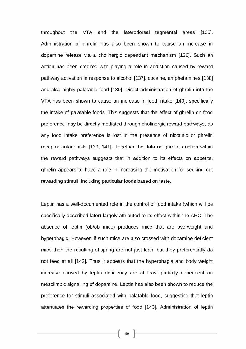
throughout the VTA and the laterodorsal tegmental areas [135].
Administration of ghrelin has also been shown to cause an increase in
dopamine release via a cholinergic dependant mechanism [136]. Such an
action has been credited with playing a role in addiction caused by reward
pathway activation in response to alcohol [137], cocaine, amphetamines [138]
and also highly palatable food [139]. Direct administration of ghrelin into the
VTA has been shown to cause an increase in food intake [140], specifically
the intake of palatable foods. This suggests that the effect of ghrelin on food
preference may be directly mediated through cholinergic reward pathways, as
any food intake preference is lost in the presence of nicotinic or ghrelin
receptor antagonists [139, 141]. Together the data on ghrelin‘s action within
the reward pathways suggests that in addition to its effects on appetite,
ghrelin appears to have a role in increasing the motivation for seeking out
rewarding stimuli, including particular foods based on taste.
Leptin has a well-documented role in the control of food intake (which will be
specifically described later) largely attributed to its effect within the ARC. The
absence of leptin (ob/ob mice) produces mice that are overweight and
hyperphagic. However, if such mice are also crossed with dopamine deficient
mice then the resulting offspring are not just lean, but they preferentially do
not feed at all [142]. Thus it appears that the hyperphagia and body weight
increase caused by leptin deficiency are at least partially dependent on
mesolimbic signalling of dopamine. Leptin has also been shown to reduce the
preference for stimuli associated with palatable food, suggesting that leptin
attenuates the rewarding properties of food [143]. Administration of leptin

47
directly into the VTA causes a reduction in food intake indicating that in
addition to its actions within the hypothalamus leptin is able to act directly
within the VTA to modulate feeding behaviour [144]. However, it has also
been suggested that leptin can indirectly inhibit VTA activity by acting on
lateral hypothalamic neurons, which project onto the VTA to inhibit the release
of dopamine [145].
As described above both the hedonic and homeostatic facets to food intake
control are influenced by gastrointestinal endocrine and neural regulation. It is
therefore of paramount importance when considering the role of central
pathways in mediating food intake to consider peripheral appetite regulatory
mechanisms.

48
1.2.2 Peripheral Regulation of Food Intake
Within the periphery a number of organs including the pancreas, liver, adipose
and gastrointestinal tract detect and monitor the current metabolic status.
Changes to the metabolic state i.e. food deprivation is met with changes in
neural and hormonal signals which can act peripherally or centrally to correct
the disturbance. The peripheral signals involved in the regulation of food
intake can be broadly classified into either long-term or short-term control.
Long term control tends to be largely mediated by levels of circulating
hormones, some of which are released by adipose tissue. The short term
control of food intake is largely controlled by neural and endocrine mediators
released by the gastrointestinal tract, usually in response to food, to control
factors such as meal termination and initiation (Figure 1.5). The following is a
description of how adipose tissue and the gastrointestinal tract can influence
food intake and exert their respective roles in mediating long and short term
energy balance.

49

50
Figure 1.5: Sites of release of long term and short term mediators on
energy balance. The pancreas and adipose tissue release insulin and leptin
respectively, which provide information to the CNS regarding the metabolic
status of the body with perturbations in their levels reflecting increased or
decreased energy stores. The gastrointestinal tract releases ghrelin as a short
term signal to initiate a meal and increase food consumption. After meal
consumption an array of peptides including PYY, GLP-1, CCK and leptin are
released from cells within the gastrointestinal tract to signal satiety. On top of
mediating appetite both the long term and short term mediators also play a
role in a number of efferent pathways including regulating energy expenditure,
growth, release of other hormones, gastrointestinal motility and partitioning
available energy. PYY; peptide YY, GLP-1; glucagon-like peptide 1, CCK;
cholecystokinin. Figure adapted from [146]

51
1.2.2.1 Adipose tissue
Historically adipose tissue was viewed as a store for excess energy that the
body could mobilize to sustain body processes when food was not readily
available for consumption [147]. However, subsequently adipose tissue was
found to be capable of releasing endocrine mediators capable of mediating
feeding behaviour [148], with leptin being the first identified [149]. Currently, at
least 30 peptides have been discovered to be released from adipocytes with
many of these adipokines having roles in regulating energy homeostasis and
metabolic processes [150].
From an evolutionary perspective, adipose tissue allows animals to consume
more food than they need for their immediate use to store for later use, when
food is less abundant due to seasonal variations in food availability [151, 152].
A similar phenomenon has been observed in native and rural human
populations [153, 154]. This suggests that there must be an endogenous
mechanism that permits accumulation of fat above and beyond what could be
considered necessary for immediate survival. Studies have presented
evidence suggesting the body is aware of current and desired levels of
adiposity and this led to the paradigm of an adipostat (Figure 1.6), a signal
that would effectively defend the level of adiposity within the body [73]. This is
consistent with food restriction induced fat loss in rats being restored after re-
feeding to levels comparable with continuously ad libitum feed rats [155].
Animal models have revealed that surgically removing one fat deposit leads to
a compensatory increase in fat being stored in the remaining intact fat
deposits [156]. The same phenomenon has been observed in humans

52
undergoing lipectomy, with the removal of the subcutaneous fat leading to an
initial weight loss, however this lost weight is soon replaced with an increase
in visceral fat [157]. This indicates that the simple removal of fat is not a viable
approach for weight loss. The adipostat paradigm does not, however, explain
obesity. The adipostat model would predict that an increase in adipose mass
would increase the adiposity signal, which should result in a reduction in food
intake and a decrease in fat mass. In obesity this obviously is not occurring.
However, in accordance with the necessity to store energy when available in
an environment with an uncertain food supply, the purpose of an adipostat
may be to permit this to occur to a genetically defined upper limit, and defend
a decrease from any level reached. Unfortunately, in a modern society where
food is readily available such an adaptation would be a liability rather than
advantageous.
Adipose tissue has also been shown to anatomically and functionally change
with metabolic state. Adipocyte size has been shown to be linked to the state
of energy balance; with hypocaloric diets reducing adipocyte size but not the
number of adipocytes [158, 159]. This is important to keep in mind because
there is a strong positive relationship between adipocyte size and the release
of adipokines. For example, increased adipocyte size is associated with
increased leptin, interleukin (IL)-6 and IL-8 and a decrease in the anti-
inflammatory mediator, IL-10 [160]. Increased adipose size in obesity is also
known to increase adipocyte death [161]. This in turn results in increased
macrophage infiltration into the adipose tissue producing the chronic low
grade inflammation of adipose tissue in obesity [161-163]. However, obesity

53
can occur in the absence of adipose tissue inflammation [164]. The
inflammation occurring within adipose tissue has been proposed to be a
mechanism for insulin resistance that often occurs in obesity [165, 166]. This
in turn could be a mechanism to protect from obesity as insulin plays a role in
adipocyte differentiation [167], lipogenesis [168] and inhibition of lipolysis
[169].
There are at least 24 reported adipokines/cytokines which have elevated
circulating levels in human obesity, with their plasma levels closely associated
to visceral adiposity [170]. However, the actual source of the increased
adipokines/cytokines is uncertain, as in addition to the adipocytes they could
also be released from the increased number of macrophages in obese
adipose tissue. Macrophages are a source of many cytokines and as such
both the adipocytes and macrophages may be responsible for altered levels
of adipokines/cytokines observed in obesity [171].
As well as subcutaneous and visceral adipose tissue stores there is also sub-
serosal and sub-mucosal adipocytes within the stomach [172]. It has been
suggested that adipokines released from similar deposits in the colon could
act in a paracrine manner to play a role in mediating intestinal barrier function
[173]. This raises the hypothesis that sub-mucosal and sub-serosal gastric
adipocytes could release adipokines that may act in a paracrine fashion to
mediate gastric function or modulate intrinsic and extrinsic gastric afferents. It
still remains to be determined if such a mechanism exists and if so whether it
is altered by the adipose hypertrophy observed in obesity.

54
A NOTE:
This figure/table/image has been removed to comply with copyright regulations. It is included in the print copy of the thesis held by the University of Adelaide Library.

55
Figure 1.6: The adipostat theory for the control of body weight. This
model proposes that the size of adipose tissue controls the level of circulating
hormones which in turn act centrally to modulate food intake and energy
expenditure. So when adipose tissue is increased (left) there is an increase in
circulating hormones such as leptin that act centrally to cause an increase in
energy expenditure whilst decreasing food intake, which results in a negative
energy balance and subsequent weight loss. Alternatively weight loss leads to
decreased adipose tissue and reduced circulating anorexigenic signals and
causes an increase in food intake and a decrease in energy expenditure
leading to a positive energy balance and weight gain. Figure adapted from
[174].

56
1.2.2.2 Proximal gastrointestinal tract
Consisting of the stomach and small intestine, the proximal gastrointestinal
tract is the site for the digestion of food and absorption of nutrients. This
system relies on co-ordinated mechanical and chemical processes to achieve
its tasks. Upon food entering the stomach relaxation of the gastric muscle
allows accommodation of the food to occur [175]. This phenomenon was
dubbed adaptive relaxation and occurs within seconds of food entering the
stomach [176]. This allows for the maintenance of intragastric pressure whilst
increasing the volume available for occupation by food [177]. The ability of the
stomach to increase its volume has been shown to be relevant to the control
of food intake, as disorders of the reservoir function of the stomach are
associated with early satiety [178]. The relaxation occurs through chemical
and neural mechanisms [179, 180], which lead to activation of inhibitory
enteric motor neurons, causing relaxation of the gastric smooth muscle.
However, the role of the smooth muscle within the gastrointestinal tract is not
limited to allowing food to be accommodated. It is also vital for the transit of
the food through the entire gastrointestinal tract by rhythmically contracting
and relaxing in the form of peristaltic waves [181]. The muscle within the walls
of the stomach contract to mix the food with digestive enzymes and acid
released from epithelial and endocrine cells that line the gastric lumen [182,
183]. Peristaltic contractions propel the stomach contents toward the pylorus
where movement into the duodenum is prevented for all but the smallest
particles and liquids [183].

57
Once in the duodenum the digestive process continues to occur due to
release of digestive enzymes from the pancreas and gall bladder [184]. Whilst
originally thought of as reservoirs for the digestive process to occur within, the
small intestine and stomach have a profound ability to sense food [185, 186]
and cause changes in food intake behaviour [187, 188] as well as control the
digestive progress [183, 189]. As such the roles of the small intestine and
stomach have been expanded by an ever growing literature base to highlight
that they are involved in the regulation of local gastrointestinal function as well
as the regulation of food intake and satiety.
Appetite regulation by the gastrointestinal tract is mediated by sensing both
volume and composition of meals. This can occur through gastrointestinal
secretion of peptides as well as activation of afferent endings located within
the wall of the gastrointestinal tract. Below is an overview of the abilities of the
small intestine and stomach to sense nutrients and signal through multiple
pathways, including neuronal and hormonal pathways, to regulate appetite.
1.2.2.2.1 Nutrient sensing
1.2.2.2.1.1 Small intestine
The small intestine can initiate satiety in response to luminal nutrients [190].
The ability for this to occur has been eluded to be due to nutrient driven
release of cholecystokinin (CCK), which is an intestinal peptide released from
I cells within the proximal small intestine [191] when exposed to nutrients
(predominately protein and lipids) [192-195]. Evidence has shown that CCK
causes satiety through activating local vagal afferent endings [196-198].

58
Furthermore, nutrients in the small intestine have been shown to induce an
increase in thermogenesis (a major source of energy expenditure) in brown
adipose tissue via CCK mediated activation of local vagal afferent endings
[199]. This suggests that as well as being involved in controlling food intake,
nutrient sensing within the small intestine is a regulator of energy expenditure.
Specialised intestinal epithelial cells have been shown to express nutrient
receptors identical to those found on the surface of the tongue [200, 201],
suggesting that the intestine has the ability to ‗taste‘ nutrients in the gut. Some
cells expressing these taste receptors are co-localised with intestinal satiety
peptides [200-202] and nutrient absorption proteins like sodium-glucose
transporter 1 (SGLT1) [203]. These findings together led to the hypothesis
that specialised intestinal epithelial cells can sense specific nutrients and
respond by releasing appetite modulatory peptides to control food intake and
gastrointestinal motor function [204]. This is supported by small intestinal lipid
inducing gastric relaxation [205, 206]. Furthermore, high level of Intestinal fat
induces a feeling of nausea during gastric distension [207]. This suggests that
intestinal nutrient sensing modulates sensory signals from the stomach as
well.
1.2.2.2.1.2 Stomach
The involvement of the stomach in nutrient sensing is less clear. Early studies
found that infusion of saline elicited the same meal terminating effect as
infusions of sucrose, fructose and glucose [208]. This indicated that the
stomach was simply detecting a mechanical stimuli not a chemical one.

59
However, there is also evidence that the stomach can respond to nutrients to
control food intake [209-211] and that changes in intragastric pressure only
result in increased satiety after intragastric infusion of nutrients [212], which
casts doubts on the paradigm that gastric satiety is purely mechanical.
Further evidence for a nutrient sensing ability of the stomach is the expression
of α-gustducin [213] and the taste receptor component, T1R3 [214, 215] in
cells within epithelium brush border of the stomach. Furthermore, these taste
receptor signalling components are located on ghrelin positive cells [214, 216]
and have been show to trigger the release of ghrelin in an α-gustducin
dependant manner [216], which further contradicts the theory that the
stomach‘s role in the regulation of food intake is purely mechanical. Gastric
nutritional sensing has also been demonstrated in response to amino acids
[217] . This further strengthened the argument that taste sensing occurs within
the stomach. However, this still remains to be conclusively demonstrated.
There is evidence that suggests that gastric nutrient sensing may be
comprised in obesity. Gastric mucosal expression of the fatty acid receptor
GPR120 is increased and sweet taste receptor component T1R3 has been
shown to be decreased in morbidly obese humans [218]. This led the authors
to conclude that alterations in gastric nutrient sensing ability may play a role in
the increased food consumption seen in obesity. However, their evidence is
very preliminary and the conclusions are based on immunohistochemical and
mRNA expression levels and thus the actual phenotypic effect of such
changes needs to be determined. However, these results do show that gastric

60
nutrient sensing is likely to be dynamic and that obesity is capable of
potentially disrupting gastric satiety signals. This warrants further
investigation.
It has been demonstrated that the suppression of food intake by nutrient
infusion into the stomach with an open pyloric cuff is greater than the
suppression in food intake caused by gastric distension or intestinal nutrient
exposure alone [208]. This indicates that both locations work together to
cause a larger net reduction in food intake than either could on their own. An
example of such a mechanism is CCK, which is released from the small
intestine and has been shown to activate the same vagal afferents activated
by gastric distension [198]. This indicates the neuronal innervation of the
gastrointestinal tract is a site for convergence of both gastric and intestinal
satiety signals.
1.2.2.2.2 Innervation of the proximal gastrointestinal tract
Within the stomach and small intestine there are discrete populations of
neurons which represent extrinsic innervation (through splanchnic and vagal
nerves) and also intrinsic innervation (through the ENS; Figure 1.7). Whilst
each is able to act independently there are an abundance of synapses
between the intrinsic and extrinsic neurons highlighting the functional
relationship that exists between them. Below is an outline of the roles of these
pathways, focusing on food intake.

61
A NOTE:
This figure/table/image has been removed to comply with copyright regulations. It is included in the print copy of the thesis held by the University of Adelaide Library.

62
Figure 1.7: Anatomical organisation of the layers of the gastrointestinal
wall and intrinsic and extrinsic innervation. Within the wall of the
gastrointestinal tract there are vagal and spinal afferents which have endings
within both the muscular and mucosal layers enabling detection of multiple
stimuli. Enteric neurons are also present within the myenteric and submucosal
plexus. Whilst capable of autonomous action they can also communicate with
extrinsic neurons. Figure adapted from [185].

63
1.2.2.2.2.1 Enteric nervous system (ENS)
The gastrointestinal tract is unique due to its extensive intrinsic nervous
system, the ENS. This intrinsic innervation allows continuation of multiple
processes even after separation from the CNS; however there is substantial
penetration of vagal neurons into the ENS [219], suggesting communication
between the two systems. The ENS is organised into two plexuses. One is
located between the circular and longitudinal layers of muscle, called the
myenteric plexus. The other exists within the submucosal layer called the
submucosal plexus. Within these plexuses there are primary afferent neurons,
interneurons and motor neurons that are able to act on a variety of targets
including smooth muscle, blood glands, epithelium, pace maker cells, mucosal
glands and extrinsic neurons. The ENS has multiple roles within the
gastrointestinal tract including regulating gastric acid secretion [220],
epithelium proliferation [221], immune function [222], endocrine release and
determining the patterns of movement along the gastrointestinal tract [223].
Given its involvement in multiple processes that occur within the
gastrointestinal tract it is not surprising that dysfunction of the ENS is
associated with a number of detrimental conditions [224].
In terms of involvement in the regulation of food intake, enteric neurons have
a couple of desirable properties. Firstly, they express receptors for appetite
modulatory peptides [225]. Secondly, the ENS contains first order neurons
that are able to sense and respond to food within the lumen of the
gastrointestinal tract and are in direct contact with enteroendocrine cells such
as CCK secreting I cells [226]. The physiological relevance of this has been

64
highlighted by the finding that CCK administration causes a dose dependent
increase in c-Fos activation in both small intestinal myenteric and submucosal
neurons [227]. This effect is abolished when administered in the presence of a
CCK1R antagonist [228] and is not mediated via a vagal pathway or hindbrain
activation [229]. CCK has been shown to activate choline acetyltransferase
(ChAT) immunoreactive neurons within the myenteric plexus which project to
the sphincter located at the duodenal orifice of the bile duct [230]. Activation
of these neurons causes a relaxation of the sphincter and increase bile
release from the gall bladder into the duodenum [230]. This suggests that
digestion and the subsequent absorption of nutrients could be modulated by
activation of enteric neurons.
Enteric neurons also express 5-hydroxytryptamine (5-HT)3 receptors within
both the myenteric plexus and submucosal plexus [231]. Given that the
majority of 5-HT exists within enterochromaffin cells of the gastrointestinal
tract [232] and that enteric neurons themselves have been shown to be a
major site of 5-HT release it seems logical that the ENS is involved in the
regulation of gastrointestinal function and is modulated by locally released
modulators including 5-HT [233].
The function and responsiveness of the ENS to chemical mediators including
CCK and ghrelin has been demonstrated to be altered by feeding status [234].
Like extrinsic afferents there is also evidence that enteric neurons are able to
sense and modulate their activity in response to changes in glucose levels
[235, 236]. Such modulation may explain, at least in part, why acute changes

65
to glycaemia can substantially alter gastric motor function [237], which in turn
would alter the availability for nutrient absorption. Consistent with this is a
state of hyperexcitability in enteric neurons and increased frequency of
peristaltic activity in fasted mice that have been re-fed [234]. There has also
been demonstrations of nutrient specific adaptations of enteric neuron
function with chronic high fat diet feeding associated with reduced enteric
neuron response to intestinal oleate [238]. This reduction in enteric activation
was associated with a reduced level of satiety induced by intestinal oleate
[238]. However, it was not possible to determine what, if any, the role the
reduced enteric response to oleate played in the reduced level of satiety.
Therefore, although there is a lack of evidence to suggest that the ENS can
directly influence food intake, by controlling gastrointestinal motility it is
reasonable to suggest the ENS plays an indirect role in the control of food
intake by responding to nutrient and chemical stimuli to mediate the amount of
nutrient available for absorption at any one time.
1.2.2.2.2.2 Spinal Afferents
Whilst the majority of peripheral food intake regulatory signals are believed to
be conveyed through vagal pathways, it is important to recognise that there
are still significant levels of spinal afferents within the upper gastrointestinal
tract, including the stomach [239] and small intestine [240]. The purpose of
these spinal afferents is believed to be to convey noxious stimuli such as pain
[241], however there is evidence that these pathways are also involved in
food intake regulation and gastric function [242, 243]. For instance, the

66
peptide bombesin has been shown to have receptors located throughout the
CNS and also within the gastrointestinal tract [244, 245]. Bombesin causes
release of gastrin [246] and also causes a reduction in food intake and meal
size [247, 248]. This bombesin induced reduction in food intake is abolished
by severing the spinal and vagal nerves, indicating that the food intake
modulatory effect is mediated through extrinsic gastrointestinal neuronal
pathways [249]. However, bombesin was still able to reduce food intake by
virtually the same amount with only the vagal or spinal innervation disrupted
compared to both being intact [249]. This indicates that bombesin can reduce
food intake through independent spinal and vagal afferents.
Furthermore, dorsal root ganglia (DRG; location of the spinal afferent cell
bodies) have receptors for ghrelin [250] and CCK [251]. Spinal afferents have
also been implicated in the control of motor functions of the upper
gastrointestinal tract with ablation of the spinal pathways causing a reduction
in gastric motility [252, 253]. Gastric electrical stimulation, which has
previously been shown to reduce food intake [254], elicits an overall excitatory
effect on spinal afferent pathways, both in the presence and absence of intact
vagal nerves [255]. This provides extra support to the notion that spinal
afferents may respond to gastric stimuli to modulate feeding behaviour, but
this remains to be conclusively determined.
Both chemosensitive and mechanosensitive afferent endings have been
identified in spinal afferents [256, 257]. These mechanosensitive endings
have been very well characterised, however the majority of the investigation

67
has occurred within the colon [258], where vagal innervation is sparse. Spinal
mucosal, muscular and serosal mechanosensitive afferents have been
identified in the colon [258], with the latter being responsive to high intensity
stimuli and suggested to be involved in detecting noxious stimuli [259].
Similarly, in the stomach of rats there are populations of high threshold spinal
afferents, which only respond to distension within the noxious range [260].
Thus, it is possible that gastric distension may activate specific populations of
spinal afferents to signal pain in the event of over distension of the stomach,
as well as providing input regarding normal distension of the stomach [261].
1.2.2.2.2.2.3 Vagal afferents
Tracing studies have revealed the vagus is predominately comprised of
afferent fibres, which originate from much of the viscera, including the
gastrointestinal tract [262]. The vagus has its cell bodies located within the
nodose ganglia, from which the vagus projects centrally to the NTS [263]. In
addition to the vagus the NTS receives inputs from the facial and
glossopharyngeal nerves [263] with the latter two being involved in oral
gustatory signalling [264-266]. Therefore the NTS appears to be a ‗hub‘ for
the convergence of neuronal activity resulting from food intake. This is
supported by stimulation of the vagus increasing activation of c-Fos within the
NTS [267] and suppressing subsequent food intake [268].
In addition to having central projections (described earlier), the neurons within
the NTS that are activated by vagal afferents are able to activate neurons
within the dorsal motor nucleus of the vagus (DMV; Figure 1.8) [269, 270].

68
Activated DMV neurons can induce gastric relaxation via activation of a non-
adrenergic, non-cholinergic (NANC) pathway, leading to the release of nitric
oxide or vasoactive intestinal peptide from intragastric neurons [271].
Alternatively, a cholinergic excitatory pathway can be activated, resulting in
muscarinic receptor dependent increases in gastric motility and tone [272].
Much of the past research into the role of the vagus in controlling food intake
involved some form of vagotomy to remove all vagal innervation. This crude
method involves severing the vagal nerves by hand. Such an approach could
lead to different effects based on the location the nerves severed. This may
explain why reports on the effects of vagotomy on body weight vary
considerably, with some reporting a decrease in weight [273, 274], whilst
others report no difference [275] or even an increase [276]. Perivagal
application of capsaicin has been used to selectively destroy vagal afferent
fibres [277]. However, evidence suggests that perivagal capsaicin does not
necessarily only destroy afferent fibres [278]. These approaches allow no
specific evidence to be extracted from the results in regards to the role of
gastric innervation in the control of food intake [279].Furthermore, a vagtomy
also reduces gastric relaxation [280], motility [281] and secretions i.e. acid
and gastrin [282]. All of these can alter the rate that nutrient absorption can
occur. This adds further difficulty in identifying mechanisms behind changes in
food intake behaviour caused by a vagotomy.
Closure of the pyloric sphincter (to prevent any intestinal response to eating)
showed an attenuation of food intake following gastric loads [283]. This

69
suggests that there is a distinct gastric mechanism that is likely being
conveyed through vagal pathways to trigger satiety. This concept was further
strengthened by findings that gastric vagal electrical stimulation caused a
reduction in food intake and a subsequent reduction of body weight in rats
[284], with a similar technique being implemented in humans as a treatment
for obesity [285]. This suggests that not only are vagal afferents involved in
regulation of food intake, but they also present a possible target for weight
reducing therapies.
Whilst it was previously discussed that nutrient sensing within the stomach is
a likely possibility; there are also specialised vagal afferent endings that are
responsive to mechanical stimuli. Upon entering the stomach, food is detected
by vagal afferent fibre endings in the mucosa that are sensitive to mucosal
touch [286, 287]. The volume of food is also sensed by afferent endings
located in the deeper muscular layers of the stomach, which are sensitive to
stretch and tension [288]. Activation of the latter type of afferent has been
shown to trigger satiation [289]. Gastric mechanosensitive vagal afferents
have previously been investigated in depth and below is a brief overview of
their anatomical and functional characteristics.
1.2.2.2.2.3.1 Tension receptors
There have been two populations of vagal neurons identified in the stomach
which are thought to respond to stretch. The first are intraganglionic laminar
endings (IGLEs). These are vagal afferent endings located within the
connective tissue capsule of the myenteric plexus ganglia between the outer
longitudinal and inner circular muscle layers [290]. They respond to muscle

70
tension generated by passive stretch of the stomach as well as active
contractions of the stomach [291] and are believed to be the
mechanotransduction sites of tension sensitive vagal afferents [291]. This
particular type of afferent ending is found in very large numbers throughout
the oesophagus [292] and stomach [293] and are believed to have roles in
generating and maintaining vagal tone.
Intramuscular arrays (IMAs) are the second class of tension sensitive afferent
ending, however they are exclusively located within the circular and
longitudinal muscle layers [294]. They have been postulated to detect
changes in muscle length [295] and respond to stretching of the stomach
walls with a sustained high rate of action potentials which is still present after
muscular tension is reduced through gastric accommodation [296]. If correct,
such a trait suggests that they may be a prime candidate for providing
feedback regarding stomach fullness, which could help signal satiety.
However, to date most speculations are based on anatomical evidence with a
distinct lack of morphological evidence linking IMAs with a
mechanotransduction site [291]. Therefore, it still remains to be confirmed
whether IMAs do in fact play a role in detecting gastric stretch and satiation.
1.2.2.2.2.3.2 Mucosal receptors
Vagal afferent fibres also have specialised endings in the mucosa [287].
These endings are commonly found to be closely associated to endocrine and
epithelial cells containing peptides with known effects on food intake. This
suggests they may be readily modulated by locally released peptides in vivo.

71
These endings have also been shown to have mechanical sensitivity,
although unlike IMAs and IGLEs are not sensitive to tension [287]. However,
when stroked over they respond with a burst of activity. These afferents have
been suggested to be important in detecting food particle size and have been
linked to delaying of gastric emptying. This was supported by an increase in
food particle size being passed into the small intestine of dogs which had
neuronal innervation of the gastric mucosa destroyed [297].
Using a flat sheet in vitro electrophysiological preparation (Figure 1.9) it is
possible to record the responses of both classes of afferents in mice (Figure
1.10). This approach has led to significant information being discovered
regarding the effects of different endogenous modulators on the response of
these afferents. For example nitric oxide [298] and GABA [299] have been
shown to have inhibitory effects on the mechanosensitivity of gastric vagal
afferent endings. Furthermore, peptides such as galanin [300] and ghrelin
[301] have also been shown to have modulatory effects on these endings.
These studies have provided important mechanistic information about how the
stomach may regulate peripheral satiety signalling as well as other visceral
signals through peripherally acting mediators acting on afferent pathways.
However, no information exists in regards to whether the sensitivity of vagal
afferents to mechanical stimuli or gut peptides such as leptin and ghrelin are
altered under states of energy deprivation or excess, which given the role of
the vagus in mediating food intake is worthy of investigation and forms part of
the current thesis.

72

73
Figure 1.8: Vagal afferent connection within the brainstem. Activation of
mechanosensitive or chemosensitive vagal afferents in the gastrointestinal
tract leads to activation of second order neurons within the nucleus tractus
solitarii (NTS). The second order neurons can activate high regions of the
central nervous system (CNS) where they can mediate satiety and energy
expenditure. Alternatively they can activate neurons within the dorsal motor
nucleus of the vagus (DMV) which can cause contraction or relaxation of
gastric smooth muscle through cholinergic (ACh) or non-adrenergic non-
cholinergic (NANC) pathways respectively. Descending input into the NTS
occurs but is not shown. Dashed lines with arrows indicate the direction signal
is travelling. AP; area postrema, CCK; cholecystokinin, CCK1R; CCK receptor
1.

74

75
Figure 1.9: Schematic representation of gastro-oesophageal vagal
afferent recording flat sheet preparation. This setup is used to record
gastric vagal afferent responses to mechanical stimulation. Drugs are applied
via the krebs solution which is superfused over the tissue. .

76

77
Figure 1.10: Original electrophysiological traces showing the two types
of mechanically sensitive gastric vagal afferent endings that have been
identified in the mouse. The first class of afferent are tension receptors
(top). These afferents exist in the muscular layers of the stomach wall and
respond to mucosal stroking with a burst of action potentials, but also respond
to tension with a sustained graded burst of action potentials in proportion to
the size of the stimuli. The second class are mucosal receptors (bottom)
which respond to mucosal stroking with calibrated von Frey hairs with a burst
of action potentials each time the receptive field is stroked. However, unlike
tension receptors, these afferents do not respond to stretch.

78
1.2.2.2.2.4 Vagal Afferent Mediators
In addition to having mechanosensitive afferents that can be activated by the
presence of food, the gastrointestinal tract also contains a great amount of
endocrine and epithelial cells which have been shown to release peptides in
response to feeding [302-304]. Below is a summary of a selection of
mediators that are released from these cells and have been suggested to
have roles in regulating food intake through vagal pathways. This is by no
means a full list of peptides/hormones released from the gastrointestinal tract
but is simply an illustration of the important role gastrointestinal peptides play
in control of food intake.
1.2.2.2.2.4.1 Gastric mediators
Leptin: Classically described to be released from adipose tissue into the
blood, leptin plasma concentrations are proportional to the amount of adipose
tissue [305]. This suggests that leptin‘s primary role is to provide information
to the CNS in regards to whether energy storage is sufficient. It has been
described that leptin, upon entering the circulation, travels to the brain where
it crosses the BBB in a saturable receptor mediated fashion [92]. Upon
crossing the BBB leptin can bind to its receptor, which is present on a number
of crucial brain centres, including the hippocampus [306] and, more relevant
to food intake, the hypothalamus [307]. Here it causes modulation of neural
activity, specifically inhibiting the NPY [308] and activating POMC [99]
neurons within the arcuate nucleus, which results in a reduction in food
consumption, an increase in energy expenditure and a preferential oxidation
of fat [309-311].

79
There are six isoforms of the leptin receptor (LepRa-f) [312], which are closely
related to the class 1 cytokine receptor family. LepRa-d and LepRf all have
the same extracellular and transmembrane domains. LepRe represents a
soluble form of the leptin receptor and does not have a known role in leptin
signalling [312]. However, it has been shown to protect leptin from
degradation in the gastrointestinal tract [313]. Therefore, it is likely to be
involved in controlling the circulating level of leptin [314], as the relative
quantities of free and receptor bound leptin are likely to impact on the level of
leptin signalling that can occur [315]. Of LepRa-d and LepRf only LepRb has a
long intracellular domain [312]. Truncation of the LepRb isoform such that it
possesses an intracellular domain similar to that exhibited by LepRa results in
an obese phenotype similar to that observed in leptin receptor knockout mice
(db/db mice) [312]. This suggests that the long intracellular domain is
responsible for the anorexigenic effect of leptin.
The process of leptin signalling begins with receptor dimerisation [316, 317].
Without this occurring leptin cannot cause any intracellular changes. However,
the short receptor isoforms also form dimers [316]. Given the short receptors
do not have leptin signalling capability indicates that receptor dimerisation,
whilst vital for signalling to occur, has no effect on the activity of the receptor
itself. Upon binding to the long form of its receptor leptin causes Janus kinase
(JAK) mediated phosphorylation of signal transducer and activator of
transcription (STAT) 3 [318], which forms a dimer [319] and translocates to
the nucleus. From here it acts as a transcription factor [320] for a number of

80
genes including suppressor of cytokine signalling (SOCS) 3, which in turn acts
to negatively regulate the activity of the leptin receptor [321] (Figure 1.11). On
top of transcriptional activation, leptin has been demonstrated to inhibit
neuronal activity by activating ion channels such as ATP activated potassium
(KATP) [322] and large conductance calcium activated potassium (BKCa)
channels [323]. In addition to neuronal inhibition, leptin has been shown to
activate canonical transient receptor potential (TRPC) channels [324] via a
phosphatidylinositide 3-kinase (PI3K) medicated mechanism increasing
neuronal excitability. Activation of a cation channel has been suggested to be
the mechanism by which leptin can induce meal termination [325].Given the
evidence that leptin can reduce food intake via a vagal mechanism [326], this
thesis will investigate whether leptin activates modulates mechanosensitive
vagal afferents via TRPC dependent pathway.

81

82
Figure 1.11: Schematic representation of leptin receptor signalling. Upon
binding to the long form of its receptor (LepRb), leptin causes phosphorylation
of signal transducer and activator of transcription 3 (STAT3) molecules, which
dimerise and translocate to the nucleus where they act as a transcription
factor. This causes an increase in suppressor of cytokine signalling 3
(SOCS3) expression, which inhibits the phosphorylation of STAT3 by Janus
kinase 2 (JAK2). JAK2 also causes activation of phosphatidylinositide 3-
kinases (PI3K) through insulin receptor substrate (IRS)1/2 which has been
linked with the modulation of ion channel activity in a variety of neurons.
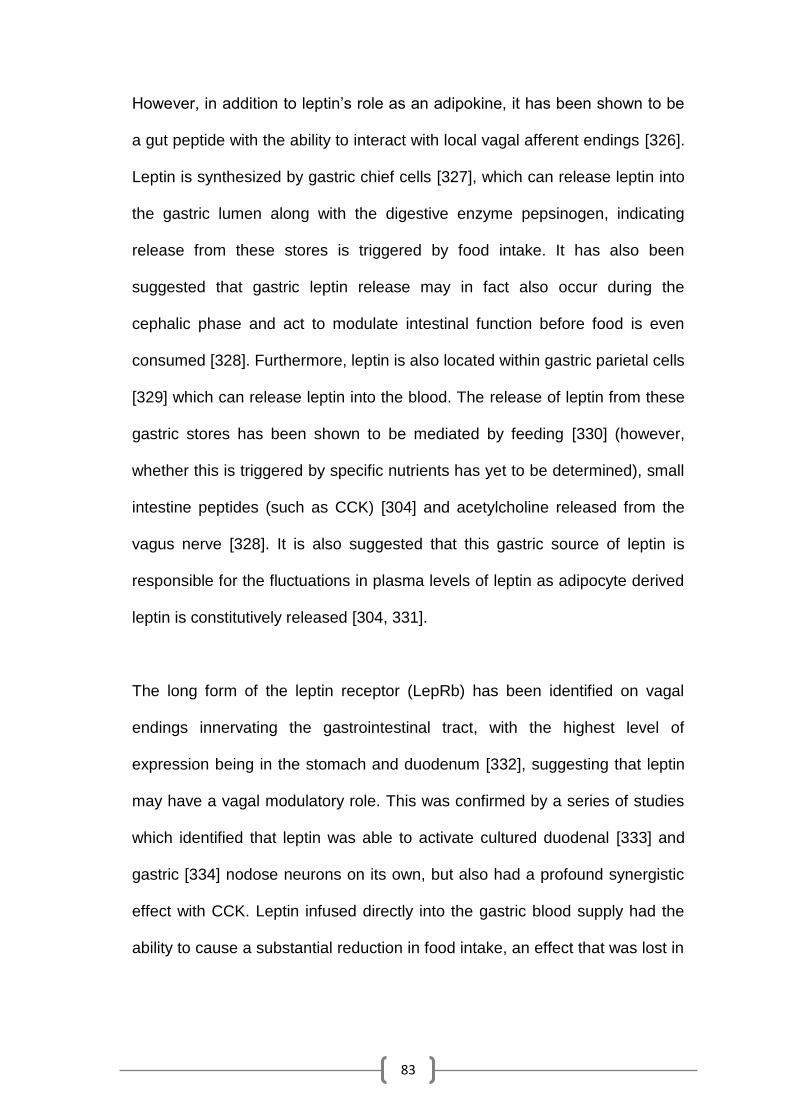
83
However, in addition to leptin‘s role as an adipokine, it has been shown to be
a gut peptide with the ability to interact with local vagal afferent endings [326].
Leptin is synthesized by gastric chief cells [327], which can release leptin into
the gastric lumen along with the digestive enzyme pepsinogen, indicating
release from these stores is triggered by food intake. It has also been
suggested that gastric leptin release may in fact also occur during the
cephalic phase and act to modulate intestinal function before food is even
consumed [328]. Furthermore, leptin is also located within gastric parietal cells
[329] which can release leptin into the blood. The release of leptin from these
gastric stores has been shown to be mediated by feeding [330] (however,
whether this is triggered by specific nutrients has yet to be determined), small
intestine peptides (such as CCK) [304] and acetylcholine released from the
vagus nerve [328]. It is also suggested that this gastric source of leptin is
responsible for the fluctuations in plasma levels of leptin as adipocyte derived
leptin is constitutively released [304, 331].
The long form of the leptin receptor (LepRb) has been identified on vagal
endings innervating the gastrointestinal tract, with the highest level of
expression being in the stomach and duodenum [332], suggesting that leptin
may have a vagal modulatory role. This was confirmed by a series of studies
which identified that leptin was able to activate cultured duodenal [333] and
gastric [334] nodose neurons on its own, but also had a profound synergistic
effect with CCK. Leptin infused directly into the gastric blood supply had the
ability to cause a substantial reduction in food intake, an effect that was lost in

84
vagotomised animals [326]. This suggested that the effect of leptin on acute
food intake is mediated through vagal afferent pathways.
The discovery of leptin was expected to lead to the production of a ‗cure‘ for
obesity. However, these hopes were diminished after the discovery that the
majority of obese humans have sustained hyperleptinemia and are unable to
respond to leptin administration with the expected anorexigenic effects.
Indeed animals and humans who have defects in either the leptin or leptin
receptor gene exhibit early onset morbid obesity, which highlighted just how
important the biological activity of this peptide is. Of particular interest is that
obesity has now been shown to be linked with leptin resistance in vagal
afferent neurons [335, 336]. This indicates that the dysfunction in leptin
signalling associated with obesity is not localised to the CNS. However, this
leptin resistance in vagal afferents was concluded based on the whole nodose
ganglia [336]. As such, it is difficult to conclude more specifically whether
gastric mechanically sensitive vagal afferents exhibit this leptin resistance.
Investigating whether leptin modulation of gastric vagal afferent
mechanosensitivity is altered under diet-induced obesity forms a major focus
of this thesis.
Neuropeptide W (NPW): NPW is a recently discovered peptide that activates
the orphan G-protein coupled receptors GPR7 and GPR8 [337]. NPW was
originally believed to exert a modulatory role on food intake centrally since
central administration of NPW into the rat brain stimulated food intake [338,
339]. However, on top of the central targets NPW has also been shown to be

85
present in antral G cells of rodent and human stomach where it is co-localized
with gastrin [340]. The expression of NPW in these G cells varies with fed
status with fasted animals having a reduction in NPW content further
indicating a role in modulating food intake [341]. In addition GPR7 knockout
mice (the only NPW receptor subtype in mice) develop hyperphagia and
obesity [342]. This suggests an anorectic role for NPW, however there is
evidence which suggests that NPW in fact increases food intake [338] and
has the ability to inhibit the responses of vagal afferent to mechanical stimuli
[343].
Ghrelin: Ghrelin is a 28-amino acid peptide secreted from X/A cells within the
gastric fundus [344]. It acts as an endogenous mediator of growth hormone
release [345]. Ghrelin has also been shown to be expressed along the length
of the gastrointestinal tract; however the level of expression diminishes distally
from the stomach [346]. Ghrelin has a unique structure with an octanyl group
present, which is vital for activation of its receptor (the growth hormone
secretagogue receptor- GHS-R). Cleavage of this group causes instant
deactivation of ghrelin and as the enzyme responsible for this cleavage freely
circulates ghrelin has a very short active half life in blood.
Post transcriptional processing of GHS-R leads to the production of two splice
variants, GHS-R1a and GHS-R1b [347, 348]. The GHS-R1a consists of 366
amino acids and forms a receptor with seven transmembrane domains linked
with alternating intracellular and extracellular loops [347]. Alternatively, GHS-
R1b is truncated at the carboxyl end resulting in only five transmembrane
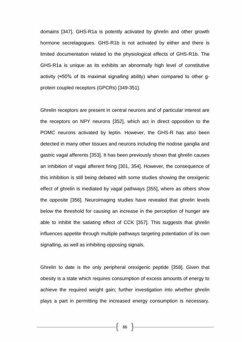
86
domains [347]. GHS-R1a is potently activated by ghrelin and other growth
hormone secretagogues. GHS-R1b is not activated by either and there is
limited documentation related to the physiological effects of GHS-R1b. The
GHS-R1a is unique as its exhibits an abnormally high level of constitutive
activity (≈50% of its maximal signalling ability) when compared to other g-
protein coupled receptors (GPCRs) [349-351].
Ghrelin receptors are present in central neurons and of particular interest are
the receptors on NPY neurons [352], which act in direct opposition to the
POMC neurons activated by leptin. However, the GHS-R has also been
detected in many other tissues and neurons including the nodose ganglia and
gastric vagal afferents [353]. It has been previously shown that ghrelin causes
an inhibition of vagal afferent firing [301, 354]. However, the consequence of
this inhibition is still being debated with some studies showing the orexigenic
effect of ghrelin is mediated by vagal pathways [355], where as others show
the opposite [356]. Neuroimaging studies have revealed that ghrelin levels
below the threshold for causing an increase in the perception of hunger are
able to inhibit the satiating effect of CCK [357]. This suggests that ghrelin
influences appetite through multiple pathways targeting potentiation of its own
signalling, as well as inhibiting opposing signals.
Ghrelin to date is the only peripheral orexigenic peptide [358]. Given that
obesity is a state which requires consumption of excess amounts of energy to
achieve the required weight gain; further investigation into whether ghrelin
plays a part in permitting the increased energy consumption is necessary.

87
Currently, there is some evidence suggesting that within the ARC ghrelin
resistance occurs in obesity [359]. However, there is no information regarding
potential changes to the peripheral orexigenic effect of ghrelin in obesity. The
fact that ghrelin still increases energy intake in obese humans [360] suggests
that ghrelin must still be able to activate an orexigenic mechanism. As stated
previously, it is already established that ghrelin reduces the
mechanosensitivity of gastric vagal tension receptors [301]. As part of this
thesis I will determine if these effects on gastric vagal afferent
mechanosensitivity are altered under conditions of diet restriction and excess.
1.2.2.2.2.4.2 Intestinal mediators
CCK: CCK is released from I-cells located largely within the proximal small
intestine. The release of CCK is mediated by the presence of luminal
nutrients, with a preference for the digestion products of amino acids and fatty
acids rather than those of carbohydrates [193, 194, 361]. Fatty acids are
required to have an acyl chain length greater than 12 carbon atoms in order to
trigger the release of and elicit the well documented meal termination effects
of CCK [362, 363]. The link between luminal fatty acid and CCK release is
believed to be mediated through the fatty acid receptor GPR40. The GPR40
ligand, linoleic acid, causes release of CCK from I-cell populations with but not
without GPR40 [202].
There is an abundance of evidence for CCK having a food intake modulatory
effect, with its exogenous administration causing a reduction in food intake as
well as a slowing of gastric emptying, both of which are lost after vagotomy

88
[364, 365]. The same loss of effect of exogenous CCK administration is also
seen in mice with CCK1R blockade [366, 367]. The expression of CCK1R
does not appear to be effected by acute nutritional changes (i.e. fasting) [368],
but there are examples of upregulation in response to high fat diet feeding
[369] although this is not consistently reported [370]. The ability for CCK to
activate CCK1 receptors on vagal afferents is inhibited by the orexigenic
peptides ghrelin, orexin A and anandamide [353, 371-373]. In addition to its
own anorexigenic effect on food intake, CCK also appears limit ghrelin
induced drive to eat [374]. Therefore, CCK seems to both activate
anorexigenic and suppress orexigenic signals in order to control food intake.
However, CCK is more than just a satiety signal as it appears to be a crucial
component in the absorption of fats in the intestine. This is highlighted by
CCK knockout mice having higher levels of lipid in their faeces [375]. These
mice have a reduced body weight even though they are on the high fat diet
and consuming equal amounts of food [375].
It is believed that CCK acts predominately as a short term modulator of food
intake as the main effect of CCK appears to be meal termination. With chronic
use there is no change in weight, as the shorter meals are merely offset by an
increase in the number of meals [376, 377]. However, CCK exists in a number
of different lengths with the short form, CCK-8 being the most commonly
studied. There is evidence to suggest that whilst this shorter and more studied
isoform is indeed involved in limiting meal size [378] the longer forms of CCK
may contribute to determining meal to meal intervals [379].

89
The ability for CCK to reduce food intake appears to be compromised in
response to chronic high fat diet feeding with the absence of increased weight
[380]. This suggests that poor nutrition may cause changes that create a
susceptibility to obesity before any weight gain is observed. This may be due
to a reduced ability of CCK to activate intestinal vagal afferents as previously
described in diet-induced obese mice [381].
GLP-1: GLP-1 is an incretin hormone released from intestinal L-cells [382],
which have the ability to respond and broadly detect the digestion products of
carbohydrates, fats and proteins [383, 384]. GLP-1 immunoreactivity is found
throughout the gastrointestinal tract; however there is substantially greater
density within the distal small intestine [385, 386]. The release of GLP-1 has
been shown to be tightly controlled and is released in proportion to caloric
load [387] and also to the amount of small intestine that is being exposed to
the nutrient [388]. Its release causes a decrease in food intake [389],
stimulation of insulin release [390], reduction of glucagon secretion [391] and
a reduction in gastric emptying [392]. GLP-1 positive cells have also been
shown to be co-localised with GPCRs activated by sweet stimulants [201].
Therefore it has been hypothesised that the presence of sugars within the
intestinal lumen causes activation of these GPCRs which triggers the release
of GLP-1 from the L-cells. However, given that glucose does not normally
reach the distal parts of the small intestine in high concentrations and fatty
acids do it is likely that fatty acids have a more physiologically significant
effect on the release of GLP-1 from the distal intestinal L-cells. Supporting this
view is the finding that fatty acid receptors are present on the surface of L-

90
cells and the administration of agonists to these receptors causes an increase
in the release of GLP-1 [393].
GLP-1 can activate vagal afferents [394] and this is believed to be the
mechanism responsible for its effects on insulin release and food intake [395].
However, there is some debate over whether the vagal pathway is the main
effector pathway by which GLP-1 signals to the CNS. Subdiaphragmatic
vagotomy has been shown to have no effect on the ability of GLP-1 to reduce
food intake when administered into the hepatic portal vein, but when
administered intraperitoneally, subdiaphragmatic vagotomy abolishes the
ability for GLP-1 to reduce food intake [396]. This has led to the suggestion
that local and circulating GLP-1 may have different effector locations. GLP-1
receptor has been localised on the endings of vagal afferents [397], within the
nodose ganglia [398] and on neurons within the dorsal vagal complex within
the brainstem [399].
Originally it was believed that the satiation effect of GLP-1 was mediated
through an endocrine effect with intestinally released GLP-1 acting on central
receptors. However, this paradigm is unlikely to be true due to the fact that
GLP-1 is rapidly degraded within the systemic circulation [400] to the point
that systemic levels of GLP-1 do not remain elevated after consumption of a
solid mixed nutrient meal [401]. Therefore it would by highly unlikely that
systemic GLP-1 would be able to have a significant effect on the observed
termination of meals [401]. However, there has been central release of GLP-1
documented [402, 403] indicating the possibility of a direct activation of GLP-1

91
receptors within the CNS. The importance of this direct CNS mechanism is in
doubt as blockade of central GLP-1 receptors does not inhibit intraperitoneal
(IP) GLP-1 induced anorexia, whereas a peripheral GLP-1 receptor blockade
induced an increase in food intake [404]. This further supports the concept
that the role of GLP-1 in reducing food intake is largely mediated through a
peripheral vagal mechanism.
Obese mice have been reported to have reduced plasma GLP-1 and reduced
release of GLP-1 in response to oral glucose [405]. Similarly in humans
obesity has been associated with attenuated GLP-1 release in response to
oral carbohydrate but not lipid [406]. This finding was later suggested to be
due to altered gastric emptying by a subsequent study demonstrating no
difference in GLP-1 release in response to carbohydrate or lipid in obese men
[407]. It therefore appears that GLP-1 signalling is likely to be maintained in
obesity, which is supported by GLP-1 induced suppression of hunger and
food intake being unchanged in obesity [408]. This has led to GLP-1
analogues being developed as potential treatments for diabetes and obesity
[409, 410].
PYY: Just like GLP-1, PYY is released from intestinal L-cells [411]. There is
PYY present throughout the intestines, with very low levels in the proximal
small intestine, increasing substantially in the ileum and even more into the
colon [412]. This is reflected by a marked attenuation of PYY release after the
removal of the colon [413, 414]. The mechanisms regulating the release of
PYY appear to involve both direct contact with luminal nutrients and also

92
through CCK release in response to more proximal exposure to fat [415]. PYY
release is also related to caloric load and is triggered by carbohydrate, fatty
acid and to a lesser extent amino acid presence in the lumen [416]. It acts to
slow gastric emptying as well as promote satiation [417, 418]. There are two
endogenous forms of PYY [419]. Initially PYY is released as PYY1-36, however
once in the circulation the first two amino acids are cleaved to form PYY3-36,
the major circulating type [420]. PYY binds to specific GPCRs of the Y
receptor family, Y1, 2, 4, 5 and 6 with PYY3-36 showing a preferential affinity
for Y2 and to a lesser extent Y1 and Y5 [411].
IP administration of PYY3-36 has been shown to have an anorectic effect in
rodents [421], which is completely abolished by sub-diaphragmatic vagotomy
[422, 423]. The Y2 receptor has been identified on both intestinal vagal
afferents [424] as well as neurons within the arcuate nucleus [425], indicating
that PYY3-36 may elicit its anorectic effects through either a central, peripheral
or a combination of pathways.
A decrease in Y2 receptor expression in nodose ganglia of fasted rats and an
increase in re-fed rats or in fasted rats that have received an infusion of CCK
has previously been reported [424]. This upregulation of the Y2 receptor is
believed to be due to increased CART expression in response to CCK, with
CART acting in an autocrine fashion to upregulate Y2 expression [371]. A
blockade of the Y2 receptor has been shown to abolish the anorectic effects
of PYY3-36 [426] and Y2 knockout mice exhibit hyperphagia and obesity

93
highlighting the importance of this pathway in the long term control of body
weight [427].
In human obesity the postprandial release of PYY is impaired [428] and diet-
induced obese rats exhibit reduced plasma PYY [429]. There is also reduced
expression of Y2 receptors in vagal afferent neurons [335], which together
suggests that in obesity there may be blunted intestinal satiety signals
conveyed via PYY3-36. However, exogenous PYY3-36 still reduced food intake
in obese mice [428]. It still remains to be determined whether obesity affects
the ability for endogenous PYY to modulate food intake and whether the effect
of PYY on vagal pathways is altered.
1.2.2.2.2.5 The vagus in obesity
Given the ability of the vagus to mediate homeostatic food intake processes, it
is logical to consider that the vagal innervation of the gastrointestinal tract is
an important site for control of food intake. As such it may be a site where
adaptations occur to allow for the consumption of the higher level of food
required to maintain an obese state
Obese people have been shown to have altered eating habits [430] and this is
also seen in rodents with obese rats consuming larger meals [431]. This
suggests that obesity may be associated with an altered perception of
stomach distension to accommodate larger meals. Consistent with this is
findings that obese humans can tolerate an increased level of intragastric
volume before discomfort is felt [432]. This would suggest that there is an

94
altered sensitivity of gastric mechanoreceptors in obesity which allow for
greater distension, but currently there is no evidence which conclusively
demonstrates such a vagal adaptation.
It has also been demonstrated that in high fat diet-induced obesity the
previously described activation of satiety circuits in response to intestinal
luminal nutrients is decreased, suggesting a possible inability to detect when
adequate nutrients have been consumed [433]. It is also reported that the
mechanosensitivity of small intestinal vagal afferents is reduced in obesity
[381]. Together this raises the possibility that diet-induced obesity may disturb
normal vagal afferent signals from the gastrointestinal tract in a way which
could promote excess energy consumption to maintain an obese state.
However, currently there is no evidence whether gastric vagal
mechanosensitive mechanisms are altered by diet-induced obesity and as
such forms a major focus of this thesis.
In obesity some of the mechanisms that would normally act to maintain a
healthy weight by limiting meal size and frequency are not doing so.
Considering the ability of the vagus to respond to a wide array of meal related
stimuli (both mechanical and chemical) and that it has central inputs into
regions that are critical to multiple facets of food consumption regulation; it is
a logical step to investigate the role of peripheral vagal afferent endings in
food intake regulation in health and obesity.

95
1.3 HYPOTHESES
Given the substantial evidence implicating vagal afferents as important
mediators in regulating food intake at a variety of levels, this thesis is
designed to investigate the possibility that gastric vagal afferents act as a site
of adaptation in obesity that act to protect a higher level of adiposity and
perpetuates an obese phenotype. As such experiments were setup to address
a number of hypotheses, which were:
Addressing these hypotheses will provide vital information on the changes
that are occurring in neuronal circuitry upon the development of obesity,
highlight possible new and novel targets for the treatment of obesity and also
explain why obesity can be such a troublesome condition to rid oneself of.
1. Food restriction and diet-induced obesity will reduce the response of
mechanosensitive gastric vagal afferent fibres to mechanical stimuli.
2. The ability of the gut peptides ghrelin and leptin to modulate vagal
afferent mechanosensitivity will be altered by food restriction and obesity
to permit increased food intake.
3. The gut peptides leptin, ghrelin and NPW regulate the expression of
their own and other appetite peptide receptors and these effects are
altered by diet-induced obesity to increase the potential for orexigenic
signalling.
4. The effects of diet-induced obesity on vagal afferent mechanosensitivity
are not reversible upon return to a ‗normal‘ diet.

96
CHAPTER 2
DIET INDUCED ADAPTATION OF VAGAL
AFFERENT FUNCTION
Kentish S, Li H, Philp LK, O'Donnell TA, Isaacs
NJ, Young RL, Wittert GA, Blackshaw LA,
Page AJ.
Nerve Gut Research Laboratory, University of
Adelaide, Adelaide, SA 5005, Australia
Published in The Journal of Physiology
2012 Jan; 590(1):209-221
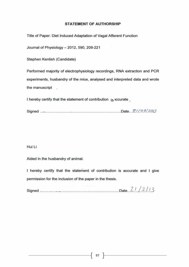
97
.
.

98

99

100
.

101
2.1 ABSTRACT
Afferent signals from the stomach play an important role in inhibition of food
intake during a meal. The gastric hormone ghrelin can influence gastric satiety
signalling by altering the sensitivity of gastric vagal afferents. Changes in diet,
including food restriction and high fat diet (HFD) induced obesity, alter satiety
signalling. We hypothesised that the function of gastric vagal afferent endings
are affected by both a period of food restriction and a high fat diet, and that
the inhibitory effect of ghrelin on vagal afferents is influenced by the different
feeding conditions. We found that both fasting and HFD reduced the
responses of gastric vagal tension receptors to distension, but not responses
of mucosal receptors to mucosal contact. We traced vagal afferents
anterogradely to their terminals in the mucosa where we found they were in
close apposition to ghrelin containing cells. Ghrelin receptor mRNA was
expressed in vagal afferent cell bodies of the nodose ganglia, and increased
in response to caloric restriction, but decreased in HFD mice. In control mice,
ghrelin decreased the sensitivity of tension but not mucosal receptors. After
caloric restriction or high fat diet, ghrelin inhibited mucosal receptors, and the
inhibition of mechanosensitive tension receptors was enhanced. Therefore,
both caloric restriction and HFD decrease mechanosensory vagal afferent
signals, and augment the inhibitory effect of ghrelin on vagal afferents, but
different mechanisms mediate the short- and longer-term changes.

102
2.2 INTRODUCTION
It is well established that signals generated in the gastrointestinal tract are
able to influence food intake [434]. One of these signals is mechanical
distension of the stomach which inhibits feeding via stimulation of vagal
afferents [435]. However, there are two distinct subtypes of vagal afferent
mechanoreceptors, mucosal receptors and tension receptors [287, 436, 437]
which are classified according to their response to specific types of
mechanical stimulation. Mucosal receptors respond to fine tactile stimulation
whereas tension receptors respond to stretch [436]. The role of gastric
distension in the control of food intake is clear but that of mucosal contact is
less obvious. In addition to generating sensations of nausea and vomiting, in
response to chemical stimuli, [438] mucosal receptors may be important in
detecting particle density, a factor which influences the rate of gastric
emptying, which in turn relates inversely to satiety [439, 440]. In response to
nutrients in the small intestine, enteroendocrine cells release peptides that
activate adjacent mucosal vagal afferents to elicit satiation [193]. There are
also enteroendocrine cells in the gastric mucosa that contain and release
peptides known to affect appetite [304, 327, 441]. The afferents innervating
the gastric mucosa are in an ideal position to detect both mechanical stimuli
and locally released hormones such as ghrelin.
Ghrelin is a 28 amino acid orexigenic peptide released from exocrine X/A cells
in the stomach [442-444]. The acute effect of ghrelin administration is a rapid
increase in food intake [345, 445-449] and chronic administration of ghrelin

103
increases body weight in rodents [448]. The data suggest that ghrelin is
involved in both the initiation of food intake and the signalling of energy deficit
and thus plays an important role in regulation of energy homeostasis [445,
448, 449]. At least part of the signalling mechanism for ghrelin involves the
vagus nerve and brain stem nuclei that ultimately signal to the hypothalamus
[355, 445]. There is controversy about the role of the vagus nerves in the
orexic effects of ghrelin. [355] showed that either truncal or selective gastric
vagotomy or perivagal capsaicin abolished its action when given intravenously
in rats. Asakawa et al. showed that ghrelin inhibited the resting discharge in
whole vagal nerve recordings and that lesion of vagal afferent fibres inhibited
the appetite-stimulating actions of ghrelin [445], whereas others have shown
no effect of vagotomy on the acute effects of ghrelin administered
intraperitoneally . The receptor for ghrelin, growth hormone secretagogue
(GHS) 1 receptor, is found in vagal afferents that project to the stomach [450]
and is expressed in mouse nodose ganglia [301]. We have reported that
ghrelin selectively inhibited a subpopulation of mechanosensitive gastric vagal
afferents [301]. Therefore there is an integral relationship between ghrelin and
gastric vagal afferent satiety signals.
In addition to the acute effect of gastrointestinal peptides on vagal afferent
satiety signals it is becoming apparent that they undergo longer-term changes
in neurochemical phenotype with changes in nutritional status [451]. For
example, it has been shown that withdrawal of food is responsible for changes
in expression of G-protein coupled receptors such as melanin-concentrating
hormone receptor-1 in vagal afferent neurons projecting to the gastrointestinal

104
tract [452]. High fat diets have also been reported to change the expression in
nodose ganglia of transcripts encoding receptors known to be involved in
appetite regulation, such as cholecystokinin (CCK1) and cannabinoid (CB1)
receptors [369, 453]. We have shown that ghrelin modulates the
mechanosensitivity of gastric vagal afferents in vitro [301] but it has not been
established whether feeding state affects this relationship between ghrelin
and gastric vagal afferents. Ghrelin release and plasma ghrelin levels depend
significantly on the feeding state; fasting causes increased plasma ghrelin
levels, whereas a high fat diet reduces plasma ghrelin levels in mice [441].
Using an in vitro gastro-oesophageal vagal afferent preparation to accurately
evaluate different populations of mechanosensitive vagal afferent fibres, we
investigated the effect of feeding state on the mechanosensitivity of gastric
vagal afferents. In addition, we determined the effect of three feeding states
on the neuromodulatory action of ghrelin on gastric vagal afferents. To
establish if any changes in function of ghrelin could be the result of changes in
receptor expression we determined the relative expression of ghrelin receptor
in the vagal afferent cell bodies in the nodose ganglia.

105
2.3 MATERIALS AND METHODS
2.3.1 Ethical approval
All experimental studies were performed with the approval of the animal ethics
committee of the Institute of Medical and Veterinary Science and the
University of Adelaide, and in accordance with the Australian Code of Practice
for the Care and Use of Animals for Scientific Purposes.
2.3.2 Short term restriction of food intake
Eight week old female C57/BL6 mice were randomly assigned to one of two
groups. The first group (controls) were fed ad libitum and allowed free access
to a standard laboratory diet. The second group were fasted for 14 hours
(fasted). Food was removed but they still had free access to water.
2.3.3 High fat diet model
Eight week old female C57/BL6 mice were randomly assigned to one of two
groups. The first group (controls) were fed ad libitum and allowed free access
to a standard laboratory diet (SLD) comprised of 7% energy from fat. The
second group were fed ad libitum and allowed free access to a high fat diet
(HFD; Specialty foods, Glen Forrest, Western Australia) comprised of 60%
energy from fat. Water was freely available for both SLD and HFD fed mice.
The mice were kept on either the SLD or the HFD for a period of 12 weeks
during which time food intake was measured. The weight of the mice was
monitored weekly. Blood was collected from a subset of these mice at the 12
week point, for determination of plasma glucose and insulin concentration.

106
2.3.4 In vitro mouse gastro-oesophageal afferent preparation
C57/BL6 mice were humanely killed via CO2 inhalation and the thorax was
opened by a midline incision. The stomach and oesophagus with attached
vagal nerves were removed and placed in a modified Krebs solution
containing (mM): NaCl 118.1, KCl 4.7, NaHCO3 25.1, NaH2PO4 1.3,
MgSO4.7H2O 1.2, CaCl2 1.5, citric acid 1.0 and glucose 11.1, bubbled with
95% O2 – 5% CO2 at 4ºC during dissection to prevent metabolic degradation.
After further dissection, the preparation was opened out longitudinally along
the oesophagus and greater curve of the stomach. The preparation was then
placed mucosal side up in the organ bath. This preparation has been
described in detail previously [300, 436]. Nifedipine (1 µM) was also added to
the Krebs solution to prevent smooth muscle contraction. In a previous
preliminary study we have shown that nifedipine has no effect on the
mechanical sensitivity of gastro-oesophageal vagal afferents [454].
2.3.5 Characterisation of gastro-oesophageal vagal afferent properties
Two types of mechanosensitive afferent fibre were studied, those responding
to mucosal stroking but not circular tension (mucosal receptors) and those
responding to mucosal stroking and circular tension (tension receptors) as
reported previously [436].
Location of receptive fields of all types of afferent fibre was determined by
mechanical stimulation throughout the preparation with a brush. Accurate
quantification of mechanical responses was performed differently according to

107
the primary adequate stimulus for the type of fibre. Mechanical thresholds of
both types of fibre were determined using calibrated von Frey hairs. The most
reproducible, stimulus-dependent responses of these afferents to mucosal
stroking were evoked when the probe was moved at a rate of 5 mm s-1 across
the receptive field rather than being kept static. Due to the fact that the
receptive fields were small (< 1 mm2), a single test at each intensity is prone
to missing the centre of the receptive field on some occasions. Therefore, we
minimised experimenter error by measuring the mean response to the middle
eight of ten standard strokes given at 1 s intervals. Because the von Frey hair
was bent throughout the stroking stimulus, the receptive field was subjected to
an even force as the hair passed over it. Tension-response curves were also
obtained and used in combination with von Frey thresholds to determine
whether the receptive fields of fibres were located in the mucosa or the
muscle layer. Tension stimuli were applied via fine suture silk attached via a
hook to an unpinned point adjacent to the mechanoreceptive fields. The
thread was attached to a cantilever via a pulley close to the preparation.
Reference standard weights were then placed on the opposite end of the
cantilever. Each weight was applied as a step and maintained for 1 min, and
the response was measured as the mean discharge evoked over this period.
Due to the fact that all responses to tension adapted slowly, this method of
assessment was considered representative of physiological responsiveness.
The tension-response curves were produced by applying weights to the
cantilever system in the range of 1-5 g. A recovery period of at least 1 min
was allowed between each tension stimulus. The effect of diet and
mechanical stimuli on responses of tension and mucosal receptors was

108
determined using a two-way ANOVA. For some animals the
mechanosensitivity of multiple receptors were determined. For analysis these
were averaged to provide one replicate per receptor type per animal.
2.3.6 Effect of ghrelin on the mechanosensitivity of vagal afferents
After mechanical sensitivity of the gastric and oesophageal vagal afferents
had been established, the effect of ghrelin on mechanical sensitivity was
determined. Ghrelin (1 nM) was added to the superfusing solution and
allowed to equilibrate for 20 min after which time the tension-response and
stroke-response curves were re-determined. This equilibration period was
observed so as to ensure penetration of the drug into all layers of the tissue.
After this time the tension-response and stroke-response curves were re-
determined. This procedure was repeated for ghrelin at increasingly higher
doses (3-10 nM). Time-controlled experiments were performed in which there
was no significant change in the mechanical responses over a comparable
duration. The effect of ghrelin and mechanical stimuli on the
mechanosensitivity of tension and mucosal receptors was determined using a
two-way ANOVA. The effect of diet on the modulatory action of ghrelin on
responses to mechanical stimulation was determined by accessing the
response to either stroking (50mg von Frey hair; mucosal receptors) or
tension (3g; tension receptors) at different concentrations of ghrelin (0, 1, 3, &
10nM). Significant differences between diets and ghrelin concentration were
assessed using a two-way ANOVA.

109
2.3.7 Drugs
Stock solution of the peptide ghrelin (0.1mM; Sigma, Australia) was kept
frozen (-80°C) and diluted to its final concentration in Krebs solution on the
day of the experiment.
2.3.8 Quantitative reverse transcription polymerase chain reaction
Nodose ganglia were removed bilaterally from the same animals used for the
in vitro mouse gastro-oesophageal vagal afferent preparation. RNA was
isolated using an RNeasy Micro RNA extraction kit as per the manufacturer‘s
instructions (Qiagen, Sydney, Australia). This process utilised RNeasy
MinElute SpinElute Columns, allowing adsorption of RNA to the silica
membrane, an on-column DNase digestion treatment, removal of
contaminants with simple wash steps and elution of the RNA with RNase-free
water. RNA quantification was determined by measuring the absorbance at
260 nm (A260) via a spectrophotometer (Bio-Rad, Regents Park, New South
Wales, Australia). RNA quality was estimated by the A260 and A280 nm ratio.
Quantitative RT-PCR (QRT-PCR) reactions were performed as described in
detail previously [455]. Briefly, QRT-PCR reactions were performed by using a
Chromo4 (MJ Research, Bio-Rad) real-time instrument attached to a PTC-200
Peltier thermal cycler (MJ Research) and analysed with Opticon Monitor
software (MJ Research). QRT-PCR reactions were performed with a Qiagen
QuantiTect SYBRgreen RT-PCR one-step RT-PCR kit (Qiagen) according to
the manufacturer‘s specifications with specific Quantitect Primer Assays
(Qiagen) optimised for the detection of the known sequence of mouse ghrelin

110
receptor and β-tubulin transcripts contained in the NCBI reference sequence
database (www.ncbi.nlm.nih.gov/RefSeq). These primer assays were used
under the following conditions: reverse transcription, 50°C for 30 min; initial
PCR activation, 95°C for 15 min; PCR cycles 94°C for 15 s, 55°C for 30 s, and
72°C for 30 s repeated for 50 cycles. A melting curve program verified the
specificity and identity of the RT-PCR products and no primer dimers were
observed. Confirmation of the amplified products was resolved by 3% agarose
gel electrophoresis and visualised via ethidium bromide staining. Each assay
was run in at least triplicate in separate experiments. Control PCRs were
performed by substituting RNA template with distilled RNAse-free water. All
assays were validated for linearity of amplification efficiency and quantitative
standard curves obtained by serial dilutions of RNA. Calculations for relative
mRNA transcripts expression were performed by the comparative CT method,
comparing to the internal reference gene β-tubulin using the equation ∆CT (CT
of target transcript - CT of β-tubulin). To determine the relative expression of
these transcripts in whole nodose ganglia from high fat fed mice compared to
standard laboratory fed mice and fasted mice compared to mice fed ad
libitum, the ∆∆CT was calculated by using the formula ∆∆CT= ∆CT (ghrelin
receptor in HFD or fasted mice) - ∆CT (ghrelin receptor in SLD or fed ad
libitum mice respectively). As such fold differences were calculated relative to
the levels of β-tubulin RNA in the same sample. The relative fold differences
were calculated by using the modified version of 2-∆∆CT [301, 456] correcting
for PCR efficiencies. Quantitative data are expressed as means ± SEM, and
significant differences in transcript expression were determined using a
nonparametric Mann-Whitney test at a significance level of p<0.05.

111
2.3.9 Tracing studies
Adult (8 weeks old) female C57BL/6 mice had ad libitum access to water and
a SLD and were fasted overnight before experimentation.
Anterograde tracing
The mice were anaesthetised with isoflurane (1-1.5% in oxygen). The left
nodose was exposed and 0.5μL of wheat-germ agglutinin conjugated
horseradish peroxidise [WGA-HRP (4mg.ml-1); Vector Laboratories] was
pressure injected into the nodose ganglia via a glass micropipette (ID=25μm).
The injection site was then dried, the skin incision closed and antibiotics
(Terramycin; 10mg/kg) and analgesic (butorphanol; 5mg/kg) administered
subcutaneously. Two days following injection, mice were anaesthetised with
pentobarbitone (60mg/kg, i.p.) and transcardially perfused with heparinised
saline at 40°C, then 4% paraformaldehyde in 0.1M PBS (PFA-PBS) at 4°C.
The stomach was then removed and cryoprotected with 30% sucrose for a
minimum of 18 hours. The stomach was then frozen and 10μm transverse
sections cut. Permanent visualisation of WGA-HRP was achieved using
tyramide signal amplification (TSA). Briefly, the tissue sections were rinsed in
TNT buffer (0.05% Tween 20, 0.15M NaCl, and 0.1M Tris-HCl, pH 7.5),
blocked for 30 min in TNB (prepared as per the directions provided with the
Perkin-Elmer TSA biotin reagent kit), and then incubated for 20 min with
tyramide-biotin, diluted 1:50 in amplification diluent. The tissue was then
rinsed in TNT buffer and reacted with streptavidin conjugated to Alexa Fluor®
647 (Invitrogen) for 2 hr at room temperature. Following ghrelin

112
immunohistochemistry (below) sections were coverslipped using ProLong
antifade mounting media (Invitrogen).
Immunohistochemistry
Immunoreactivity for ghrelin was detected in anterograde traced stomach
sections using a chicken polyclonal antibody to ghrelin (Abcam). Primary
antibody was visualised using goat anti-chicken secondary antibody
conjugated to Alexa Fluor®488 (Invitrogen). Briefly, sections were air dried at
room temperature and rinsed in PBS + 0.2% Triton X-100 (Sigma-Aldrich;
PBS-T, pH7.4) to facilitate antibody penetration. Primary antibody was diluted
1:1000 in PBS-T and incubated at 4°C overnight. Unbound antibody was then
removed with PBS-T washes and sections were incubated for 1hr at room
temperature with secondary antibody (1:200 in PBS-T). The sections were
given final PBS-T washes, drained and mounted with ProLong Antifade
(Invitrogen). Slides where the primary antibody was omitted showed no
labelling and served as negative controls.
Visualisation
Slide sections were visualised using an epifluorescence microscope (BX-51,
Olympus) equipped with filters for AF647 and AF488, with images acquired by
a Cool-Snapfx monochrome digital camera (Roper Scientific). Individual
fluorochromes were pseudo-coloured using Olympus imaging analySis
software: however the luminance of images was not adjusted.

113
2.4 RESULTS
2.4.1 Short term restriction of food intake reduces mechanosensitivity of
vagal afferents
The effect of mechanical stimulation on gastric and oesophageal vagal
afferents from control and fasted mice is illustrated in Figure 2.1. The
mechanosensitivity of mucosal receptors was similar in both groups of mice
(Figure 2.1Ai and ii respectively). In contrast, the responses of gastric tension
receptors from fasted mice were significantly smaller than those in control
mice (p<0.0001: diet effect, two-way ANOVA; Figure 2.1Bi). Oesophageal
tension receptors were more variably affected, and although there appears to
be a similar reduction in the responses of oesophageal tension receptors from
fasted mice the difference in response did not differ significantly from those in
control mice (Figure 2.1Bii: p>0.05: diet effect, two-way ANOVA).
2.4.2 Effects of long term alterations in diet
Mice on the HFD increased in weight by 6.57 ± 0.3g (N=51) over the 12 week
period that they were on the diet (N=51) and gained significantly more weight
(p<0.001; unpaired t-test) than SLD mice who gained 5.22 ± 0.12g (N=52).
This is likely to be due to the significant increase (p<0.0001; unpaired t-test) in
energy intake, over the 12 week period of the HFD mice (4798.97 ±
149.49MJ; N=11) compared to the SLD mice (3434.82 ± 99.72MJ; N=12) in
accord with previous reports [457]. The gonadal fat pad weight was
significantly heavier (p<0.01; unpaired t-test) in HFD mice (0.55±0.07g; N=17)
compared to SLD mice (0.32±0.03g; N=16). Blood glucose and insulin levels

114
were not significantly different between SLD (7.34 ± 0.64mmol/L and 0.41 ±
0.08pmol/L respectively; N=11) and HFD (5.96 ± 0.48mmol/L and 0.66 ±
0.15pmol/L respectively; N=11) indicating that the HFD group did not
developed diabetes over the 12 week diet period.
The effect of mechanical stimulation on gastric and oesophageal vagal
afferents from both groups of mice is illustrated in Figure 2.2. The
mechanosensitivity of mucosal receptors was not significantly different
between SLD and HFD mice (Figure 2.2Ai and ii). However, the response of
gastric and oesophageal tension receptors of mice fed a HFD was
significantly less than the responses of mice fed a SLD (p<0.001: diet effect,
two-way ANOVA; Figure 2.2Bi and ii respectively). Thus the effect of a HFD
on the mechanosensitivity of gastro-oesophageal tension and mucosal
receptors appears similar to the effect of fasting (Figure 2.1).
2.4.3 Anatomy of vagal afferent endings and ghrelin containing cells in
the gastric mucosa
We determined the anatomical relationship of ghrelin immunopositive cells in
the gastric mucosa with identified vagal afferent endings. Ghrelin-like
immunoreactivity was restricted to isolated cells at the base of submucosal
glands with apical surfaces in the lumen (Figure 2.3Aii) which had morphology
consistent with enteroendocrine X/A cells. Anterograde tracing of vagal
afferents from the nodose ganglion revealed intraepithelial fibres within the
mucosal layer of the stomach (Figure 2.3Ai), as previously described in the
small intestine [458]. These were closely apposed (within a few micrometres)

115
to ghrelin containing cells (Figure 2.3Aiii). Identified vagal endings also
surrounded myenteric ganglia, consistent with previously characterised
intraganglionic laminar endings [288]. These endings were located at a
distance from ghrelin-containing cells separated by the lamina propria,
submucosa, muscularis mucosae and circular smooth muscle (data not
shown).
2.4.4 Ghrelin receptor expression in vagal afferent pathways
QRT-PCR was used to determine expression of ghrelin receptor in vagal
afferent neurons in the nodose ganglia. Quantitative analysis revealed that
ghrelin receptor mRNA levels in the nodose ganglia of mice fasted for 14
hours were 1.53-fold higher than in fed mice (p<0.05: Mann-Whitney test;
Figure 2.3B). There was no significant difference in the expression of ghrelin
receptor mRNA detected in the nodose ganglia of HFD mice compared to
SLD mice (Figure 2.3B). Agarose gel electrophoresis confirmed amplification
of housekeeper (β-tubulin) and ghrelin receptor with product sizes of 118 and
121bp respectively (Figure 2.3C).
2.4.5 Vagal afferent responses to ghrelin are altered by changes in food
intake
An overnight fast had major effects on the ghrelin sensitivity of gastric
mucosal receptors (Figure 2.5). The response to mucosal stroking of gastric
mucosal receptors was significantly reduced in fed mice, but only at the
highest dose of ghrelin (10nM; Figure 2.4Ai; p<0.05: ghrelin effect, two-way
ANOVA). In fasted mice ghrelin (1-10nM) significantly reduced the responses

116
of gastric mucosal receptors to mucosal stroking with calibrated von Frey
hairs (10-1000mg; p<0.01: ghrelin effect, two-way ANOVA; Figure 2.5Aii).
When the response to 50mg von Frey hair at varying concentrations of ghrelin
(0-10nM) was plotted (Figure 2.4B) it was evident that diet significantly altered
the response to mechanical stimulation in the presence of ghrelin (P<0.01;diet
effect, two-way ANOVA). Therefore gastric mucosal receptor sensitivity to
ghrelin was significantly increased following an overnight fast.
Ghrelin (3-10nM) significantly and dose-dependently reduced the response of
gastric (Figure 2.5Ai & Figure, 2.5Bi) tension receptors to circular tension
(p<0.05: ghrelin effect, two-way ANOVA; 1-5g). In fasted mice ghrelin (1-
10nM) also significantly reduced the response of gastric tension receptors
(Figure 2.5Aii & 2.5Bii) to circular tension even at the lowest dose tested of
1nM (p<0.001: ghrelin effect, two-way ANOVA). When response to a 3g load
was plotted against increasing concentrations of ghrelin there was no
significant difference between the fed and fasted curves (data not shown).
Therefore the sensitivity of gastric tension receptors to ghrelin was unaffected
by a short term restriction of food intake.
The effect of longer term changes in food intake on ghrelin‘s neuromodulatory
action on vagal afferent responses to mechanical stimulation is illustrated in
figures 2.6 and 2.7. Ghrelin (1-10nM) did not significantly affect the response
of gastric (Figure 2.6Ai & 2.6Ci) mucosal receptors to mucosal stroking in SLD
mice. In contrast, ghrelin significantly reduced the response to mechanical
stimulation of gastric (Figure 2.6Aii & 2.6Cii) mucosal receptors (p<0.05:

117
ghrelin effect, two-way ANOVA) in HFD mice, thus mimicking responses seen
in the fasted state. When the response to stroking with a 50mg von Frey hair
was plotted against varying concentrations of ghrelin (0-10nM; Figure 2.6B) it
was evident that diet significantly altered the response to mechanical
stimulation in the presence of ghrelin (P<0.01: diet effect, two-way ANOVA).
Therefore gastric mucosal receptor sensitivity to ghrelin was significantly
increased after a HFD.
Ghrelin significantly reduced the response of gastric tension receptors to
circular stretch (1-5g) in SLD and.HFD mice (Figure 2.7i & ii respectively;
p<0.05: ghrelin effect, two-way ANOVA). When the response to a 3g load was
plotted against increasing concentrations of ghrelin there was no significant
difference between the SLD and HFD curves (data not shown). Therefore the
sensitivity of gastric tension receptors to ghrelin was unaffected by a HFD.

118
2.5 DISCUSSION
These data provide the first evidence that signalling of gastric distension by
vagal afferents is significantly reduced by both food withdrawal and a high fat
diet. Moreover, ghrelin inhibits mucosal vagal afferents that are normally
insensitive to it. Thus, not only is the gastric vagal afferent satiety signal
reduced by food withdrawal and high fat feeding, but the orexigenic effect of
ghrelin is also enhanced.
Short-term restriction of food intake selectively reduced the
mechanosensitivity of tension receptors and in particular gastric tension
receptors. As these afferents detect distension of the stomach [288, 290, 296]
their reduced sensitivity to stretch may delay satiety signalling, leading to
increased food intake of an individual in order to achieve satiation; this would
constitute a natural adaptive response after a fast. Importantly we found that
in mice chronically fed a high fat diet there was also a reduction in the
mechanosensitivity of tension receptors suggesting that the satiety signal
would also be attenuated in response to a high fat diet. Such a mechanism
may have evolved to maximise assimilation of energy from calorie-rich foods
in anticipation of famine. A number of studies have shown that obese humans
have increased gastric capacity [432, 459] which results in increased energy
intake. This could occur due to a reduction in the gastric distension-stimulated
vagal afferent satiety signal. A recent publication has revealed that the
sensitivity of jejunal afferents to satiety stimuli is reduced after a HFD [381].
Further, the major mechanism for this decrease in sensitivity was due to a

119
reduction in excitability of the neuronal cell membrane [381]. This reduced
excitability of all vagal afferent cell membranes in HFD mice may explain the
reduced mechanosensitivity of oesophageal afferents observed in the present
study.
The mechanisms leading to reduced mechanosensitivity of vagal afferents are
currently unknown both in terms of its precise causes and how it is manifested
at the molecular level. It has been shown that fasting alters the neurochemical
phenotype of vagal afferent neurons that project to the gastrointestinal tract in
terms of the G-protein coupled receptors and peptide neurotransmitters they
express [368, 424, 452, 460]. High fat diets have also been reported to modify
vagal afferents transcript expression of specific G-protein coupled receptors
and growth factors [369, 453, 461]. Together with changes in the levels of
plasma satiety hormones these adaptations are likely to profoundly alter vagal
satiety signals during a high fat diet. For example, the plasma levels of leptin
are significantly elevated in mice fed a high fat diet [462] and this adipokine
may directly influence vagal afferent mechanosensitivity while also acting to
modulate their responsiveness to ghrelin. Our data now adds new knowledge
that diet induces changes in the fundamental mechanosensory properties of
gastric vagal afferents. Now that we have demonstrated that reduced
mechanosensitivity is a robust outcome in response to a HFD, we are in a
good position to investigate the mechanism of this change, which may reveal
more empirical targets for the treatment of obesity.

120
In the present study we confirmed our earlier findings [301] that ghrelin
selectively and dose-dependently reduces the response of tension receptors
in normal control mice fed ad libitum on a SLD. This effect of ghrelin on
tension receptors was maintained after food withdrawal and after a HFD. The
effect of ghrelin was not selective for gastric vagal afferents as it also reduced
the mechanosensitivity of oesophageal tension sensitive afferents which are
distant from the site of ghrelin release (data not shown), suggesting this would
be a systemic action in vivo. The concentration of ghrelin used in our in vitro
organ bath preparation is comparable with the concentration of ghrelin in
mouse plasma which ranges from 1 – 3 nM depending on the feeding state
[441]. Plasma ghrelin levels are significantly higher after withdrawal of food for
18 hours and significantly reduced in mice fed a HFD [441]. Therefore, based
on plasma levels and the fact that sensitivity of gastric vagal tension receptors
to ghrelin did not significantly change in both conditions, it could be predicted
that ghrelin inhibition of tension receptors would be increased after food
withdrawal, but decreased in mice fed a high fat diet.
The response of mucosal receptors to mucosal stroking was unaffected by
both a short-term restriction in food intake and a high fat diet. However, where
these particular vagal afferent fibres become relevant is their innervation of
the gastric mucosa, where they are also likely to detect locally released
hormones such as ghrelin. Indeed, we observed that vagal afferents
innervating the gastric mucosa are in close apposition to ghrelin containing
epithelial cells. However, in agreement with our earlier observations [301],
mucosal receptors were unaffected by ghrelin in control mice other than at the

121
highest dose used. After food withdrawal ghrelin potently reduced the
response of gastric vagal mucosal receptors to mucosal stroking. This effect
was seen also after a high fat diet, so mucosal receptors acquire ghrelin
sensitivity. Gastric mucosal receptors are considered to be important in
detecting particle size, which reduces gastric emptying and food intake [439,
440], so ghrelin could act to reduce this signal in fasting and obesity. In fasted
healthy volunteers, ghrelin administration has been shown to increase the rate
of gastric emptying in addition to elevating hunger ratings [463]; this may be a
consequence of the decreased mechanosensitivity of gastric mucosal
receptors by ghrelin.
The expression of ghrelin receptor transcript in the nodose ganglia was
significantly increased in mice that were fasted for 14 hours. An increase in
ghrelin receptor expression may explain the increase in effect of ghrelin on
gastric mucosal receptors. Correspondingly it has been established that
growth hormone secretagogue (GHS) receptor containing vagal neurons
project to the stomach [450] although reports vary on changes in ghrelin
receptor expression after food withdrawal. One group has reported that the
transcript levels of ghrelin receptor in the rat nodose ganglia are unaltered
[353] while another group has shown a doubling of transcript expression in
fasted compared to fed rats [464]. Previously we reported that ghrelin reduced
the mechanosensitivity of mucosal receptors in the ferret and concluded that
this was a possible species difference [301]. The difference is most likely due
to the fact that ferrets were fasted prior to experimentation. The expression of
ghrelin receptor in the nodose ganglia of mice fed a HFD was not significantly

122
different from mice fed a SLD, and therefore the increased effect of ghrelin on
gastric mucosal receptors cannot be explained by an increase in ghrelin
receptor expression unlike the effect of fasting. It is possible that in the longer
term, changes occur in the coupling of ghrelin receptors to specific
intracellular pathways, rather than in absolute levels of ghrelin receptor
expression.
In conclusion, we have established that the basic mechanosensitivity of
gastro-oesophageal vagal afferents, particularly gastric afferents, is
significantly reduced by both restriction and excess of energy intake. A
consequence of this decreased mechanosensitivity would be an undesirable
dampening of the satiety signal in obesity. In addition, the inhibitory effect of
ghrelin on vagal afferents becomes less selective after food withdrawal and a
high fat diet and is therefore amplified. The additional inhibitory effect of
ghrelin on gastric mucosal receptors would reinforce the inhibitory effect of
ghrelin on tension receptors and enhance its orexigenic effect. This data
indicates the plasticity in the mechanism of action of ghrelin under different
feeding states, and the complex role of gastric mechanisms in food intake
regulation.

123

124
Figure 2.1: The effect of short term restriction in food intake on the
response of gastric and oesophageal mechanosensitive vagal afferents
to mechanical stimulation. Stimulus-response functions of mucosal (A) and
tension sensitive (B) gastric (i) and oesophageal (ii) vagal afferents from mice
fed ad libitum (●) and mice fasted for 14 hours (○). ***P<0.001 compared with
fed mice (Diet effect, two-way ANOVA).

125

126
Figure 2.2: The effect of a high fat diet on the response of gastric and
oesophageal mechanosensitive vagal afferents to mechanical
stimulation. Stimulus-response functions of mucosal (A) and tension
sensitive (B) gastric (i) and oesophageal (ii) vagal afferents from mice fed a
standard laboratory diet (SLD; ●) and mice fed a high fat diet (HFD; ○).
*P<0.05, ***P<0.001 compared with mice fed a standard laboratory diet (Diet
effect, two-way ANOVA).

127

128
Figure 2.3: Relationship between vagal afferents and ghrelin containing
cells in the gastric mucosa. Combined ghrelin immunohistochemistry and
anterogradely traced vagal afferent fibres in the mouse stomach (Ai-iii). The
stomach sections show a cross section through a gland. (Ai) Anterogradely
traced (WGA-HRP) vagal afferent fibre (yellow arrow). (Aii) Epithelial cells
immunopositive for ghrelin in stomach villi (white arrows). (Aiii) Overlay of i
and ii showing close proximity of ghrelin-containing epithelial cells and
anterograde labeled vagal afferent fibres. (B) Relative expression of growth
hormone secretagogue 1 (GHS) receptor mRNA in nodose from HFD mice
compared with SLD mice (open bar) and fasted mice compared with fed mice
(closed bar). The relative expression values were calculated using ∆∆CT (∆CT
HFD - ∆CT SLD or ∆CT fasted - ∆CT fed ad libitum) of ghrelin receptor
transcript. The fold difference in transcript expression in HFD nodose relative
to SLD and nodose from fasted mice compared to mice fed ad libitum was
calculated using the formula 2-∆∆CT. In nodose from mice fasted for 14hr there
is a 1.53 fold increase in transcript compared to fed mice (closed bar,
*P<0.05; Mann Whitney test). In nodose from HFD mice there is a small but
not significant 0.06 fold decrease in transcript compared to SLD mice (open
bar). (C) Agarose gel electrophoresis confirmed the amplified product sizes.

129

130
Figure 2.4: Fasting increases the effect of ghrelin on the response of
gastric mucosal receptors. (A) The responses of gastric mucosal receptors
to mucosal stroking with calibrated von Frey hairs (10-1000mg) in the
absence (●) and presence of ghrelin (1 (○), 3 (■) and 10nM (□). These
recordings were taken from fed mice (i) or fasted mice (ii) prior to the
experiment. *P<0.05, **P<0.01, ***P<0.001 compared with control (●; Ghrelin
effect, two-way ANOVA). (B) The effect of ghrelin on the responses of
mucosal receptors to stoking with a 50mg von Frey hair in mice fed ad libitum
(●) and mice fasted (○) for 14 hours. Diet significantly enhanced the inhibitory
action of ghrelin (**P<0.01; diet effect, two-way ANOVA).

131

132
Figure 2.5: The effect of ghrelin on the response of gastric tension
receptors. (A) The responses of gastric tension sensitive receptor to
circumferential stretch (1-5g) in the absence (●) and presence of ghrelin (1
(○), 3 (■) and 10nM (□). These recordings were taken from fed (i) or fasted
mice (ii). *p<0.05, **P<0.01, ***P<0.001 compared with control (●; ghrelin
effect, two-way ANOVA). There was no significant difference in the inhibitory
effect of ghrelin between the two diets. (B) Typical traces of tension receptors
from a fed (i) and a fasted (ii) mouse.

133

134
Figure 2.6: High fat diet increases the effect of ghrelin on the response
of gastric (A & C) mucosal receptors. (A & C) The responses of gastric
mucosal receptors to mucosal stroking with calibrated von Frey hairs (10-
1000mg) in the absence (●) and presence of ghrelin (1 (○), 3 (■) and 10nM
(□). These recordings were taken from mice fed a standard laboratory diet (Ai
& Ci) or mice fed a high fat diet (Aii & Cii) for 12 weeks prior to the
experiment. **P<0.01 compared with control (●; ghrelin effect, two-way
ANOVA). (B) The effect of ghrelin on the response of mucosal receptors to
stoking with a 50mg von Frey hair in mice fed a SLD (●) and mice fed a HFD
(○). Diet significantly enhanced the inhibitory action of ghrelin (**P<0.01; diet
effect, two-way ANOVA).

135

136
Figure 2.7: The effect of ghrelin on the response of gastric tension
receptors. The responses of gastric tension receptors to circumferential
stretch (1-5g) in the absence (●) and presence of ghrelin (1 (○), 3 (■) and
10nM (□)). These recordings were taken from mice fed a standard laboratory
diet (i) or mice fed a high fat diet (ii) for 12 weeks prior to the experiment.
*p<0.05, **P<0.01 compared with control (●; ghrelin effect, two-way ANOVA).
There was no significant difference in the inhibitory effect of ghrelin between
the two diets.
137
CHAPTER 3
GASTRIC VAGAL AFFERENT MODULATION BY
LEPTIN IS INFLUENCED BY FOOD INTAKE
STATUS.
Kentish SJ, O'Donnell TA, Isaacs NJ, Young RL,
Li H, Harrington AM, Brierley SM, Wittert GA,
Blackshaw LA, Page AJ.
Nerve Gut Research Laboratory, University of
Adelaide, Adelaide, SA 5005, Australia
Published in The Journal of Physiology
2013 April; 591(7):1921-1934

138
.
.

139

140

141

142
3.1 ABSTRACT
Energy intake is strongly influenced by vagal afferent signals from the
stomach, and is also modulated by leptin. Leptin may be secreted from gastric
epithelial cells, so we aimed to determine the direct effect of leptin on gastric
vagal afferents under different feeding conditions. Female C57BL/6 mice were
fed standard laboratory diet, high fat diet or were food restricted. The
expression of leptin receptor and its signal transduction molecules in vagal
afferents was determined by retrograde tracing and reverse-transcription
polymerase chain reaction (RT-PCR), and the relationship between leptin
immunopositive cells and gastric vagal afferent endings determined by
anterograde tracing and leptin immunohistochemistry. An in vitro preparation
was used to determine the functional effects of leptin on gastric vagal
afferents and the second messenger pathways involved. Leptin potentiated
vagal mucosal afferent responses to tactile stimuli, and epithelial cells
expressing leptin were found close to vagal mucosal endings. After fasting or
diet-induced obesity, potentiation of mucosal afferents by leptin was lost and
leptin receptor expression reduced in the cell bodies of gastric mucosal
afferents. These effects in diet-induced obese mice were accompanied by a
reduction in anatomical vagal innervation of the gastric mucosa. In striking
contrast, after fasting or diet-induced obesity, leptin actually inhibited
responses to distension in tension receptors. The inhibitory effect on gastric
tension receptors was mediated through PI3K-dependent activation of BKCa
channels. The excitatory effect of leptin on gastric mucosal vagal afferents
was mediated by PLC-dependent activation of TRPC1 channels. These data

143
suggest the effect of leptin on gastric vagal afferent excitability is dynamic and
related to the feeding state. Paradoxically, in obesity, leptin may reduce
responses to gastric distension following food intake.

144
3.2 INTRODUCTION
The satiety hormone, leptin, which is secreted from adipose tissue provides a
signal to the hypothalamus reflecting the amount of fat stored [465, 466] and
serves as a long-term regulator of nutrient intake, adiposity and body weight
[467]. Levels of circulating leptin reflect the degree of adiposity [466].
However, despite elevated leptin levels obese individuals do not show
diminished appetite, suggesting a relative resistance to leptin [305]. An
alternative explanation may be that leptin permits fat stores to be maintained
at or around a set point, that is genetically determined but, susceptible to
environmental modification [174].
Leptin is also expressed in, and secreted by, chief cells [304] and parietal (P)
cells [468] in the gastric mucosa. Leptin released from this site could act on
adjacent gastric vagal afferent endings to modulate peripheral signals in
response to food intake. Indeed, leptin has been shown to directly activate
cultured gastric and duodenal vagal afferents from rats [333, 334] and the
leptin receptor (Lep-R) is expressed in rat vagal afferent neurons [469, 470],
many of which terminate in the stomach. A vagal action of gastric leptin may
serve to augment the acute appetite suppressant effects of circulating leptin
[326]. However, recent evidence shows that the effects of leptin on vagal
afferents are lost with chronic high fat diet feeding [335, 336].
Within the stomach wall there are two classes of mechanically sensitive
gastric vagal afferents. In the muscular layer, tension receptors detect fullness

145
by responding to distension and contraction of the stomach wall [471, 472]. In
contrast, mucosal receptors are excited by mechanical contact of larger food
particles with the epithelium, and may contribute to the discrimination of
particle size. The net effect of activating mucosal afferents is to trigger vagal
reflexes which slow gastric emptying and facilitate mechanical digestion in the
stomach [297]. It is currently unknown whether leptin has differential effects
on gastric vagal afferent subclasses or whether any such effects can be
modified by caloric restriction or excess. Furthermore, the mechanism by
which Lep-Rs trigger changes in the electrical activity of vagal afferent
endings has not been established.
We therefore determined the effect of leptin on the mechanosensitivity of
gastric vagal afferents under different feeding conditions, including a short
term restriction in food intake and long term consumption of a high fat diet
(HFD), and examined the second messenger signalling pathways activated by
leptin. In addition, we established the relationship between leptin containing
cells in the gastric mucosa and vagal afferent endings.

146
3.3 MATERIALS AND METHODS
3.3.1 Ethical approval
All studies were approved and performed in accordance with the guidelines of
the Animal Ethics Committees of the University of Adelaide and SA Pathology,
Adelaide, Australia.
3.3.2 Mice
All mice used for this study were housed with littermates in groups of four,
unless otherwise stated. For the fed and fasted treatment groups, 8wk old
female C57BL/6 mice, housed individually, were fed ad libitum or fasted for 14
hours prior to experimentation. In diet studies, 7 week old female C57BL/6
mice were allowed to acclimatize for a period of one week after which control
mice were continued on a standard laboratory mouse diet (SLD (7% energy
from fat), n=52), whilst the HFD mice (n=51) were placed on a diet high in fat
(60% of energy from fat, Specialty Feeds, Glen Forrest, WA, Australia) for a
further 12 weeks. During this period the mice were weighed weekly. We have
previously determined that female C57BL/6 mice fed a HFD have increased
total body weight and fat mass over a 12 week feeding period compared to
SLD mice [473]. Reduced mechanosensitivity of gastric afferents observed in
female HFD mice [473] has also been observed in intestinal afferents of male
mice [381] suggesting that diet induced modulation of vagal afferent satiety
signals are independent of gender.

147
3.3.3 In vitro mouse gastro-oesophageal afferent preparation
This preparation has been described in detail previously [436]. In short,
C57BL/6 mice were killed via CO2 inhalation and the thorax opened by a
midline incision. The stomach and oesophagus, with intact vagal nerves, were
removed. The stomach and oesophagus were opened out longitudinally mid
way between the two main vagal branches, either side of the oesophagus,
and along the greater curvature of the stomach. The tissue was then pinned
down mucosa side up in an organ bath containing a modified Krebs solution
composing of (in mM): 118.1 NaCl, 4.7 KCl, 25.1 NaHCO3, 1.3 NaH2PO4, 1.2
MgSO4.7H2O, 1.5 CaCl2, 1.0 citric acid, 11.1 glucose and 0.001 Nifidipine,
bubbled with 95% O2- 5% CO2. The dissection process was carried out at
4°C.
3.3.4 Characterization of gastric vagal afferent properties
Receptive fields of mechanically sensitive vagal afferents, described earlier,
were initially located using mechanical stimulation by brushing gently along
the mouse gastro-oesophageal preparation. Once located specific stimuli
were then applied. Mucosal stroking was performed using calibrated von Frey
hairs (10-1000mg) which were stroked across the mucosa at a rate of 5mm/s.
Each receptive field was stroked ten times and mechanical responses from
the middle eight strokes were taken for analysis. Circular tension was applied
using a threaded hook attached to an underpinned point adjacent to the
receptive field. The threaded hook was attached to a cantilever via a pulley
close to the preparation. Standard weights (0.5-5g) were then placed on the
opposite end of the cantilever. Each weight was applied for one minute with a

148
break of another minute between removing one weight and applying the next.
After analysing the two stimulus response curves, we classified a receptive
field as either a mucosal or tension receptor.
3.3.5 Single unit vagal afferent recordings
Single units were discriminated on the basis of action potential shape,
duration, and amplitude by use of Spike 2 software (Cambridge Electronic
Design, Cambridge, UK). After mechanosensitivity of a receptive field was
established the effect of leptin on this mechanosensitivity was assessed by
adding leptin (0.1nM) to the Krebs and allowing it to equilibrate for 20 minutes.
This equilibration period was observed so as to ensure penetration of the drug
into all layers of the tissue. After this time the tension-response and stroke-
response curves were re-determined. This procedure was repeated for leptin
at increasingly higher doses (1-10nM). Time-controlled experiments were
performed in which there was no significant change in the mechanical
responses over a comparable duration. The second messenger system used
by leptin to elicit its effects on mechanosensitivity was established using
inhibitors of Janus kinase 2 (JAK2) - AG490 (5µM) [474], phosphatidylinositol
3-kinases (PI3K) -wortmannin (5nM) [475], phosphodiesterase 3 (PDE3) -
cilostamide (5µM) [476], phospholipase C (PLC) - U73122 (10µM) [477], large
conductance calcium activated potassium channels (BKCa) - iberiotoxin
(100nM) [478] and canonical transient receptor potential cation channels
(TRPC) channels - 2-APB (30µM) [479], at concentrations previously reported
in the literature before and after the addition of leptin (10nM) in a separate
series of experiments to the leptin dose-response experiments.

149
3.3.6 Drugs
Stock solutions of all drugs were kept frozen at -80°C and diluted to their final
concentration in Krebs immediately prior to their use in the experiment. Leptin
was obtained from Sigma-Aldrich (Sydney, Australia). Iberiotoxin, wortmannin,
cilostamide, U73122, 2-APB and AG490 were obtained from Tocris
Bioscience (Bristol, UK).
3.3.7 Nodose ganglia quantitative RT-PCR
Nodose ganglia were removed bilaterally from mice from all 4 experimental
groups. Total RNA was extracted using an RNeasy Micro Kit (Qiagen,
Doncaster, Australia) according to the manufacturer‘s instructions. RNA was
quantified by measuring the absorbance at 260nm (A260) using a
NanoDrop™ ND 1000 spectrophotometer (Thermo Scientific) and RNA purity
was estimated via the 260/280 absorbance ratio. Quantitative RT-PCR
reactions were performed as described in detail previously [455]. Primers
targeting Lep-R, TRPC1, TRPC5 and β-tubulin were pre-designed Quantitect
Primer assays (Qiagen). The primer pairs for KCa1.1 were designed using
Primer 3.0 software (Applied Biosystems; Foster City, USA). Forward primer
(TCTACTTTGGCTTGCGGTTT) and reverse primer
(CAAGCCAAGCCAACTTCTGT) for KCa1.1 were supplied by Geneworks
(Adelaide, Australia). RT-PCR reactions were carried out using 10ng of RNA
under the following conditions: reverse transcription, 50°C for 30min; initial
PCR activation, 95°C for 15min: PCR cycles 94°C for 15s, 55°C for 30s and
72°C for 30s repeated for 50 cycles. A melt curve was obtained to confirm the

150
specificity of the products produced and products were visualised using 3%
agarose gel electrophoresis with ethidium bromide staining. Relative RNA
levels were calculated using the comparative CT method as described
previously [456]. Quantitative data are expressed as mean ± SEM.
3.3.8 Anterograde tracing
This procedure has been previously documented [298]. In brief, mice from the
SLD (n=7) and HFD (n=7) groups were anaesthetised with isoflurane (1-1.5%
in oxygen). The left nodose ganglion was exposed, and 0.5 µl of a
horseradish peroxidase conjugate of wheat-germ agglutinin (WGA-HRP, 4
mg/ml; Vector Laboratories) was pressure injected into the nodose ganglion
via a glass micropipette (ID 25 µm). The injection site was dried and skin
incision closed. Antibiotic (terramycin; 10 mg/kg) and analgesic (butorphanol;
5 mg/kg) were administered subcutaneously. Two days following injection of
tracer (WGA-HRP), into the left nodose, the stomach was removed and
dissected into ventral and dorsal flat sheets along the greater and lesser
curvature. Frozen serial transverse sections of ventral flat sheet (10 µM) were
then cut for immunohistochemistry. The dorsal flat sheet had submucosa and
mucosa gently removed leaving the smooth muscle layers and myenteric
plexus intact.
Permanent visualization of WGA-HRP was carried out using tyramide signal
amplification (Perkin Elmer, USA) [298]. Briefly, dorsal sheet whole mounts
and on-slide sections were rinsed in TNT buffer (0.05% Tween 20, 0.15 M
NaCl, and 0.1 M Tris-HCl, pH 7.5), blocked for 30min in TNB (supplied with

151
Perkin-Elmer TSA reagent kit), and then reacted for 20min with tyramide-
biotin, diluted 1:50 in amplification diluent. The tissue was then rinsed in TNT
buffer and reacted with streptavidin conjugated to Alexa Fluor® 647 (1:200,
Invitrogen) for 1-2hrs at room temperature. Frozen sections were
subsequently processed for leptin immunohistochemistry, then both sections
and wholemount specimens were mounted on slides and cover slipped using
ProLong antifade (Invitrogen).
3.3.9 Retrograde tracing
Cell bodies of gastric vagal afferents innervating specific stomach layers were
identified using differential tracing from the stomach as previously
documented [298].
Gastric muscle: SLD (n = 5) and HFD (n=5) mice were anaesthetised with
isoflurane (1-1.5% in oxygen), a laparotomy performed, and an Alexa Fluor®
555 conjugate of cholera toxin β-subunit (CTB-AF555 (0.5%); Invitrogen)
injected subserosally into the muscularis externa of the proximal stomach
using a 30 ga Hamilton syringe. Multiple equally spaced injections of 2 µl were
made parallel to and 1–2 mm from the lesser curvature on both dorsal and
ventral surfaces (total volume 10 µl). The injection sites were dried, the
laparotomy incision was closed, and antibiotic and analgesic were
administered as above.
Gastric mucosa: SLD (n = 5) and HFD (n=5) mice were anaesthetised with
isoflurane (1-1.5% in oxygen), a laparotomy performed and a mucolytic (10%

152
N-acetylcysteine; 200 µl) was injected into the stomach lumen, then removed
via syringe after 5min, followed by two saline rinses (200 µl each).
Subsequently, 10µl of 0.5% CTB-AF555 was injected into the proximal gastric
lumen via a 30 ga Hamilton syringe and the proximal stomach walls gently
opposed to expose the dorsal and ventral surfaces to tracer. The laparotomy
incision was then closed and antibiotic and analgesic administered as above.
Food and water were withheld for 2hrs postoperatively to maximize exposure
of tracer.
After two days, traced mice were anaesthetised and perfused as indicated
above. The left and right nodose ganglia were then removed and fixed in 4%
PFA/PB at room temperature for 4hrs and subsequently cryoprotected in 30%
sucrose at 4°C for 24hrs. Frozen serial transverse sections (10 µM) were then
cut for immunohistochemistry.
Due to the ethical consideration of restricting food intake in mice a few days
after surgery, these studies were only performed on SLD and HFD mice. We
use the tracer WGA-HRP because WGA is widely accepted as one of the
optimal anterograde tracers for filling of fine varicosity endings in vagal
afferent pathways, while HRP allows for tyramide signal amplification to
resolve fine anatomical structures [480].
3.3.10 Laser capture microdissection
Retrogradely traced nodose ganglion (NDG) were removed and dissociated
before being cultured on a duplex dish for 2hrs at 37°C in 5% CO2. Cells were
153
then subject to laser-capture microdissection, performed on a P.A.L.M.®
microbeam microdissection system (Carl Zeiss, Jena, Germany). Fluorescent
labelled nodose neurons were microdissected and catapulted directly into a
lysis and stabilization buffer (Buffer RLT, RNeasy Micro RNA extraction Kit,
Qiagen) containing 0.14M β-Mercaptoethanol (Sigma-Aldrich, Australia). The
number of neurons captured varied with diet so that the maximum neurons
possible were captured in order to maximise the RNA yield. RNA was
extracted from these cells using the same protocol as for whole NDG [455].
There was no significant difference in RNA yield between the diet groups.
3.3.11 Immunohistochemistry
Immunohistochemistry was carried out in anterograde-traced stomach
sections as described previously [298]. The primary anti-leptin antibody (rabbit
polyclonal, Santa Cruz) was diluted 1:200 and a chicken anti-rabbit Alexa
Fluor® 488 secondary antibody (1:200; Invitrogen) was used. Slide sections
and whole mounts were visualized using an epifluorescence microscope (BX-
51, Olympus, Australia) equipped with filters for Alexa Fluor® 488 and 647,
with images acquired by a CoolSnapfx monochrome digital camera (Roper
Scientific, Tuscon, AZ). Pseudocoloured fluorescence images were overlaid
and fluorescence imaging software (Analysis LifeScience, Olympus) used to
calculate the percentage of complete traced NDG neurons and density of
traced vagal afferent endings in the stomach. Luminance of images was not
adjusted. Counts were performed on a minimum of 10 sections per animal.

154
3.3.12 Statistical analysis
All data in graphs are expressed as mean ± SEM with n= the number of
individual animals used. Vagal afferent stimulus-response curves and weight
change were analysed using two-way analysis of variance (two-way ANOVA)
and Bonferroni post hoc tests. We compared the effects of leptin and
mechanical stimuli on the response of tension and mucosal receptors using a
two-way ANOVA, to establish if leptin affected the response to mechanical
stimulation. The effect of diet on the modulatory action of leptin on responses
to mechanical stimulation was determined by assessing the response to either
stroking (50mg von Frey hair; mucosal receptors) or tension (3g; tension
receptors) at different concentrations of leptin (0.1, 1 and 10nM). Significant
difference between diets and leptin concentration were assessed using two-
way ANOVA to determine if diet caused a change in leptin effect. RNA levels,
fat mass and vagal afferent profiles were analysed using unpaired t-tests.
Significance was defined at P<0.05.

155
3.4 RESULTS
3.4.1 Effect of leptin on gastric vagal afferent mechanosensitivity in fed
and fasted mice
As previously reported [473], short-term restriction of food intake consistently
decreased the mechanosensitivity of tension receptors to circumferential
stretch (p<0.05; two-way ANOVA: data not shown), but not mucosal afferent
responses to stroking.
In normal fed mice, leptin (0.1-10nM) potentiated the mechanosensitivity of
mucosal receptors (p<0.01; leptin effect: two-way ANOVA, Figure 3.1Ai&C).
This effect was completely lost in fasted mice (Figure 3.1Aii). When the
response to mucosal stroking (50mg) at varying concentrations of leptin was
plotted (Figure 3.1Aiii) it was evident that fasting reduces mucosal receptor
sensitivity to leptin (p<0.001; diet effect; two-way ANOVA). Leptin had no
effect on tension receptor mechanosensitivity in fed mice (Figure 3.1Bi),
however in fasted mice the mechanosensitivity of these receptors was
reduced by leptin (p<0.05; leptin effect: two-way ANOVA, Figure 3.1Bii&D).
When the percentage change in response to a 3g load was plotted against
varying concentrations of leptin (Figure 3.1Biii) it was evident that a restricted
diet increased sensitivity of tension receptors to inhibition by leptin (p<0.01;
diet effect; two-way ANOVA). Recordings were also made from oesophageal
receptors which showed effects of leptin similar to those in gastric afferents
(Supplementary Figure 3.1A&B).
156
3.4.2 Effect of leptin in diet-induced obese mice
Mice fed the HFD gained a greater amount of body weight (p<0.001; two-way
ANOVA) and fat mass (p<0.01; unpaired t-test) than age-matched controls
(Supplementary Figure 3.2).
As previously reported [473], the mechanosensitivity of tension receptors was
reduced in HFD compared to SLD mice (p<0.05, two-way ANOVA) and there
was no difference in the mechanosensitivity of gastric mucosal receptors
between HFD and SLD mice (data not shown).
In SLD mice, leptin (0.1-10nM) increased the mechanosensitivity of mucosal
receptors (p<0.001; leptin effect: two-way ANOVA, Figure 3.2Ai&C), a result
similar to that reported in 8wk old fed mice (above), indicating no change in
leptin effect with age. Leptin failed to increase the mechanosensitivity of
mucosal receptors in HFD mice (Figure 3.2Aii). When the response to
mucosal stroking (50mg) was plotted against leptin concentration (Figure
3.2Aiii) it was evident that a HFD diet reduced the sensitivity of gastric
mucosal receptors to leptin (p<0.05; diet effect; two-way ANOVA).
Correspondingly, the effect of leptin on tension receptor mechanosensitivity
was switched from no effect in SLD (Figure 3.2Bi) to inhibition in HFD mice
(p<0.001; leptin effect: two-way ANOVA, Figure 3.2Bii&D). When the
percentage change in response to 3g load was plotted against concentration
of leptin (Figure 3.2Biii) it was evident that a HFD diet increased the sensitivity
of tension receptors to leptin (p<0.01; diet effect; two-way ANOVA).

157
Oesophageal vagal afferents were also recorded, and their modulation by
leptin closely mirrored that reported for gastric afferents (Supplementary
Figure 3.1C&D).
3.4.3 Leptin receptor expression
To determine if the differential effects of leptin were due to changes in Lep-R
expression we first compared mRNA levels of Lep-R in whole NDG. We found
there was no difference in Lep-R expression in the fed and fasted mice
(Figure 3.3Ai), or SLD and HFD mice (Figure 3Aii). However, following
differential retrograde tracing to identify muscular or mucosal gastric afferents
[298], we observed a 94% reduction in Lep-R expression in mucosal afferents
from HFD compared to SLD mice (p<0.001; unpaired t-test: Figure 3.3Bi). In
contrast, Lep-R expression in muscular afferents was similar in mice fed a
HFD or SLD (p>0.05: Figure 3.3Bii). It appears, therefore, that Lep-R
expression is regulated by feeding state only in mucosal afferents. QRT-PCR
products were confirmed to be of appropriate size by agarose gel
electrophoresis (Figure 3.3C).
3.4.4 Leptin localization
To determine if the effects of leptin on mechanosensitivity of mucosal
afferents can occur locally, we examined the relationship between leptin
immunopositive cells in the gastric mucosa and vagal afferent endings.
Anterogradely traced mucosal vagal afferents were abundant in SLD mice
and were closely associated with leptin immunopositive cells (Figure 3.3Di). In
contrast, the number of mucosal afferents traced in HFD mice was reduced by

158
80% (p<0.001; unpaired t-test: Figure 3.3Diii) and they were located at an
increased distance from leptin immunopositive cells (Figure 3.3Dii). In
retrograde labelling experiments, the number of mucosal afferents that could
be traced to the NDG was 50% lower in HFD compared to SLD mice (p<0.01;
unpaired t-test: Figure 3.3Ei-iii), presumably due to the increased distance of
the afferent endings from the lumen of the stomach where the tracer is added.
In contrast, there was no change in the location or number of muscular
afferents in either the stomach or NDG (Supplementary Figure 3.3).
3.4.5 The second messaging systems utilized by leptin
The potentiating effect of leptin on mucosal afferents (Fed (8wk old), SLD
(20wk old) mice) and the inhibitory effect of leptin on tension sensitive
afferents (Fasted (8wk old), HFD (20wk old) mice) suggests that the Lep-R is
coupled to different second messenger systems. Evidence from previous work
indicates that JAK2, PI3K, PDE3, PLC and TRPC channels may be involved
in the potentiating effects of leptin on hypothalamic neuronal function [318,
324, 325, 481]. Using a range of blockers for these molecules, we show that
the potentiating effect of leptin on afferent mechanosensitivity is abolished
when any of these targets were inhibited individually (Fed-Figure 3.4Ai-v;
SLD-Figure 3.5Ai-v: indicating no age effect). However, the potentiating effect
of leptin was not affected by the BKCa blocker iberiotoxin (Fed-Figure 3.4Avi;
SLD-Figure 3.5Avi). This suggests that the potentiating effect of leptin may
involve in-series activation of signal transduction molecules illustrated in
Figure 3.5B. The presence of TRPC channels in vagal afferents was
corroborated by QRT-PCR on whole NDG. We assessed levels of TRPC1 and

159
TRPC5 previously shown to be expressed in NDG [482] and suggested to be
activated by leptin in hypothalamic POMC neurons [324]. There was no
difference in mRNA transcript expression of TRPC5 in whole NDG of fed and
fasted mice (p>0.05: Figure 3.4B). However, transcript expression of TRPC1
was higher in fed compared to fasted mice (p<0.05; unpaired t-test: Figure
3.4C) and SLD compared to HFD mice (p<0.01; unpaired t-test: data not
shown), suggesting this may be the channel subtype activated by leptin to
potentiate function of mucosal afferents.
The inhibitory effect of leptin on mechanosensitivity of tension receptors
(fasted, HFD mice) was eliminated by inhibitors targeting JAK2, PI3K and
BKCa (Fasted-Figure 3.6Ai-iii; HFD-Figure 3.7Ai-iii), but was unaffected by
inhibitors targeting PDE3, PLC and TRPC (Fasted-Figure 3.6Aiv-vi; HFD-
Figure 3.7Aiv-vi). These data demonstrate a divergence in the signalling
pathway prior to the effector channel as illustrated in Figure 3.7B. Performing
QRT-PCR targeting the KCa1.1 subunit of BKCa we found that transcript
levels were higher in the whole NDG of fasted compared to fed mice (p<0.05;
unpaired t-test: Figure 3.6B) and HFD compared to SLD mice (p<0.05;
unpaired t-test: data not shown).

160
3.5 DISCUSSION
We have shown that the appetite suppressant peptide leptin, has a potent
potentiating effect on mucosal vagal afferents during feeding, which is lost in
response to fasting or HFD-induced obesity. In contrast, the
mechanosensitivity of muscular vagal afferents was not affected by leptin
during feeding but was reduced by leptin after fasting or HFD-induced obesity.
The varying effect of leptin on specific vagal afferent subtypes is due to
coupling of the Lep-R to alternative second messenger systems. The changes
in effect of leptin with feeding status is due to alterations in the coupling of
Lep-R to these alternate second messenger systems and partly through
reduced Lep-R expression. The data strongly suggest that leptin has
important but opposite effects on vagal afferent signalling in normal and
energy imbalanced states.
The observation that leptin increases mechanosensitivity of mucosal receptors
in fed mice, but not in fasted or HFD fed mice, indicates that at least in part,
the anorexigenic effect of leptin may be peripherally mediated. Consistent with
this notion is the observation that leptin acts synergistically with
cholecystokinin (CCK) on peripheral intestinal vagal afferent neurons to
potentiate the effect of CCK on satiety [483, 484]. In our preparation, leptin
potentiates the mechanosensitivity of gastric vagal mucosal afferents. This
mechanism of action makes sense, since it is well described that the tactile
sensitivity of mucosal receptors facilitates detection of food particle size in the
gastric lumen and can initiate sensory feedback to delay gastric emptying and

161
food intake via a vagal pathway [197, 297]. It has been reported that only 15
percent of vagal afferent cell bodies projecting to the rat fundus are leptin
receptor immunoreactive [485] whereas, in the current study, all mucosal
afferents from fed and SLD mice were potentiated by leptin. We speculate
that, other than the possibility of species differences, leptin receptor protein is
rapidly trafficked to vagal peripheral endings, upon which leptin released from
the gastric mucosa elicits modulation, and that the residual low levels of Lep-
R in vagal cell bodies are difficult to detect with current immunohistochemical
techniques.
The loss of effect of leptin on mucosal receptors in HFD mice is consistent
with existing evidence of leptin resistance in obesity [305, 335, 336] which we
extend here to show is due to a specific reduction in Lep-R expression in
mucosal vagal afferents, which may be paired with reduced expression of the
putative effector channel, TRPC1. Furthermore, we identified a marked
reduction in the close apposition of mucosal afferents to leptin secreting cells
in HFD mice. Such re-organisation of peripheral afferent endings would
reduce the likelihood of gastric leptin-vagal signalling per se, further
contributing to the net loss of signal from vagal afferents due to reduced leptin
potentiation of individual mucosal afferents. We observed that Lep-R
transcript expression in mouse whole NDG was unaffected by a short-term
restriction in food intake or a HFD. In contrast, it has previously been
demonstrated that Lep-R protein expression in the rat whole NDG is
increased after fasting [470]. These differences may be species specific, or
differences between transcript and protein regulation. In our traced vagal

162
afferents we observed a quantitative reduction in Lep-R expression in
mucosal afferents from HFD mice, which is in accord with the lack of effect of
leptin on these afferents.
Evidence here of an inhibitory effect of leptin on tension receptors in fasting
and HFD mice contrasts significantly with the extensive literature on leptin
resistance in obesity [305, 335, 486]. There is evidence that leptin can inhibit
neurons, for example, leptin has been shown to inhibit hypothalamic appetite
stimulatory NPY neurons in rats, whilst also activating appetite suppressing
POMC neurons [99, 308, 324]. The novel observation in the current study is
that the peripheral inhibitory effect of leptin on muscular afferents appears to
occur in mutual exclusion to its excitatory effect on mucosal afferents,
dependent on feeding status. However, due to the limited time points studied
it is not possible to determine whether there is a point where both the
potentiating and inhibiting effects of leptin are present. We have previously
shown that mechanosensitivity of tension receptors is reduced after fasting or
HFD [473]. Our current findings reveal that leptin causes a further decrease in
the mechanosensitivity of tension receptors in both fasted and HFD mice.
Tension receptors in the stomach detect distension [188] following the
ingestion of food. Gastric distension has been shown to activate satiety
circuitry in the human brain and increase feelings of fullness [471]. Therefore,
reduced mechanosensitivity of these afferents by leptin may permit ingestion
of more food before satiation is reached, and although we have yet to
undertake the behavioural studies to confirm this, rodents with diet induced
obesity have been shown to consume larger meals [487]. This may be an
163
adaptive function to allow an increase in energy intake to occur when food is
readily available and perceived as ―abundant‖. The exact triggers for this
‗switch‘ in effect of leptin requires further investigation but lead candidates are
specific nutrients and appetite regulating hormones/peptides.
It is well established that the Lep-R is linked to JAK2 [318] signalling through
a PI3K pathway [325]. Our data indicate that the modulatory effect of leptin on
gastric afferent mechanosensitivity is, likewise mediated through JAK2 and
PI3K. There appears, however, to be a divergence in the second messenger
pathways utilised by tension and mucosal afferents after this point.
Importantly, our study suggests that the excitatory effect of leptin on gastric
mucosal afferents may be mediated through PLC activation of a TRP channel,
possibly TRPC1, which has been shown to be preferentially expressed in
capsaicin sensitive vagal afferents [482]. This view is further supported by
studies showing that the short-term appetite suppressive effect of leptin is
abolished upon capsaicin pre-treatment, and by reports that TRPC1 is highly
expressed in POMC neurons which are leptin activated [324]. TRPC1
knockout mice are also known to gain more weight than their wild-type
counterparts, indicating a role for TRPC1 in appetite regulation [488]. The
potentiating effect of leptin on gastric mucosal afferents also appears to rely
on activity of PDE3. This could be due to an indirect activation of TRPC1, as
cAMP and PKA can inhibit PLC [489, 490] and would therefore prevent PLC
activation of TRP channels. Accordingly, PDE3 may assist in mediating
excitation by inhibiting activity of molecules which oppose excitatory
responses to leptin.

164
Leptin can activate ATP-activated potassium channels, via a PDE3 mediated
pathway [322, 491], which represents a candidate mechanism for the
inhibitory action of leptin on tension receptors in HFD and fasted mice.
However, in the present study, PDE3 inhibition did not alter the inhibitory
effect of leptin and therefore this pathway is less likely to be involved. Another
report demonstrated that leptin can cause neuronal inhibition via BKCa [492].
Consistent with this we found that the inhibitory effect of leptin on gastric
tension sensitive afferents was abolished by blockade of BKCas. This was
further supported by an increase in KCa1.1 expression seen in the whole
NDG of fasted and HFD mice, suggesting that the channel responsible for
signalling the inhibitory effect of leptin is up-regulated by food intake.
The in vitro effects of leptin on gastric vagal afferents were also observed in
oesophageal afferents indicating that changes in afferent sensitivity following
a HFD extend more generally to gastrointestinal afferent populations.
However, the close apposition of vagal afferent endings and leptin containing
P-cells in the stomach mean that only the gastric afferent populations would
be likely to undergo in vivo signalling changes based on leptin sensitivity. This
selectivity, however, may be reduced in obesity where the plasma levels of
leptin are significantly increased [305] and needs to be considered in any
potential targeted pharmacotherapy for obesity.
In conclusion, we have established that leptin has an excitatory effect on
gastric mucosal vagal afferents which is abolished after both food restriction
165
and an excess of energy intake. In contrast, leptin has an inhibitory effect on
gastric tension sensitive vagal afferents, evident only after food restriction or
energy excess. This data highlights the plasticity in the mechanism of action
of leptin under different feeding conditions and provides potential new targets
for novel peripherally acting pharmacotherapies to modify food intake in
obesity.

166

167
Figure 3.1: Fasting changes the effects of leptin on gastric vagal afferent
mechanosensitivity. Single fibre recording stimulus response curves of
mucosal (Ai, n=5 & Aii, n=6) and tension (Bi, n= 5 & Bii, n=5) receptors to
mucosal stroking and circular tension respectively in fed (Ai&Bi) and fasted
mice (Aii&Bii) before ○ and after exposure to leptin 0.1nM □, 1nM Δ and 10nM
◊. Aiii&Biii: Percentage change in response to a 50mg von Frey hair and 3g
tension respectively compared to control at varying concentrations of leptin in
fed (●) and fasted (■) mice. *p<0.05, **p<0.01, ***p<0.001 vs control (two-way
ANOVA). Data expressed as mean ± SEM. (C) Original recording, from a fed
mouse, of a mucosal receptor response to mucosal stroking with a 50mg von
Frey hair (i) prior to leptin and (ii) after addition of leptin(10nM). (D) Original
recording, from a fasted mouse, of a tension receptor response to 3g circular
tension (i) prior to leptin, (ii) after addition of leptin (10nM) and (iii) the average
spike shape of the tension receptor prior to leptin (solid line) and after addition
of leptin (dashed line) illustrating that both responses were obtained from the
same unit.

168

169
Figure 3.2: Leptin‟s effect on gastric vagal afferent mechanosensitivity is
modified by high fat diet-induced obesity. Single fibre recording stimulus
response curves of mucosal (Ai, n=5 & Aii, n=6) and tension (Bi, n=5 & Bii,
n=5) receptors to mucosal stroking and circular tension respectively in SLD
(Ai&Bi) and HFD fed mice (Aii&Bii) before ○ and after exposure to leptin
0.1nM □, 1nM Δ and 10nM ◊. Aiii&Biii: Percentage change in response to
50mg von Frey hair and 3g tension respectively compared to control at
varying concentrations of leptin in SLD (●) and HFD (■) mice. *p<0.05,
**p<0.01, ***p<0.001 vs control (two-way ANOVA). Data are expressed as
mean ± SEM. (C) Original recording, from a SLD mouse, of a mucosal
receptor response to mucosal stroking with a 50mg von Frey hair (i) prior to
leptin and (ii) after addition of leptin(10nM). (D) Original recording, from a HFD
mouse, of a tension receptor response to 3g circular tension (i) prior to leptin,
(ii) after addition of leptin(10nM) and (iii) the average spike shape of the
tension receptor prior to leptin (solid line) and after addition of leptin (dashed
line) illustrating that both responses were obtained from the same unit.

170

171
Figure 3.3: Changes in leptin receptor expression and neuronal
innervation of the mucosa induced by fasting and diet induced obesity.
(Ai) Expression of Lep-R mRNA in whole NDG of fed (n=6) or fasted (n=7)
8wk old female C57BL/6 mice. (Aii) Expression of Lep-R mRNA in SLD (n=7)
or HFD (n=7) fed mice. (Bi&ii) Expression of Lep-R mRNA in gastric mucosal
and tension afferent neurons respectively in SLD (n=5) or HFD (n=6) mice.
***p<0.001 vs SLD (unpaired t-test). (C) Agarose gel electrophoresis
confirming QRT-PCR product size from the mucosal traced cells. (D)
Combined leptin immunohistochemistry and anterogradely traced vagal
afferent fibres in the mouse stomach (i-SLD; ii-HFD), showing the relationship
between leptin-containing epithelial cells (white arrows) and anterograde
labeled vagal afferent fibres (yellow arrows). The stomach sections show a
cross section through glands at the base of villi. (Diii) Proportion of vagal
afferent profiles in gastric mucosa of SLD (n=6) or HFD (n=6) mice following
anterograde tracing (***p<0.001; unpaired t-test). (E) Vagal afferent cell
bodies (white arrows) traced from the gastric mucosa in the nodose ganglia of
SLD (i) or HFD (ii) mice. (Eiii) Proportion of vagal afferent cell bodies labelled
in SLD (n=3) or HFD (n=3) mice (**p<0.01; unpaired t-test). Scale 50 µm in all
instances. Graphed data expressed as mean ± SEM.

172

173
Figure 3.4: The excitatory effect of leptin on mucosal afferents from fed
mice is conveyed through PLC mediated activation of a TRPC channel.
Single fibre recording stimulus response curves of mucosal receptors to
mucosal stroking in fed mice (Ai-vi, n=5) before ○ and after exposure to
second messenger inhibitors ((i) AG490-5µM, (ii) wortmannin-5nM, (iii)
cilostamide-5µM, (iv) 2-APB-30µM, (v) U73122-10µM, (vi) iberiotoxin-100nM)
□ and in the presence of the respective second messenger inhibitor and leptin
10nM Δ, ***p<0.001 vs control (leptin effect: two-way ANOVA). (B&C)
Expression of TRPC5&C1 mRNA respectively in whole NDG of fed and fasted
mice (*p<0.05; unpaired t-test). Data expressed as mean ± SEM.

174

175
Figure 3.5: The excitatory effect of leptin on mucosal afferents from 20
week old SLD mice is conveyed through PLC mediated activation of a
TRPC channel and therefore not affected by age. (A) Single fibre recording
stimulus response curves of mucosal receptors to mucosal stroking in SLD
(Ai-vi, n=5) before ○ and after exposure to second messenger inhibitors ((i)
AG490-5µM, (ii) wortmannin-5nM, (iii) cilostamide-5µM, (iv) 2-APB-30µM, (v)
U73122-10µM, (vi) iberiotoxin-100nM) □ and in the presence of the respective
second messenger inhibitor and leptin 10nM Δ . ***p<0.001 vs control (leptin
effect: two-way ANOVA). Data expressed as mean ± SEM. (B) Schematic
showing proposed signal transduction used by leptin to exert potentiation.

176

177
Figure 3.6: Leptin inhibits gastric tension receptors through PI3K
activation of BKCa in fasted mice. Single fibre recording stimulus response
curves of tension receptors to circular tension in fasted mice (Ai-vi, n=5)
before ○ and after exposure to the second messenger inhibitors ((i) AG490-
5µM, (ii) wortmannin-5nM, (iii) iberiotoxin-100nM, (iv) cilostamide-5µM, (v)
U73122-10µM, (vi) 2-APB-30µM) □ and in the presence of the respective
second messenger inhibitor and leptin 10nM Δ, **p<0.01, ***p<0.001 vs
control (leptin effect: two-way ANOVA). (B) Expression of KCa1.1 mRNA in
whole NDG of fed and fasted mice (*p<0.05; unpaired t-test). Data expressed
as mean ± SEM.

178
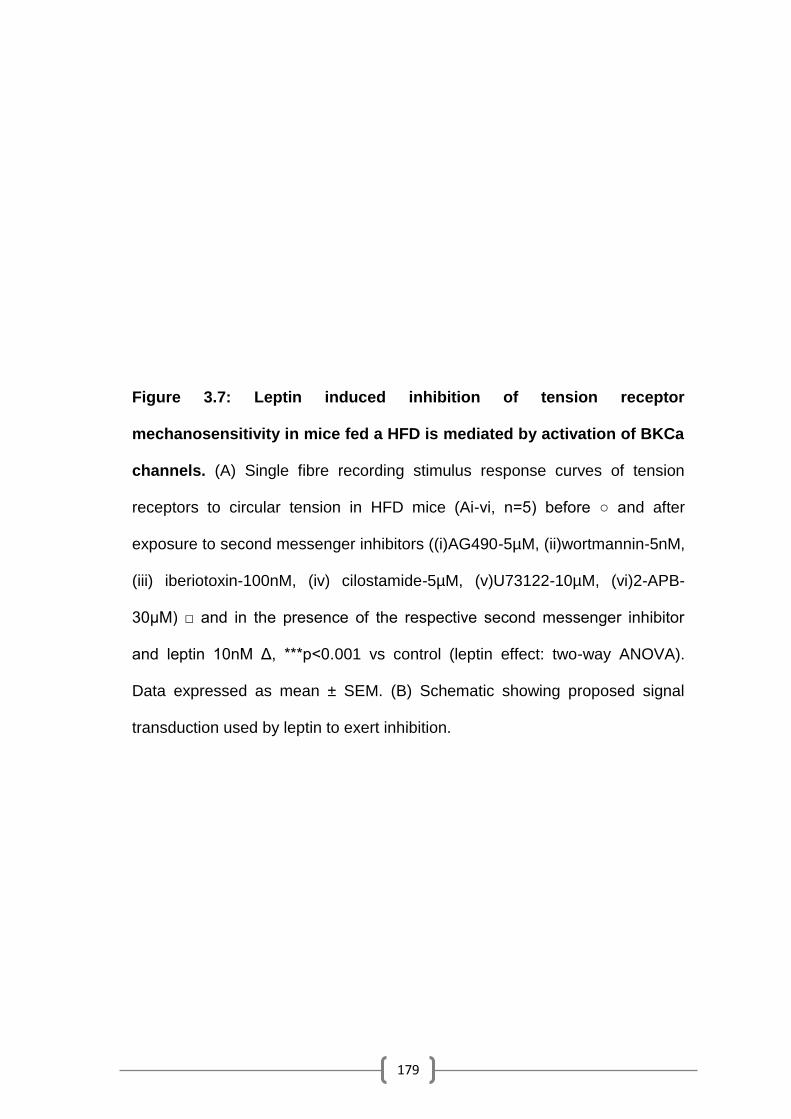
179
Figure 3.7: Leptin induced inhibition of tension receptor
mechanosensitivity in mice fed a HFD is mediated by activation of BKCa
channels. (A) Single fibre recording stimulus response curves of tension
receptors to circular tension in HFD mice (Ai-vi, n=5) before ○ and after
exposure to second messenger inhibitors ((i)AG490-5µM, (ii)wortmannin-5nM,
(iii) iberiotoxin-100nM, (iv) cilostamide-5µM, (v)U73122-10µM, (vi)2-APB-
30µM) □ and in the presence of the respective second messenger inhibitor
and leptin 10nM Δ, ***p<0.001 vs control (leptin effect: two-way ANOVA).
Data expressed as mean ± SEM. (B) Schematic showing proposed signal
transduction used by leptin to exert inhibition.

180

181
Supplementary Figure 3.1: Leptin modulates esophageal vagal afferent
mechanosensitivity in a similar fashion to gastric afferents. (A) Stimulus-
response curves of oesophageal mucosal afferents from fed (i, n=6) and
fasted mice (ii, n=6) before ○ and after exposure to leptin (0.1nM □, 1nM Δ,
10nM ◊). (B) Stimulus-response curves of oesophageal tension receptors from
fed (i, n=5) and fasted mice (ii, n=6) before ○ and after exposure to leptin
(0.1nM □, 1nM Δ, 10nM ◊). (C) Stimulus-response curves of oesophageal
mucosal receptors from mice fed a SLD (i, n=5) and a HFD (ii, n=5) before ○
and after exposure to leptin (0.1nM □, 1nM Δ, 10nM ◊). (D) Stimulus-response
curves of oesophageal tension receptors from mice fed a SLD (i, n=7) and a
HFD (ii, n=13) before ○ and after exposure to leptin (0.1nM □, 1nM Δ, 10nM
◊). * p<0.05, **p<0.01, ***p<0.001. Data are expressed as mean±SEM.

182

183
Supplementary Figure 3.2: HFD feeding caused an increase in body
weight and fat mass. (A) Change in body weight over 12 week period on
either SLD (●) (n=52) or HFD (○) (n=51). (B) Gonadal fat pad mass in mice
fed either SLD (n=16) or a HFD (n=17). *p<0.05, **p<0.01, ***p<0.001
compared with mice fed a standard laboratory diet. Data are expressed as
mean±SEM.

184

185
Supplementary Figure 3.3: A HFD does not change muscular afferent
profile in the stomach or nodose ganglia. (A) Anterograde traced vagal
afferent fibres and intraganglionic laminar endings in wholemount stomach of
the mouse (i, SLD; ii, HFD). (B) Number of vagal afferent profiles identified in
muscular layers of the stomach in mice fed a SLD (n=6) or HFD (n=6). (C)
Anterograde traced vagal afferent fibres and intramuscular arrays in the
wholemount stomach of the mouse (i, SLD; ii, HFD). (D) Vagal afferents
(white arrows) traced from gastric muscle (serosa) in the nodose ganglia of
mice fed a SLD (i) or HFD (ii). (E) Proportion of vagal afferents traced from
gastric muscle in nodose ganglia of mice fed a SLD or HFD. Scale 50µm in all
instances. Graphed data are expressed as mean±SEM.

186
CHAPTER 4
A CHRONIC HIGH FAT DIET ALTERS THE
HOMOLOGOUS AND HETEROLOGOUS
CONTROL OF SATIETY PEPTIDE RECEPTOR
EXPRESSION
Kentish SJ, Wittert GA, Blackshaw LA and Page
AJ.
Nerve Gut Research Laboratory, University of
Adelaide, Adelaide, SA 5005, Australia
Published in Peptides 2013 Aug; 46: 150–158

187
.
.

188

189
4.1 ABSTRACT
Leptin, ghrelin and neuropeptide W (NPW) modulate vagal afferent activity,
which may underlie their appetite regulatory actions. High fat diet (HFD)
induced obesity induces changes in the plasma levels of these peptides and
alters the expression of receptors on vagal afferents. We investigated
homologous and heterologous receptor regulation by leptin, ghrelin and NPW.
Mice were fed (12-weeks) a standard laboratory diet (SLD) or HFD. Nodose
ganglia were cultured overnight in the presence or absence of each peptide.
Leptin (LepR), ghrelin (GHS-R), NPW (GPR7) and cholecystokinin type-1
(CCK1R) receptor mRNA, and plasma peptide levels were measured. SLD:
leptin reduced LepR, GPR7, increased GHS-R and CCK1R mRNA; ghrelin
increased LepR, GPR7, CCK1R, and decreased GHS-R. HFD: leptin
decreased GHS-R and GPR7, ghrelin increased GHS-R and GPR7. NPW
decreased all receptors except GPR7 which increased with HFD. Plasma
leptin was higher and NPW lower in HFD. Thus, HFD-induced obesity disrupts
inter-regulation of appetite regulatory receptors in vagal afferents.

190
4.2 INTRODUCTION
Vagal nerves innervate the gastrointestinal tract where they have both
chemosensitive and mechanosensitive endings [436]. In the stomach these
endings respond to mechanical stimuli such as distension [471], and activate
neurons within the nucleus tractus solitarius [268] and regions of the brain
involved in the regulation of food intake such as the arcuate nucleus [493] and
paraventricular nucleus [494]. The activity of gastric vagal nerve endings can
be modulated by locally released peptides, such as leptin, ghrelin and
neuropeptide W (NPW), which are secreted by gastric endocrine and
epithelial cells [340, 473, 495]. These three peptides have well documented
effects on appetite, food intake and vagal afferent mechanosensitivity. Leptin
potentiates vagal activity [333, 496] and reduces appetite [326], whilst ghrelin
inhibits vagal afferent firing [301] and promotes food intake [497]. NPW
reduces vagal afferent activity (Li et al., 2011), but its effects on food intake
remain unresolved, with evidence for both anorexigenic [340] and orexigenic
effects [498].
Previously it has been shown that the ability of the stomach to respond to
mechanical stimuli through vagal pathways is altered by the induction of high
fat diet (HFD) induced obesity [473] and obese humans have diminished
perception of gastric distension [432]. Furthermore, the anorexigenic
response of vagal afferents to leptin and cholecystokinin (CCK) has also been
shown to be abrogated under diet induced obese conditions [336].

191
The receptors for leptin [496], ghrelin [473],NPW [499] and CCK [369] are
synthesized in the cell bodies of the vagal afferents located in the nodose
ganglia and transported to the neuron endings. The expression of the
receptors in vagal afferents appears modified by feeding status [369]. For
instance, ghrelin receptor expression is increased in fasted mice [473] and
obese rats [369]. These changes in receptor expression are associated with
decreased (fasted) or increased (obese) plasma leptin levels and increased
(fasted) or decreased (obese) plasma ghrelin levels [462, 500]. It has not
been established whether the change in circulating peptides is linked to the
change in their receptor expression in the vagal afferent neurons. This led us
to hypothesise that a change in peptide exposure may drive a change in the
expression of receptors involved in the regulation of appetite. This notion is
supported indirectly by leptin‘s ability to cause suppression of ghrelin [501]
and NPW [498] release, whilst increasing the release of the anorexigenic
vagal afferent modulator CCK [502], which together may augment the
anorexigenic effect of leptin. Similarly, appetite hormones, including leptin and
ghrelin can regulate expression and receptor abundance of their cognate and
other receptors in a variety of tissues, including vagal afferents [353, 424,
503]. Accordingly we sought to determine (i) the effects of leptin, ghrelin and
NPW on their own and each other‘s receptor expression in cultured nodose
ganglia, and (ii) whether any homologous or heterologous regulation of
receptor expression is altered by diet-induced obesity.

192
4.3 MATERIALS AND METHODS
4.3.1 Ethical approval
All experimental studies were performed with the approval of the animal ethics
committee of the Institute of Medical and Veterinary Science and the
University of Adelaide, and in accordance with the Australian Code of Practice
for the Care and Use of Animals for Scientific Purposes.
4.3.2 High fat diet model
Eight week old female C57BL/6 mice (N=12/group) were randomly assigned
to consume, for 12 weeks, either a standard laboratory diet (SLD) with 7%
energy from fat, or HFD (Specialty foods, Glen Forrest, Western Australia)
with 60% energy from fat . Water and food was available ad libitum for both
groups. Mice were housed under 12 hour light dark conditions with lights on at
06:00. Weights of mice were monitored weekly and gonadal fat pads collected
and weighed after euthanasia. All tissue was collected at 08:00.
4.3.3 Cell culture
After 12 weeks on their respective diet the mice were anesthetized with
isoflurane (1-1.5% in oxygen) and decapitated after exsanguination via the
abdominal aorta. Both nodose ganglia from each animal were harvested and
placed immediately together, into a tube containing F12 complete nutrient
medium (+ foetal calf serum and penicillin/streptomycin) (Invitrogen,
Mulgrave, Australia) and stored on ice. Nodose ganglia were dissociated by
incubating at 37°C in a 3mg/ml solution of dispase and collagenase II made

193
up in Hanks Balanced Salt Solution (HBSS) (Invitrogen) with agitation at 5
minute intervals. After 30 minutes the dispase/collagenase solution was
removed, and the ganglia were incubated in a 3mg/ml solution of collagenase
II in HBSS for 15 minutes. After this incubation the cells were rinsed in cold
HBSS and F12 and then further dissociated by passing them through a fire
polished Pasteur pipette until no cell clumps were visible. The cells were then
pelleted, rinsed in HBSS, and resuspended in Neurobasal A medium (NBA)
containing B-27 supplement (Invitrogen). A cell count was performed using
Tryphan blue exclusion. An equal number of cells were plated as a dot in the
centre of six wells in a 48 well cell culture plate with the wells coated with
poly-D-lysine and laminin (Sigma, Castel Hill, Australia), to promote cell
adhesion. The plate was placed in a 5% CO2 incubator set at 37°C for two
hours to allow cell adherence. After two hours the wells were filled with warm
(37°C) NBA medium. Two wells were left with just NBA in them as a control
and leptin (0.1nM and 1nM) was added to the other wells (two wells for each
concentration). The plate was then returned to the incubator for a 14 hour
incubation which has previously been shown to be of sufficient length to see a
change in receptor mRNA expression in nodose ganglia in vivo [473].
The above process was repeated using 4 SLD and 4 HFD mice with ghrelin (1
and 3nM) and neuropeptide W (1 and 3nM) in the place of leptin (N=4 for
each peptide and each diet). Leptin was obtained from Sigma and ghrelin and
NPW were obtained from Tocris (Bristol, UK). The concentrations used were
based on values found within previous literature that have been shown to
cause modulation of vagal afferent activity [343, 441, 462, 473].

194
4.3.4 RNA isolation
After 14 hours the plate was removed and culture medium drawn off. The
wells were washed using sterile PBS. Cells were lysed in the wells by addition
of buffer RLT+ βMe (Qiagen, Doncaster, Australia). Cells lysate was
processed and RNA isolated using an RNeasy Micro kit (Qiagen) following the
manufacturer‘s instructions. This process utilized RNeasy MinElute SpinElute
Columns, allowing adsorption of RNA to the silica membrane, an on-column
DNase digestion treatment, removal of contaminants with wash steps and
elution of the RNA with RNase-free water. RNA quantification was determined
by measuring the absorbance at 260 nm (A260) via a NanoDrop™ ND 1000
spectrophotometer (Thermo Scientific, Australia). RNA quality was estimated
by the A260 and A280 nm ratio.
4.3.5 Quantitative RT-PCR
Expression of leptin receptor (LepR), ghrelin receptor (GHS-R), neuropeptide
W receptor (GPR7) and CCK receptor (CCK1R) was carried out using
Quantitative RT-PCR (QRT-PCR) reactions performed as described in detail
previously [455]. Briefly, QRT-PCR reactions were performed by using a
Chromo4 (MJ Research, Bio-Rad) real-time instrument attached to a PTC-200
Peltier thermal cycler (MJ Research) and analysed with Opticon Monitor
software (MJ Research). QRT-PCR reactions were performed with a Qiagen
QuantiTect SYBRgreen RT-PCR one-step RT-PCR kit (Qiagen) according to
the manufacturer‘s specifications with specific Quantitect Primer Assays
(Qiagen) optimized for the detection of the known sequence of mouse LepR,

195
GHS-R, GPR7, CCK1R and β-tubulin transcripts contained in the NCBI
reference sequence database (www.ncbi.nlm.nih.gov/RefSeq). These primer
assays were used under the following conditions: reverse transcription, 50°C
for 30 min; initial PCR activation, 95°C for 15 min; PCR cycles 94°C for 15
sec, 55°C for 30 sec, and 72°C for 30 sec repeated for 50 cycles. A melting
curve program verified the specificity and identity of the RT-PCR products and
no primer dimers were observed. Each assay was run in triplicate in two
separate experiments. Control PCRs were performed by substituting RNA
template with distilled RNase-free water. All assays were validated for linearity
of amplification efficiency and quantitative standard curves obtained by serial
dilutions of RNA. Calculations for relative mRNA transcripts expression were
performed by the comparative CT method, comparing to the internal reference
gene β-tubulin using the equation ∆CT (CT of target transcript - CT of β-
tubulin). The relative fold differences were calculated by using the modified
version of 2-∆∆CT [456] correcting for PCR efficiencies.
4.3.6 Quantification of plasma concentrations of peptides
Blood withdrawn from the aorta during euthanasia was placed into pre-chilled
K2EDTA coated tubes and spun for 15 minutes at 4°C 1000xg to separate the
plasma from the cells. The plasma concentrations of ghrelin and leptin were
determined by ELISA using kits supplied by Millipore (Billerica, USA) and
NPW-23 was quantified using a kit supplied by Phoenix Pharmaceuticals
(Burlingame, USA) according to the manufacturers‘ instruction. The leptin and
ghrelin assays have previously been used for plasma quantification by other
groups [504, 505]. The sensitivities of the assays were 0.05ng/ml (leptin), 0.04

196
ng/ml (ghrelin) and 0.2ng/ml (NPW), respectively. The intra-assay variations
were 1.4% (leptin), 1.4% (ghrelin) and 3.7% (NPW) and the inter-assay
variations were 3.8% (leptin), 2.6% (ghrelin) and 5.3% (NPW).
4.3.7 Data analysis
Change in mouse weight were analysed by two-way ANOVA with Bonferroni
post hoc test. Gonadal fat weights and plasma peptide levels were analysed
with unpaired t-test. The sample expression levels are expressed as either
expression relative to β-tubulin ± SEM to analyse the effect of peptide
exposure and significance determined using a one-way ANOVA with Tukey
post hoc test or as n-fold difference relative to the control non-exposed cell
expression level ± SEM. Significant differences in transcript expression were
determined by a two-way ANOVA with Bonferroni post hoc test to determine
whether the effect of the peptide was altered by high fat diet feeding.
Differences were considered significant if p<0.05 vs. untreated cells or SLD.

197
4.4 RESULTS
4.4.1 Effect of high fat diet on body weight and plasma hormones
Body weight (Figure 4.1A) and body fat (Figure 4.1B) increased more in the
HFD than SLD mice (both p<0.001). Plasma leptin levels were 3.906 ± 0.2928
ng/ml and 2.286 ± 0.1826 ng/ml in the HFD and SLD mice (N=12/group)
respectively (p<0.01) (Figure 4.1C). Plasma ghrelin levels were 3.961 ±
0.3600 ng/ml and 4.234 ± 0.3188 ng/ml in the SLD and HFD mice respectively
(p>0.05) (Figure 4.1D). Plasma NPW-23 concentrations were 9.68 ± 0.6947
ng/ml and 5.867 ± 0.5069 ng/ml in the SLD and HFD mice respectively
(p<0.001) (Figure 4.1E).
4.4.2 Effect of leptin on expression of LepR, GHS-R, GPR7 and CCK1R in
SLD and HFD vagal cell bodies
Compared to control, leptin (0.1nM, 1nM) caused a 0.45 and 0.73 fold
reduction in expression of LepR mRNA (both p<0.05) in neurons from SLD
mice but was without effect in those from HFD mice (Figure 4.2Ai; p>0.05).
Receptor expression in SLD and HFD neurons under control conditions for
some peptides was significantly different. Therefore, in order to determine an
effect of diet itself, receptor expression was plotted and analysed relative to
control. A HFD abolished the effect of leptin on LepR expression in vagal
afferent cell bodies (Figure 4.2Aii; p<0.001). GHS-R expression was up
regulated by the addition of leptin only at the 1nM concentration in the SLD
mice with a 2.04 fold increase in GHS-R mRNA being present in the leptin
(1nM) exposed cells than the control cells (Figure 4.2Bi; p<0.001). In contrast,

198
exposure of leptin (0.1nM, 1nM) to vagal afferent cell bodies from HFD mice
resulted in a 0.42 and 0.70 fold decrease in ghrelin receptor expression
(Figure 4.2Bi; p<0.05 and p<0.001 respectively). Thus a HFD switched the
effect of leptin on GHS-R mRNA expression from an increase, observed in
SLD nodose neurons, to a decrease (Figure 4.2Bii; p<0.01). Expression of the
NPW receptor, GPR7 was almost completely abolished when leptin was
added to the culture medium with 0.1nM and 1nM of leptin both causing a
0.99 fold reduction in the expression of GPR7 (Figure 4.2Ci; p<0.001). In the
vagal afferent cell bodies of HFD fed mice leptin (0.1nM and 1nM) still caused
a significant reduction (0.90 and 0.97 fold respectively) in the expression of
GPR7 (Figure 4.2Ci; p<0.001). However, a HFD significantly attenuated the
inhibitory effect of leptin on GPR7 mRNA expression (Figure 4.2Cii; p<0.01).
Exposure to the highest dose of leptin (1nM) caused a 2.61 fold increase in
the expression of CCK1R in vagal afferent cell bodies from SLD mice (Figure
4.2Di; p<0.001), however in vagal afferent cell bodies from HFD mice leptin
failed to have any effect on CCK1R expression (Figure 4.2Di; p>0.05).
Therefore a HFD abolished the excitatory effect of leptin on CCK1R
expression (Figure 4.2Dii; p<0.05).
4.4.3 Effect of ghrelin on expression of LepR, GHS-R, GPR7 and CCK1R
in SLD and HFD vagal cell bodies
Ghrelin (3nM) caused a significant increase in LepR expression of 1.64 fold
(Figure 4.3Ai; p<0.01). In the HFD fed mice cells ghrelin 1nM no longer had
an effect on expression of LepR (p>0.05), but ghrelin 3nM caused a 0.44 fold
reduction in expression (Figure 4.3Ai; p<0.001). Therefore a HFD switched

199
the effect of ghrelin on LepR mRNA expression from an increase observed in
SLD nodose neurons to a decrease (Figure 4.3Aii; p<0.01). Ghrelin (3nM)
caused a 0.70 fold reduction in GHS-R expression (Figure 4.3Bi; p<0.05) in
the cells from SLD mice, whilst exposure to ghrelin (1nM, 3nM) caused an
increase of GHS-R expression (1.86 and 2.50 fold respectively) in HFD vagal
cell bodies (Figure 4.3Bi; p<0.05 and p<0.001 respectively). Thus the HFD
switched the effect of ghrelin on GHS-R mRNA transcript expression from a
decrease as seen in SLD nodose neurons to an increase in the HFD neurons
(Figure 4.3Bii; p<0.001). Ghrelin (3nM) caused a 3.00 fold increase in GPR7
expression (Figure 4.3Ci; p<0.05). In HFD vagal cell bodies ghrelin caused a
6.66 (1nM) and 23.00 fold (3nM) increase in expression of GPR7 (Figure
4.3Ci; p<0.01 and p<0.001 respectively). Demonstrating that HFD feeding
causes a potentiation of the increase in GPR7 mRNA transcript expression
caused by ghrelin (Figure 4.3Cii; p<0.001). Ghrelin (3nM) caused a 2.02 fold
increase in CCK1R expression in SLD vagal cell bodies (p<0.05), however
when HFD cell bodies were exposed to ghrelin (1nM and 3nM) there was a
significant reduction in the expression of CCK1R (0.67 and 0.64 fold reduction
respectively) (Figure 4.3Di; both p<0.01). Therefore a HFD causes ghrelin to
switch its effect from causing an increase in CCK1R mRNA expression in SLD
to a decrease in HFD vagal cell bodies (Figure 4.3Dii; p<0.001).
4.4.4 Effect of NPW on expression of LepR, GHS-R, GPR7 and CCK1R in
SLD and HFD vagal cell bodies
When NPW (1nM, 3nM) was added to the cell culture medium it caused a
reduction in LepR in cultured cells from both SLD (1nM - 0.63, 3nM - 0.89 fold

200
reduction; both p<0.001) and HFD (1nM - 0.60, 3nM - 0.75 fold reduction;
p<0.05 and p<0.01 respectively) mice (Figure 4.4Ai). Thus a HFD did not alter
the effect of NPW on LepR mRNA expression (Figure 4.4Aii; p>0.05). NPW
(3nM) caused a significant 0.85 fold reduction in GHS-R expression in SLD
cultured cells (Figure 4.4Bi; p<0.01). In the vagal cell bodies from HFD mice
NPW still caused a decrease in expression of GHS-R, however the reduction
caused by NPW (3nM) was now only 0.36 fold (Figure 4.4Bi; p<0.05). This
indicates that a HFD significantly ablated the down regulatory effect of NPW
on GHS-R mRNA expression (Figure 4.4Bii; p<0.001). NPW (3nM) caused a
0.70 fold reduction in expression of GPR7, in SLD cell bodies (p<0.05; Figure
4.4Ci). In the HFD cells NPW (1nM, 3nM) caused an increase in GPR7
expression of 1.87 and 2.05 fold respectively (Figure 4.4Ci; both p<0.05). This
demonstrated that a HFD caused the effect of NPW on GPR7 mRNA
expression in vagal cell bodies to be altered from a decrease to an increase
(Figure 4.4Cii; p<0.05). NPW (1nM, 3nM) caused a significant reduction in
CCK1R expression of 0.36 and 0.40 fold in SLD vagal cell bodies respectively
(Figure 4.4Di; both p<0.05) and 0.52 and 0.47 fold reduction in HFD cell
bodies respectively (Figure 4.4Di; both p<0.01). Therefore the effect of NPW
on CCK1R mRNA expression was unaffected by HFD feeding (Figure 4.4Dii;
p>0.05).

201
4.5. DISCUSSION
These data show that the appetite regulating peptides, leptin, ghrelin and
NPW modify the expression of their own, and each other‘s receptors. Chronic
high fat diet feeding alters the magnitude and direction of these effects, which
may alter effects mediated by gastric vagal afferent signalling, including
satiety.
Leptin has previously been shown to decrease the expression of its cognate
receptor [506, 507]. The observation that this is abrogated in nodose ganglia
in response to HFD induced obesity is consistent with previous data [369,
370, 496]. In contrast to what occurs in vagal afferents, leptin induces an
increase in LepR mRNA in the arcuate nucleus of lean mice [508-510], an
effect that is also lost in HFD induced obesity [511]. In obese mice leptin
increases SOCS3 [512], but the phosphorylation of STAT3 is reduced [335,
513, 514]. Therefore it seems likely that receptor binding occurs, but there is a
reduction in downstream signalling. However, we have not differentiated
between the isoforms of LepR, which have been shown to follow different
patterns of regulation [515]. Therefore, although the full length isoform of the
receptor is considered the functional receptor [516], further investigation into
the possible contributions of all LepR isoforms is required.
In addition to its central effects, leptin can decrease food intake by acting
alone or in concert with CCK to activate gastrointestinal vagal afferents [326,
333, 334, 517]. The finding in this study that leptin increases the expression of

202
CCK1R provides additional support for the notion that leptin may reduce food
intake in part via a CCK-mediated vagal mechanism of action [334, 335]. Our
finding that leptin no longer increased CCK1R expression in vagal afferent cell
bodies from HFD fed mice may contribute to the dampened vagally mediated
CCK satiety signal that is observed in obese rodents [335, 518].
Leptin has previously been shown to increase GHS-R expression in human T-
cells [503]. We now show that leptin increases GHS-R in vagal afferent cell
bodies. Leptin reducing GHS-R mRNA in HFD fed mice may represent a
mechanism of suppressing orexigenic signals. Similarly in obese mice [359]
and rats [519] hypothalamic GHS-R is decreased. However, in isolated
pituitary cells from obese leptin knockout mice (ob/ob) GHS-R mRNA
expression increases in response to increasing concentrations of leptin [520].
This may reflect tissue or culture specific effects or alternatively, a nutrient
driven change in receptor expression regulation, as the ob/ob mice used were
maintained on a chow diet.
The observations that leptin decreased mRNA expression for its own receptor
at a concentration lower than that required to change CCK1R and GHS-R
mRNA expression and that leptin has no effect on CCK1R or LepR mRNA,
whilst still effecting GHS-R and GPR7 in the HFD vagal afferents suggests
that leptin alters transcriptional regulation through multiple pathways.
However, this possibility requires further investigation.

203
Ghrelin is orexigenic and believed to be primarily involved in the initiation of
meals; increasing plasma concentrations precede food intake and fall
dramatically thereafter [521]. It has previously been shown that obese male
and female mice have reduced plasma ghrelin [359, 441], which is
independent of leptin levels [522]. In contrast, the current study showed no
difference in plasma ghrelin levels in lean and obese mice, which has also
been shown previously [504]. There are a number of possible explanations for
this, including feeding status and sampling differences. As plasma ghrelin
levels decrease after eating, plasma level measurements depend on the
timing of blood collection in relation to meal intake. Plasma ghrelin levels have
also been shown to oscillate in a circadian manner [523]. The degree to which
plasma ghrelin levels change over the course of a day is altered in obesity
[523]. Our plasma samples were collected at 08:00, which has previously
been shown to be a time when there is no difference in plasma ghrelin
between lean and obese mice [523]. Previous studies have not provided a
measure of recent food intake, or detailed the time plasma samples were
collected which makes comparisons difficult.
In lean mice the decreased GHS-R mRNA in response to ghrelin may
decrease ghrelin signalling after meal initiation. The concomitant increase of
LepR and CCK1R mRNA, may serve to enhance meal termination signals.
The increase in LepR and CCK1R mRNA is consistent with previous reports
showing an increased proportion of vagal neurons expressing LepR and
CCK1R in nodose ganglia from fasted mice [424, 470], with elevated plasma
ghrelin [524, 525]. Given the opposing effect of leptin and ghrelin on food

204
intake, it seems contradictory that leptin and ghrelin have the same effect on
CCK1R mRNA in vagal cell bodies from SLD mice. This may be due to the
predominately short term role of ghrelin and long term role for leptin in the
regulation of food intake [526]. Thus we could speculate that ghrelin increases
CCK1R expression to ensure that vagal afferents are always primed to
respond to CCK release in response to nutrients and that leptin released from
adipose tissue increases CCK1R expression when there is sufficient energy
stores.
In vagal cell bodies from HFD mice, the increase in GHS-R mRNA in
response to ghrelin, may serve to maintain ghrelin signalling in obesity, where
levels have been found to be decreased [359]. Plasma ghrelin continues to
show preprandial rises in obesity [527]. This suggests ghrelin is still involved
in the initiation of meals in obesity. The switch in the effect of ghrelin on
regulation of GHS-R mRNA in mice with diet induced obesity may be
mediated by activation of an alternate signalling pathway, as we have
previously described for leptin [496], that results in different mRNA transcript
levels. The ghrelin induced decrease in LepR and CCK1R in the HFD vagal
afferent cell bodies may blunt meal termination in obesity, which is consistent
with previous findings that obese mice eat longer meals [377] and have
reduced satiation in response to leptin and CCK [335, 336, 380, 518].
However, this is speculative and the relationship between altered receptor
regulation and reduced CCK and leptin mediated satiety signalling needs to
be clarified with more specific experiments.

205
In the current study, NPW suppressed receptor mRNA expression for
anorexigenic and orexigenic peptides. Similarly in obese cell bodies NPW
decreased expression of all receptors with the exception of its own receptor.
However, the regulation of GPR7 in vivo in lean and obese mice still needs to
be determined. Thus it remains unclear as to what the physiological role of the
peptinergic regulation of GPR7 in lean and obese mice is. We also reported a
decrease in the plasma concentration of NPW-23 in obese mice. Comparison
with previous values was not possible due to conflicting models and
methodological approaches.
The static cultures used do not reflect the oscillations in plasma levels of leptin
and ghrelin that occur over a 24 hour period [528, 529], or the combined
effects of changes in plasma nutrients and other gut peptides that occur in
vivo. Therefore, although we have established that the gut peptides studied
have direct regulatory effect on appetite receptor regulation in vagal afferent
cell bodies that are modified by diet induced obesity; the physiological
relevance in vivo remains to be determined.

206

207
Figure 4.1: High fat diet induced changes in weight and plasma peptides.
(A) Mice on a HFD (○ N=12) gained a significantly greater proportion of body
weight over the 12 week diet period compared to the SLD mice (● N=12). (B)
The HFD fed mice also had significantly heavier gonadal fat pads. (C) HFD
mice had elevated plasma leptin levels, (D) no difference in plasma ghrelin
and (E) decreased plasma NPW. Weight analysed using two-way ANOVA
with Bonferroni post hoc test, fat mass and plasma peptide levels compared
using unpaired t-tests. **p<0.01, ***p<0.001.

208

209
Figure 4.2: Expression of appetite regulatory peptide receptors is altered
by leptin exposure. Changes in expression of LepR (Ai), GHS-R (Bi), GPR7
(Ci) and CCK1R (Di) relative to β-tubulin in the absence and presence of
leptin (0.1, 1nM). Changes in expression of LepR (Aii), GHS-R (Bii), GPR7
(Cii) and CCK1R (Dii) mRNA relative to untreated cells from SLD (●) and HFD
(○) mice. Data are means ± SEM (N=4 in all instances), Leptin effect (i) within
group analysed using one-way ANOVA with Tukey post hoc test with
significance being determined if p<0.05. Diet effect (ii) analysed with two-way
ANOVA with Bonferroni post-hoc test. *p<0.05, **p<0.01, ***p<0.001.

210

211
Figure 4.3: Effect of ghrelin exposure of expression of appetite
modulator peptide receptor expression. Ghrelin (1nM, 3nM) induced
changes in expression of LepR (Ai), GHS-R (Bi), GPR7 (Ci) and CCK1R (Di)
in SLD and HFD cell bodies relative to β-tubulin. Changes in expression of
LepR (Aii), GHS-R (Bii), GPR7 (Cii) and CCK1R (Dii) mRNA relative to
untreated cells from SLD (●) and HFD (○) mice. Data are means ± SEM (N=4
in all instances), Ghrelin effect (i) within group analysed using one-way
ANOVA with Tukey post hoc test with significance being determined if p<0.05.
Diet effect (ii) analysed with two-way ANOVA with Bonferroni post-hoc test.
*p<0.05, **p<0.01, ***p<0.001.

212

213
Figure 4.4: NPW changes the expression of appetite regulatory
receptors. NPW (1nM, 3nM) induced changes in expression of LepR (Ai),
GHS-R (Bi), GPR7 (Ci) and CCK1R (Di) in SLD and HFD cell bodies relative
to β-tubulin. Changes in expression of LepR (Aii), GHS-R (Bii), GPR7 (Cii)
and CCK1R (Dii) mRNA relative to untreated cells from SLD (●) and HFD (○)
mice. Data are means ± SEM (N=4 in all instances), NPW effect (i) within
group analysed using one-way ANOVA with Tukey post hoc test with
significance being determined if p<0.05. Diet effect (ii) analysed with two-way
ANOVA with Bonferroni post-hoc test. *p<0.05, **p<0.01, ***p<0.001.

214
CHAPTER 5
ALTERED GASTRIC VAGAL
MECHANOSENSITIVITY IN DIET-INDUCED
OBESITY PERSISTS ON RETURN TO NORMAL
CHOW AND IS ACCOMPANIED BY INCREASED
FOOD INTAKE
Kentish SJ, O‟Donnell TA, Li H, Wittert GA,
Page AJ.
Nerve Gut Research Laboratory, University of
Adelaide, Adelaide, SA 5005, Australia
Submitted to The International Journal of
Obesity

215

216
217
5.1 ABSTRACT
Background and aims: Gastric vagal afferents convey satiety signals in
response to mechanical stimuli. The sensitivity of these afferents is decreased
in diet induced obesity. Leptin, secreted from gastric epithelial cells,
potentiates the response of vagal afferents to mechanical stimuli in lean mice,
but has an inhibitory effect in high fat diet (HFD) induced obese mice. We
sought to determine whether changes in vagal afferent function and response
to leptin in obesity were reversible by returning obese mice consuming a HFD
to standard laboratory chow diet (SLD).
Methods: 8wk old female C57BL/6 mice were either fed a SLD (N=20) or HFD
(N=20) for 24wks. A third group was fed a HFD for 12wks and then a SLD for
a further 12wks (RFD, N=18). An in vitro gastro-oesophageal vagal afferent
preparation was used to determine the mechanosensitivity of gastric vagal
afferents and the modulatory effect of leptin (0.1-10nM) was examined.
Retrograde tracing and quantitative RT-PCR were used to determine the
expression of leptin receptor (LepR) mRNA in whole nodose and specific cell
bodies traced from the stomach.
Results: After 24wks both the HFD and RFD mice had increased body weight,
gonadal fat mass, plasma leptin, plasma insulin and daily energy consumption
compared to the SLD mice. The HFD and RFD mice had reduced tension
receptor mechanosensitivity and leptin further inhibited responses to tension
in HFD, RFD but not SLD mice. Mucosal receptors from both the SLD and
RFD mice were potentiated by leptin, an effect not seen in HFD mice. LepR

218
expression was unchanged in the whole nodose, but was reduced in the
mucosal afferents of the HFD and RFD mice.
Conclusions: Disruption to gastric vagal afferent function by HFD induced
obesity is only partially reversible by dietary change, which provides a
potential mechanism preventing maintenance of weight loss.

219
5.2 INTRODUCTION
The experience of most obese people is that following diet induced weight
loss, they return at least to their previous weight within two years [68, 530-
532]. There is evidence from both animal [73] and human studies [533] that
fat mass is permitted to increase to what is presumably a genetically
determined upper limit. At any particular point along that trajectory, the fat
mass reached is defended such that if it decreases, mechanisms are
activated to ensure that it is restored. The mechanism responsible for this
defence of body fat has been suggested to be mediated by prolonged
changes in the level of appetite regulating gastrointestinal hormones, known
to be altered following weight loss and changed in such a way as to promote
weight regain [78].
Neurally mediated signals from the gastrointestinal tract play a role in the
regulation of food intake [534-536]. For example, cognitive perception of
fullness following food intake is reliant on two vagally mediated mechanisms.
One pathway relies on the presence of nutrients, which triggers
gastrointestinal hormone endocrine and paracrine secretions from the
stomach and small intestine [537, 538]. The other pathway is via mechanical
distension of the stomach, which in conjunction with small intestinal nutrient
signals can induce satiety [472, 539]. Two classes of vagal afferent
mechanoreceptors have been identified in the stomach [436]. The first are
tension receptors which respond to distension and contraction of the stomach.

220
The second class are mucosal receptors, which are located close to the
lumen, are responsive to contact by food particles with the mucosa and are
believed to participate in the control of gastric motor function [281, 297, 436,
471]. Previously it has been shown that diet induced obesity caused a
reduction in the ability of gastric and intestinal vagal afferents to respond to
stretch/distension, suggesting in obesity there is reduced gastrointestinal
signalling in response to food intake [381, 473].
Leptin is a peptide released from both adipocytes[540] and gastric epithelial
cells [304]. Ordinarily leptin reduces food intake largely attributed to actions
within the arcuate nucleus[541]. However, leptin receptor (LepR) is also
present in gastric vagal afferent neurons [470] and leptin has been shown to
play a role in regulating food intake through a vagally mediated
mechanism[326]. In diet-induced obesity this effect is lost [335, 336].
Previously, we have demonstrated that leptin has a potentiating effect on
gastric vagal mucosal receptors [496]. Furthermore, we have shown that
obesity causes leptin to lose its potentiating effect and instead have an
inhibitory effect on vagal tension receptors [496]. This indicates that leptin
may act within the stomach to modulate peripheral appetite signals and its
ability to do so is disrupted by obesity to a degree that defends the obese
state. We sought to determine the effect of returning these mice to a standard
laboratory diet (SLD) for 12 weeks on weight, food intake, gastric
mechanosensitivity, and the vagal afferent response to leptin. To establish
whether any change in the effect of leptin was associated with receptor

221
expression changes we determined the expression of LepR in both whole
nodose and mucosal traced gastric vagal afferents.

222
5.3 MATERIALS AND METHODS
5.3.1 Ethical approval
All studies were approved and performed in accordance with the guidelines of
the Animal Ethics Committees of the University of Adelaide and Institute for
Medical and Veterinary Science, Adelaide, Australia. Every attempt was made
to limit the number of animals used and minimize their suffering.
5.3.2 Animals
All mice in these studies were obtained from Animal Resource Centre
(Canning Vale, Australia) and group housed under a 12 hour light: dark cycle
(lights on at 06:00) with free access to food and water. Female C57BL/6 mice
were obtained at 7 weeks of age and assigned randomly to either a 24 week
SLD group (7% energy from fat; N=20), a 24 week high fat diet (HFD) group
(60% energy from fat; N=20) or a group that was fed a HFD for 12 weeks and
then returned to a SLD for a further 12 weeks (RFD; N=18). All mice were
allowed to acclimatize for one week before being started on their respective
diet. The mice were weighed weekly and had food intake monitored over the
24 week diet period. Blood glucose level and gonadal fat pad mass were
determined from all mice on the day they were used for the in vitro mouse
gastro-oesophageal afferent preparation.
5.3.3 In vitro mouse gastro-oesophageal afferent preparation
This preparation has been described in detail previously [436]. In short,
female C57BL/6 mice on the SLD, HFD or RFD diets were anaesthetised with

223
isoflurane (1-1.5% in oxygen) and killed via exsanguination. The stomach and
oesophagus, with intact vagal nerves, were removed and placed mucosa side
up in an organ bath containing a modified Krebs solution comprised of (in
mM): 118.1 NaCl, 4.7 KCl, 25.1 NaHCO3, 1.3 NaH2PO4, 1.2 MgSO4.7H2O, 1.5
CaCl2, 1.0 citric acid, 11.1 glucose and 0.001 nifidipine, bubbled with 95% O2-
5% CO2. The dissection process was carried out at 4°C to prevent metabolic
breakdown.
5.3.4 Characterization of gastric vagal afferent properties
In mice two types of mechanosensitive gastric vagal afferent have been
reported [436], those that respond to mucosal stroking, but not to circular
tension (mucosal receptors) and those that respond to both mucosal stroking
and circular tension (tension receptors). Receptive fields of these receptors
were first located using mechanical stimulation with a brush in the mouse
gastro-oesophageal preparation. Once located specific stimuli were then
applied. Mucosal stroking was performed using calibrated von Frey hairs (10-
1000mg) which were stroked across the mucosa at a rate of 5mms-1. Each
receptive field was stroked ten times and mechanical responses from the
middle eight strokes taken for analysis. Circular tension was applied using a
threaded hook attached to an underpinned point adjacent to the receptive
field. The threaded hook was attached to a cantilever via a pulley close to the
preparation. Standard weights (0.5-5g) were then placed on the opposite end
of the cantilever. Each weight was applied for one minute with a break of
another minute between removing one weight and applying the next. After

224
analysing the two stimulus response curves, we would classify a receptive
field as either a mucosal or tension receptor.
5.3.5 Effect of leptin on the mechanosensitivity of vagal afferents
After the mechanosensitivity of a receptive field was determined, the effect of
leptin was assessed. Leptin (0.1nM, Sigma-Aldrich, Castle Hill, Australia) was
added to the superfusing Krebs solution and allowed to equilibrate for 20 min
after which the stimulus response curves were re-determined. This procedure
was repeated for leptin 1 and 10nM.
5.3.6 Data recording
Afferent impulses were amplified with a biological amplifier (DAM 50, World
Precision Instruments, Sarasota, FL), and filtered (band-pass filter 932, CWE,
Ardmore, PA, USA). Single units were discriminated on the basis of action
potential shape, duration, and amplitude by use of Spike 2 software
(Cambridge Electronic Design, Cambridge, UK).
5.3.7 Nodose ganglia quantitative RT-PCR
Nodose ganglia were removed bilaterally from mice from all 3 experimental
groups. Total RNA was extracted using an RNeasy Micro Kit (Qiagen,
Doncaster, Australia) according to the manufacturer‘s instructions. RNA was
quantified by measuring the absorbance at 260nm (A260) using a
NanoDrop™ ND 1000 spectrophotometer (Thermo Fisher Scientific,
Scoresby, Australia) and RNA purity was estimated via the 260/280
absorbance ratio. Quantitative RT-PCR (QRT-PCR) reactions were performed

225
as described in detail previously [455]. In short, reactions were performed
using a Chromo4 (MJ Research, Bio-Rad, Gladesville, Australia) real-time
instrument attached to a PTC-200 Peltier thermal cycler (MJ Research) and
analysed with Opticon Monitor software (MJ Research). QRT-PCR reactions
were performed using a QuantiTect SYBRgreen RT-PCR one-step RT-PCR
kit (Qiagen) according to the manufacturer‘s instructions. All primers used
were pre-designed validated Quantitect Primer assays (Qiagen) targeting
leptin receptor and β-tubulin. RT-PCR reactions were carried out under the
following conditions: reverse transcription, 50°C for 30min; initial PCR
activation, 95°C for 15min; PCR cycles 94°C for 15sec, 55°C for 30sec and
72°C for 30sec repeated for 50 cycles. A melt curve was obtained to confirm
the specificity of the products produced. Each assay was run in triplicate and
repeated on separate days. Control PCRs were carried out substituting
RNase-free water for template RNA. Relative RNA levels were calculated
using the comparative CT method as described previously [456].
5.3.8 Retrograde tracing
This procedure has been detailed previously [473]. In short; to identify specific
gastric mucosal afferents SLD (N = 4), HFD (N=4) and RFD (N=4) mice were
anesthetized with isoflurane (1-1.5% in oxygen), a laparotomy performed, and
a mucolytic (10% N-acetylcysteine; 200µl) was injected into the stomach
lumen then removed via syringe after 5 min, this was followed by two saline
rinses (200µl each). Subsequently, 10µl of 0.5% Alexa Fluor® 555 conjugate
of cholera toxin β-subunit (CTB-AF555); Invitrogen, Mulgrave, Australia) was
injected into the proximal gastric lumen via a 30 ga Hamilton syringe and the

226
proximal stomach walls gently opposed to expose the dorsal and ventral
surfaces to the tracer. The injection site was dried and skin incision closed.
Antibiotic (Baytril; 50μl of 50mg/ml) and analgesic (butorphanol; 5mg/kg) were
administered subcutaneously. Food and water were withheld for 2 hours
postoperatively to maximize exposure of tracer. All mice recovered well from
surgery and were routinely monitored. Isolation of tension afferents is
possible, however we have previously determined that diet induced obesity
has no effect on the expression of LepR mRNA in this sub-population [496].
5.3.9 Laser capture microdissection
After 2 days, retrogradely traced mice were anesthetized with pentobarbitone
(60 mg/kg, i.p.) their left and right nodose ganglion was then removed and
dissociated before being cultured on a duplex dish for 2 hours at 37°C in 5%
CO2 [496]. Cells were then subject to laser-capture microdissection,
performed on a P.A.L.M.® microbeam microdissection system (Carl Zeiss,
Jena, Germany). Fluorescent labelled nodose neurons were microdissected
and catapulted directly into a lysis and stabilization buffer (Buffer RLT,
RNeasy Micro RNA extraction Kit, Qiagen) containing 0.14M
β-Mercaptoethanol (Sigma-Aldrich). RNA was extracted and QRT-PCR was
performed on these cells using the same protocol as for whole nodose
ganglia.
5.3.10 Plasma hormone measurements
Blood samples from all groups of mice were collected on the day of
experiment from the abdominal aorta, under anaesthesia. Blood was placed

227
into K2EDTA coated tubes and spun for 15min at 1000xg to separate the
plasma which was stored at -80°C until needed. Plasma leptin and insulin
were determined using commercially available ELISA assay kits (Millipore,
Billerica, MA, USA) according to manufacturers‘ instructions. The sensitivities
of the assays were 0.05ng/ml (leptin) and 0.2ng/ml (insulin); the intra-assay
variations were 1.4% (leptin), 4.65% (insulin).
5.3.11 Statistical analysis
All data in graphs are expressed as mean values ± SEM with N= number of
animals studied and n= number of individual afferents analysed. Vagal
afferent stimulus-response curves and weight change were analysed using
two-way analysis of variance (2-way ANOVA) and Bonferroni post hoc tests.
RNA levels, fat mass, food/energy intake and plasma peptide levels were
analysed using one-way ANOVA‘s with Tukey post hoc tests. Significance
was defined at p<0.05.

228
5.4 RESULTS
5.4.1 Diet induced changes to mouse weight, fat mass, food
consumption and plasma peptide levels
The HFD mice gained more weight than both the SLD and RFD mice (Figure
5.1A, both p<0.001). The RFD mice initially lost some weight upon return to
the SLD, but by the end of the 24th week they had gained back all lost weight
and on average weighed more than at the point they were removed from the
HFD (Figure 5.1A, p<0.001 SLD vs. RFD). Gonadal fat mass of the HFD
(2.71±0.37g) mice was greater than that of both the RFD (0.62±0.10g) and
SLD (0.34±0.02g) mice (Figure 5.1B, both p<0.001), however the RFD fat
mass was also greater than the SLD mice (Figure 5.1B, p<0.01 SLD vs.
RFD). Blood glucose levels were higher in both the HFD and RFD mice
(12.78±0.82mM and 11.07±0.79mM respectively) compared to the SLD mice
(6.45±0.73mM) (Figure 5.1C, both p<0.01 vs. SLD). The average 24-hour
food consumption of the RFD mice (4.8± 0.43g) was significantly greater than
that of the SLD (2.78±0.23g) and HFD (3.14±.0.19g) mice (Figure 5.1D,
p<0.001 vs. SLD, p<0.01 vs. HFD) at the end of the 24 week period. The
energy intake of the RFD mice (16.23 ±1.41kcal) was similar to the HFD:
(16.42±0.99kcal), and both were greater than the SLD mice (9.29±0.77kcal)
(Figure 5.1E, HFD vs. SLD p<0.001, RFD vs. SLD p<0.01). Plasma leptin
levels (Figure 5.1F) were higher in the HFD (23.09±1.27ng/ml, p<0.001 vs.
SLD and RFD) and RFD (16.33±1.83ng/ml, p<0.001 vs. SLD) mice compared
to the SLD mice (5.01±0.31ng/ml). Plasma insulin (Figure 5.1G) was also

229
elevated in the HFD (0.97±0.08ng/ml p<0.001 vs. SLD p<0.01 vs. RFD) and
RFD (0.69±0.03ng/ml p<0.01 vs. SLD) relative to SLD mice (0.55±0.13ng/ml).
5.4.2 High fat diet induced changes in gastric mechanosensitivity are not
altered by reverting to „normal‟ chow feed
Gastric tension mechanosensitivity of the HFD and RFD mice was reduced by
50% (at 5g) compared to the SLD mice (Figure 5.2A, both p<0.001 vs. SLD).
There was no significant difference between the responses of the gastric
tension receptors of the HFD mice compared to the RFD mice (p>0.05). There
was no difference in the response to mucosal stroking by mucosal receptors
between any of the groups of mice (Figure 5.2B, p>0.05).
5.4.3 Leptin‟s effects on gastric vagal afferent mechanosensitivity are
dependent on diet
Leptin (0.1-10nM) potentiated gastric mucosal mechanosensitivity (Figure
5.3A & E p<0.001 vs. Control) in SLD mice, but had no effect in HFD mice
(Figure 5.3B & F, p>0.05). In the RFD mice leptin increased mucosal receptor
mechanosensitivity (Figure 5.3C & G, p<0.001 vs. Control). The level of
potentiation induced by leptin (0.1-10nM) in response to 50mg stroking was
different in all three groups of mice (Figure 5.3D, p<0.001 SLD vs. HFD,
p<0.01 RFD vs. HFD and SLD).

230
Leptin had no effect on the mechanosensitivity of gastric tension receptors (1-
5g) in SLD mice (Figure 5.4A & E, p>0.05), however caused a reduction in
HFD (Figure 5.4B & F, p<0.001 vs. Control) and RFD (Figure 5.4C & G,
p<0.001 vs. Control) mice. The level of inhibition caused by leptin in response
to 3g stretch was highest in the HFD mice followed by the RFD and SLD mice
(Figure 5.4D, p<0.05 HFD vs. RFD, p<0.001 HFD and RFD vs. SLD).
5.4.4 Diet induced changes in the expression of leptin receptor in vagal
afferents
There was no difference in LepR expression in whole nodose ganglia
between any of the groups of mice (Figure 5.5A, p>0.05). However, when
LepR mRNA expression was quantified specifically in mucosal afferents there
was a 99% reduction in LepR mRNA in the HFD mice compared to the SLD
mice (Figure 5.5B, p<0.001 SLD vs. HFD). The RFD mucosal afferents had a
68% reduction in LepR mRNA relative to the SLD mice (p<0.001 SLD vs.
RFD). The RFD LepR mRNA content was greater than that observed in the
HFD mice (p<0.05 HFD vs. RFD).

231
5.5 DISCUSSION
These data show when HFD induced obese mice received a standard chow
diet; brief weight loss was followed by a return to their previously established
weight, associated with an increase in food intake. The reduced
mechanosensitivity of gastric vagal afferent tension receptors, in HFD obesity,
was maintained. However, the switch in effect of leptin, from excitatory on
gastric mucosal receptors in SLD mice to inhibitory on gastric tension
receptors in HFD mice, is partially reversed upon return to a standard chow
diet.
The reduction in tension receptor mechanosensitivity is consistent with the
reduced neural activation seen in the hypothalamus of obese humans in
response to gastric distension [123]. Its persistence on return to the standard
chow diet is presumably part of the mechanism that facilitates the increased
food intake required to maintain the higher body weight. Although the amount
of food consumed increased, the absolute caloric intake matched that
consumed on the HFD. It is well established that chronic feeding of a
palatable, high fat, energy dense diet, induces obesity [542], which once
obtained, is defended against perturbations in body weight [543, 544]. These
data demonstrate that after 12 weeks on a high fat diet, there appears be
changes in gastric mechanosensitivity which serve to protect an increased
body weight, but it remains to be determined whether the increased food
intake will see the RFD mice obtain the same level of adiposity and weight as
the HFD mice or settle at a point between the HFD and SLD mice.

232
The partial restoration of leptin sensitivity, on mucosal receptors, is of interest
since it may preclude the ongoing increase in weight occurring in mice that
continue on the HFD, and facilitate maintenance of the weight reached at the
time the switch occurred. Leptin sensitivity has previously been shown to be
restored both centrally and peripherally in diet induced obese rats resuming a
chow diet [545]. However, we only observed a partial restoration in the
potentiating effect of leptin. This may represent either a new level of leptin
sensitivity or a transient effect with restoration to normal after a longer period
back on the standard chow diet. The mechanisms responsible for the varying
levels of potentiation of mucosal receptors by leptin still need to be elucidated,
but we speculate that it may, at least, involve differential expression of the
LepR as we observed a 99% reduction in LepR mRNA in mucosal afferents
from HFD mice, and a 68% reduction of LepR mRNA in the RFD mice. It
remains to be determined whether these changes at the transcript level,
reflect changes in functional protein at the cell surface.
Similar to the effect of leptin on mucosal receptors, the RFD mice exhibited a
partial restoration of the effect of leptin on tension receptors to the lean
phenotype. Previously we have shown that LepR expression in muscular
vagal afferents is unchanged in obese mice [496], however changes in LepR
protein levels have been observed in cultured Caco-2 cells in the absence of
any changes in mRNA [515] and it remains to be determined whether such a
change in LepR protein exists in these afferents. Alternatively there may be a
change in the ability for leptin to activate a downstream signalling protein,
233
which previously has been suggested to involve activation of the large
conductance calcium activated potassium channel [496].
It has been demonstrated in humans that following diet-induced weight loss
there are persistent long term changes to gut peptides, with increased levels
of the orexigenic peptide ghrelin, and decreased levels of the anorexigenic
peptides, cholecystokinin, glucagon-like peptide 1 and peptide YY present
one year later [78]. The lack of adaptation of gastric tension receptors upon
return to a normal diet could therefore exacerbate the situation making it more
difficult to lose weight. Whether the reduced mechanosensitivity of gastric
tension receptors are also part of the mechanism favouring weight regain after
diet-induced weight loss in obese humans and the extent to which the
remarkable efficacy of bariatric surgery, even in individuals with hypothalamic
obesity [546, 547], depends on circumventing this decrease, remains to be
determined.
In conclusion we have established that changes in regards to leptin‘s vagal
afferent modulatory action are not fully reversible by placing obese mice on a
standard chow diet for a period of 12 weeks. Furthermore, they continue to
exhibit decreased response to stretch, which could indicate a mechanism that
promotes the over consumption of food in an attempt to maintain the obese
state and thus combat successful weight loss. Further studies including a
longer time course and pair-feeding experiments are required to determine
whether the observed changes are driven by the macronutrient content of the

234
diet, the inducement of obesity, or a combination of the two, as well the timing
of the onset of the irreversibility, and potential for reversibility over time.

235

236
Figure 5.1: Diet dependent changes to mouse body parameters. (A) The
weight gain of mice that were either fed a chow diet for 24 weeks (SLD, N=16)
(●), a high fat diet for 24 weeks (HFD, N= 16) (■) or a HFD for 12 weeks
followed by 12 weeks on a chow diet (RFD, N=14) (○). The gonadal fat
deposit weight (B), blood glucose levels (C), 24 hour food consumption (D),
24 hour energy consumption (E), plasma leptin (F) and insulin (G)
concentration of SLD, HFD & RFD mice at the end of the 24 week diet regime.
***p<0.001 vs. SLD, ** p<0.01 vs. SLD, ### p<0.001 vs. HFD, ## p<0.01 vs.
HFD.
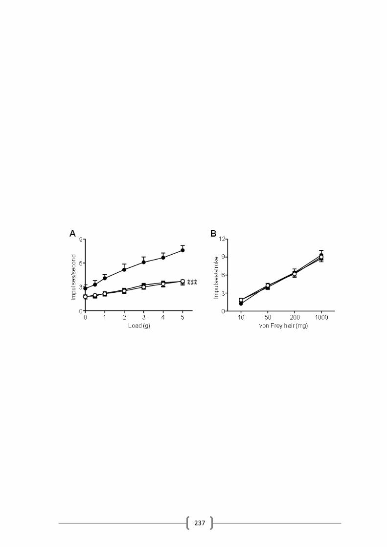
237

238
Figure 5.2: Selective high fat diet suppression of gastric tension
receptor mechanosensitivity is maintained after chow diet feeding.
Stimulus response functions of tension-sensitive (A) and mucosal (B) gastric
vagal afferents from mice fed a SLD (●, A: n=16 B: n=18), HFD (■, A: n=25,
B: n=29) or RFD (○ A: n=18, B: n=22). *** p<0.001 vs. SLD.

239

240
Figure 5.3: Mucosal receptor sensitivity to leptin is partially restored
upon reverting to a chow diet. The responses of gastric mucosal receptors
to stroking with calibrated von Frey hairs (10-1000mg) in the absence (●) and
presence of leptin 0.1 (○), 1 (□) and 10nM (Δ) from mice fed either a SLD for
24 weeks (A, N=8), HFD for 24 weeks (B, N=9) or a HFD for 12 weeks
followed by 12 weeks of SLD (C, N=6).*** p<0.001 vs. afferents prior to leptin
exposure. (D) The effect of leptin on the response to mucosal stroking with a
50mg von Frey hair in SLD (●), HFD (■) and RFD (○) mice. Diet significantly
modulated the potentiating ability of leptin. *** p<0.001 vs. SLD, ** p<0.01 vs.
SLD, ## p<0.01 vs. HFD. Original recordings of a mucosal receptor in
response to 50mg stroking before (i) and after addition of leptin 10nM (ii) from
SLD (E), HFD (F) and RFD (G) mice.

241

242
Figure 5.4: Tension receptor inhibition by leptin is reduced by
subsequent chow feeding. The responses of gastric tension receptors to
circular stretch (1-5g) in the absence (●) and presence of leptin 0.1 (○), 1 (□)
and 10nM (Δ) from mice fed either a SLD for 24 weeks (A, N=8), HFD for 24
weeks (B, N=9) or a HFD for 12 weeks followed by 12 weeks of SLD (C,
N=10). *** p<0.001 vs. afferents prior to leptin exposure. (D) The effect of
leptin on the response to 3g circular tension in SLD (●), HFD (■) and RFD (○)
mice. Diet significantly modulated the inhibiting ability of leptin. *** p<0.001 vs.
SLD, # p<0.05 vs. HFD. Original recordings of a tension receptor in response
to 3g tension before (i) and after addition of leptin 10nM (ii) from SLD (E),
HFD (F) and RFD (G) mice.

243

244
Figure 5.5: Diet induced changes in leptin receptor mRNA expression in
nodose ganglia and mucosal afferent cell bodies. Leptin receptor mRNA in
whole nodose ganglia was no different in any of the groups of mice (all groups
N=8). In mucosal traced afferents leptin receptor mRNA was reduced in
afferents from HFD and RFD mice (N=4, all groups). *** p<0.001 vs. SLD, #
p<0.05 vs. HFD.

245
CHAPTER 6: CONCLUSIONS
246
This thesis highlights specific mechanisms that may underlie peripheral satiety
signalling from the molecular basis of receptor regulation through to changes
observed in live animals in terms of feeding behaviour. As a whole these
studies demonstrate that the endogenous gastric peptides, leptin and ghrelin,
have distinct effects on specific populations of mechanosensitive gastric vagal
afferents, which are consistent with their previously observed general effects
on feeding and vagal afferent modulation [301, 326, 355, 548]. Previously, it
has been established that the expression profile of appetite regulatory
peptides and their receptors, on rodent vagal afferents, is dependent on fed
status [368, 369, 424, 452, 453, 460, 461]. This thesis now demonstrates that
the ability of mechanosensitive vagal afferents to respond to mechanical
stimuli and the endogenous peptides leptin and ghrelin is also dependent on
fed status.
In lean mice the effect of ghrelin on vagal afferent mechanosensitivity is
consistent with previous observations [301, 326, 355]. The opposing effects of
leptin and ghrelin on gastric vagal afferent mechanosensitivity in lean mice
may play a role in their respective anorexigenic and orexigenic effects.
However, the maintenance of an obese state depends upon a sustained
increase in food intake, which would require a shift in food intake signals away
from anorexigenic and towards orexigenic. Consistent with this idea is the
orexigenic effect of ghrelin potentially being amplified by gaining an inhibitory
effect on mucosal afferents, which is not seen in lean mice, and leptin losing
its potentiating and possibly anorexigenic effect and gaining an inhibitory and
potentially orexigenic effect. The potential dysfunction of peripheral satiety

247
signalling observed in the electrophysiological data was complimented by
equally dramatic changes in terms of receptor transcript regulation with gut
peptides, ghrelin and NPW causing decreases in anorexigenic leptin and CCK
receptor in vagal afferent cell bodies from obese mice, which could cause a
further suppression of anorexigenic signals and increase food consumption. It
is important to remember that this study only examined mRNA levels which,
as demonstrated previously, does not necessarily translate to changes in the
amount of functional protein at the cell surface [549]. However, these
adaptations may permit increased food intake to occur to protect a new higher
level of adiposity, if reflected at the functional protein level.
The electrophysiological studies revealed that the changes observed in
obesity in terms of baseline mechanosensitivity and the effects leptin and
ghrelin on vagal afferents were the same as observed in acutely food
restricted mice. This appears counterintuitive as the two states are
fundamentally opposite, one is a lack of food availability and energy stores,
the other is an excess of energy stores in the presence of food availability.
However, this may point to a potential mechanism to increase food intake to
the higher level necessary to maintain the obese state just as a food deprived
animal would want to consume extra food to restore any lost adiposity.
Whilst it is yet to be determined whether the observed changes in leptin and
ghrelin on gastric vagal afferents in obesity causes increased orexigenic
signalling, there is some evidence which suggests that ghrelin stimulates a
greater increase in food intake in obese humans compared to lean ones [360],

248
which would be consistent with increased orexigenic potency of ghrelin in
obesity. Furthermore, obesity is associated with an increased hedonic hunger
[550], which as discussed earlier are modulated by peripheral neuronal
signals (via the NTS and ARC), as well as directly by ghrelin and leptin. It
remains to be determined if, similar to gastric vagal afferents, there are altered
reward centre responses to leptin and ghrelin in obesity. There are also
changes to food patterns associated with obesity including changes to meal
size and frequency. Both the change in tension receptor mechanosensitivity
and effect of leptin and ghrelin on the gastric vagal afferents from obese mice
suggests meal size may be increased, which is consistent with meal pattern
analysis in rats [431, 487] and humans [551].
Leptin resistance is believed to occur in the majority of obese individuals
[552]. Mouse studies have revealed that this is probably, at least in part,
caused by an inability of leptin to activate neurons. This has been
demonstrated both centrally, in the ARC [553] and peripherally in the vagus
[336]. This thesis confirms the presence of leptin resistance in vagal afferents,
but in terms of gastric mechanosensitive vagal afferents, leptin resistance only
occurred within mucosal afferents. Tension sensitive gastric vagal afferents
did not exhibit leptin resistance in obese mice, but instead leptin exhibited an
inhibitory effect on tension receptor mechanosensitivity. This suggests that
leptin resistance occurs in a very specific manner in which only
subpopulations of nerves are affected. This also suggests that more refined
approaches need to be taken to treat obesity. In this instance, trying to
increase leptin signalling in obesity through the activation of the leptin

249
receptor could lead to a reduction in response to gastric distension, which
could potentially cause a further unwanted increase in food intake. A more
detailed analysis of the role leptin plays in appetite regulation and the
plasticity of this system in obesity is needed before the therapeutic potential of
this system can be fully determined. Downstream second messenger
molecules or effector channels of leptin receptors may offer more suitable and
effective therapeutic targets for obesity. In this case inhibiting BKCa channels
(which may be responsible for the inhibitory action of leptin) or activating
TRPC1 channels (which may be responsible for the potentiating effect of
leptin) may prove to be better targets than leptin and its receptor in the
treatment of obesity. However, both these channels have functional purposes
in a variety of tissue types and locations including smooth muscle [554] and
the heart [555]. This suggests that they would need to be specifically targeted
in the vagal afferents innervating the stomach to avoid the systemic side
effects that have plagued previous pharmacological approaches [556].
Whilst plenty of information exists regarding obesity induced dysfunction of
appetite regulatory systems, there is a lack of detailed studies looking at the
plasticity of these changes. Weight regain in obese people trying to lose
weight is exceptionally common [557-560]. This suggests that after attaining
the obese state adaptations occur that favour the maintenance of this obese
state [78]. This thesis has identified that the mechanosensitivity of gastric
tension receptors is reduced in obesity. This would decrease gastric
distension induced satiety signalling and may serve to potentiate or maintain
the obese state. In addition, it has been established that these alterations are

250
not readily reversible. Whilst the only intervention performed was changing the
diet this may provide a suitable comparison for the common human weight
loss experience. It demonstrates that simply changing dietary composition
may not be enough to cause sustained weight loss as persisting obesity
induced adaptations, that decrease the satiety signal, may lead to increased
food intake and subsequent weight gain even in the absence of an
obesogenic diet [78, 558]. This facet of the thesis will need further expansion
including looking for the presence of a theoretical point of no return, a point in
the development of obesity where the observed obesity induced adaptations
may become permanent as previously suggested [561, 562]. Furthermore,
other interventions need to be explored including pair feeding to determine
whether it is the diet or the adiposity that causes these adaptations. More
precise meal analysis needs to be undertaken, as currently the food intake
data featured in this thesis used a rather crude method of measurement. The
approach used, in this thesis, made it impossible to determine specific eating
parameters such as meal size, number, duration, inter-meal duration as well
as energy expenditure, all of which are vitally important when looking at
weight control, as there are substantially different effects of consuming the
same number of calories in a different pattern [563, 564].
Given that currently the most effective treatment for obesity is bariatric
surgery, with the roux-en-y gastric bypass (RYGP) being the gold standard of
weight loss procedures; this thesis highlights mechanisms that may underlie
its efficacy in weight loss. The nature of the gastrotomy in a RYGP means that
the bypassed section of the stomach would be left without vagal innervation.

251
The gastric pouch on the other hand would still retain its innervation meaning
that vagal satiety signals could still be transmitted. There would likely be an
enhanced response to distension of the stomach by an isovolumetric meal
given the substantially reduced size of the gastric pouch, which could
overcome the reduced sensitivity of the gastric tension receptors to trigger
early satiety. RYGB patients also often exhibit reduced ghrelin secretion [565],
which is proposed to be caused by the removal of autonomic input to the
ghrelin containing cells of the stomach (the vast majority of which are in the
bypassed portion of the stomach) [566]. It would be interesting and worthwhile
to examine the responsiveness of gastric vagal afferents to mechanical
stimuli, as well as leptin and ghrelin, in a mouse model of the RYGB to
determine whether the effects documented in this thesis are reversed or
merely counteracted.
Whilst it is well accepted that gastric vagal afferents are involved in the control
of food intake [567, 568], they are also involved in the regulation of wider
gastrointestinal function [210, 467]. This is highlighted by the fact that they are
targeted in the treatment of multiple gastrointestinal disorders including
functional dyspepsia and gastro-oesophageal reflux disease [284, 285, 438,
569-572]. It is not known whether there are discrete and identifiable
subpopulations of vagal afferents that are responsible for initiating different
feedback signals. Thus whilst the studies within this thesis suggest that in
obesity leptin and ghrelin may act to mediate an increase in food intake it is
possible that their effects on gastric vagal afferents are in fact mediating
changes in other functions. Therefore, it is imperative that future studies

252
determine the behavioural effect of modulating specific populations of gastric
vagal mechanosensitive afferents.
Regardless of the limitations of these studies they have identified novel and
new effects of previously studied molecules in a common disease. The results
highlight possible new specific targets for obesity treatments, as well as
providing potential mechanisms and explanations for adaptations that favour
weight gain/maintenance when weight loss is desirable, further adding to the
ever expanding understanding of obesity.

253
REFERENCES

254
[1] Nguyen T, Lau DCW. The Obesity Epidemic and Its Impact on Hypertension. Canadian Journal of Cardiology. 2012;28:326-33.
[2] Finucane MM, Stevens GA, Cowan MJ, Danaei G, Lin JK, Paciorek CJ, et al. National, regional, and global trends in body-mass index since 1980: systematic analysis of health examination surveys and epidemiological studies with 960 country-years and 9·1 million participants. The Lancet. 2011;377:557-67.
[3] Wang YC, McPherson K, Marsh T, Gortmaker SL, Brown M. Health and economic burden of the projected obesity trends in the USA and the UK. The Lancet. 2011;378:815-25.
[4] Ogunbode AM, Ladipo M, Ajayi IO, Fatiregun AA. Obesity: an emerging disease. Nigerian journal of clinical practice. 2011;14:390-4.
[5] Popkin BM. The Nutrition Transition and Obesity in the Developing World. The Journal of Nutrition. 2001;131:871S-3S.
[6] Caballero B. Introduction. The Journal of Nutrition. 2001;131:866S-70S.
[7] Jones COH, White NG. Adiposity in Aboriginal people from Arnhem Land, Australia: variation in degree and distribution associated with age, sex and lifestyle. Annals of Human Biology. 1994;21:207-27.
[8] McGarvey ST. Obesity in Samoans and a perspective on its etiology in Polynesians. The American Journal of Clinical Nutrition. 1991;53:1586S-94S.
[9] Bhatnagar D, Anand IS, Durrington PN, Patel DJ, Wander GS, Mackness MI, et al. Coronary risk factors in people from the Indian subcontinent living in west London and their siblings in India. Lancet. 1995;345:405-9.
[10] Popkin BM, Keyou G, Zhai F, Guo X, Ma H, Zohoori N. The nutrition transition in China: a cross-sectional analysis. European journal of clinical nutrition. 1993;47:333-46.
[11] Murphy NJ, Schraer CD, Thiele MC, Boyko EJ, Bulkow LR, Doty BJ, et al. Dietary Change and Obesity Associated with Glucose Intolerance in Alaska Natives. Journal of the American Dietetic Association. 1995;95:676-82.
[12] Curb JD, Marcus EB. Body fat and obesity in Japanese Americans. The American Journal of Clinical Nutrition. 1991;53:1552S-5S.
[13] Ravussin E, Valencia ME, Esparza J, Bennett PH, Schulz LO. Effects of a Traditional Lifestyle on Obesity in Pima Indians. Diabetes Care. 1994;17:1067-74.

255
[14] Young LR, Nestle M. The Contribution of Expanding Portion Sizes to the US Obesity Epidemic. American Journal of Public Health. 2002;92:246-9.
[15] Rolls BJ, Roe LS, Meengs JS, Wall DE. Increasing the portion size of a sandwich increases energy intake. Journal of the American Dietetic Association. 2004;104:367-72.
[16] Rolls BJ, Morris EL, Roe LS. Portion size of food affects energy intake in normal-weight and overweight men and women. The American Journal of Clinical Nutrition. 2002;76:1207-13.
[17] Rolls BJ, Engell D, Birch LL. Serving Portion Size Influences 5-Year-Old but Not 3-Year-Old Children's Food Intakes. Journal of the American Dietetic Association. 2000;100:232-4.
[18] Young LR, Nestle M. Expanding portion sizes in the US marketplace: Implications for nutrition counseling. Journal of the American Dietetic Association. 2003;103:231-4.
[19] Nielsen S PBM. PAtterns and trends in food portion sizes, 1977-1998. JAMA : the journal of the American Medical Association. 2003;289:450-3.
[20] Rozin P, Kabnick K, Pete E, Fischler C, Shields C. The Ecology of Eating: Smaller Portion Sizes in France Than in the United States Help Explain the French Paradox. Psychological Science. 2003;14:450-4.
[21] Schneider H, Dietrich ES, Venetz WP. Trends and stabilization up to 2022 in overweight and obesity in Switzerland, comparison to France, UK, US and Australia. Int J Environ Res Public Health. 2010;7:460-72.
[22] Keast DR, Fulgoni VL, 3rd, Nicklas TA, O'Neil CE. Food sources of energy and nutrients among children in the United States: National Health and Nutrition Examination Survey 2003-2006. Nutrients. 2013;5:283-301.
[23] Duffey KJ, Popkin BM. Energy Density, Portion Size, and Eating Occasions: Contributions to Increased Energy Intake in the United States, 1977–2006. PLoS Med. 2011;8:e1001050.
[24] Bell EA, Castellanos VH, Pelkman CL, Thorwart ML, Rolls BJ. Energy density of foods affects energy intake in normal-weight women. The American Journal of Clinical Nutrition. 1998;67:412-20.
[25] Kral TV, Roe LS, Rolls BJ. Combined effects of energy density and portion size on energy intake in women. The American Journal of Clinical Nutrition. 2004;79:962-8.
[26] Milanski M, Arruda AP, Coope A, Ignacio-Souza LM, Nunez CE, Roman EA, et al. Inhibition of Hypothalamic Inflammation Reverses Diet-Induced Insulin Resistance in the Liver. Diabetes. 2012;61:1455-62.
[27] Milanski M, Degasperi G, Coope A, Morari J, Denis R, Cintra DE, et al. Saturated Fatty Acids Produce an Inflammatory Response Predominantly

256
through the Activation of TLR4 Signaling in Hypothalamus: Implications for the Pathogenesis of Obesity. The Journal of Neuroscience. 2009;29:359-70.
[28] De Souza CT, Araujo EP, Bordin S, Ashimine R, Zollner RL, Boschero AC, et al. Consumption of a Fat-Rich Diet Activates a Proinflammatory Response and Induces Insulin Resistance in the Hypothalamus. Endocrinology. 2005;146:4192-9.
[29] Johnstone AM, Murison SD, Duncan JS, Rance KA, Speakman JR. Factors influencing variation in basal metabolic rate include fat-free mass, fat mass, age, and circulating thyroxine but not sex, circulating leptin, or triiodothyronine. The American Journal of Clinical Nutrition. 2005;82:941-8.
[30] Astrup A, Gøtzsche PC, van de Werken K, Ranneries C, Toubro S, Raben A, et al. Meta-analysis of resting metabolic rate in formerly obese subjects. The American Journal of Clinical Nutrition. 1999;69:1117-22.
[31] Astrup A, Buemann B, Toubro S, Ranneries C, Raben A. Low resting metabolic rate in subjects predisposed to obesity: a role for thyroid status. The American Journal of Clinical Nutrition. 1996;63:879-83.
[32] Vosselman MJ, van Marken Lichtenbelt WD, Schrauwen P. Energy dissipation in brown adipose tissue: From mice to men. Molecular and cellular endocrinology. 2013.
[33] Tremblay A, Royer MM, Chaput JP, Doucet E. Adaptive thermogenesis can make a difference in the ability of obese individuals to lose body weight. International journal of obesity (2005). 2012.
[34] French SA, Story M, Jeffery RW. Environmental Influences on Eating and Physical Activity. Annual Review of Public Health. 2001;22:309-35.
[35] Webber J. Energy balance in obesity. Proceedings of the Nutrition Society. 2003;62:539-43.
[36] Sharma S, Kavuru M. Sleep and Metabolism: An Overview. International Journal of Endocrinology. 2010;2010.
[37] López-García E, Faubel R, León-Muñoz L, Zuluaga MC, Banegas JR, Rodríguez-Artalejo F. Sleep duration, general and abdominal obesity, and weight change among the older adult population of Spain. The American Journal of Clinical Nutrition. 2008;87:310-6.
[38] Watanabe M, Kikuchi H, Tanaka K, Takahashi M. Association of short sleep duration with weight gain and obesity at 1-year follow-up: a large-scale prospective study. Sleep. 2010;33:161-7.
[39] Nedeltcheva AV, Kilkus JM, Imperial J, Schoeller DA, Penev PD. Insufficient Sleep Undermines Dietary Efforts to Reduce Adiposity. Annals of Internal Medicine. 2010;153:435-41.
257
[40] Booth JN, Bromley LE, Darukhanavala AP, Whitmore HR, Imperial JG, Penev PD. Reduced physical activity in adults at risk for type 2 diabetes who curtail their sleep. Obesity (Silver Spring, Md). 2012;20:278-84.
[41] St-Onge M-P, Roberts AL, Chen J, Kelleman M, O'Keeffe M, RoyChoudhury A, et al. Short sleep duration increases energy intakes but does not change energy expenditure in normal-weight individuals. The American Journal of Clinical Nutrition. 2011;94:410-6.
[42] Nedeltcheva AV, Kilkus JM, Imperial J, Kasza K, Schoeller DA, Penev PD. Sleep curtailment is accompanied by increased intake of calories from snacks. The American Journal of Clinical Nutrition. 2009;89:126-33.
[43] Schmid SM, Hallschmid M, Jauch-Chara K, Wilms B, Benedict C, Lehnert H, et al. Short-term sleep loss decreases physical activity under free-living conditions but does not increase food intake under time-deprived laboratory conditions in healthy men. The American Journal of Clinical Nutrition. 2009;90:1476-82.
[44] Brondel L, Romer MA, Nougues PM, Touyarou P, Davenne D. Acute partial sleep deprivation increases food intake in healthy men. The American Journal of Clinical Nutrition. 2010;91:1550-9.
[45] Spiegel K, Tasali E, Penev P, Cauter EV. Brief Communication: Sleep Curtailment in Healthy Young Men Is Associated with Decreased Leptin Levels, Elevated Ghrelin Levels, and Increased Hunger and Appetite. Annals of Internal Medicine. 2004;141:846-50.
[46] Magee CA, Huang X-F, Iverson DC, Caputi P. Acute sleep restriction alters neuroendocrine hormones and appetite in healthy male adults. Sleep and Biological Rhythms. 2009;7:125-7.
[47] Spiegel K, Leproult R, Van Cauter E. Impact of sleep debt on metabolic and endocrine function. The Lancet. 1999;354:1435-9.
[48] Buxton OM, Pavlova M, Reid EW, Wang W, Simonson DC, Adler GK. Sleep Restriction for 1 Week Reduces Insulin Sensitivity in Healthy Men. Diabetes. 2010;59:2126-33.
[49] Nedeltcheva AV, Kessler L, Imperial J, Penev PD. Exposure to Recurrent Sleep Restriction in the Setting of High Caloric Intake and Physical Inactivity Results in Increased Insulin Resistance and Reduced Glucose Tolerance. Journal of Clinical Endocrinology & Metabolism. 2009;94:3242-50.
[50] Yaggi HK, Araujo AB, McKinlay JB. Sleep Duration as a Risk Factor for the Development of Type 2 Diabetes. Diabetes Care. 2006;29:657-61.
[51] Müssig K, Remer T, Maser-Gluth C. Brief review: Glucocorticoid excretion in obesity. The Journal of Steroid Biochemistry and Molecular Biology. 2010;121:589-93.

258
[52] Pi-Sunyer FX. The obesity epidemic: pathophysiology and consequences of obesity. Obesity research. 2002;10 Suppl 2:97S-104S.
[53] Herrera B, Lindgren C. The Genetics of Obesity. Curr Diab Rep. 2010;10:498-505.
[54] Bouchard C, Tremblay A, Després J-P, Nadeau A, Lupien PJ, Thériault G, et al. The Response to Long-Term Overfeeding in Identical Twins. New England Journal of Medicine. 1990;322:1477-82.
[55] Stunkard AJ, Sørensen TIA, Hanis C, Teasdale TW, Chakraborty R, Schull WJ, et al. An Adoption Study of Human Obesity. New England Journal of Medicine. 1986;314:193-8.
[56] Huszar D, Lynch CA, Fairchild-Huntress V, Dunmore JH, Fang Q, Berkemeier LR, et al. Targeted Disruption of the Melanocortin-4 Receptor Results in Obesity in Mice. Cell. 1997;88:131-41.
[57] Drel VR, Mashtalir N, Ilnytska O, Shin J, Li F, Lyzogubov VV, et al. The Leptin-Deficient (ob/ob) Mouse: A New Animal Model of Peripheral Neuropathy of Type 2 Diabetes and Obesity. Diabetes. 2006;55:3335-43.
[58] Lowell BB, V SS, Hamann A, Lawitts JA, Himms-Hagen J, Boyer BB, et al. Development of obesity in transgenic mice after genetic ablation of brown adipose tissue. Nature. 1993;366:740-2.
[59] Strobel A, Issad T, Camoin L, Ozata M, Strosberg AD. A leptin missense mutation associated with hypogonadism and morbid obesity. Nature genetics. 1998;18:213-5.
[60] Montague CT, Farooqi IS, Whitehead IP, Soos MA, Rau H, Wareham NJ, et al. Congenital leptin deficiency is associated with severe early-onset obesity in humans. Nature. 1997;387:903-8.
[61] Clement K, Vaisse C, Lahlou N, Cabrol S, Pelloux V, Cassuto D, et al. A mutation in the human leptin receptor gene causes obesity and pituitary dysfunction. Nature. 1998;392:398-401.
[62] Krude H, Grüters A. Implications of Proopiomelanocortin (POMC) Mutations in Humans: The POMC Deficiency Syndrome. Trends in Endocrinology & Metabolism. 2000;11:15-22.
[63] Yeo GS, Farooqi IS, Aminian S, Halsall DJ, Stanhope RG, O'Rahilly S. A frameshift mutation in MC4R associated with dominantly inherited human obesity. Nature genetics. 1998;20:111-2.
[64] Scheermeyer E. Prader-Willi syndrome Care of adults in general practice. Australian Family Physician. 2013;42:51-4.
[65] Katsanis N, Lupski JR, Beales PL. Exploring the molecular basis of Bardet–Biedl syndrome. Human Molecular Genetics. 2001;10:2293-9.

259
[66] Bettini V, Maffei P, Pagano C, Romano S, Milan G, Favaretto F, et al. The progression from obesity to type 2 diabetes in Alström syndrome. Pediatric Diabetes. 2012;13:59-67.
[67] Goldstone AP, Beales PL. Genetic obesity syndromes. Frontiers of hormone research. 2008;36:37-60.
[68] Wadden TA, Frey DL. A multicenter evaluation of a proprietary weight loss program for the treatment of marked obesity: A five-year follow-up. International Journal of Eating Disorders. 1997;22:203-12.
[69] Simpson SJ, Raubenheimer D. Obesity: the protein leverage hypothesis. Obesity Reviews. 2005;6:133-42.
[70] Griffioen-Roose S, Mars M, Siebelink E, Finlayson G, Tomé D, de Graaf C. Protein status elicits compensatory changes in food intake and food preferences. The American Journal of Clinical Nutrition. 2012;95:32-8.
[71] Gosby AK, Conigrave AD, Lau NS, Iglesias MA, Hall RM, Jebb SA, et al. Testing Protein Leverage in Lean Humans: A Randomised Controlled Experimental Study. PloS one. 2011;6:e25929.
[72] de Castro JM. Behavioral genetics of food intake regulation in free-living humans. Nutrition. 1999;15:550-4.
[73] Kennedy GC. The Role of Depot Fat in the Hypothalamic Control of Food Intake in the Rat. Proceedings of the Royal Society of London Series B, Biological Sciences. 1953;140:578-92.
[74] Mrosovsky N. Body fat: What is regulated? Physiology & behavior. 1986;38:407-14.
[75] Keesey RE, Hirvonen MD. Body Weight Set-Points: Determination and Adjustment. The Journal of Nutrition. 1997;127:1875S-83S.
[76] Hu FB, Chen C, Wang B, Stampfer MJ, Xu X. Leptin concentrations in relation to overall adiposity, fat distribution, and blood pressure in a rural Chinese population. Int J Obes Relat Metab Disord. 2001;25:121.
[77] Gray DS, Fisler JS, Bray GA. Effects of repeated weight loss and regain on body composition in obese rats. The American Journal of Clinical Nutrition. 1988;47:393-9.
[78] Sumithran P, Prendergast LA, Delbridge E, Purcell K, Shulkes A, Kriketos A, et al. Long-Term Persistence of Hormonal Adaptations to Weight Loss. New England Journal of Medicine. 2011;365:1597-604.
[79] Crujeiras AB, Goyenechea E, Abete I, Lage M, Carreira MC, Martínez JA, et al. Weight Regain after a Diet-Induced Loss Is Predicted by Higher Baseline Leptin and Lower Ghrelin Plasma Levels. Journal of Clinical Endocrinology & Metabolism. 2010;95:5037-44.

260
[80] Holm JC, Gamborg M, Ward L, Ibsen KK, Gammeltoft S, Sørensen TIA, et al. Longitudinal Analysis of Leptin Variation during Weight Regain after Weight Loss in Obese Children. Obesity Facts. 2009;2:243-8.
[81] Mars M, de Graaf C, de Groot CP, van Rossum CT, Kok FJ. Fasting leptin and appetite responses induced by a 4-day 65%-energy-restricted diet. International journal of obesity (2005). 2006;30:122-8.
[82] Wyrwicka W, Dobrzecka C. Relationship between feeding and satiation centers of the hypothalamus. Science. 1960;132:805-6.
[83] Hollifield G, Parson W. Studies of the Satiety Response in Mice. The Journal of Clinical Investigation. 1957;36:1638-41.
[84] Li A-J, Dinh TT, Ritter S. Hyperphagia and obesity produced by arcuate injection of NPY–saporin do not require upregulation of lateral hypothalamic orexigenic peptide genes. Peptides. 2008;29:1732-9.
[85] Baldwin BA. Neural and hormonal mechanisms regulating food intake. The Proceedings of the Nutrition Society. 1985;44:303-11.
[86] Bellinger LL, Bernardis LL. The dorsomedial hypothalamic nucleus and its role in ingestive behavior and body weight regulation: Lessons learned from lesioning studies. Physiology & behavior. 2002;76:431-42.
[87] Anand BK, Brobeck JR. Localization of a ―Feeding Center‖ in the Hypothalamus of the Rat. Proceedings of the Society for Experimental Biology and Medicine Society for Experimental Biology and Medicine (New York, NY). 1951;77:323-5.
[88] Debons AF, Silver L, Cronkite EP, Johnson HA, Brecher G, Tenzer D, et al. Localization of gold in mouse brain in relation to gold thioglucose obesity. The American journal of physiology. 1962;202:743-50.
[89] Peruzzo B, Pastor FE, Blázquez JL, Schöbitz K, Peláez B, Amat P, et al. A second look at the barriers of the medial basal hypothalamus. Experimental Brain Research. 2000;132:10-26.
[90] Nonaka N, Shioda S, Niehoff ML, Banks WA. Characterization of Blood-Brain Barrier Permeability to PYY3-36 in the Mouse. Journal of Pharmacology and Experimental Therapeutics. 2003;306:948-53.
[91] Kastin A, Akerstrom V, Pan W. Interactions of glucagon-like peptide-1 (GLP-1) with the blood-brain barrier. Journal of Molecular Neuroscience. 2002;18:7-14.
[92] Banks WA, Kastin AJ, Huang W, Jaspan JB, Maness LM. Leptin enters the brain by a saturable system independent of insulin. Peptides. 1996;17:305-11.
[93] Woods SC, Seeley RJ, Porte D, Schwartz MW. Signals That Regulate Food Intake and Energy Homeostasis. Science. 1998;280:1378-83.

261
[94] Marsh DJ, Hollopeter G, Kafer KE, Palmiter RD. Role of the Y5 neuropeptide Y receptor in feeding and obesity. Nature medicine. 1998;4:718-21.
[95] Clark JT, Kalra PS, Crowley WR, Kalra SP. Neuropeptide Y and human pancreatic polypeptide stimulate feeding behavior in rats. Endocrinology. 1984;115:427-9.
[96] Légrádi G, Lechan RM. The Arcuate Nucleus Is the Major Source for Neuropeptide Y-Innervation of Thyrotropin-Releasing Hormone Neurons in the Hypothalamic Paraventricular Nucleus. Endocrinology. 1998;139:3262-70.
[97] Bai FL, Yamano M, Shiotani Y, Emson PC, Smith AD, Powell JF, et al. An arcuato-paraventricular and -dorsomedial hypothalamic neuropeptide Y-containing system which lacks noradrenaline in the rat. Brain Research. 1985;331:172-5.
[98] Csiffa´ry A, Go¨rcs TJ, Palkovits M. Neuropeptide Y innervation of ACTH-immunoreactive neurons in the arcuate nucleus of rats: a correlated light and electron microscopic double immunolabeling study. Brain Research. 1990;506:215-22.
[99] Cowley MA, Smart JL, Rubinstein M, Cerdán MG, Diano S, Horvath TL, et al. Leptin activates anorexigenic POMC neurons through a neural network in the arcuate nucleus. Nature. 2001;411:480-4.
[100] Ellacott KLJ, Halatchev IG, Cone RD. Characterization of Leptin-Responsive Neurons in the Caudal Brainstem. Endocrinology. 2006;147:3190-5.
[101] Pritchard L, Turnbull A, White A. Pro-opiomelanocortin processing in the hypothalamus: impact on melanocortin signalling and obesity. Journal of Endocrinology. 2002;172:411-21.
[102] Rossi M, Kim MS, Morgan DGA, Small CJ, Edwards CMB, Sunter D, et al. A C-Terminal Fragment of Agouti-Related Protein Increases Feeding and Antagonizes the Effect of Alpha-Melanocyte Stimulating Hormone in Vivo. Endocrinology. 1998;139:4428-31.
[103] Chen YY, Pelletier G. Demonstration of contacts between proopiomelanocortin neurons in the rat hypothalamus. Neuroscience Letters. 1983;43:271-6.
[104] Eskay RL, Giraud P, Oliver C, Brown-Stein MJ. Distribution of alpha-melanocyte-stimulating hormone in the rat brain: evidence that alpha-MSH-containing cells in the arcuate region send projections to extrahypothalamic areas. Brain Res. 1979;178:55-67.
[105] Cowley MA, Smith RG, Diano S, Tschöp M, Pronchuk N, Grove KL, et al. The Distribution and Mechanism of Action of Ghrelin in the CNS Demonstrates a Novel Hypothalamic Circuit Regulating Energy Homeostasis. Neuron. 2003;37:649-61.

262
[106] Date Y, Shimbara T, Koda S, Toshinai K, Ida T, Murakami N, et al. Peripheral ghrelin transmits orexigenic signals through the noradrenergic pathway from the hindbrain to the hypothalamus. Cell metabolism. 2006;4:323-31.
[107] Xu L, Gao S, Guo F, Sun X. Effect of motilin on gastric distension sensitive neurons in arcuate nucleus and gastric motility in rat. Neurogastroenterology & Motility. 2011;23:265-e121.
[108] Jeanningros R. Modulation of lateral hypothalamic single unit activity by gastric and intestinal distension. Journal of the autonomic nervous system. 1984;11:1-11.
[109] Zhu JN, Guo CL, Li HZ, Wang JJ. Dorsomedial hypothalamic nucleus neurons integrate important peripheral feeding-related signals in rats. Journal of neuroscience research. 2007;85:3193-204.
[110] Ueta Y, Kannan H, Yamashita H. Gastric afferents to the paraventricular nucleus in the rat. Experimental Brain Research. 1991;84:487-94.
[111] Mazda T, Yamamoto H, Fujimura M, Fujimiya M. Gastric distension-induced release of 5-HT stimulates c-fos expression in specific brain nuclei via 5-HT3 receptors in conscious rats. American Journal of Physiology - Gastrointestinal and Liver Physiology. 2004;287:G228-G35.
[112] Singru PS, Wittmann G, Farkas E, Zséli G, Fekete C, Lechan RM. Refeeding-Activated Glutamatergic Neurons in the Hypothalamic Paraventricular Nucleus (PVN) Mediate Effects of Melanocortin Signaling in the Nucleus Tractus Solitarius (NTS). Endocrinology. 2012;153:3804-14.
[113] Zheng H, Patterson LM, Phifer CB, Berthoud H-R. Brain stem melanocortinergic modulation of meal size and identification of hypothalamic POMC projections. American Journal of Physiology - Regulatory, Integrative and Comparative Physiology. 2005;289:R247-R58.
[114] Barsh GS, Schwartz MW. Genetic approaches to studying energy balance: perception and integration. Nature reviews Genetics. 2002;3:589-600.
[115] Yoshida M, Yokoo H, Mizoguchi K, Kawahara H, Tsuda A, Nishikawa T, et al. Eating and drinking cause increased dopamine release in the nucleus accumbens and ventral tegmental area in the rat: Measurement by in vivo microdialysis. Neuroscience Letters. 1992;139:73-6.
[116] Dahan L, Astier B, Vautrelle N, Urbain N, Kocsis B, Chouvet G. Prominent burst firing of dopaminergic neurons in the ventral tegmental area during paradoxical sleep. Neuropsychopharmacology. 2007;32:1232-41.
[117] Nishino H, Ono T, Muramoto K, Fukuda M, Sasaki K. Neuronal activity in the ventral tegmental area (VTA) during motivated bar press feeding in the monkey. Brain Research. 1987;413:302-13.

263
[118] Wise RA. Addictive drugs and brain stimulation reward. Annual review of neuroscience. 1996;19:319-40.
[119] Stratford TR, Kelley AE. Evidence of a Functional Relationship between the Nucleus Accumbens Shell and Lateral Hypothalamus Subserving the Control of Feeding Behavior. The Journal of Neuroscience. 1999;19:11040-8.
[120] Alhadeff AL, Rupprecht LE, Hayes MR. GLP-1 Neurons in the Nucleus of the Solitary Tract Project Directly to the Ventral Tegmental Area and Nucleus Accumbens to Control for Food Intake. Endocrinology. 2012;153:647-58.
[121] Manta S, El Mansari M, Debonnel G, Blier P. Electrophysiological and neurochemical effects of long-term vagus nerve stimulation on the rat monoaminergic systems. The International Journal of Neuropsychopharmacology. 2013;16:459-70.
[122] Ziomber A, Thor P, Krygowska-Wajs A, Zalecki T, Moskala M, Romanska I, et al. Chronic impairment of the vagus nerve function leads to inhibition of dopamine but not serotonin neurons in rat brain structures. Pharmacological reports : PR. 2012;64:1359-67.
[123] Tomasi D, Wang G-J, Wang R, Backus W, Geliebter A, Telang F, et al. Association of Body Mass and Brain Activation during Gastric Distention: Implications for Obesity. PloS one. 2009;4:e6847.
[124] Epstein LH, Leddy JJ, Temple JL, Faith MS. Food reinforcement and eating: a multilevel analysis. Psychological bulletin. 2007;133:884-906.
[125] Oswald KD, Murdaugh DL, King VL, Boggiano MM. Motivation for palatable food despite consequences in an animal model of binge eating. International Journal of Eating Disorders. 2011;44:203-11.
[126] Goldfield GS, Lumb AB, Colapinto CK. Relative reinforcing value of energy-dense snack foods in overweight and obese adults. Canadian journal of dietetic practice and research : a publication of Dietitians of Canada = Revue canadienne de la pratique et de la recherche en dietetique : une publication des Dietetistes du Canada. 2011;72:170-4.
[127] Stice E, Spoor S, Bohon C, Veldhuizen MG, Small DM. Relation of reward from food intake and anticipated food intake to obesity: a functional magnetic resonance imaging study. Journal of abnormal psychology. 2008;117:924-35.
[128] Geiger BM, Behr GG, Frank LE, Caldera-Siu AD, Beinfeld MC, Kokkotou EG, et al. Evidence for defective mesolimbic dopamine exocytosis in obesity-prone rats. The FASEB Journal. 2008;22:2740-6.
[129] Volkow ND, Wang G-J, Telang F, Fowler JS, Thanos PK, Logan J, et al. Low dopamine striatal D2 receptors are associated with prefrontal metabolism in obese subjects: Possible contributing factors. NeuroImage. 2008;42:1537-43.

264
[130] Papp M, Bal A. Separation of the motivational and motor consequences of 6-hydroxydopamine lesions of the mesolimbic or nigrostriatal system in rats. Behavioural brain research. 1987;23:221-9.
[131] Berridge KC, Valenstein ES. What psychological process mediates feeding evoked by electrical stimulation of the lateral hypothalamus? Behavioral neuroscience. 1991;105:3-14.
[132] Davis JF, Tracy AL, Schurdak JD, Tschop MH, Lipton JW, Clegg DJ, et al. Exposure to elevated levels of dietary fat attenuates psychostimulant reward and mesolimbic dopamine turnover in the rat. Behavioral neuroscience. 2008;122:1257-63.
[133] Zhou Q-Y, Palmiter RD. Dopamine-deficient mice are severely hypoactive, adipsic, and aphagic. Cell. 1995;83:1197-209.
[134] Kobayashi M, Iaccarino C, Saiardi A, Heidt V, Bozzi Y, Picetti R, et al. Simultaneous absence of dopamine D1 and D2 receptor-mediated signaling is lethal in mice. Proceedings of the National Academy of Sciences of the United States of America. 2004;101:11465-70.
[135] Zigman JM, Jones JE, Lee CE, Saper CB, Elmquist JK. Expression of ghrelin receptor mRNA in the rat and the mouse brain. The Journal of Comparative Neurology. 2006;494:528-48.
[136] Jerlhag E, Janson AC, Waters S, Engel JA. Concomitant Release of Ventral Tegmental Acetylcholine and Accumbal Dopamine by Ghrelin in Rats. PloS one. 2012;7:e49557.
[137] Jerlhag E, Egecioglu E, Landgren S, Salomé N, Heilig M, Moechars D, et al. Requirement of central ghrelin signaling for alcohol reward. Proceedings of the National Academy of Sciences. 2009;106:11318-23.
[138] Jerlhag E, Egecioglu E, Dickson S, Engel J. Ghrelin receptor antagonism attenuates cocaine- and amphetamine-induced locomotor stimulation, accumbal dopamine release, and conditioned place preference. Psychopharmacology. 2010;211:415-22.
[139] Egecioglu E, Jerlhag E, Salomé N, Skibicka KP, Haage D, Bohlooly-Y M, et al. Ghrelin increases intake of rewarding food in rodents. Addiction Biology. 2010;15:304-11.
[140] Naleid AM, Grace MK, Cummings DE, Levine AS. Ghrelin induces feeding in the mesolimbic reward pathway between the ventral tegmental area and the nucleus accumbens. Peptides. 2005;26:2274-9.
[141] Dickson SL, Hrabovszky E, Hansson C, Jerlhag E, Alvarez-Crespo M, Skibicka KP, et al. Blockade of central nicotine acetylcholine receptor signaling attenuate ghrelin-induced food intake in rodents. Neuroscience. 2010;171:1180-6.

265
[142] Szczypka MS, Rainey MA, Palmiter RD. Dopamine is required for hyperphagia in Lep(ob/ob) mice. Nature genetics. 2000;25:102-4.
[143] Figlewicz DP, Higgins MS, Ng-Evans SB, Havel PJ. Leptin reverses sucrose-conditioned place preference in food-restricted rats. Physiology & behavior. 2001;73:229-34.
[144] Hommel JD, Trinko R, Sears RM, Georgescu D, Liu Z-W, Gao X-B, et al. Leptin Receptor Signaling in Midbrain Dopamine Neurons Regulates Feeding. Neuron. 2006;51:801-10.
[145] Krügel U, Schraft T, Kittner H, Kiess W, Illes P. Basal and feeding-evoked dopamine release in the rat nucleus accumbens is depressed by leptin. European Journal of Pharmacology. 2003;482:185-7.
[146] Flier JS. Obesity Wars: Molecular Progress Confronts an Expanding Epidemic. Cell. 2004;116:337-50.
[147] Ferris WF, Crowther NJ. Once fat was fat and that was that: our changing perspectives on adipose tissue. Cardiovascular journal of Africa. 2011;22:147-54.
[148] Kershaw EE, Flier JS. Adipose Tissue as an Endocrine Organ. Journal of Clinical Endocrinology & Metabolism. 2004;89:2548-56.
[149] Maffei M, Fei H, Lee GH, Dani C, Leroy P, Zhang Y, et al. Increased expression in adipocytes of ob RNA in mice with lesions of the hypothalamus and with mutations at the db locus. Proceedings of the National Academy of Sciences. 1995;92:6957-60.
[150] Ahima RS, Lazar MA. Adipokines and the Peripheral and Neural Control of Energy Balance. Molecular Endocrinology. 2008;22:1023-31.
[151] Mercer JG. Regulation of appetite and body weight in seasonal mammals. Comparative Biochemistry and Physiology Part C: Pharmacology, Toxicology and Endocrinology. 1998;119:295-303.
[152] Otis JP, Raybould HE, Carey HV. Cholecystokinin activation of central satiety centers changes seasonally in a mammalian hibernator. General and Comparative Endocrinology. 2011;171:269-74.
[153] Prentice AM, Whitehead RG, Roberts SB, Paul AA. Long-term energy balance in child-bearing Gambian women. The American Journal of Clinical Nutrition. 1981;34:2790-9.
[154] Sabbağ Ç. Seasonal BMI Changes of Rural Women Living in Anatolia. International Journal of Environmental Research and Public Health. 2012;9:1159-70.
[155] Velkoska E, Morris MJ, Burns P, Weisinger RS. Leptin reduces food intake but does not alter weight regain following food deprivation in the rat.

266
International journal of obesity and related metabolic disorders : journal of the International Association for the Study of Obesity. 2003;27:48-54.
[156] Hope PJ, Wittert GA, Horowitz M, Morley JE. Feeding patterns of S. crassicaudata (Marsupialia:Dasyuridae): role of gender, photoperiod, and fat stores. American Journal of Physiology - Regulatory, Integrative and Comparative Physiology. 1997;272:R78-R83.
[157] Benatti F, Solis M, Artioli G, Montag E, Painelli V, Saito F, et al. Liposuction Induces a Compensatory Increase of Visceral Fat Which Is Effectively Counteracted by Physical Activity: A Randomized Trial. Journal of Clinical Endocrinology & Metabolism. 2012;97:2388-95.
[158] Löfgren P, Andersson I, Adolfsson B, Leijonhufvud BM, Hertel K, Hoffstedt J, et al. Long-term prospective and controlled studies demonstrate adipose tissue hypercellularity and relative leptin deficiency in the postobese state. Journal of Clinical Endocrinology and Metabolism. 2005;90:6207-13.
[159] Spalding KL, Arner E, Westermark PO, Bernard S, Buchholz BA, Bergmann O, et al. Dynamics of fat cell turnover in humans. Nature. 2008;453:783-7.
[160] Skurk T, Alberti-Huber C, Herder C, Hauner H. Relationship between Adipocyte Size and Adipokine Expression and Secretion. Journal of Clinical Endocrinology & Metabolism. 2007;92:1023-33.
[161] Cinti S, Mitchell G, Barbatelli G, Murano I, Ceresi E, Faloia E, et al. Adipocyte death defines macrophage localization and function in adipose tissue of obese mice and humans. Journal of Lipid Research. 2005;46:2347-55.
[162] Greenberg AS, Obin MS. Obesity and the role of adipose tissue in inflammation and metabolism. The American Journal of Clinical Nutrition. 2006;83:461S-5S.
[163] Lumeng CN, DelProposto JB, Westcott DJ, Saltiel AR. Phenotypic Switching of Adipose Tissue Macrophages With Obesity Is Generated by Spatiotemporal Differences in Macrophage Subtypes. Diabetes. 2008;57:3239-46.
[164] Shin MJ, Hyun YJ, Kim OY, Kim JY, Jang Y, Lee JH. Weight loss effect on inflammation and LDL oxidation in metabolically healthy but obese (MHO) individuals: Low inflammation and LDL oxidation in MHO women. International Journal of Obesity. 2006;30:1529-34.
[165] Koppaka S, Kehlenbrink S, Carey M, Li W, Sanchez E, Lee D-E, et al. Reduced Adipose Tissue Macrophage Content Is Associated With Improved Insulin Sensitivity in Thiazolidinedione-Treated Diabetic Humans. Diabetes. 2013.

267
[166] Xu H, Barnes GT, Yang Q, Tan G, Yang D, Chou CJ, et al. Chronic inflammation in fat plays a crucial role in the development of obesity-related insulin resistance. The Journal of Clinical Investigation. 2003;112:1821-30.
[167] Zhang HH, Huang J, Düvel K, Boback B, Wu S, Squillace RM, et al. Insulin Stimulates Adipogenesis through the Akt-TSC2-mTORC1 Pathway. PloS one. 2009;4:e6189.
[168] Gathercole LL, Morgan SA, Bujalska IJ, Hauton D, Stewart PM, Tomlinson JW. Regulation of Lipogenesis by Glucocorticoids and Insulin in Human Adipose Tissue. PloS one. 2011;6:e26223.
[169] Choi SM, Tucker DF, Gross DN, Easton RM, DiPilato LM, Dean AS, et al. Insulin Regulates Adipocyte Lipolysis via an Akt-Independent Signaling Pathway. Molecular and Cellular Biology. 2010;30:5009-20.
[170] Pou KM, Massaro JM, Hoffmann U, Vasan RS, Maurovich-Horvat P, Larson MG, et al. Visceral and Subcutaneous Adipose Tissue Volumes Are Cross-Sectionally Related to Markers of Inflammation and Oxidative Stress: The Framingham Heart Study. Circulation. 2007;116:1234-41.
[171] Fain JN, Buehrer B, Bahouth SW, Tichansky DS, Madan AK. Comparison of messenger RNA distribution for 60 proteins in fat cells vs the nonfat cells of human omental adipose tissue. Metabolism. 2008;57:1005-15.
[172] Rao C, Rana SS, Lal A, Kumar M, Behera A, Dahiya D, et al. Large gastric lipoma presenting with GI bleeding. Gastrointestinal Endoscopy. 2013;77:512-3.
[173] John BJ, Abulafi AM, Poullis A, Mendall MA. Chronic subclinical bowel inflammation may explain increased risk of colorectal cancer in obese people. Gut. 2007;56:1034-5.
[174] Speakman JR, Levitsky DA, Allison DB, Bray MS, de Castro JM, Clegg DJ, et al. Set points, settling points and some alternative models: theoretical options to understand how genes and environments combine to regulate body adiposity. Disease models & mechanisms. 2011;4:733-45.
[175] Villanova N, Azpiroz F, Malagelada JR. Gastrogastric reflexes regulating gastric tone and their relationship to perception. American Journal of Physiology - Gastrointestinal and Liver Physiology. 1997;273:G464-G9.
[176] Cannon WB, Lieb CW. The receptive relaxation of the stomach. American Journal of Physiology. 1911;29:267-73.
[177] Schwizer W, Steingötter A, Fox M, Zur T, Thumshirn M, Bösiger P, et al. Non-invasive measurement of gastric accommodation in humans. Gut. 2002;51:i59-i62.
[178] Tack J, Piessevaux H, Coulie B, Caenepeel P, Janssens J. Role of impaired gastric accommodation to a meal in functional dyspepsia. Gastroenterology. 1998;115:1346-52.

268
[179] Takahashi T, Owyang C. Characterization of vagal pathways mediating gastric accommodation reflex in rats. The Journal of Physiology. 1997;504:479-88.
[180] Mearadji, Straathof, Lamers, Masclee. Effect of gastrin on proximal gastric motor function in humans. Neurogastroenterology & Motility. 1999;11:449-55.
[181] Sanders KM, Koh SD, Ro S, Ward SM. Regulation of gastrointestinal motility--insights from smooth muscle biology. Nature reviews Gastroenterology & hepatology. 2012;9:633-45.
[182] Kito Y. The functional role of intramuscular interstitial cells of Cajal in the stomach. Journal of Smooth Muscle Research. 2011;47:47-53.
[183] Janssen P, Vanden Berghe P, Verschueren S, Lehmann A, Depoortere I, Tack J. Review article: the role of gastric motility in the control of food intake. Alimentary pharmacology & therapeutics. 2011;33:880-94.
[184] Croom WJ, Bull LS, Taylor IL. Regulation of Pancreatic Exocrine Secretion in Ruminants: A Review. The Journal of Nutrition. 1992;122:191-202.
[185] Furness JB, Kunze WAA, Clerc N. II. The intestine as a sensory organ: neural, endocrine, and immune responses. American Journal of Physiology - Gastrointestinal and Liver Physiology. 1999;277:G922-G8.
[186] Nakamura E, Hasumura M, San Gabriel A, Uneyama H, Torii K. New frontiers in gut nutrient sensor research: luminal glutamate-sensing cells in rat gastric mucosa. Journal of pharmacological sciences. 2010;112:13-8.
[187] Meyer JH, Tabrizi Y, DiMaso N, Hlinka M, Raybould HE. Length of intestinal contact on nutrient-driven satiety. American Journal of Physiology - Regulatory, Integrative and Comparative Physiology. 1998;275:R1308-R19.
[188] Powley TL, Phillips RJ. Gastric satiation is volumetric, intestinal satiation is nutritive. Physiol Behav. 2004;82:69-74.
[189] Maljaars PWJ, Peters HPF, Mela DJ, Masclee AAM. Ileal brake: A sensible food target for appetite control. A review. Physiology & behavior. 2008;95:271-81.
[190] Zittel TT, De Giorgio R, Sternini C, Raybould HE. Fos protein expression in the nucleus of the solitary tract in response to intestinal nutrients in awake rats. Brain Research. 1994;663:266-70.
[191] Polak JM, Bloom SR, Rayford PL, Pearse AGE, Buchan AMJ, Thompson JC. Identifacation of Cholecystokinin-Secreting Cells. The Lancet. 1975;306:1016-8.

269
[192] Sharara AI, Bouras EP, Misukonis MA, Liddle RA. Evidence for indirect dietary regulation of cholecystokinin release in rats. American Journal of Physiology - Gastrointestinal and Liver Physiology. 1993;265:G107-G12.
[193] Cummings DE, Overduin J. Gastrointestinal regulation of food intake. The Journal of Clinical Investigation. 2007;117:13-23.
[194] Rehfeld JF. Immunochemical studies on cholecystokinin. II. Distribution and molecular heterogeneity in the central nervous system and small intestine of man and hog. Journal of Biological Chemistry. 1978;253:4022-30.
[195] Lo C-M, Ma L, Zhang DM, Lee R, Qin A, Liu M, et al. Mechanism of the induction of brain c-Fos-positive neurons by lipid absorption. American Journal of Physiology - Regulatory, Integrative and Comparative Physiology. 2007;292:R268-R73.
[196] Schwartz GJ, Moran TH. CCK Elicits and Modulates Vagal Afferent Activity Arising from Gastric and Duodenal Sites. Annals of the New York Academy of Sciences. 1994;713:121-8.
[197] Raybould HE, Tache Y. Cholecystokinin inhibits gastric motility and emptying via a capsaicin-sensitive vagal pathway in rats. American Journal of Physiology - Gastrointestinal and Liver Physiology. 1988;255:G242-G6.
[198] Raybould H, Gayton R, Dockray G. Mechanisms of action of peripherally administered cholecystokinin octapeptide on brain stem neurons in the rat. The Journal of Neuroscience. 1988;8:3018-24.
[199] Blouet C, Schwartz GJ. Duodenal Lipid Sensing Activates Vagal Afferents to Regulate Non-Shivering Brown Fat Thermogenesis in Rats. PloS one. 2012;7:e51898.
[200] Sutherland K, Young RL, Cooper NJ, Horowitz M, Blackshaw LA. Phenotypic characterization of taste cells of the mouse small intestine. American Journal of Physiology - Gastrointestinal and Liver Physiology. 2007;292:G1420-G8.
[201] Jang H-J, Kokrashvili Z, Theodorakis MJ, Carlson OD, Kim B-J, Zhou J, et al. Gut-expressed gustducin and taste receptors regulate secretion of glucagon-like peptide-1. Proceedings of the National Academy of Sciences. 2007;104:15069-74.
[202] Liou AP, Lu X, Sei Y, Zhao X, Pechhold S, Carrero RJ, et al. The G-Protein−Coupled Receptor GPR40 Directly Mediates Long-Chain Fatty Acid−Induced Secretion of Cholecystokinin. Gastroenterology. 2011;140:903-12.e4.
[203] Margolskee RF, Dyer J, Kokrashvili Z, Salmon KSH, Ilegems E, Daly K, et al. T1R3 and gustducin in gut sense sugars to regulate expression of Na+-glucose cotransporter 1. Proceedings of the National Academy of Sciences. 2007;104:15075-80.

270
[204] Glatzle J, Darcel N, Rechs AJ, Kalogeris TJ, Tso P, Raybould HE. Apolipoprotein A-IV stimulates duodenal vagal afferent activity to inhibit gastric motility via a CCK1 pathway. American Journal of Physiology - Regulatory, Integrative and Comparative Physiology. 2004;287:R354-R9.
[205] Feinle C, Grundy D, Read NW. Effects of duodenal nutrients on sensory and motor responses of the human stomach to distension. American Journal of Physiology - Gastrointestinal and Liver Physiology. 1997;273:G721-G6.
[206] Barbera R, Feinle C, Read N. Nutrient-specific modulation of gastric mechanosensitivity in patients with functional dyspepsia. Digestive diseases and sciences. 1995;40:1636-41.
[207] Caldarella MP, Azpiroz F, Malagelada J-R. Selective effects of nutrients on gut sensitivity and reflexes. Gut. 2007;56:37-42.
[208] Phillips RJ, Powley TL. Gastric volume rather than nutrient content inhibits food intake. American Journal of Physiology - Regulatory, Integrative and Comparative Physiology. 1996;271:R766-R9.
[209] Deutsch JA, Wang ML. The Stomach as a Site for Rapid Nutrient Reinforcement Sensors. Science. 1977;195:89-90.
[210] Deutsch JA, Young WG, Kalogeris TJ. The stomach signals satiety. Science. 1978;201:165-7.
[211] Deutsch JA, Gonzalez MF. Gastric nutrient content signals satiety. Behavioral and Neural Biology. 1980;30:113-6.
[212] Janssen P, Verschueren S, Tack J. Intragastric pressure as a determinant of food intake. Neurogastroenterology & Motility. 2012;24:612-e268.
[213] Höfer D, Püschel B, Drenckhahn D. Taste receptor-like cells in the rat gut identified by expression of alpha-gustducin. Proceedings of the National Academy of Sciences. 1996;93:6631-4.
[214] Hass N, Schwarzenbacher K, Breer H. T1R3 is expressed in brush cells and ghrelin-producing cells of murine stomach. Cell and tissue research. 2010;339:493-504.
[215] Haid D, Widmayer P, Breer H. Nutrient sensing receptors in gastric endocrine cells. J Mol Hist. 2011;42:355-64.
[216] Janssen S, Laermans J, Verhulst P-J, Thijs T, Tack J, Depoortere I. Bitter taste receptors and α-gustducin regulate the secretion of ghrelin with functional effects on food intake and gastric emptying. Proceedings of the National Academy of Sciences. 2011;108:2094-9.
[217] Nakamura E, Hasumura M, Uneyama H, Torii K. Luminal Amino Acid-Sensing Cells in Gastric Mucosa. Digestion. 2011;83:13-8.

271
[218] Widmayer P, Kuper M, Kramer M, Konigsrainer A, Breer H. Altered expression of gustatory-signaling elements in gastric tissue of morbidly obese patients. International journal of obesity (2005). 2012;36:1353-9.
[219] Powley TL. Vagal input to the enteric nervous system. Gut. 2000;47:iv30-iv2.
[220] Schubert ML. Gastric secretion. Current opinion in gastroenterology. 2002;18:639-49.
[221] Toumi F, Neunlist M, Cassagnau E, Parois S, Laboisse CL, Galmiche JP, et al. Human submucosal neurones regulate intestinal epithelial cell proliferation: evidence from a novel co-culture model. Neurogastroenterology & Motility. 2003;15:239-42.
[222] Genton L, Kudsk KA. Interactions between the enteric nervous system and the immune system: role of neuropeptides and nutrition. The American Journal of Surgery. 2003;186:253-8.
[223] Waterman SA, Costa M. The role of enteric inhibitory motoneurons in peristalsis in the isolated guinea-pig small intestine. The Journal of Physiology. 1994;477:459-68.
[224] De Giorgio R, Guerrini S, Barbara G, Cremon C, Stanghellini V, Corinaldesi R. New insights into human enteric neuropathies. Neurogastroenterology & Motility. 2004;16:143-7.
[225] Näslund E, Ehrström M, Ma J, Hellström PM, Kirchgessner AL. Localization and effects of orexin on fasting motility in the rat duodenum. American Journal of Physiology - Gastrointestinal and Liver Physiology. 2002;282:G470-G9.
[226] Sayegh AI, Reeve JR, Jr., Lampley ST, Hart B, Gulley S, Esdaile AR, et al. Role for the enteric nervous system in the regulation of satiety via cholecystokinin-8. Journal of the American Veterinary Medical Association. 2005;226:1809-16.
[227] Sayegh AI, Ritter RC. CCK-A receptor activation induces Fos expression in myenteric neurons of rat small intestine. Regulatory Peptides. 2000;88:75-81.
[228] Schutte IWM, M.A. Akkermans L, Kroese ABA. CCKA and CCKB receptor subtypes both mediate the effects of CCK-8 on myenteric neurons in the guinea-pig ileum. Journal of the autonomic nervous system. 1997;67:51-9.
[229] Sayegh AI, Ritter RC. Vagus nerve participates in CCK-induced Fos expression in hindbrain but not myenteric plexus. Brain Research. 2000;878:155-62.
[230] Kennedy AL, Mawe GM. Duodenal Sensory Neurons Project to Sphincter of Oddi Ganglia in Guinea Pig. The Journal of Neuroscience. 1998;18:8065-73.
272
[231] Glatzle J, Sternini C, Robin C, Zittel TT, Wong H, Reeve Jr.§ JR, et al. Expression of 5-HT3 receptors in the rat gastrointestinal tract. Gastroenterology. 2002;123:217-26.
[232] Gershon MD, Tack J. The Serotonin Signaling System: From Basic Understanding To Drug Development for Functional GI Disorders. Gastroenterology. 2007;132:397-414.
[233] Raybould HE, Glatzle J, Robin C, Meyer JH, Phan T, Wong H, et al. Expression of 5-HT3 receptors by extrinsic duodenal afferents contribute to intestinal inhibition of gastric emptying. American Journal of Physiology - Gastrointestinal and Liver Physiology. 2003;284:G367-G72.
[234] Roosen L, Boesmans W, Dondeyne M, Depoortere I, Tack J, Vanden Berghe P. Specific hunger- and satiety-induced tuning of guinea pig enteric nerve activity. The Journal of Physiology. 2012;590:4321-33.
[235] Grabauskas G, Zhou S-Y, Lu Y, Song I, Owyang C. Essential Elements for Glucosensing by Gastric Vagal Afferents: Immunocytochemistry and Electrophysiology Studies in the Rat. Endocrinology. 2013;154:296-307.
[236] Liu M-t, Seino S, Kirchgessner AL. Identification and Characterization of Glucoresponsive Neurons in the Enteric Nervous System. The Journal of Neuroscience. 1999;19:10305-17.
[237] MacGregor IL, Gueller R, Watts HD, Meyer JH. The effect of acute hyperglycemia on gastric emptying in man. Gastroenterology. 1976;70:190-6.
[238] Covasa M, Grahn J, Ritter RC. Reduced hindbrain and enteric neuronal response to intestinal oleate in rats maintained on high-fat diet. Autonomic Neuroscience. 2000;84:8-18.
[239] Grhkn T, J. Dockray G. Characterization of the peptidergic afferent innervation of the stomach in the rat, mouse and guinea-pig. Neuroscience. 1988;25:181-93.
[240] El Ouazzani T, Mei N. Sensory innervation of the gastro-intestinal junction: new electrophysiological, histological and histochemical data. Comptes rendus des seances de la Societe de biologie et de ses filiales. 1978;172:283-8.
[241] Grundy D. Neuroanatomy of visceral nociception: vagal and splanchnic afferent. Gut. 2002;51:i2-i5.
[242] Zittel TT, Lloyd KCK, Rothenhöfer I, Wong H, Walsh JH, Raybould HE. Calcitonin gene-related peptide and spinal afferents partly mediate postoperative colonic ileus in the rat. Surgery. 1998;123:518-27.
[243] Holzer HH, Raybould HE. Vagal and splanchnic sensory pathways mediate inhibition of gastric motility induced by duodenal distension. American Journal of Physiology - Gastrointestinal and Liver Physiology. 1992;262:G603-G8.

273
[244] Severi C, Jensen RT, Erspamer V, D'Arpino L, Coy DH, Torsoli A, et al. Different receptors mediate the action of bombesin-related peptides on gastric smooth muscle cells. The American journal of physiology. 1991;260:G683-90.
[245] Wada E, Wray S, Key S, Battey J. Comparison of gene expression for two distinct bombesin receptor subtypes in postnatal rat central nervous system. Molecular and Cellular Neuroscience. 1992;3:446-60.
[246] Delle Fave G, Kohn A, De Magistris L, Annibale B, Bruzzone R, Sparvoli C, et al. Effects of bombesin on gastrin and gastric acid secretion in patients with duodenal ulcer. Gut. 1983;24:231-5.
[247] Lieverse RJ, Jansen JB, van de Zwan A, Samson L, Masclee AA, Rovati LC, et al. Bombesin reduces food intake in lean man by a cholecystokinin-independent mechanism. The Journal of clinical endocrinology and metabolism. 1993;76:1495-8.
[248] Denbow DM. Centrally and peripherally administered bombesin decreases food intake in turkeys. Peptides. 1989;10:275-9.
[249] Stuckey JA, Gibbs J, Smith GP. Neural disconnection of gut from brain blocks bombesin-induced satiety. Peptides. 1985;6:1249-52.
[250] Jia YD, Chen X, Tang M, Jiang ZY. Expression of growth hormone secretagogue receptor type 1a in visceral vagal and spinal afferent parthways. Sheng li xue bao : [Acta physiologica Sinica]. 2008;60:149-55.
[251] Ghilardi J, Allen C, Vigna S, McVey D, Mantyh P. Trigeminal and dorsal root ganglion neurons express CCK receptor binding sites in the rat, rabbit, and monkey: possible site of opiate- CCK analgesic interactions. The Journal of Neuroscience. 1992;12:4854-66.
[252] Zittel TT, Reddy SN, Plourde V, Raybould HE. Role of spinal afferents and calcitonin gene-related peptide in the postoperative gastric ileus in anesthetized rats. Annals of surgery. 1994;219:79-87.
[253] Raybould HE. Visceral perception: sensory transduction in visceral afferents and nutrients. Gut. 2002;51 Suppl 1:i11-4.
[254] Li S, Maude-Griffin R, Sun Y, Starkebaum W, Chen JZ. Food Intake and Body Weight Responses to Intermittent vs. Continuous Gastric Electrical Stimulation in Diet-Induced Obese Rats. Obesity surgery. 2013;23:71-9.
[255] Qin C, Chen JD, Zhang J, Foreman RD. Modulatory effects and afferent pathways of gastric electrical stimulation on rat thoracic spinal neurons receiving input from the stomach. Neuroscience research. 2007;57:29-39.
[256] Jones RCW, Xu L, Gebhart GF. The Mechanosensitivity of Mouse Colon Afferent Fibers and Their Sensitization by Inflammatory Mediators Require Transient Receptor Potential Vanilloid 1 and Acid-Sensing Ion Channel 3. The Journal of Neuroscience. 2005;25:10981-9.
274
[257] Khayutin VM, Baraz LA, Lukoshkova EV, Sonina RS, Chernilovskaya PE. Chemosensitive Spinal Afferents: Thresholds of Specific and Nociceptive Reflexes as Compared with Thresholds of Excitation for Receptors and Axons. In: Iggo A, Ilyinsky OB, editors. Progress in Brain Research: Elsevier; 1976. p. 293-306.
[258] Brierley SM, Jones Iii RCW, Gebhart GF, Blackshaw LA. Splanchnic and pelvic mechanosensory afferents signal different qualities of colonic stimuli in mice. Gastroenterology. 2004;127:166-78.
[259] Hughes PA, Brierley SM, Blackshaw LA. Post-inflammatory modification of colonic afferent mechanosensitivity. Clinical and experimental pharmacology & physiology. 2009;36:1034-40.
[260] Ozaki N, Gebhart GF. Characterization of mechanosensitive splanchnic nerve afferent fibers innervating the rat stomach. American Journal of Physiology - Gastrointestinal and Liver Physiology. 2001;281:G1449-G59.
[261] Qin C, Chandler MJ, Miller KE, Foreman RD. Responses and afferent pathways of C1–C2 spinal neurons to gastric distension in rats. Autonomic Neuroscience. 2003;104:128-36.
[262] Berthoud H-R, Kressel M, Raybould H, Neuhuber W. Vagal sensors in the rat duodenal mucosa: distribution and structure as revealed by in vivo DiI-tracing. Anatomy and Embryology. 1995;191:203-12.
[263] Torvik A. Afferent connections to the sensory trigeminal nuclei, the nucleus of the solitary tract and adjacent structures. An experimental study in the rat. The Journal of Comparative Neurology. 1956;106:51-141.
[264] Kim GR. The Facial Nerve and Gustatory Function. Yonsei Med J. 1971;12:49-54.
[265] Li ET, Anderson GH. A role for vagus nerve in regulation of protein and carbohydrate intake. American Journal of Physiology - Endocrinology And Metabolism. 1984;247:E815-E21.
[266] Treesukosol Y, Blonde GD, Jiang E, Gonzalez D, Smith JC, Spector AC. Necessity of the glossopharyngeal nerve in the maintenance of normal intake and ingestive bout size of corn oil by rats. American journal of physiology Regulatory, integrative and comparative physiology. 2010;299:R1050-8.
[267] Cunningham JT, Mifflin SW, Gould GG, Frazer A. Induction of c-Fos and DeltaFosB immunoreactivity in rat brain by Vagal nerve stimulation. Neuropsychopharmacology. 2008;33:1884-95.
[268] Gil K, Bugajski A, Thor P. Electrical vagus nerve stimulation decreases food consumption and weight gain in rats fed a high-fat diet. J Physiol Pharmacol. 2011;62:637-46.

275
[269] Hornby PJ. II. Excitatory amino acid receptors in the brain-gut axis. American Journal of Physiology - Gastrointestinal and Liver Physiology. 2001;280:G1055-G60.
[270] Browning KN, Travagli RA. Plasticity of vagal brainstem circuits in the control of gastric function. Neurogastroenterology & Motility. 2010;22:1154-63.
[271] Zhou S-Y, Lu Y-X, Yao H, Owyang C. Spatial organization of neurons in the dorsal motor nucleus of the vagus synapsing with intragastric cholinergic and nitric oxide/VIP neurons in the rat. American Journal of Physiology - Gastrointestinal and Liver Physiology. 2008;294:G1201-G9.
[272] Travagli RA, Hermann GE, Browning KN, Rogers RC. Brainstem circuits regulating gastric function. Annual review of physiology. 2006;68:279-305.
[273] Stearns AT, Balakrishnan A, Radmanesh A, Ashley SW, Rhoads DB, Tavakkolizadeh A. Relative Contributions of Afferent Vagal Fibers to Resistance to Diet-Induced Obesity. Digestive diseases and sciences. 2012;57:1281-90.
[274] Opsahl CA, Powley TL. Body weight and gastric acid secretion in rats with subdiaphragmatic vagotomy and lateral hypothalamic lesions. J Comp Physiol Psychol. 1977;91:1284-96.
[275] Kanno H, Kiyama T, Fujita I, Kato S, Uchida E, Tajiri T. Vagotomy Suppresses Body Weight Gain in a Rat Model of Gastric Banding. Digestive Surgery. 2010;27:302-6.
[276] Andrews PL, Rothwell NJ, Stock MJ. Effects of subdiaphragmatic vagotomy on energy balance and thermogenesis in the rat. J Physiol. 1985;362:1-12.
[277] Czaja K, Burns GA, Ritter RC. Capsaicin-induced neuronal death and proliferation of the primary sensory neurons located in the nodose ganglia of adult rats. Neuroscience. 2008;154:621-30.
[278] Browning KN, Babic T, Holmes GM, Swartz E, Travagli RA. A critical re-evaluation of the specificity of action of perivagal capsaicin. J Physiol. 2013;591:1563-80.
[279] Ritter RC. Gastrointestinal mechanisms of satiation for food. Physiology & behavior. 2004;81:249-73.
[280] Takasugi S, Ueda T, Kurata Y, Kodama M, Ezaki H, Fujh K. Neural and humoral factors influence gastric receptive relaxation in dogs. The Japanese Journal of Surgery. 1982;12:208-13.
[281] Tanaka T, Kendrick ML, Zyromski NJ, Meile T, Sarr MG. Vagal innervation modulates motor pattern but not initiation of canine gastric migrating motor complex. American Journal of Physiology - Gastrointestinal and Liver Physiology. 2001;281:G283-G92.

276
[282] Clark CG, Lewin MR, Stagg BH, Wyllie JH. Effect of proximal gastric vagotomy on gastric acid secretion and plasma gastrin. Gut. 1973;14:293-9.
[283] Lorenz DN. Effects of gastric filling and vagotomy on ingestion, nipple attachment, and weight gain by suckling rats. Developmental Psychobiology. 1983;16:469-83.
[284] Laskiewicz J, Krolczyk G, Zurowskid D, Sobocki J, Matyja A, Thor PJ. Effects of vagal neuromodulation and vagotomy on control of food intake and body weight in rats. J Physiol Pharmacol. 2003;54:603-10.
[285] Toouli J, Collins J, Wray N, Billington C, Knudson M, Pulling C, et al. Vagal blocking for obesity control. Obes Surg. 2007;17:1043-.
[286] Berthoud H-R, Lynn PA, Blackshaw LA. Vagal and spinal mechanosensors in the rat stomach and colon have multiple receptive fields. American Journal of Physiology - Regulatory, Integrative and Comparative Physiology. 2001;280:R1371-R81.
[287] Page AJ, Blackshaw LA. An in vitro study of the properties of vagal afferent fibres innervating the ferret oesophagus and stomach. J Physiol. 1998;512:907-16.
[288] Phillips RJ, Powley TL. Tension and stretch receptors in gastrointestinal smooth muscle: re-evaluating vagal mechanoreceptor electrophysiology. Brain Research Reviews. 2000;34:1-26.
[289] Phillips RJ, Powley TL. Gastric volume detection after selective vagotomies in rats. American Journal of Physiology - Regulatory, Integrative and Comparative Physiology. 1998;274:R1626-R38.
[290] Berthoud H-R, Neuhuber WL. Functional and chemical anatomy of the afferent vagal system. Autonomic Neuroscience. 2000;85:1-17.
[291] Zagorodnyuk VP, Chen BN, Brookes SJH. Intraganglionic laminar endings are mechano-transduction sites of vagal tension receptors in the guinea-pig stomach. J Physiol. 2001;534:255-68.
[292] Neuhuber WL, Kressel M, Stark A, Berthoud HR. Vagal efferent and afferent innervation of the rat esophagus as demonstrated by anterograde DiI and DiA tracing: focus on myenteric ganglia. Journal of the autonomic nervous system. 1998;70:92-102.
[293] Berthoud HR, Patterson LM, Neumann F, Neuhuber WL. Distribution and structure of vagal afferent intraganglionic laminar endings (IGLEs) in the rat gastrointestinal tract. Anatomy and Embryology. 1997;195:183-91.
[294] Berthoud HR, Powley TL. Vagal Afferent Innervation of the Rat Fundic Stomach: Morphological Characterization of the Gastric Tension Receptor. J Comp Neurol. 1992;319:261-76.

277
[295] Wang FB, Powley TL. Topographic inventories of vagal afferents in gastrointestinal muscle. The Journal of Comparative Neurology. 2000;421:302-24.
[296] Berthoud HR. Vagal and hormonal gut–brain communication: from satiation to satisfaction. J Neurogastroenterol Motil. 2008;20:64-72.
[297] Becker JM, Kelly KA. Antral control of canine gastric emptying of solids. Am J Physiol Gastrointest Liver Physiol. 1983;8:334-8.
[298] Page AJ, O‘Donnell T, Cooper NJ, Young RL, Blackshaw LA. Nitric Oxide as an Endogenous Peripheral Modulator of Visceral Sensory Neuronal Function. J Neurosci. 2009;29:7246-55.
[299] Page AJ, Blackshaw LA. GABAB Receptors Inhibit Mechanosensitivity of Primary Afferent Endings. J Neurosci. 1999;19:8597–602.
[300] Page AJ, Slattery JA, O'Donnell TA, Cooper NJ, Young RL, Blackshaw LA. Modulation of gastro-oesophageal vagal afferents by galanin in mouse and ferret. J Physiol. 2005;563:809-19.
[301] Page AJ, Slattery JA, Milte C, Laker R, O'Donnell T, Dorian C, et al. Ghrelin selectively reduces mechanosensitivity of upper gastrointestinal vagal afferents. Am J Physiol Gastrointest Liver Physiol. 2007;292:G1376 –G84.
[302] Dockray GJ, Varro A, Dimaline R. Gastric endocrine cells: gene expression, processing, and targeting of active products. Physiological Reviews. 1996;76:767-98.
[303] Ariyasu H, Takaya K, Tagami T, Ogawa Y, Hosoda K, Akamizu T, et al. Stomach Is a Major Source of Circulating Ghrelin, and Feeding State Determines Plasma Ghrelin-Like Immunoreactivity Levels in Humans. J Clin Endocrinol Metab. 2001;86:4753-8.
[304] Bado A, Levasseur S, Attoub S, Kermorgant S, Laigneau J, Bortoluzzi MI, et al. The stomach is a source of leptin. Nature. 1998;394:790- 3.
[305] Frederich RC, Hamann A, Anderson S, Lollmann B, Lowell BB, Flier JS. Leptin levels reflect body lipid content in mice: Evidence for diet-induced resistance to leptin action. Nature medicine. 1995;1:1311-4.
[306] Kanoski SE, Hayes MR, Greenwald HS, Fortin SM, Gianessi CA, Gilbert JR, et al. Hippocampal Leptin Signaling Reduces Food Intake and Modulates Food-Related Memory Processing. Neuropsychopharmacology. 2011;36:1859-70.
[307] Morton GJ. Hypothalamic leptin regulation of energy homeostasis and glucose metabolism. The Journal of Physiology. 2007;583:437-43.
[308] Morrison CD, Morton GJ, Niswender KD, Gelling RW, Schwartz MW. Leptin inhibits hypothalamic Npy and Agrp gene expression via a mechanism

278
that requires phosphatidylinositol 3-OH-kinase signaling. Am J Physiol Endocrinol Metab. 2005;289:1051-7.
[309] Wang T, Hartzell DL, Rose BS, Flatt WP, Hulsey MG, Menon NK, et al. Metabolic Responses to Intracerebroventricular Leptin and Restricted Feeding. Physiology & behavior. 1998;65:839-48.
[310] Doring H, Schwarzer K, Nuesslein-Hildesheim B, Schmidt I. Leptin selectively increases energy expenditure of food-restricted lean mice. International journal of obesity and related metabolic disorders : journal of the International Association for the Study of Obesity. 1998;22:83-8.
[311] Scarpace PJ, Matheny M, Pollock BH, Tumer N. Leptin increases uncoupling protein expression and energy expenditure. American Journal of Physiology - Endocrinology And Metabolism. 1997;273:E226-E30.
[312] Lee GH, Proenca R, Montez JM, Carroll KM, Darvishzadeh JG, Lee JI, et al. Abnormal splicing of the leptin receptor in diabetic mice. Nature. 1996;379:632-5.
[313] Cammisotto PG, Gingras ED, Renaud C, Levy E, Bendayan M. Secretion of soluble leptin receptors by exocrine and endocrine cells of the gastric mucosa. Am J Physiol Gastrointest Liver Physiol. 2005;290:G242-G9.
[314] Huang L, Wang Z, Li C. Modulation of Circulating Leptin Levels by Its Soluble Receptor. Journal of Biological Chemistry. 2001;276:6343-9.
[315] Brabant G, Horn R, von zur Mühlen A, Mayr B, Wurster U, Heidenreich F, et al. Free and protein bound leptin are distinct and independently controlled factors in energy regulation. Diabetologia. 2000;43:438-42.
[316] Devos R, Guisez Y, Van der Heyden J, White DW, Kalai M, Fountoulakis M, et al. Ligand-independent Dimerization of the Extracellular Domain of the Leptin Receptor and Determination of the Stoichiometry of Leptin Binding. Journal of Biological Chemistry. 1997;272:18304-10.
[317] White DW, Tartaglia LA. Evidence for ligand-independent homo-oligomerization of leptin receptor (OB-R) isoforms: A proposed mechanism permitting productive long-form signaling in the presence of excess short-form expression. Journal of Cellular Biochemistry. 1999;73:278-88.
[318] Jiang L, Li Z, Rui L. Leptin Stimulates Both JAK2-dependent and JAK2-independent Signaling Pathways. Journal of Biological Chemistry. 2008;283:28066-73.
[319] Bendinelli P, Maroni P, Pecori Giraldi F, Piccoletti R. Leptin activates Stat3, Stat1 and AP-1 in mouse adipose tissue. Molecular and cellular endocrinology. 2000;168:11-20.
[320] Darnell JE. STATs and Gene Regulation. Science. 1997;277:1630-5.

279
[321] Dunn SL, Björnholm M, Bates SH, Chen Z, Seifert M, Myers MG. Feedback Inhibition of Leptin Receptor/Jak2 Signaling via Tyr1138 of the Leptin Receptor and Suppressor of Cytokine Signaling 3. Molecular Endocrinology. 2005;19:925-38.
[322] Harvey J, McKenna F, Herson PS, Spanswick D, Ashford ML. Leptin activates ATP-sensitive potassium channels in the rat insulin-secreting cell line, CRI-G1. J Physiol. 1997;504:527-35.
[323] Yang M, Wang F, Wang J, Wu W, Hu Z, Cheng J, et al. PI3K integrates the effects of insulin and leptin on large-conductance Ca2+-activated K+ channels in neuropeptide Y neurons of the hypothalamic arcuate nucleus. Am J Physiol Endocrinol Metab. 2010;298:193-201.
[324] Qiu J, Fang Y, Rønnekleiv OK, Kelly MJ. Leptin Excites Proopiomelanocortin Neurons via Activation of TRPC Channels. J Neurosci. 2010;30:1560-5.
[325] Hill JW, Williams KW, Ye C, Luo J, Balthasar N, Coppari R, et al. Acute effects of leptin require PI3K signaling in hypothalamic proopiomelanocortin neurons in mice. The Journal of Clinical Investigation. 2008;118:1796-805.
[326] Peters JH, McKay BM, Simasko SM, Ritter RC. Leptin-induced satiation mediated by abdominal vagal afferents. Am J Physiol Regul Integr Comp Physiol. 2005;288:879-84.
[327] Sobhani I, Bado A, Vissuzaine C, Buyse M, Kermorgant S, Laigneau J-P, et al. Leptin secretion and leptin receptor in the human stomach. Gut. 2000;47:178-83.
[328] Sobhani I, Buyse M, Goiot H, Weber N, Laigneau JP, Henin D, et al. Vagal stimulation rapidly increases leptin secretion in human stomach. Gastroenterology. 2002;122:259-63.
[329] Cinti S, De Matteis R, Ceresi E, Pico C, Oliver J, Oliver P, et al. Leptin in the human stomach. Gut. 2001;49:155.
[330] Attoub S, Lev Asseur S, Buyse M, Goiot H, Laigneau J-P, Moizo L, et al. Physiological Role of Cholecystokinin B/Gastrin Receptor in Leptin Secretion. Endocrinology. 1999;140:4406-10.
[331] Cammisotto PG, Bukowiecki LJ, Deshaies Y, Bendayan M. Leptin biosynthetic pathway in white adipocytes. Biochemistry and Cell Biology. 2006;84:207-14.
[332] Covasa M. CCK- and leptin-induced vagal afferent activation: a model for organ-specific endocrine modulation of visceral sensory information. American Journal of Physiology - Regulatory, Integrative and Comparative Physiology. 2006;290:R1542-R3.

280
[333] Peters JH, Karpiel AB, Ritter RC, Simasko SM. Cooperative Activation of Cultured Vagal Afferent Neurons by Leptin and Cholecystokinin. Endocrinology. 2004;145:3652-7.
[334] Peters JH, Ritter RC, Simasko SM. Leptin and CCK selectively activate vagal afferent neurons innervating the stomach and duodenum. Am J Physiol Regul Integr Comp Physiol. 2006;290:1544-9.
[335] de Lartigue G, Barbier de la Serre C, Espero E, Lee J, Raybould HE. Leptin resistance in vagal afferent neurons inhibits cholecystokinin signaling and satiation in diet induced obese rats. PloS one. 2012;7:e32967.
[336] de Lartigue G, Barbier de la Serre C, Espero E, Lee J, Raybould HE. Diet-induced obesity leads to the development of leptin resistance in vagal afferent neurons. Am J Physiol Endocrinol Metab. 2011;301:E187-E95.
[337] Tanaka H, Yoshida T, Miyamoto N, Motoike T, Kurosu H, Shibata K, et al. Characterization of a family of endogenous neuropeptide ligands for the G protein coupled receptors GPR7 and GPR8. PNAS. 2003;100:6251-6.
[338] Levine AS, Winsky-Sommerer R, Huitron-Resendiz S, Grace MK, de Lecea L. Injection of neuropeptide W into paraventricular nucleus of hypothalamus increases food intake. Am J Physiol Regul Integr Comp Physiol. 2005;288:R1727-R32.
[339] Shimomura Y, Harada M, Goto M, Sugo T, Matsumoto Y, Abe M, et al. Identification of Neuropeptide W as the Endogenous Ligand for Orphan G-protein-coupled Receptors GPR7 and GPR8. Journal of Biological Chemistry. 2002;277:35826-32.
[340] Mondal MS, Yamaguchi H, Date Y, Toshinai K, Kawagoe T, Tsuruta T, et al. Neuropeptide W is present in antral G cells of rat, mouse, and human stomach. J Endocrinol. 2006;188:49-57.
[341] Caminos JE, Bravo SB, García-Rendueles MER, Ruth González C, Garcés MF, Cepeda LA, et al. Expression of neuropeptide W in rat stomach mucosa: Regulation by nutritional status, glucocorticoids and thyroid hormones. Regulatory Peptides. 2008;146:106-11.
[342] Ishii M, Fei H, Friedman JM. Targeted disruption of GPR7, the endogenous receptor for neuropeptides B and W, leads to metabolic defects and adult-onset obesity. Proc Natl Acad Sci USA. 2003;100:10540-5.
[343] Li H, Kentish S, O'Donnell TA, Wittert G, Blackshaw LA, Page AJ. Modulatory Effect of Npw on Mechanosensitivity of Vagal Afferents in Obesity. Gastroenterology. 2011;140:S-34.
[344] Date Y, Kojima M, Hosoda H, Sawaguchi A, Mondal MS, Suganuma T, et al. Ghrelin, a Novel Growth Hormone-Releasing Acylated Peptide, Is Synthesized in a Distinct Endocrine Cell Type in the Gastrointestinal Tracts of Rats and Humans. Endocrinology. 2000;141:4255-61.
281
[345] Wren AM, Small CJ, Ward HL, Murphy KG, Dakin CL, Taheri S, et al. The Novel Hypothalamic Peptide Ghrelin Stimulates Food Intake and Growth Hormone Secretion. Endocrinology. 2000;141:4325-8.
[346] Yang J, Brown MS, Liang G, Grishin NV, Goldstein JL. Identification of the Acyltransferase that Octanoylates Ghrelin, an Appetite-Stimulating Peptide Hormone. Cell. 2008;132:387-96.
[347] Howard AD, Feighner SD, Cully DF, Arena JP, Liberator PA, Rosenblum CI, et al. A Receptor in Pituitary and Hypothalamus That Functions in Growth Hormone Release. Science. 1996;273:974-7.
[348] Petersenn S, Rasch AC, Penshorn M, Beil FU, Schulte HM. Genomic Structure and Transcriptional Regulation of the Human Growth Hormone Secretagogue Receptor. Endocrinology. 2001;142:2649-59.
[349] Holst B, Cygankiewicz A, Jensen TH, Ankersen M, Schwartz TW. High Constitutive Signaling of the Ghrelin Receptor—Identification of a Potent Inverse Agonist. Molecular Endocrinology. 2003;17:2201-10.
[350] Damian M, Marie J, Leyris J-P, Fehrentz J-A, Verdie P, Martinez J, et al. High constitutive activity is an intrinsic feature of the ghrelin receptor protein. J Biol Chem. 2011.
[351] Holst B, Holliday ND, Bach A, Elling CE, Cox HM, Schwartz TW. Common Structural Basis for Constitutive Activity of the Ghrelin Receptor Family. Journal of Biological Chemistry. 2004;279:53806-17.
[352] Willesen MG, Kristensen P, Rømer J. Co-Localization of Growth Hormone Secretagogue Receptor and NPY mRNA in the Arcuate Nucleus of the Rat. Neuroendocrinology. 1999;70:306-16.
[353] Burdyga G, Varro A, Dimaline R, Thompson DG, Dockray GJ. Ghrelin receptors in rat and human nodose ganglia: putative role in regulating CB-1 and MCH receptor abundance. Am J Physiol Gastrointest Liver Physiol. 2006;290:G1289-G97.
[354] Murray CDR, Booth CE, Bulmer DCE, Kamm MA, Emmanuel AV, Winchester WJ. Ghrelin augments afferent response to distension in rat isolated jejunum. Neurogastroenterology & Motility. 2006;18:1112-20.
[355] Date Y, Murakami N, Toshinai K, Matsukura S, Niijima A, Matsuo H, et al. The role of the gastric afferent vagal nerve in ghrelin-induced feeding and growth hormone secretion in rats. Gastroenterology. 2002;123:1120-8.
[356] Arnold M, Mura A, Langhans W, Geary N. Gut Vagal Afferents Are Not Necessary for the Eating-Stimulatory Effect of Intraperitoneally Injected Ghrelin in the Rat. The Journal of Neuroscience. 2006;26:11052-60.
[357] Jones RB, McKie S, Astbury N, Little TJ, Tivey S, Lassman DJ, et al. Functional neuroimaging demonstrates that ghrelin inhibits the central nervous system response to ingested lipid. Gut. 2012.

282
[358] Perez-Tilve D, Heppner K, Kirchner H, Lockie SH, Woods SC, Smiley DL, et al. Ghrelin-induced adiposity is independent of orexigenic effects. The FASEB Journal. 2011;25:2814-22.
[359] Briggs DI, Enriori PJ, Lemus MB, Cowley MA, Andrews ZB. Diet-Induced Obesity Causes Ghrelin Resistance in Arcuate NPY/AgRP Neurons. Endocrinology. 2010;151:4745-55.
[360] Druce MR, Wren AM, Park AJ, Milton JE, Patterson M, Frost G, et al. Ghrelin increases food intake in obese as well as lean subjects. International journal of obesity (2005). 2005;29:1130-6.
[361] Liou AP, Sei Y, Zhao X, Feng J, Lu X, Thomas C, et al. The extracellular calcium-sensing receptor is required for cholecystokinin secretion in response to l-phenylalanine in acutely isolated intestinal I cells. American Journal of Physiology - Gastrointestinal and Liver Physiology. 2011;300:G538-G46.
[362] McLaughlin J, Grazia Luca M, Jones MN, D'Amato M, Dockray GJ, Thompson DG. Fatty acid chain length determines cholecystokinin secretion and effect on human gastric motility. Gastroenterology. 1999;116:46-53.
[363] Feltrin KL, Little TJ, Meyer JH, Horowitz M, Smout AJ, Wishart J, et al. Effects of intraduodenal fatty acids on appetite, antropyloroduodenal motility, and plasma CCK and GLP-1 in humans vary with their chain length. American journal of physiology Regulatory, integrative and comparative physiology. 2004;287:R524-33.
[364] Brennan IM, Little TJ, Feltrin KL, Smout AJ, Wishart JM, Horowitz M, et al. Dose-dependent effects of cholecystokinin-8 on antropyloroduodenal motility, gastrointestinal hormones, appetite, and energy intake in healthy men. American journal of physiology Endocrinology and metabolism. 2008;295:E1487-94.
[365] Sullivan CN, Raboin SJ, Gulley S, Sinzobahamvya NT, Green GM, Reeve JR, Jr., et al. Endogenous cholecystokinin reduces food intake and increases Fos-like immunoreactivity in the dorsal vagal complex but not in the myenteric plexus by CCK1 receptor in the adult rat. American journal of physiology Regulatory, integrative and comparative physiology. 2007;292:R1071-80.
[366] Beglinger C, Degen L, Matzinger D, D'Amato M, Drewe J. Loxiglumide, a CCK-A receptor antagonist, stimulates calorie intake and hunger feelings in humans. American journal of physiology Regulatory, integrative and comparative physiology. 2001;280:R1149-54.
[367] Moran TH, Ameglio PJ, Schwartz GJ, McHugh PR. Blockade of type A, not type B, CCK receptors attenuates satiety actions of exogenous and endogenous CCK. The American journal of physiology. 1992;262:R46-50.

283
[368] Burdyga G, Lal S, Varro A, Dimaline R, Thompson DG, Dockray GJ. Expression of Cannabinoid CB1 Receptors by Vagal Afferent Neurons Is Inhibited by Cholecystokinin. The Journal of Neuroscience. 2004;24:2708-15.
[369] Paulino G, Barbier de la Serre C, Knotts TA, Oort PJ, Newman JW, Adams SH, et al. Increased expression of receptors for orexigenic factors in nodose ganglion of diet-induced obese rats. Am J Physiol Endocrinol Metab. 2009;296:898-903.
[370] Duca FA, Swartz TD, Sakar Y, Covasa M. Decreased intestinal nutrient response in diet-induced obese rats: role of gut peptides and nutrient receptors. International journal of obesity (2005). 2013;37:375-81.
[371] de Lartigue G, Dimaline R, Varro A, Raybould H, De La Serre CB, Dockray GJ. Cocaine- and Amphetamine-Regulated Transcript Mediates the Actions of Cholecystokinin on Rat Vagal Afferent Neurons. Gastroenterology. 2010;138:1479-90.
[372] Burdyga G, Lal S, Spiller D, Jiang W, Thompson D, Attwood S, et al. Localization of orexin-1 receptors to vagal afferent neurons in the rat and humans. Gastroenterology. 2003;124:129-39.
[373] Burdyga G, Varro A, Dimaline R, Thompson DG, Dockray GJ. Expression of cannabinoid CB1 receptors by vagal afferent neurons: kinetics and role in influencing neurochemical phenotype. American Journal of Physiology - Gastrointestinal and Liver Physiology. 2010;299:G63-G9.
[374] Lee J, Martin E, Paulino G, de Lartigue G, Raybould HE. Effect of ghrelin receptor antagonist on meal patterns in cholecystokinin type 1 receptor null mice. Physiology & behavior. 2011;103:181-7.
[375] Lo CM, King A, Samuelson LC, Kindel TL, Rider T, Jandacek RJ, et al. Cholecystokinin knockout mice are resistant to high-fat diet-induced obesity. Gastroenterology. 2010;138:1997-2005.
[376] West DB, Fey D, Woods SC. Cholecystokinin persistently suppresses meal size but not food intake in free-feeding rats. American Journal of Physiology - Regulatory, Integrative and Comparative Physiology. 1984;246:R776-R87.
[377] Donovan MJ, Paulino G, Raybould HE. CCK1 receptor is essential for normal meal patterning in mice fed high fat diet. Physiol Behav. 2007;92:969-74.
[378] Lateef DM, Washington MC, Sayegh AI. The short term satiety peptide cholecystokinin reduces meal size and prolongs intermeal interval. Peptides. 2011;32:1289-95.
[379] Washington MC, Coggeshall J, Sayegh AI. Cholecystokinin-33 inhibits meal size and prolongs the subsequent intermeal interval. Peptides. 2011;32:971-7.

284
[380] Covasa M, Ritter RC. Rats maintained on high-fat diets exhibit reduced satiety in response to CCK and bombesin. Peptides. 1998;19:1407-15.
[381] Daly DM, Park SJ, Valinsky WC, Beyak MJ. Impaired intestinal afferent nerve satiety signalling and vagal afferent excitability in diet induced obesity in the mouse. The Journal of Physiology. 2011;589:2857-70.
[382] Lee YC, Brubaker PL, Drucker DJ. Developmental and Tissue-Specific Regulation of Proglucagon Gene Expression. Endocrinology. 1990;127:2217-22.
[383] Elliott RM, Morgan LM, Tredger JA, Deacon S, Wright J, Marks V. Glucagon-like peptide-1(7–36)amide and glucose-dependent insulinotropic polypeptide secretion in response to nutrient ingestion in man: acute post-prandial and 24-h secretion patterns. Journal of Endocrinology. 1993;138:159-66.
[384] Cordier-Bussat M, Bernard C, Levenez F, Klages N, Laser-Ritz B, Philippe J, et al. Peptones stimulate both the secretion of the incretin hormone glucagon-like peptide 1 and the transcription of the proglucagon gene. Diabetes. 1998;47:1038-45.
[385] Eissele R, GÖKe R, Willemer S, Harthus HP, Vermeer H, Arnold R, et al. Glucagon-like peptide-1 cells in the gastrointestinal tract and pancreas of rat, pig and man. European Journal of Clinical Investigation. 1992;22:283-91.
[386] Bryant MG, Bloom SR, Polak JM, Hobbs S, Domschke W, Domschke S, et al. Measurement of gut hormonal peptides in biopsies from human stomach and proximal small intestine. Gut. 1983;24:114-9.
[387] Schirra J, Katschinski M, Weidmann C, Schafer T, Wank U, Arnold R, et al. Gastric emptying and release of incretin hormones after glucose ingestion in humans. J Clin Invest. 1996;97:92-103.
[388] Little TJ, Doran S, Meyer JH, Smout AJPM, O'Donovan DG, Wu K-L, et al. The release of GLP-1 and ghrelin, but not GIP and CCK, by glucose is dependent upon the length of small intestine exposed. American Journal of Physiology - Endocrinology And Metabolism. 2006;291:E647-E55.
[389] Gutzwiller J-P, Göke B, Drewe J, Hildebrand P, Ketterer S, Handschin D, et al. Glucagon-like peptide-1: a potent regulator of food intake in humans. Gut. 1999;44:81-6.
[390] Fridolf T, Ahren B. GLP-1(7-36) amide stimulates insulin secretion in rat islets: studies on the mode of action. Diabetes research (Edinburgh, Scotland). 1991;16:185-91.
[391] Drucker DJ. The biology of incretin hormones. Cell metabolism. 2006;3:153-65.
[392] Delgado-Aros S, Kim D-Y, Burton DD, Thomforde GM, Stephens D, Brinkmann BH, et al. Effect of GLP-1 on gastric volume, emptying, maximum

285
volume ingested, and postprandial symptoms in humans. American Journal of Physiology - Gastrointestinal and Liver Physiology. 2002;282:G424-G31.
[393] Reimann F. Molecular mechanisms underlying nutrient detection by incretin-secreting cells. International dairy journal / published in association with the International Dairy Federation. 2010;20:236-42.
[394] Gaisano GG, J. Park S, Daly DM, Beyak MJ. Glucagon-like peptide-1 inhibits voltage-gated potassium currents in mouse nodose ganglion neurons. Neurogastroenterology & Motility. 2010;22:470-e111.
[395] Hayes MR, Kanoski SE, De Jonghe BC, Leichner TM, Alhadeff AL, Fortin SM, et al. The common hepatic branch of the vagus is not required to mediate the glycemic and food intake suppressive effects of glucagon-like-peptide-1. American journal of physiology Regulatory, integrative and comparative physiology. 2011;301:R1479-85.
[396] Rüttimann EB, Arnold M, Hillebrand JJ, Geary N, Langhans W. Intrameal Hepatic Portal and Intraperitoneal Infusions of Glucagon-Like Peptide-1 Reduce Spontaneous Meal Size in the Rat via Different Mechanisms. Endocrinology. 2009;150:1174-81.
[397] Bucinskaite V, Tolessa T, Pedersen J, Rydqvist B, Zerihun L, Holst JJ, et al. Receptor-mediated activation of gastric vagal afferents by glucagon-like peptide-1 in the rat. Neurogastroenterology and motility : the official journal of the European Gastrointestinal Motility Society. 2009;21:978-e78.
[398] Nakagawa A, Satake H, Nakabayashi H, Nishizawa M, Furuya K, Nakano S, et al. Receptor gene expression of glucagon-like peptide-1, but not glucose-dependent insulinotropic polypeptide, in rat nodose ganglion cells. Autonomic Neuroscience. 2004;110:36-43.
[399] Washington MC, Raboin SJ, Thompson W, Larsen CJ, Sayegh AI. Exenatide reduces food intake and activates the enteric nervous system of the gastrointestinal tract and the dorsal vagal complex of the hindbrain in the rat by a GLP-1 receptor. Brain Research. 2010;1344:124-33.
[400] Vilsbøll T, Agersø H, Krarup T, Holst JJ. Similar Elimination Rates of Glucagon-Like Peptide-1 in Obese Type 2 Diabetic Patients and Healthy Subjects. Journal of Clinical Endocrinology & Metabolism. 2003;88:220-4.
[401] Punjabi M, Arnold M, Geary N, Langhans W, Pacheco-López G. Peripheral Glucagon-like Peptide-1 (GLP-1) and Satiation. Physiology & Behavior. 2011;105:71-6.
[402] Merchenthaler I, Lane M, Shughrue P. Distribution of pre-pro-glucagon and glucagon-like peptide-1 receptor messenger RNAs in the rat central nervous system. The Journal of Comparative Neurology. 1999;403:261-80.
[403] Llewellyn-Smith IJ, Reimann F, Gribble FM, Trapp S. Preproglucagon neurons project widely to autonomic control areas in the mouse brain. Neuroscience. 2011;180:111-21.

286
[404] Williams DL, Baskin DG, Schwartz MW. Evidence that Intestinal Glucagon-Like Peptide-1 Plays a Physiological Role in Satiety. Endocrinology. 2009;150:1680-7.
[405] Anini Y, Brubaker PL. Role of Leptin in the Regulation of Glucagon-Like Peptide-1 Secretion. Diabetes. 2003;52:252-9.
[406] Ranganath LR, Beety JM, Morgan LM, Wright JW, Howland R, Marks V. Attenuated GLP-1 secretion in obesity: cause or consequence? Gut. 1996;38:916-9.
[407] Feinle C, Chapman IM, Wishart J, Horowitz M. Plasma glucagon-like peptide-1 (GLP-1) responses to duodenal fat and glucose infusions in lean and obese men. Peptides. 2002;23:1491-5.
[408] Naslund E, Barkeling B, King N, Gutniak M, Blundell JE, Holst JJ, et al. Energy intake and appetite are suppressed by glucagon-like peptide-1 (GLP-1) in obese men. International journal of obesity and related metabolic disorders : journal of the International Association for the Study of Obesity. 1999;23:304-11.
[409] Shyangdan DS, Royle P, Clar C, Sharma P, Waugh N, Snaith A. Glucagon-like peptide analogues for type 2 diabetes mellitus. Cochrane database of systematic reviews (Online). 2011:CD006423.
[410] Jelsing J, Vrang N, Hansen G, Raun K, Tang-Christensen M, Bjerre Knudsen L. Liraglutide: short-lived effect on gastric emptying—long lasting effects on body weight. Diabetes, Obesity and Metabolism. 2012;14:531-8.
[411] Karra E, Chandarana K, Batterham RL. The role of peptide YY in appetite regulation and obesity. The Journal of Physiology. 2009;587:19-25.
[412] Adrian TE, Ferri GL, Bacarese-Hamilton AJ, Fuessl HS, Polak JM, Bloom SR. Human distribution and release of a putative new gut hormone, peptide YY. Gastroenterology. 1985;89:1070-7.
[413] P. L. H. van Battum WPMH, J. M. J. I. Salemans, J. H. C. Kuijpers, F. M. Nagengast, J. B. M. J. Jansen. Impaired Release of Peptide YY in Patients with Proctocolectomy and Ileal Pouch-Anal Anastomosis. Scandinavian Journal of Gastroenterology. 1999;34:404-8.
[414] Imamura M, Nakajima H, Mikami Y, Yamauchi H. Morphological and Immunohistochemical Changes in Intestinal Mucosa and PYY Release Following Total Colectomy with Ileal Pouch-Anal Anastomosis in Dogs. Digestive diseases and sciences. 1999;44:1000-7.
[415] Greeley GH, Jr., Jeng YJ, Gomez G, Hashimoto T, Hill FL, Kern K, et al. Evidence for regulation of peptide-YY release by the proximal gut. Endocrinology. 1989;124:1438-43.
[416] Pilichiewicz AN, Papadopoulos P, Brennan IM, Little TJ, Meyer JH, Wishart JM, et al. Load-dependent effects of duodenal lipid on

287
antropyloroduodenal motility, plasma CCK and PYY, and energy intake in healthy men. American journal of physiology Regulatory, integrative and comparative physiology. 2007;293:R2170-8.
[417] Batterham RL, Heffron H, Kapoor S, Chivers JE, Chandarana K, Herzog H, et al. Critical role for peptide YY in protein-mediated satiation and body-weight regulation. Cell metabolism. 2006;4:223-33.
[418] MacIntosh CG, Andrews JM, Jones KL, Wishart JM, Morris HA, Jansen JB, et al. Effects of age on concentrations of plasma cholecystokinin, glucagon-like peptide 1, and peptide YY and their relation to appetite and pyloric motility. The American Journal of Clinical Nutrition. 1999;69:999-1006.
[419] Grandt D, Schimiczek M, Beglinger C, Layer P, Goebell H, Eysselein VE, et al. Two molecular forms of peptide YY (PYY) are abundant in human blood: characterization of a radioimmunoassay recognizing PYY 1-36 and PYY 3-36. Regul Pept. 1994;51:151-9.
[420] Mentlein R, Dahms P, Grandt D, Kruger R. Proteolytic processing of neuropeptide Y and peptide YY by dipeptidyl peptidase IV. Regul Pept. 1993;49:133-44.
[421] Batterham RL, Cowley MA, Small CJ, Herzog H, Cohen MA, Dakin CL, et al. Gut hormone PYY(3-36) physiologically inhibits food intake. Nature. 2002;418:650-4.
[422] Koda S, Date Y, Murakami N, Shimbara T, Hanada T, Toshinai K, et al. The role of the vagal nerve in peripheral PYY3-36-induced feeding reduction in rats. Endocrinology. 2005;146:2369-75.
[423] Abbott CR, Monteiro M, Small CJ, Sajedi A, Smith KL, Parkinson JR, et al. The inhibitory effects of peripheral administration of peptide YY(3-36) and glucagon-like peptide-1 on food intake are attenuated by ablation of the vagal-brainstem-hypothalamic pathway. Brain Res. 2005;1044:127-31.
[424] Burdyga G, de Lartigue G, Raybould HE, Morris R, Dimaline R, Varro A, et al. Cholecystokinin Regulates Expression of Y2 Receptors in Vagal Afferent Neurons Serving the Stomach. J Neurosci. 2008;28:11583-92.
[425] Broberger C, Landry M, Wong H, Walsh JN, Hokfelt T. Subtypes Y1 and Y2 of the neuropeptide Y receptor are respectively expressed in pro-opiomelanocortin- and neuropeptide-Y-containing neurons of the rat hypothalamic arcuate nucleus. Neuroendocrinology. 1997;66:393-408.
[426] Scott V, Kimura N, Stark JA, Luckman SM. Intravenous Peptide YY3-36 and Y2 Receptor Antagonism in the Rat: Effects on Feeding Behaviour. Journal of Neuroendocrinology. 2005;17:452-7.
[427] Naveilhan P, Hassani H, Canals JM, Ekstrand AJ, Larefalk A, Chhajlani V, et al. Normal feeding behavior, body weight and leptin response require the neuropeptide Y Y2 receptor. Nature medicine. 1999;5:1188-93.

288
[428] le Roux CW, Batterham RL, Aylwin SJB, Patterson M, Borg CM, Wynne KJ, et al. Attenuated Peptide YY Release in Obese Subjects Is Associated with Reduced Satiety. Endocrinology. 2006;147:3-8.
[429] Rahardjo GL, Huang X-F, Tan YY, Deng C. Decreased Plasma Peptide YY Accompanied by Elevated Peptide YY and Y2 Receptor Binding Densities in the Medulla Oblongata of Diet-Induced Obese Mice. Endocrinology. 2007;148:4704-10.
[430] Ma Y, Bertone ER, Stanek EJ, Reed GW, Hebert JR, Cohen NL, et al. Association between Eating Patterns and Obesity in a Free-living US Adult Population. American Journal of Epidemiology. 2003;158:85-92.
[431] Furnes M, Zhao C-M, Chen D. Development of Obesity is Associated with Increased Calories per Meal Rather than per Day. A Study of High-Fat Diet-Induced Obesity in Young Rats. Obesity surgery. 2009;19:1430-8.
[432] Geliebter A. Gastric distension and gastric capacity in relation to food intake in humans. Physiol Behav. 1988;44:665-8.
[433] Donovan MJ, Paulino G, Raybould HE. Activation of hindbrain neurons in response to gastrointestinal lipid is attenuated by high fat, high energy diets in mice prone to diet-induced obesity. Brain Research. 2009;1248:136-40.
[434] Bray GA. Afferent signals regulating food intake. Proceedings of the Nutrition Society. 2000;59:373-84.
[435] Deutsch JA. Dietary control and the stomach. Progress in Neurobiology. 1983;20:313-32.
[436] Page AJ, Martin CM, Blackshaw LA. Vagal mechanoreceptors and chemoreceptors in mouse stomach and esophagus. J Neurophysiol. 2002;87:2095-103.
[437] Iggo A. Tension receptors in the stomach and the urinary bladder. The Journal of Physiology. 1955;128:593-607.
[438] Andrews PLR, Sanger GJ. Abdominal vagal afferent neurones: an important target for the treatment of gastrointestinal dysfunction. Current Opinion in Pharmacology. 2002;2:650-6.
[439] Tuleu C, Andrieux C, Boy P, Chaumeil JC. Gastrointestinal transit of pellets in rats: effect of size and density. International Journal of Pharmaceutics. 1999;180:123-31.
[440] McIntyre A, Vincent RM, Perkins AC, Spiller RC. Effect of bran, ispaghula, and inert plastic particles on gastric emptying and small bowel transit in humans: the role of physical factors. Gut. 1997;40:223-7.
[441] Moesgaard SG, Ahrén B, Carr RD, Gram DX, Brand CL, Sundler F. Effects of high-fat feeding and fasting on ghrelin expression in the mouse stomach. Regul Pept. 2004;120:261-7.

289
[442] Gnanapavan S, Kola B, Bustin SA, Morris DG, McGee P, Fairclough P, et al. The Tissue Distribution of the mRNA of Ghrelin and Subtypes of Its Receptor, GHS-R, in Humans. Journal of Clinical Endocrinology & Metabolism. 2002;87:2988.
[443] Hosoda H, Kojima M, Matsuo H, Kangawa K. Ghrelin and Des-acyl Ghrelin: Two Major Forms of Rat Ghrelin Peptide in Gastrointestinal Tissue. Biochemical and Biophysical Research Communications. 2000;279:909-13.
[444] Kojima M, Hosoda H, Date Y, Nakazato M, Matsuo H, Kangawa K. Ghrelin is a growth-hormonereleasing acylated peptide from stomach. Nature. 1999;402:656-60.
[445] Asakawa A, Inui A, Kaga T, Yuzuriha H, Nagata T, Ueno N, et al. Ghrelin is an appetite-stimulatory signal from stomach with structural resemblance to motilin. Gastroenterology. 2001;120:337-45.
[446] Druce MR, Neary NM, Small CJ, Milton J, Monteiro M, Patterson M, et al. Subcutaneous administration of ghrelin stimulates energy intake in healthy lean human volunteers. Int J Obes (Lond). 2006;30:293-6.
[447] Nakazato M, Murakami N, Date Y, Kojima M, Matsuo H, Kangawa K, et al. A role for ghrelin in the central regulation of feeding. Nature. 2001;409:194-8.
[448] Tschop M, Smiley DL, Heiman ML. Ghrelin induces adiposity in rodents. Nature. 2000;407:908-13.
[449] Wren AM, Seal LJ, Cohen MA, Brynes AE, Frost GS, Murphy KG, et al. Ghrelin Enhances Appetite and Increases Food Intake in Humans. Journal of Clinical Endocrinology & Metabolism. 2001;86:5992.
[450] Sakata I, Yamazaki M, Inoue K, Hayashi Y., Kangawa K, Sakai T. Growth hormone secretagogue receptor expression in the cells of the stomach-projected afferent nerve in the rat nodose ganglion. Neurosci Lett. 2003;342:183-6.
[451] Dockray GJ, Burdyga G. Plasticity in vagal afferent neurones during feeding and fasting: mechanisms and significance*. Acta Physiologica. 2011;201:313-21.
[452] Burdyga G, Varro A, Dimaline R, Thompson DG, Dockray GJ. Feeding-dependent depression of melanin-concentrating hormone and melanin-concentrating hormone receptor-1 expression in vagal afferent neurones. Neuroscience. 2006;137:1405-15.
[453] Nefti W, Chaumontet C, Fromentin G, Tome´ D, Darcel N. A high-fat diet attenuates the central response to within-meal satiation signals and modifies the receptor expression of vagal afferents in mice. Am J Physiol Regul Integr Comp Physiol. 2009;296:1681-6.

290
[454] Slattery JA, Page AJ, Dorian CL, Brierley SM, Blackshaw LA. Potentiation of mouse vagal afferent mechanosensitivity by ionotropic and metabotropic glutamate receptors. The Journal of Physiology. 2006;577:295-306.
[455] Hughes PA, Brierley SM, Young RL, Blackshaw LA. Localization and comparative analysis of acid-sensing ion channel (ASIC1, 2, and 3) mRNA expression in mouse colonic sensory neurons within thoracolumbar dorsal root ganglia. J Comp Neurol. 2007;500:863-75.
[456] Pfaffl MW, Horgan GW, Dempfle L. Relative expression software tool (REST) for group-wise comparison and statistical analysis of relative expression results in real-time PCR. Nucleic Acids Res. 2002;30:e36.
[457] Gardiner JV, Campbell D, Patterson M, Kent A, Ghatei MA, Bloom SR, et al. The Hyperphagic Effect of Ghrelin Is Inhibited in Mice by a Diet High in Fat. Gastroenterology. 2010;138:2468-76.e1.
[458] Williams RM, Berthoud HR, Stead RH. Vagal afferent nerve fibres contact mast cells in rat small intestinal mucosa. Neuroimmunomodulation. 1997;4:266-70.
[459] Kim DY, Camilleri M, Murray JA, Stephens DA, Levine JA, Burton DD. Is there a role for gastric accommodation and satiety in asymptomatic obese people? Obesity research. 2001;9:655-61.
[460] de Lartigue G, Dimaline R, Varro A, Dockray GJ. Cocaine- and Amphetamine-Regulated Transcript:Stimulation of Expression in Rat Vagal Afferent Neurons by Cholecystokinin and Suppression by Ghrelin. J Neurosci. 2007;27:2876-82.
[461] Zeeni N, Chaumontet C, Moyse E, Fromentin G, Tardivel C, Tome D, et al. A positive change in energy balance modulates TrkB expression in the hypothalamus and nodose ganglia of rats. Brain Research. 2009;1289:49-55.
[462] Ahren B, Mansson S, Gingerich RL, Havel PJ. Regulation of plasma leptin in mice influence of age, high-fat diet, and fasting. Am J Physiol Regul Integr Comp Physiol. 1997;273:113-20.
[463] Falkén Y, Hellström PM, Sanger GJ, Dewit O, Dukes G, Grybäck P, et al. Actions of prolonged ghrelin infusion on gastrointestinal transit and glucose homeostasis in humans. Neurogastroenterology & Motility. 2010;22:e192-e200.
[464] Sato M, Nakahara K, Miyazato M, Kangawa K, Murakami N. Regulation of GH secretagogue receptor gene expression in the rat nodose ganglion. J Endocrinol. 2007;194:41-6.
[465] Yannakoulia M, Yiannakouris N, Blüher S, Matalas A-L, Klimis-Zacas D, Mantzoros CS. Body Fat Mass and Macronutrient Intake in Relation to Circulating Soluble Leptin Receptor, Free Leptin Index, Adiponectin, and

291
Resistin Concentrations in Healthy Humans. Journal of Clinical Endocrinology & Metabolism. 2003;88:1730-6.
[466] Considine RV, Sinha MK, Heiman ML, Kriauciunas A, Stephens TW, Nyce MR, et al. Serum Immunoreactive-Leptin Concentrations in Normal-Weight and Obese Humans. New England Journal of Medicine. 1996;334:292-5.
[467] Owyang C, Heldsinger A. Vagal control of satiety and hormonal regulation of appetite. Journal of neurogastroenterology and motility. 2011;17:338-48.
[468] Mix H, Widjaja A, Jandl O, Cornberg M, Kaul A, Göke M, et al. Expression of leptin and leptin receptor isoforms in the human stomach. Gut. 2000;47:481-6.
[469] Burdyga G, Spiller D, Morris R, Lal S, Thompson DG, Saeed S, et al. Expression of the Leptin Receptor in Rat and Human Nodose Ganglion Neurones. Neuroscience. 2002;109:339-57.
[470] Buyse M, Ovesjö M-L, Goïot H, Guilmeau S, Péranzi G, Moizo L, et al. Expression and regulation of leptin receptor proteins in afferent and efferent neurons of the vagus nerve. Eur J Neurosci. 2001;14:64-72.
[471] Wang G, Tomasi D, Backus W, Wang R, Telang F, Geliebter A, et al. Gastric distention activates satiety circuitry in the human brain. Neuroimage. 2008;39:1824 – 31.
[472] Blackshaw LA, Grundy D, Scratcherd T. Vagal afferent discharge from gastric mechanoreceptors during contraction and relaxation of the ferret corpus. Journal of the autonomic nervous system. 1987;18:19-24.
[473] Kentish S, Li H, Philp LK, O‘Donnell TA, Isaacs NJ, Young RL, et al. Diet-induced adaptation of vagal afferent function. J Physiol. 2012;590:209-21.
[474] Liu Z, Qiu YH, Li B, Ma SH, Peng YP. Neuroprotection of interleukin-6 against NMDA-induced apoptosis and its signal-transduction mechanisms. Neurotoxicity research. 2011;19:484-95.
[475] Shyu KG, Wang BW, Chen WJ, Kuan P, Hung CR. Mechanism of the inhibitory effect of atorvastatin on endoglin expression induced by transforming growth factor-beta1 in cultured cardiac fibroblasts. European journal of heart failure. 2010;12:219-26.
[476] Williams JL, Jr., Malik KU. Forskolin stimulates prostaglandin synthesis in rabbit heart by a mechanism that requires calcium and is independent of cyclic AMP. Circulation research. 1990;67:1247-56.
[477] Rogers RC, Hermann GE. Mechanisms of action of CCK to activate central vagal afferent terminals. Peptides. 2008;29:1716-25.

292
[478] Womack MD, Khodakhah K. Characterization of large conductance Ca2+-activated K+ channels in cerebellar Purkinje neurons. The European journal of neuroscience. 2002;16:1214-22.
[479] Lievremont JP, Bird GS, Putney JW, Jr. Mechanism of inhibition of TRPC cation channels by 2-aminoethoxydiphenylborane. Molecular pharmacology. 2005;68:758-62.
[480] Kressel M. Tyramide amplification allows anterograde tracing by horseradish peroxidase-conjugated lectins in conjunction with simultaneous immunohistochemistry. The journal of histochemistry and cytochemistry : official journal of the Histochemistry Society. 1998;46:527-33.
[481] Zhoa AZ, Huan J, Gupta S, Pal R, Sahu A. A phosphatidylinositol 3-kinase-phosphodiesterase 3B-cyclic AMP pathway in hypothalamic action of leptin on feeding. Nat Neurosci. 2002;5:727-8.
[482] Zhoa H, Sprunger LK, Simasko SM. Expression of transient receptor potential channels and two-pore potassium channels in subtypes of vagal afferent neurons in rat. Am J Physiol Gastrointest Liver Physiol. 2009;298:G212-G21.
[483] de Lartigue G, Lur G, Dimaline R, Varro A, Raybould H, Dockray GJ. EGR1 Is a target for cooperative interactions between cholecystokinin and leptin, and inhibition by ghrelin, in vagal afferent neurons. Endocrinology. 2010;151:3589-99.
[484] Barrachina MD, Martinez V, Wang L, Wei JY, Tache Y. Synergistic interaction between leptin and cholecystokinin to reduce short-term food intake in lean mice. Proc Natl Acad Sci U S A. 1997;94:10455-60.
[485] Peiser C, Springer J, Groneberg DA, McGregor GP, Fischer A,
Lang RE. Leptin receptor expression in nodose ganglion cells projecting to the rat gastric fundus. Neurosci Lett. 2002;320:41-4.
[486] de Lartigue G, de La Serre CB, Raybould HE. Vagal afferent neurons in high fat diet-induced obesity; intestinal microflora, gut inflammation and cholecystokinin. Physiology & Behavior. 2011;105:100-5.
[487] Farley C, Cook JA, Spar BD, Austin TM, Kowalski TJ. Meal Pattern Analysis of Diet-Induced Obesity in Susceptible and Resistant Rats. Obesity. 2003;11:845-51.
[488] Dietrich A, Kalwa H, Storch U, Schnitzler MM, Salanova B, Pinkenburg O, et al. Pressure-induced and store-operated cation influx in vascular smooth muscle cells is independent of TRPC1. Pflugers Arch. 2007;455:465-77.
[489] Yada Y, Nagao S, Okano Y, Nozawa Y. Inhibition by cyclic AMP of guanine nucleotide-induced activation of phosphoinositide-specific phospholipase C in human platelets. FEBS Lett. 1989;242:368-72.

293
[490] Campbell MD, Subramaniam S, Kotlikoff MI, Williamson JR, Fluharty SJ. Cyclic AMP Inhibits Inositol Polyphosphate Production and Calcium Mobilization in Neuroblastoma x Glioma NG1 08-15 Cells. Mol Pharmacol. 1990;38:282-8.
[491] Williams KW, Zsombok A, Smith BN. Rapid Inhibition of Neurons in the Dorsal Motor Nucleus of the Vagus by Leptin. Endocrinology. 2007;148:1868-81.
[492] Shanley LJ, Irving AJ, Rae MG, Ashford MLJ, Harvey J. Leptin inhibits rat hippocampal neurons via activation of large conductance calciumactivated K+ channels. Nat Neurosci. 2002;5:299-300.
[493] Faipoux R, Tomé D, Gougis S, Darcel N, Fromentin G. Proteins Activate Satiety-Related Neuronal Pathways in the Brainstem and Hypothalamus of Rats. J Nutr. 2008;138:1172-8.
[494] Mönnikes H, Lauer G, Arnold R. Peripheral administration of cholecystokinin activates c-fos expression in the locus coeruleus/subcoeruleus nucleus, dorsal vagal complex and paraventricular nucleus via capsaicin-sensitive vagal afferents and CCK-A receptors in the rat. Brain Res. 1997;770:277-88.
[495] Peters JH, Simasko SM, Ritter RC. Modulation of vagal afferent excitation and reduction of food intake by leptin and cholecystokinin. Physiol Behav. 2006;89:477-85.
[496] Kentish SJ, O'Donnell TA, Isaacs NJ, Young RL, Li H, Harrington AM, et al. Gastric vagal afferent modulation by leptin is influenced by food intake status. J Physiol. 2013;591:1921-34.
[497] Levin F, Edholm T, Schmidt PT, Gryback P, Jacobsson H, Degerblad M, et al. Ghrelin Stimulates Gastric Emptying and Hunger in Normal-Weight Humans. J Clin Endocrinol Metab. 2006;91:3296-302.
[498] Date Y, Mondal MS, Kageyama H, Ghamari-Langroudi M, Takenoya F, Yamaguchi H, et al. Neuropeptide W: An Anorectic Peptide Regulated by Leptin and Metabolic State. Endocrinology. 2010;151:2200-10.
[499] Li H, Kentish S, O‘Donnell T, Wittert G, Blackshaw A, Page A. The role of gastric neuropeptide W is regulated by food intake. Obes Res Clin Pract. 2012;6 (Sup. 1):51.
[500] Shiiya T, Nakazato M, Mizuta M, Date Y, Mondal MS, Tanaka M, et al. Plasma Ghrelin Levels in Lean and Obese Humans and the Effect of Glucose on Ghrelin Secretion. J Clin Endocrinol Metab. 2002;87:240-4.
[501] Kalra SP, Ueno N, Kalra PS. Stimulation of Appetite by Ghrelin Is Regulated by Leptin Restraint: Peripheral and Central Sites of Action. J Nutr. 2005;135:1331–5.

294
[502] Guilmeau S, Buyse M, Tsocas A, Laigneau JP, Bado A. Duodenal Leptin Stimulates Cholecystokinin Secretion: Evidence of a Positive Leptin-Cholecystokinin Feedback Loop. Diabetes. 2003;52:1664-72.
[503] Dixit VD, Schaffer EM, Pyle RS, Collins GD, Sakthivel SK, Palaniappan R, et al. Ghrelin inhibits leptin- and activation-induced proinflammatory cytokine expression by human monocytes and T cells. J Clin Invest. 2004;114:57-66.
[504] Briggs DI, Lemus MB, Kua E, Andrews ZB. Diet-Induced Obesity Attenuates Fasting-Induced Hyperphagia. Journal of Neuroendocrinology. 2011;23:620-6.
[505] Bivona BJ, Park S, Harrison-Bernard LM. Glomerular filtration rate determinations in conscious type II diabetic mice. American Journal of Physiology - Renal Physiology. 2011;300:F618-F25.
[506] Benomar Y, Roy A, Aubuorg A, Djiane J, Taouis M. Cross down-regulation of leptin and insulin receptor expression and signalling in a human neuronal cell line. Biochem J. 2005;388:929-39.
[507] Wu M-H, Chuang P-C, Chen H-M, Lin C-C, Tsai S-J. Increased leptin expression in endometriosis cells is associated with endometrial stromal cell proliferation and leptin gene up-regulation. Mol Hum Reprod. 2002;8:456-64.
[508] Huang XF, Xin X, McLennan P, Storlien L. Role of fat amount and type in ameliorating diet-induced obesity: insights at the level of hypothalamic arcuate nucleus leptin receptor, neuropeptide Y and pro-opiomelanocortin mRNA expression. Diabetes Obes Metab. 2004;6:35-44.
[509] Huang X-F, Han M, Storlien LH. The level of NPY receptor mRNA expression in diet-induced obese and resistant mice. Brain Res Mol Brain Res. 2003;115:21-8.
[510] Lin S, Storlien LH, Huang X-F. Leptin receptor, NPY, POMC mRNA expression in the diet-induced obese mouse brain. Brain Res. 2000;875:89-95.
[511] Mitchell SE, Nogueiras R, Morris A, Tovar S, Grant C, Cruickshank M, et al. Leptin receptor gene expression and number in the brain are regulated by leptin level and nutritional status. J Physiol. 2009;587:3573-85.
[512] Bjørbæk C, Elmquist JK, Frantz JD, Shoelson SE, Flier JS. Identification of SOCS-3 as a Potential Mediator of Central Leptin Resistance. Molecular Cell. 1998;1:619-25.
[513] Wilsey J, Scarpace P. Caloric restriction reverses the deficits in leptin receptor protein and leptin signaling capacity associated with diet-induced obesity: role of leptin in the regulation of hypothalamic long-form leptin receptor expression. J Endocrinol. 2004;181:297-306.

295
[514] Gamber KM, Huo L, Ha S, Hairston JE, Greeley S, Bjørbæk C. Over-Expression of Leptin Receptors in Hypothalamic POMC Neurons Increases Susceptibility to Diet-Induced Obesity. PloS one. 2012;7:e30485.
[515] Cammisotto PG, Bendayan M, Levy E. Regulation of leptin receptor expression in human polarized Caco-2/15 cells. Endocr Metab Immune Disord Drug Targets. 2012;12:57-70.
[516] Frühbeck G. Intracellular signalling pathways activated by leptin. Biochem J. 2006;393:7-20.
[517] Gaigé S, Abysique A, Bouvier M. Effects of leptin on cat intestinal vagal mechanoreceptors. J Physiol. 2002;543:679-89.
[518] Savastano DM, Covasa M. Adaptation to a High-Fat Diet Leads to Hyperphagia and Diminished Sensitivity to Cholecystokinin in Rats. J Nutr. 2005;135:1953-9.
[519] Levin BE, Dunn-Meynell AA, Ricci MR, Cummings DE. Abnormalities of leptin and ghrelin regulation in obesity-prone juvenile rats. Am J Physiol Endocrinol Metab. 2003;285:949-57.
[520] Luque RM, Huang ZH, Shah B, Mazzone T, Kineman RD. Effects of leptin replacement on hypothalamic-pituitary growth hormone axis function and circulating ghrelin levels in ob/ob mice. Am J Physiol Endocrinol Metab. 2007;292:E891-E9.
[521] Cummings DE, Purnell JQ, Frayo RS, Schmidova K, Wisse BE, Weigle DS. A Preprandial Rise in Plasma Ghrelin Levels Suggests a Role in Meal Initiation in Humans. Diabetes. 2001;50:1714-9.
[522] Ueno N, Asakawa A, Inui A. Blunted metabolic response to fasting in obese mice. Endocr. 2007;32:192-6.
[523] Perreault M, Istrate N, Wang L, Nichols AJ, Tozzo E, Stricker-Krongrad A. Resistance to the orexigenic effect of ghrelin in dietary-induced obesity in mice: reversal upon weight loss. Int J Obes Relat Metab Disord. 2004;28:879-85.
[524] Wren AM, Small CJ, Abbott CR, Dhillo WS, Seal LJ, Cohen MA, et al. Ghrelin Causes Hyperphagia and Obesity in Rats. Diabetes. 2001;50:2540-7.
[525] Toshinai K, Mondal MS, Nakazato M, Date Y, Murakami N, Kojima M, et al. Upregulation of Ghrelin Expression in the Stomach upon Fasting, Insulin-Induced Hypoglycemia, and Leptin Administration. Biochem Biophys Res Commun. 2001;281:1220-5.
[526] Klok MD, Jakobsdottir S, Drent ML. The role of leptin and ghrelin in the regulation of food intake and body weight in humans: a review. Obesity Reviews. 2007;8:21-34.

296
[527] Guo Z-F, Zheng X, Qin Y-W, Hu J-Q, Chen S-P, Zhang Z. Circulating Preprandial Ghrelin to Obestatin Ratio Is Increased in Human Obesity. J Clin Endocrinol Metab. 2007;92:1875-80.
[528] Sánchez J, Oliver P, Picó C, Palou A. Diurnal rhythms of leptin and ghrelin in the systemic circulation and in the gastric mucosa are related to food intake in rats. Pflugers Arch - Eur J Physiol. 2004;448:500-6.
[529] Zvonic S, Ptitsyn AA, Conrad SA, Scott LK, Floyd ZE, Kilroy G, et al. Characterization of Peripheral Circadian Clocks in Adipose Tissues. Diabetes. 2006;55:962-70.
[530] Wing RR, Phelan S. Long-term weight loss maintenance. The American Journal of Clinical Nutrition. 2005;82:222S-5S.
[531] Skender ML, Goodrick GK, Del Junco DJ, Reeves RS, Darnell L, Gotto Jr AM, et al. Comparison of 2-Year Weight Loss Trends in Behavioral Treatments of Obesity: Diet, Exercise, and Combination Interventions. Journal of the American Dietetic Association. 1996;96:342-6.
[532] Tsai AG, Wadden TA. Systematic Review: An Evaluation of Major Commercial Weight Loss Programs in the United States. Annals of Internal Medicine. 2005;142:56-66.
[533] Woods SC, Porte D, Bobbioni E, Ionescu E, Sauter JF, Rohner-Jeanrenaud F, et al. Insulin: its relationship to the central nervous system and to the control of food intake and body weight. The American Journal of Clinical Nutrition. 1985;42:1063-71.
[534] Stanley S, Wynne K, McGowan B, Bloom S. Hormonal Regulation of Food Intake. Physiological Reviews. 2005;85:1131-58.
[535] Inui A, Asakawa A, Bowers CY, Mantovani G, Laviano A, Meguid MM, et al. Ghrelin, appetite, and gastric motility: the emerging role of the stomach as an endocrine organ. FASEB J. 2004;18:439–56.
[536] Havel PJ. Peripheral Signals Conveying Metabolic Information to the Brain: Short-Term and Long-Term Regulation of Food Intake and Energy Homeostasis. Experimental Biology and Medicine. 2001;226:963-77.
[537] Meyer JH, Hlinka M, Tabrizi Y, DiMaso N, Raybould HE. Chemical specificities and intestinal distributions of nutrient-driven satiety. American Journal of Physiology - Regulatory, Integrative and Comparative Physiology. 1998;275:R1293-R307.
[538] D'Alessio D. Intestinal Hormones and Regulation of Satiety: The Case for CCK, GLP-1, PYY, and Apo A-IV. Journal of Parenteral and Enteral Nutrition. 2008;32:567-8.
[539] Read NW. Role of gastrointestinal factors in hunger and satiety in man. The Proceedings of the Nutrition Society. 1992;51:7-11.

297
[540] Rentsch J, Levens N, Chiesi M. Recombinant OB-Gene Product Reduces Food-Intake in Fasted Mice. Biochemical and Biophysical Research Communications. 1995;214:131-6.
[541] Satoh N, Ogawa Y, Katsuura G, Hayase M, Tsuji T, Imagawa K, et al. The arcuate nucleus as a primary site of satiety effect of leptin in rats. Neuroscience Letters. 1997;224:149-52.
[542] Lemonnier D, Suquet JP, Aubert R, De Gasquet P, Pequignot E. Metabolism of the mouse made obese by a high-fat diet. Diabete & metabolisme. 1975;1:77-85.
[543] Ravussin Y, Gutman R, Diano S, Shanabrough M, Borok E, Sarman B, et al. Effects of chronic weight perturbation on energy homeostasis and brain structure in mice. American Journal of Physiology - Regulatory, Integrative and Comparative Physiology. 2011;300:R1352-R62.
[544] Levin BE, Dunn-Meynell AA. Defense of body weight against chronic caloric restriction in obesity-prone and -resistant rats. American Journal of Physiology - Regulatory, Integrative and Comparative Physiology. 2000;278:R231-R7.
[545] Apolzan JW, Harris RBS. Rapid onset and reversal of peripheral and central leptin resistance in rats offered chow, sucrose solution, and lard. Appetite. 2013;60:65-73.
[546] Gatta B, Nunes ML, Bailacq-Auder C, Etchechoury L, Collet D, Tabarin A. Is bariatric surgery really inefficient in hypothalamic obesity? Clinical Endocrinology. 2013;78:636-8.
[547] Inge TH, Pfluger P, Zeller M, Rose SR, Burget L, Sundararajan S, et al. Gastric bypass surgery for treatment of hypothalamic obesity after craniopharyngioma therapy. Nature clinical practice Endocrinology & metabolism. 2007;3:606-9.
[548] Yuan C, Attele AS, Wu JA, Zhang L, Shi ZQ. Peripheral gastric leptin modulates brain stem neuronal activity in neonates. Am J Physiol Gastrointest Liver Physiol. 1999;277:626-30.
[549] Vogel C, Marcotte EM. Insights into the regulation of protein abundance from proteomic and transcriptomic analyses. Nature reviews Genetics. 2012;13:227-32.
[550] Schultes B, Ernst B, Wilms B, Thurnheer M, Hallschmid M. Hedonic hunger is increased in severely obese patients and is reduced after gastric bypass surgery. The American Journal of Clinical Nutrition. 2010;92:277-83.
[551] Rodrigues AGM, Proença RPdC, Calvo MCM, Fiates GMR. Overweight/obesity is associated with food choices related to rice and beans, colors of salads, and portion size among consumers at a restaurant serving buffet-by-weight in Brazil. Appetite. 2012;59:305-11.

298
[552] Friedman JM. Leptin at 14 y of age: an ongoing story. The American Journal of Clinical Nutrition. 2009;89:973S-9S.
[553] Enriori PJ, Evans AE, Sinnayah P, Jobst EE, Tonelli-Lemos L, Billes SK, et al. Diet-Induced Obesity Causes Severe but Reversible Leptin Resistance in Arcuate Melanocortin Neurons. Cell Metab. 2007;5:181-94.
[554] Yang Y, Li P-Y, Cheng J, Mao L, Wen J, Tan X-Q, et al. Function of BKCa Channels Is Reduced in Human Vascular Smooth Muscle Cells From Han Chinese Patients With Hypertension. Hypertension. 2013;61:519-25.
[555] Seth M, Zhang Z-S, Mao L, Graham V, Burch J, Stiber J, et al. TRPC1 Channels Are Critical for Hypertrophic Signaling in the Heart. Circulation research. 2009;105:1023-30.
[556] Ioannides-Demos LL, Piccenna L, McNeil JJ. Pharmacotherapies for Obesity: Past, Current, and Future Therapies. Journal of Obesity. 2011;2011.
[557] Pasman WJ, Saris WH, Westerterp-Plantenga MS. Predictors of weight maintenance. Obesity research. 1999;7:43-50.
[558] Doucet E, Imbeault P, St-Pierre S, Almeras N, Mauriege P, Richard D, et al. Appetite after weight loss by energy restriction and a low-fat diet-exercise follow-up. International journal of obesity and related metabolic disorders : journal of the International Association for the Study of Obesity. 2000;24:906-14.
[559] Perri MG. The Maintenance of Treatment Effects in the Long-Term Management of Obesity. Clinical Psychology: Science and Practice. 1998;5:526-43.
[560] Wadden TA, Butryn ML, Byrne KJ. Efficacy of lifestyle modification for long-term weight control. Obesity research. 2004;12 Suppl:151S-62S.
[561] Llado I, Proenza AM, Serra F, Palou A, Pons A. Dietary-induced permanent changes in brown and white adipose tissue composition in rats. Int J Obes. 1991;15:415-9.
[562] Liu C, Elmquist JK. Tipping the scales early: probing the long-term effects of obesity. The Journal of Clinical Investigation. 2012;122:3840-2.
[563] Fuse Y, Hirao A, Kuroda H, Otsuka M, Tahara Y, Shibata S. Differential roles of breakfast only (one meal per day) and a bigger breakfast with a small dinner (two meals per day) in mice fed a high-fat diet with regard to induced obesity and lipid metabolism. Journal of Circadian Rhythms. 2012;10:4.
[564] Faibry P, Tepperman J. Meal Frequency - A Possible Factor in Human Pathology. The American Journal of Clinical Nutrition. 1970;23:1059-68.
[565] Stylopoulos N, Davis P, Pettit JD, Rattner DW, Kaplan LM. Changes in serum ghrelin predict weight loss after Roux-en-Y gastric bypass in rats. Surgical endoscopy. 2005;19:942-6.

299
[566] Cummings DE, Overduin J, Foster-Schubert KE. Gastric Bypass for Obesity: Mechanisms of Weight Loss and Diabetes Resolution. Journal of Clinical Endocrinology & Metabolism. 2004;89:2608-15.
[567] Schwartz GJ. The role of gastrointestinal vagal afferents in the control of food intake: current prospects. Nutrition. 2000;16:866-73.
[568] Berthoud H-R. The vagus nerve, food intake and obesity. Regulatory Peptides. 2008;149:15-25.
[569] Hausken T, Svebak S, Wilhelmsen I, Haug TT, Olafsen K, Pettersson E, et al. Low vagal tone and antral dysmotility in patients with functional dyspepsia. Psychosomatic Medicine. 1993;55:12-22.
[570] Holtmann G, Goebell H, Jockenhoevel F, Talley NJ. Altered vagal and intestinal mechanosensory function in chronic unexplained dyspepsia. Gut. 1998;42:501-6.
[571] Hong D, Kamath M, Wang S, Tabet J, Tougas G, Anvari M. Assessment of the afferent vagal nerve in patients with gastroesophageal reflux. Surgical Endoscopy And Other Interventional Techniques. 2002;16:1042-5.
[572] Mizrahi M, Ben Ya'acov A, Ilan Y. Gastric stimulation for weight loss. World journal of gastroenterology : WJG. 2012;18:2309-19.