oa pathogenesis & risk factors
TRANSCRIPT

OA Pathogenesis & Risk Factors
Osteoarthritis Prevention and Management in Primary Care

2O A PAT H O G E N E S I S A N D R I S K FA C T O R S
O S T E O A RT H R I T I S P R E V E N T I O N A N D M A N A G E M E N T I N P R I M A R Y C A R E
OA Pathogenesis & Risk Factors
PAT H O G E N E S I SMuch has been learned about the pathogenesis of osteoarthritis (OA) in the last two decades. While this research has not yet produced effective disease-modifying treatment options for OA, it has given direction to providers and patients alike for identifying the presence of and the risk factors for OA and helping manage the disease and slow its progression.
Osteoarthritis Research Society International (OARSI) defines osteoarthritis as: “a disorder involving movable joints characterized by cell stress and extracellular matrix degradation initiated by micro- and macro-injury that activates maladaptive repair responses including pro- inflammatory pathways of innate immunity. The disease manifests first as a molecular derange-ment (abnormal joint tissue metabolism) followed by anatomic, and/or physiologic derangements (characterized by cartilage degradation, bone remodeling, osteophyte formation, joint inflamma-tion and loss of normal joint function), that can culminate in illness.”1
H E A LT HY J O I NT
Healthy articular (hyaline) cartilage is a frictionless, aneural, and avascular substance that covers the ends of bones in a joint. Cartilage is only 2–5 mm thick and may be compressed as much as 40% when bearing a load.2 Water is the primary component of cartilage, but it also contains collagen, proteo-glycan aggregates (aggrecans), proteins, and chondrocytes. Cartilage is metabolically active and undergoes continual internal remodeling under the control of the chondrocytes. Synovial fluid, found in the joint capsule, is viscous, aids in the lubrication and movement of a joint, and allows vital nutri-ents to reach the cartilage while blocking harmful substances. Along with muscles and subchondral bone, synovial fluid is an important component for joint stress reduction or load absorp-
tion. Hyaluronic acid is a key component of both synovial fluid and cartilage which facilitates the viscosity of the synovial fluid, acts as a shock absorber under sudden joint loading, and filters potentially damaging cells and molecules.3
D I S E A S E D J O I NTFollowing joint injury (e.g., trauma or repeated loading), proin-flammatory mediators (e.g., cytokines and chemokines) are naturally produced, leading to extensive matrix degradation and loss. As part of this process proteoglycans attract water, and articular cartilage expands. Chondrocytes, which are normally dormant, begin to proliferate due to the loss of matrix. When cartilage degeneration exceeds the rate of chondrocyte re-modeling, OA occurs. When collagen is degraded and lost, the cartilage has limited ability to repair and chondrocytes die.
Within the joint, soft tissue damage leads to thickening of the joint capsule which can result in visible, but often minor joint swelling. Bone sclerosis and osteophyte (bone spur) formation can also result when OA is more moderate to severe.4 Such alterations to the joint often precede the onset of clinically or
Pain in OA is likely the result of a complex interplay of factors including mechanical, inflammatory, and centralized pain pathways.
radiographically detectable OA.5-7
Age-related changes within a joint can increase the risk of developing OA due to the joint’s susceptibility to injury and decreased capacity for repair. Cellular senescence and mito-chondrial dysfunction due to advancing age also contribute to the development of OA.4
Patient-reported symptoms—that is, the illness—do not necessarily align with this underlying disease pathology.8 Pain in OA is likely the result of a complex interplay of factors includ-ing mechanical, inflammatory, and centralized pain pathways.2

3O A PAT H O G E N E S I S A N D R I S K FA C T O R S
Reproduced with permission from: Loeser RF. Pathogenesis of osteoarthritis. In: UpToDate, Post TW (Ed), UpToDate, Waltham, MA. (Accessed on 1/23/19.) Copyright © 2019 UpToDate, Inc. For more information visit www.uptodate.com.
F I G U R E 1Osteoarthritis involves all of the joint tissues including the menisci in the knee, ligaments, synovium, articular cartilage, and bone. Damage to the menisci and ligament tears not only alter joint mechanics but, along with the inflamed synovium (synovitis), produce proinflammatory factors (cytokines and chemokines) and matrix-degrading enzymes (eg, matrix metalloproteinases [MMPs]). These factors are also produced by chondrocytes and serve to promote joint tissue destruction.
Pathogenesis of Osteoarthritis9
4O A PAT H O G E N E S I S A N D R I S K FA C T O R S
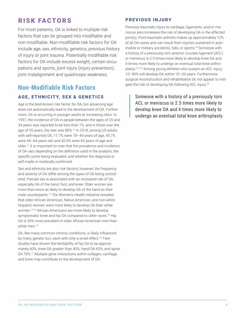
R I S K FA CTO R SFor most patients, OA is linked to multiple risk factors that can be grouped into modifiable and non-modifiable. Non-modifiable risk factors for OA include age, sex, ethnicity, genetics, previous history of injury or joint trauma. Potentially modifiable risk factors for OA include excess weight, certain occu-pations and sports, joint injury (injury prevention), joint malalignment and quadriceps weakness.
A G E, E T H N I C IT Y, S E X & G E N E T I C SAge is the best-known risk factor for OA, but advancing age does not automatically lead to the development of OA. Further-more, OA is occurring in younger adults at increasing rates. In 1997, the incidence of OA in people between the ages of 25 and 34 years was reported to be less than 1%, and in those over the age of 55 years, the rate was 80%.10 In 2018, among US adults with self-reported OA, 11.7% were 18–44 years of age, 45.7% were 45–64 years old, and 42.6% were 65 years of age and older.11 It is important to note that the prevalence and incidence of OA vary depending on the definition used in the analysis, the specific joints being evaluated, and whether the diagnosis is self-made or medically confirmed.
Sex and ethnicity are also risk factors; however, the frequency and severity of OA differ among the types of OA being consid-ered. Female sex is associated with an increased risk of OA, especially OA of the hand, foot, and knee. Older women are more than twice as likely to develop OA of the hand as their male counterparts.12 The Women’s Health Initiative revealed that older African-American, Native American, and non-white Hispanic women were more likely to develop OA than white women.13,14 African-Americans are more likely to develop symptomatic knee and hip OA compared to other races.15 Hip OA is 33% more prevalent in older African-American men than white men.12
OA, like many common chronic conditions, is likely influenced by many genetic loci, each with only a small effect.16 Twin studies have shown the heritability of hip OA to be approxi-mately 60%, knee OA greater than 40%, hand OA 65%, and spine OA 70%.17 Multiple gene interactions within collagen, cartilage, and bone may contribute to the development of OA.
Non-Modifiable Risk Factors
P R E V I O U S I N J U RYPrevious traumatic injury to cartilage, ligaments, and/or me-niscus also increases the risk of developing OA in the affected joint(s). Post-traumatic arthritis makes up approximately 12% of all OA cases and can result from injuries sustained in auto-mobile or military accidents, falls, or sports.18 Someone with a history of a previously torn anterior cruciate ligament (ACL) or meniscus is 2.5 times more likely to develop knee OA and 4 times more likely to undergo an eventual total knee arthro-plasty.2,15,19 Among young athletes who sustain an ACL injury, 10–90% will develop OA within 10–20 years. Furthermore, surgical reconstruction and rehabilitation do not appear to miti-gate the risk of developing OA following ACL injury.20
Someone with a history of a previously torn ACL or meniscus is 2.5 times more likely to develop knee OA and 4 times more likely to undergo an eventual total knee arthroplasty.
5O A PAT H O G E N E S I S A N D R I S K FA C T O R S
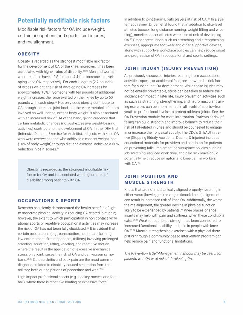
Potentially modifiable risk factorsModifiable risk factors for OA include weight, certain occupations and sports, joint injuries, and malalignment.
O B E S IT YObesity is regarded as the strongest modifiable risk factor for the development of OA of the knee; moreover, it has been associated with higher rates of disability.21,22 Men and women who are obese have a 2.8-fold and 4.4-fold increase in devel-oping knee OA, respectively. For each kilogram (2.2 pounds) of excess weight, the risk of developing OA increases by approximately 10%.21 Someone with ten pounds of additional weight increases the force exerted on their knee by up to 60 pounds with each step.23 Not only does obesity contribute to OA through increased joint load, but there are metabolic factors involved as well. Indeed, excess body weight is also associated with an increased risk of OA of the hand, giving credence that certain metabolic changes (not just excessive weight-bearing activities) contribute to the development of OA. In the IDEA trial (Intensive Diet and Exercise for Arthritis), subjects with knee OA who were overweight and who achieved a modest weight loss (10% of body weight) through diet and exercise, achieved a 50% reduction in pain scores.24
O C C U PAT I O N S & S P O RT SResearch has clearly demonstrated the health benefits of light to moderate physical activity in reducing OA-related joint pain; however, the extent to which participation in non-contact recre-ational sports or repetitive occupational activities may increase the risk of OA has not been fully elucidated.25 It is evident that certain occupations (e.g., construction, healthcare, farming, law enforcement, first responders, military) involving prolonged standing, squatting, lifting, kneeling, and repetitive motion where the result is the application of excessive mechanical stress on a joint, raises the risk of OA and can worsen symp-toms.26,27 Osteoarthritis and back pain are the most common diagnoses related to disability-caused separation from the military, both during periods of peacetime and war.27,28
High impact professional sports (e.g., hockey, soccer, and foot-ball), where there is repetitive loading or excessive force,
Obesity is regarded as the strongest modifiable risk factor for OA and is associated with higher rates of disability among patients with OA.
in addition to joint trauma, puts players at risk of OA.29 In a sys-tematic review, Driban et al found that in addition to elite-level athletes (soccer, long-distance running, weight lifting and wres-tling), nonelite soccer athletes were also at risk of developing OA.25 Proper precautions such as stretching and strengthening exercises, appropriate footwear and other supportive devices, along with supportive workplace policies can help reduce onset and progression of OA in occupational and sports settings.
J O I NT I N J U RY ( I N J U RY P R E V E NT I O N)As previously discussed, injuries resulting from occupational activities, sports, or accidental falls, are known to be risk fac-tors for subsequent OA development. While these injuries may not be entirely preventable, steps can be taken to reduce their incidence or impact in later life. Injury prevention activities such as such as stretching, strengthening, and neuromuscular train-ing exercises can be implemented in all levels of sports—from youth to professional levels—to protect athletes’ joints. See the OA Prevention module for more information. Patients at risk of falling can build strength and improve balance to reduce their risk of fall-related injuries and should be counseled to engage in or increase their physical activity. The CDC’s STEADI initia-tive (Stopping Elderly Accidents, Deaths, & Injuries) includes educational materials for providers and handouts for patients on preventing falls. Implementing workplace policies such as job-switching, reduced work time, and paid sick leave could potentially help reduce symptomatic knee pain in workers with OA.30
J O I NT P O S IT I O N A N D M U S C L E S T R E N G T HKnees that are not mechanically aligned properly- resulting in either varus (bowlegged) or valgus (knock-kneed) alignments- can result in increased risk of knee OA. Additionally, the worse the malalignment, the greater decline in physical function likely to be experienced by patients.31 Knee braces or shoe inserts may help with pain and stiffness when these conditions exist.31,32 Weaker quadriceps strength has been connected to increased functional disability and pain in people with knee OA.33,34 Muscle-strengthening exercises with a physical thera-pist or through a community-based intervention program can help reduce pain and functional limitations.
The Prevention & Self-Management handout may be useful for patients with OA or at risk of developing OA.
6O A PAT H O G E N E S I S A N D R I S K FA C T O R S
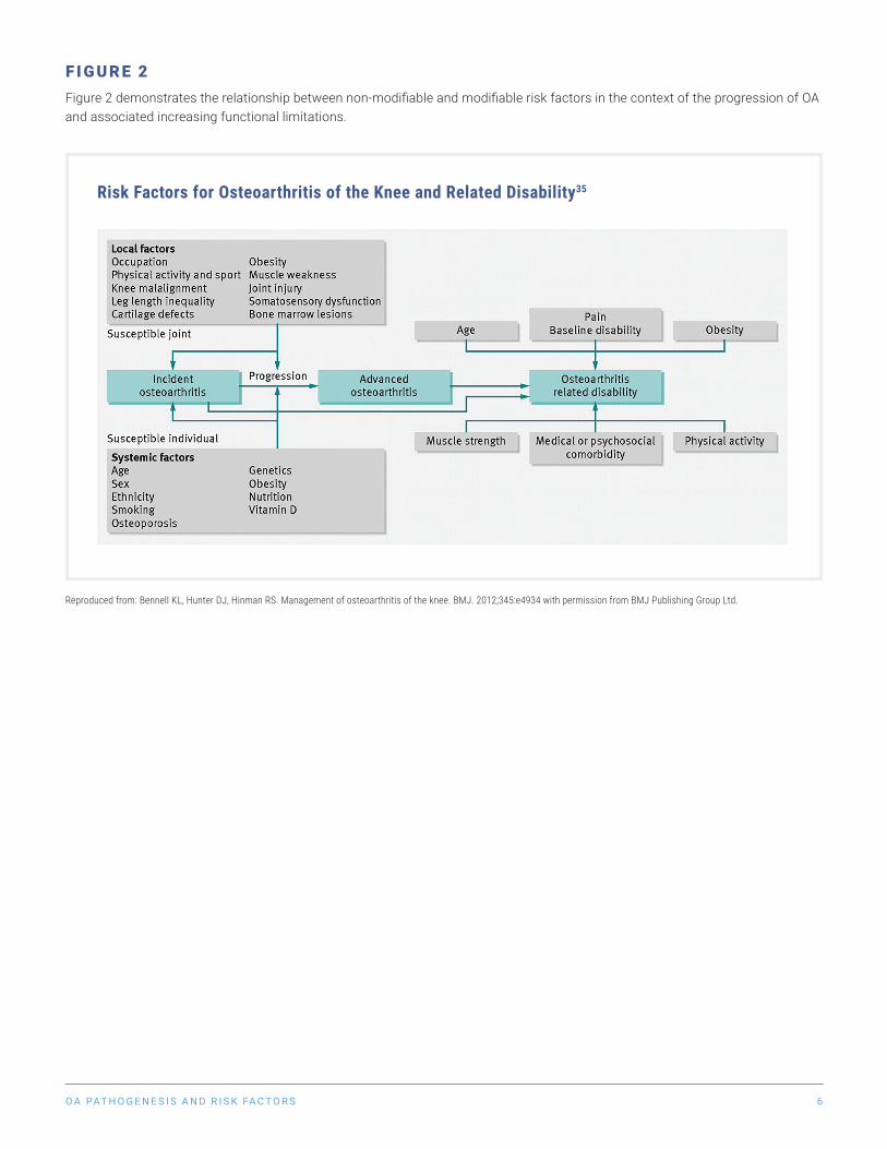
F I G U R E 2Figure 2 demonstrates the relationship between non-modifiable and modifiable risk factors in the context of the progression of OA and associated increasing functional limitations.
Risk Factors for Osteoarthritis of the Knee and Related Disability35
Reproduced from: Bennell KL, Hunter DJ, Hinman RS. Management of osteoarthritis of the knee. BMJ. 2012;345:e4934 with permission from BMJ Publishing Group Ltd.

7O A PAT H O G E N E S I S A N D R I S K FA C T O R S
A D D IT I O N A L R E A D I N GHunter DJ, Bierma-Zeinstra S. Osteoarthritis. Lancet. 2019;393(10182):1745–1759.
• OA is not simply “wear and tear” or “degeneration” of the joint, but rather a complex disorder characterized by a variety of molecular, anatomic, and physiologic changes leading to disease.
• Non-modifiable risk factors should be considered, and modifiable risk factors addressed, to reduce disease burden.
Clinical Take-Home Points
R E V I S I O N D AT E: A U G U S T 31, 2019
A C K N O W L E D G E M E NT S A N D D I S C LO S U R E SThe Osteoarthritis Prevention & Management in Primary Care Toolkit was funded, in part, by a Pfizer Independent Grant for Learning and Change and by a cooperative agreement from the Centers for Disease Control and Prevention. Toolkit contents are solely the responsibility of the Osteoarthritis Action Alliance and acknowledged Stakeholders and are based on best evidence and best practices in medicine. The OAAA expresses appreciation to U.S. Bone & Joint Initiative for their partnership in developing the Toolkit and to the field of experts comprising the Stakeholder panel for their many contributions. A list of Stakeholders and contributors can be found on the OAAA website.
8O A PAT H O G E N E S I S A N D R I S K FA C T O R S
R E F E R E N C E S
1. Osteoarthritis Research Society International. Standardization of Osteoarthritis Definitions. Available at https://www.oarsi.org/research/standardization- osteoarthritis-definitions. Published 2015. Accessed April 1, 2019.
2. Buys LM, Wiedenfeld SA. Osteoarthritis. In: DiPiro JT, Talbert RL, Yee GC, Matzke GR, Wells BG, M P, eds. Pharmacotherapy: A Pathophysiologic Approach, 10e. New York, NY: McGraw-Hill.
3. Moreland LW. Intra-articular hyaluronan (hyaluronic acid) and hylans for the treatment of osteoarthritis: mechanisms of action. Arthritis Res Ther. 2003;5(2):54–67.
4. Collins JA, Diekman BO, Loeser RF. Targeting aging for disease modification in osteoarthritis. Curr Opin Rheumatol. 2018;30(1):101–107.
5. Sharma L, Chmiel JS, Almagor O, et al. Significance of preradiographic magnetic resonance imaging lesions in persons at increased risk of knee osteoarthritis. Arthritis Rheumatol. 2014;66(7):1811–1819.
6. Roemer FW, Kwoh CK, Hannon MJ, et al. What comes first? Multitissue involvement leading to radiographic osteoarthritis: magnetic resonance imaging-based trajectory analysis over four years in the osteoarthritis initiative. Arthritis Rheumatol. 2015;67(8):2085–2096.
7. Podsiadlo P, Nevitt MC, Wolski M, et al. Baseline trabecular bone and its relation to incident radiographic knee osteoarthritis and increase in joint space narrowing score: directional fractal signature analysis in the MOST study. Osteoarthritis Cartilage. 2016;24(10):1736–1744.
8. Lane NE, Brandt K, Hawker G, et al. OARSI-FDA initiative: defining the disease state of osteoarthritis. Osteoarthritis Cartilage. 2011;19(5):478–482.
9. Loeser R. Pathogenesis of osteoarthritis. In: Post T, ed. UpToDate. Waltham, Mass: UpToDate; 2018: www.uptodate.com. Accessed January 23, 2019.
10. Brandt K. Osteoarthritis: clinical patterns and pathology. In: Kelley WN, Harris ED Jr, Ruddy S, Sledge CE, eds. Textbook of Rheumatology, 5th edition. Philadelphia: W.B. Saunders; 1997:1383.
11. United States Bone and Joint Initiative. The Burden of Musculoskeletal Diseases in the United States (BMUS). In: In. Fourth ed. Rosemont, IL. 2018: Available at https://www.boneandjointburden.org/fourth-edition. Accessed June 12, 2019.
12. Arthritis Foundation. Arthritis by the Numbers. In: Atlanta, GA: Arthritis Foun-dation; 2019: https://www.arthritis.org/Documents/Sections/About-Arthritis/arthritis-facts-stats-figures.pdf. Accessed April 5, 2019.
13. Wright NC, Riggs GK, Lisse JR, Chen Z, Women’s Health I. Self-reported osteoarthritis, ethnicity, body mass index, and other associated risk factors in postmenopausal women-results from the Women’s Health Initiative. J Am Geriatr Soc. 2008;56(9):1736–1743.
14. Eustice C. The effect of ethnicity of osteoarthritis. Verywell Health. https://www.verywellhealth.com/the-effect-of-ethnicity-on-osteoarthritis-2552101. Published 2018. Accessed June 21, 2018.
15. Vina ER, Kwoh CK. Epidemiology of osteoarthritis: literature update. Curr Opin Rheumatol. 2018;30(2):160–167.
16. Warner SC, Valdes AM. Genetic association studies in osteoarthritis: is it fairytale? Curr Opin Rheumatol. 2017;29(1):103–109.
17. Spector TD, MacGregor AJ. Risk factors for osteoarthritis: genetics. Osteoar-thritis Cartilage. 2004;12 Suppl A:S39–44.
18. Punzi L, Galozzi P, Luisetto R, et al. Post-traumatic arthritis: overview on pathogenic mechanisms and role of inflammation. RMD Open. 2016;2(2):e000279.
19. Hunter DJ, Zhang YQ, Niu JB, et al. The association of meniscal pathologic changes with cartilage loss in symptomatic knee osteoarthritis. Arthritis Rheum. 2006;54(3):795–801.
20. Padua DA, DiStefano LJ, Hewett TE, et al. National Athletic Trainers’ Association Position Statement: Prevention of Anterior Cruciate Ligament Injury. J Athl Train. 2018;53(1):5–19.
21. Garstang SV, Stitik TP. Osteoarthritis: epidemiology, risk factors, and patho-physiology. Am J Phys Med Rehabil. 2006;85(11 Suppl):S2-11; quiz S12–14.
22. Jordan JM, Luta G, Renner JB, et al. Self-reported functional status in osteoarthritis of the knee in a rural southern community: the role of sociodemographic factors, obesity, and knee pain. Arthritis Care Res. 1996;9(4):273–278.
23. Johns Hopkins Arthritis Center. Role of body weight in osteoarthritis. Available at https://www.hopkinsarthritis.org/patient-corner/disease- management/role-of-body-weight-in-osteoarthritis/. Accessed June 4, 2018.
24. Messier SP, Mihalko SL, Legault C, et al. Effects of intensive diet and exercise on knee joint loads, inflammation, and clinical outcomes among overweight and obese adults with knee osteoarthritis: the IDEA randomized clinical trial. JAMA. 2013;310(12):1263–1273.
25. Driban JB, Hootman JM, Sitler MR, Harris KP, Cattano NM. Is Participation in Certain Sports Associated With Knee Osteoarthritis? A Systematic Review. J Athl Train. 2017;52(6):497–506.
26. Yucesoy B, Charles LE, Baker B, Burchfiel CM. Occupational and genetic risk factors for osteoarthritis: a review. Work. 2015;50(2):261–273.
27. Cameron KL, Driban JB, Svoboda SJ. Osteoarthritis and the Tactical Athlete: A Systematic Review. J Athl Train. 2016;51(11):952–961.
28. Patzkowski JC, Rivera JC, Ficke JR, Wenke JC. The changing face of disability in the US Army: the Operation Enduring Freedom and Operation Iraqi Freedom effect. J Am Acad Orthop Surg. 2012;20 Suppl 1:S23–30.
29. Amoako AO, Pujalte GG. Osteoarthritis in young, active, and athletic individuals. Clin Med Insights Arthritis Musculoskelet Disord. 2014;7:27–32.
30. Chen JC, Linnan L, Callahan LF, Yelin EH, Renner JB. Workplace policies and prevalence of knee osteoarthritis: the Johnston County Osteoarthritis Project. Occup Environ Med. 2007;64(12):798–805.
31. Sharma L, Song J, Felson DT, Cahue S, Shamiyeh E, Dunlop DD. The role of knee alignment in disease progression and functional decline in knee osteoarthritis. JAMA. 2001;286(2):188–195.
32. Heidari B. Knee osteoarthritis diagnosis, treatment and associated factors of progression: part II. Caspian J Intern Med. 2011;2(3):249–255.
33. O’Reilly SC, Jones A, Muir KR, Doherty M. Quadriceps weakness in knee osteoarthritis: the effect on pain and disability. Ann Rheum Dis. 1998;57(10):588–594.
34. Bacon KL, Segal NA, Oiestad BE, et al. Thresholds in the relationship of quadriceps strength with functional limitations in women with knee osteoarthritis. Arthritis Care Res (Hoboken). 2018.
35. Bennell KL, Hunter DJ, Hinman RS. Management of osteoarthritis of the knee. BMJ. 2012;345:e4934.