(nz)ch...o contacts assist crystallization of a parb-like nuclease
TRANSCRIPT
BioMed CentralBMC Structural Biology
ss
Open AcceMethodology article(NZ)CH...O Contacts assist crystallization of a ParB-like nucleaseNeil Shaw1, Chongyun Cheng1, Wolfram Tempel2, Jessie Chang2, Joseph Ng3, Xin-Yu Wang1, Sarah Perrett1, John Rose2, Zihe Rao1,4, Bi-Cheng Wang2 and Zhi-Jie Liu*1,2Address: 1National Laboratory of Biomacromolecules, Institute of Biophysics, Chinese Academy of Sciences, Beijing, 100101, China, 2Southeast Collaboratory for Structural Genomics, Department of Biochemistry and Molecular Biology, University of Georgia, GA 30602, USA, 3Laboratory of Structural Biology and Department of Biological Sciences, University of Alabama in Huntsville, Huntsville, AL 35899, USA and 4Laboratory of Structural Biology, Tsinghua University, 100084, China
Email: Neil Shaw - [email protected]; Chongyun Cheng - [email protected]; Wolfram Tempel - [email protected]; Jessie Chang - [email protected]; Joseph Ng - [email protected]; Xin-Yu Wang - [email protected]; Sarah Perrett - [email protected]; John Rose - [email protected]; Zihe Rao - [email protected]; Bi-Cheng Wang - [email protected]; Zhi-Jie Liu* - [email protected]
* Corresponding author
AbstractBackground: The major bottleneck for determination of 3 D structures of proteins using X-raysis the production of diffraction quality crystals. Often proteins are subjected to chemicalmodification to improve the chances of crystallization
Results: Here, we report the successful crystallization of a nuclease employing a reductivemethylation protocol. The key to crystallization was the successful introduction of 44 new cohesive(NZ) CH...O contacts (3.2 – 3.7 Å) by the addition of 2 methyl groups to the side chain aminenitrogen (NZ) of 9 lysine residues of the nuclease. The new contacts dramatically altered thecrystallization properties of the protein, resulting in crystals that diffracted to 1.2 Å resolution.Analytical ultracentrifugation analysis and thermodynamics results revealed a more compactprotein structure with better solvent exclusion of buried Trp residues in the folded state of themethylated protein, assisting crystallization.
Conclusion: In this study, introduction of novel cohesive (NZ)CH...O contacts by reductivemethylation resulted in the crystallization of a protein that had previously resisted crystallization inspite of extensive purification and crystallization space screening. Introduction of (NZ)CH...Ocontacts could provide a solution to crystallization problems for a broad range of protein targets.
BackgroundThe resolution and accuracy of the structural informationprovided by X-rays is unsurpassed when compared toother techniques employed to resolve the 3 D structure ofproteins [1]. However, not all proteins can be made avail-able for X-ray diffraction studies because of the inherent
difficulty in obtaining single crystals of adequate size andquality [2]. A subset of such proteins that resist crystalliza-tion can be salvaged by employing a reductive methyla-tion protocol [3-11]. A 26 kD nuclease from Pyrococcusfuriosus used in the current study had resisted crystalliza-tion inspite of extensive purification and crystallization
Published: 7 July 2007
BMC Structural Biology 2007, 7:46 doi:10.1186/1472-6807-7-46
Received: 10 February 2007Accepted: 7 July 2007
This article is available from: http://www.biomedcentral.com/1472-6807/7/46
© 2007 Shaw et al; licensee BioMed Central Ltd. This is an Open Access article distributed under the terms of the Creative Commons Attribution License (http://creativecommons.org/licenses/by/2.0), which permits unrestricted use, distribution, and reproduction in any medium, provided the original work is properly cited.
Page 1 of 12(page number not for citation purposes)
BMC Structural Biology 2007, 7:46 http://www.biomedcentral.com/1472-6807/7/46
space screening. A number of techniques have been devel-oped to improve a given target's potential for crystalliza-tion. Truncation of disordered regions [12,13],mutagenesis of surface residues [14-16] and chemicalmodification of proteins [3] have been proven effective inthis regard and a number of protein structures have beensolved successfully. We decided to modify the surfacelysines of the nuclease by reductive methylation in anattempt to crystallize the protein. The rationale behindtargeting the amine nitrogen (NZ) of the nuclease arosefrom a number of considerations. Lysines harbouring freeNZ atoms almost always reside on the surface of proteinmolecules [17]. The thermodynamic cost for ordering thehighly flexible solvent exposed side chains of lysines isexorbitant [18]. Since crystallization is a surface phenom-enon, a disordered lysine side chain has a profound nega-tive impact on the formation of stable, uniform, intermolecular contacts essential for packaging of protein mol-ecules in a crystal lattice. Interestingly, we found thatmethylation of surface lysines resulted in a decrease in thefree energy of folding of the protein. This is consistentwith the entropic cost of reduced flexibility in the nativestate due to formation of new intra molecular interac-tions, which in turn will lower the barrier to crystalliza-tion. We further probed the effect of methylation at themolecular level and show that cohesive (NZ)CH...Obonds assisted crystallization of the nuclease.
ResultsGeneration of intra molecular contactsAfter purifying the protein to homogeneity, two methylgroups were covalently linked to the free amine nitrogenof lysine residues [3-11] (Figure 1A) by treating the pro-tein with formaldehyde and dimethylamine borane com-plex (DMAB). Electron density for only 9 amine nitrogensout of 32 (including the N-terminal amine group), couldbe unambiguously assigned as dimethylated (Figure 1B).
The methyl carbon is protonated because of the strongelectron withdrawing nature of the NZ atom. The proto-nated methyl carbon can form ionic interactions with car-bonyl and carboxyl oxygens of surrounding residues. The(NZ)CH...O bond formed by the CH2 group of MLY159and the carboxyl oxygen OE2 of Glu156 is shown in Fig-ure 1C. The length of the cohesive interaction is 3.27 Å.Forty new intra molecular (NZ)CH...O contacts in therange 3.2 to 4.0 Å were generated because of the methyla-tion, of which 25 interactions were between 3.2 to 3.8 Å(see Additional file 1). The large number of cohesive intramolecular contacts generated helps immobilize the flexi-ble regions of the protein molecules (Figure 1D), which iscrucial for the formation of stable intermolecular contactsand may lower the entropic cost of crystallization. Thetemperature factor of the MLY159NZ was 12.18. The addi-tion of methyl groups to the amine nitrogen and the for-
mation of cohesive bonds through the protonated carbonof the methyl group seemed to have significantly loweredthe B factor of the NZ atom of MLY159 indicating a local-ized side chain. Glu216 forms two strong (NZ)CH...Ocohesive bonds with the methyl groups of MLY159 (Fig-ure 2A). The B factors of the OE1 and OE2 atoms ofGlu216 involved in the formation of the hydrogen bondwere 14.3 and 12.36 respectively. The low B factors dem-onstrate immobilization of the Glu216 side chain possi-bly because of the new (NZ)CH...O bonds. Similarly, theB factors of both the carboxyl oxygens of Glu156 partici-pating in (NZ)CH...O interactions were also low. B factorsof 16.64 for the OE1 oxygen and 20.24 for the OE2 oxy-gen indicate the side chain of Glu156 to be severelyrestricted in movement. The B factors, however, need tobe interpreted cautiously in absence of a structure of theunmodified protein for comparison.
Generation of inter molecular contactsThe methyl carbons of all the dimethylated lysine residueswere involved in the formation of multiple new symme-try-generated intermolecular contacts. The synthesis of theintermolecular contacts was initiated by gradual evapora-tion of the solvent in presence of different chemicals.Commercially available sparse matrix screens under oilwere used for screening the best chemical environment forformation of intermolecular bonds [19,20]. The structurereveals 96 new symmetry-generated inter molecular con-tacts in the range 3.2 – 5.0 Å involving the (NZ)CH groupand 28 of these inter molecular contacts are of the(NZ)CH...O type and in the range 3.2 – 4.0 Å (see Addi-tional file 2). A significant number of these interactionswere within the optimal range of 3.2 – 3.7 Å for CH...Ohydrogen bonds [21]. The intermolecular contacts involv-ing the methyl groups of MLY112 and MLY201 are shownin Figures 2B and 2C respectively. The B factors for the NZatoms of MLY112 and MLY201 were 25 and 15 respec-tively. The new intra and inter molecular contacts formedby the covalently linked methyl groups with the surround-ing residues seem to have lowered the B factor values ofthe NZ atom indicating a localized side chain. This helpsthe packaging of the molecules in the crystal and the for-mation of a compact crystalline lattice.
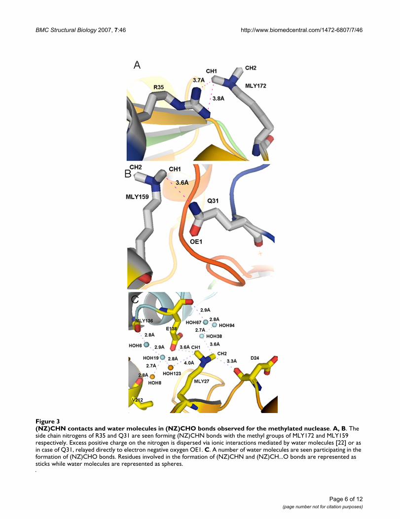
Although the contacts formed were predominantly(NZ)CH...O bonds, a few (NZ)CH...N bonds were alsoformed (Figure 3A and 3B). The side chain nitrogens ofArg35 interact with the CH1 methyl carbon of MLY172.Water molecules surrounding these interactions possiblyhelp disperse the excess positive charge on the nitrogensby bridging ionic interactions [22]. The NH1 nitrogen ofArg35 forms a hydrogen bond with water 119, which ishydrogen bonded to the carboxylic oxygens, OD1 andOD2, of Asp 39. Similarly, the NH2 nitrogen of Arg35forms a hydrogen bond with water 117, which is hydro-
Page 2 of 12(page number not for citation purposes)

BMC Structural Biology 2007, 7:46 http://www.biomedcentral.com/1472-6807/7/46
Page 3 of 12(page number not for citation purposes)
Nature of (NZ)CH...O contacts of the methylated nucleaseFigure 1Nature of (NZ)CH...O contacts of the methylated nuclease. A. Diagrammatic representation of the (NZ)CH...O bond. The side chain amine nitrogen (NZ) of lysine residues (blue circle) polarizes the covalently linked methyl carbon (green circle). The polarized methyl carbon acts like a proton donor and forms ionic interactions with neighbouring carboxyl oxygens (red circle). The optimal range for the (NZ)CH...O bond distance is between 3.2 – 3.7 Å. The angle of the approach of a pro-ton towards the lone pair of electrons is generally between 90 – 180°. In order to calculate the angle, the position of hydrogen (grey circle) for X-ray structures is usually deduced. B. Electron density for the dimethylated lysine MLY159. The 2 | Fo | – | Fc | electron density map was contoured at 1.5 σ. C. The protonated methyl carbon, CH2, of MLY159 is seen forming a 3.6 Å (NZ)CH...O bond with the carboxyl oxygen of E156. The amino acids are represented as sticks. The (NZ)CH...O bond is shown as a dashed magenta line. D. A cartoon representation of the modified protein showing the dimethylated lysines engaged in the formation of numerous intra molecular (NZ)CH...O contacts. Such contacts localize side chains and loops in space, resulting in a compact protein molecule. (NZ)CH...O bonds are shown as dashed blue lines.
A B
C
D

BMC Structural Biology 2007, 7:46 http://www.biomedcentral.com/1472-6807/7/46
Page 4 of 12(page number not for citation purposes)
Generation of intra and inter molecular (NZ)CH...O contacts for the nucleaseFigure 2Generation of intra and inter molecular (NZ)CH...O contacts for the nuclease. A. The intra molecular (NZ)CHO contacts generated for MLY159 as a result of the chemical modification of the nuclease are shown as dashed magenta lines. B, C. The symmetry generated inter molecular contacts synthesized for MLY112 and MLY201 during the crystallization of the nuclease are shown as magenta dashed lines. Residues involved in the formation of (NZ)CH...O bonds are represented as sticks while water molecules are represented as spheres.

BMC Structural Biology 2007, 7:46 http://www.biomedcentral.com/1472-6807/7/46
gen bonded to the carbonyl oxygen of Gln170. The excesspositive charge on the Arg35 nitrogens is relayed to thecarboxyl oxygens of Asp35 and carbonyl oxygen ofGln170 via water molecules (data not shown). Similarly,in case of Gln31, the excess positive charge is relayed tothe side chain OE1 oxygen atom.
In addition, a significant number of water molecules wereobserved to form (NZ)CH...O contacts (Figure 3C).
Analytical ultracentrifugationThe 26 kD protein was subjected to analytical ultracentrif-ugation analysis in order to determine the effect of thereductive methylation protocol on the purity, aggregationstate and shape of the protein. The sedimentation velocityexperiment results revealed that both non-methylated andmethylated proteins were pure, homogenous and mono-meric (Figure 4). However, a qualitative decrease in thediffusion co-efficient (D) could be observed for the meth-ylated protein (Figure 4B). This is consistent withdecreased flexibility of side chains and a more compactstructure in the methylated protein.
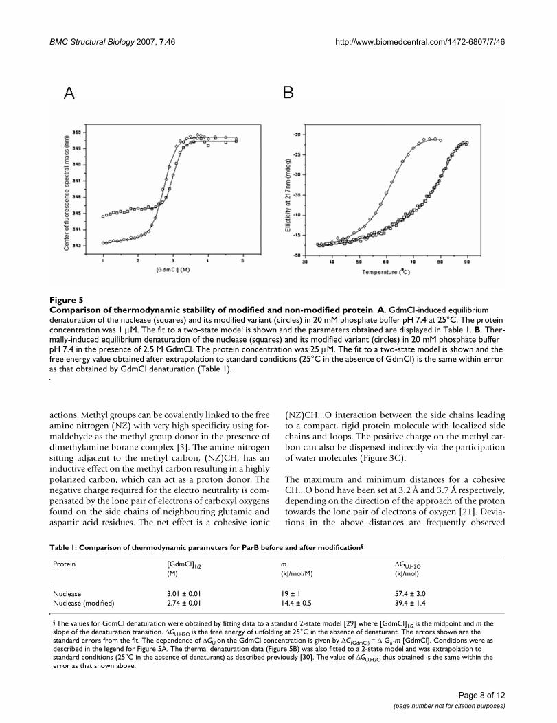
Thermodynamic stability of nuclease before and after modificationIn order to further investigate the mechanism by whichchemical modification of the protein affected the ease ofcrystallization, we performed equilibrium denaturationexperiments using heat or the chaotropic agent guanidin-ium chloride (GdmCl) as denaturants. The nuclease con-tains three Trp residues, of which two (W74 and W102)are buried in the folded structure, making intrinsic fluo-rescence a sensitive probe of global structural changes. Wealso used far-UV circular dichroism (CD), which monitorschanges in secondary structure. Both proteins were foundto be extremely thermostable and no secondary structuralchanges were detected below 90°C (not shown). How-ever, we found that the proteins could be completelyunfolded in 4 M GdmCl at 25°C (Figure 5A). Further,thermal denaturation could be achieved in the presence ofa non-denaturing concentration of GdmCl (Figure 5B).Interestingly, we found that the chemical modificationresulted in a decrease in the mid-point of unfolding inboth GdmCl and thermal denaturation (Figures 5A, B),which corresponds to a decrease in the free energy ofunfolding, Δ GU,H20, of 18 ± 3 kJ/mol (Table 1). Surfacelysines are likely to be involved in salt-bridges or otherfavourable interactions that may be lost on methylation,resulting in a decrease in stability. Another explanation isthat there is a decrease in entropy in the folded state of themodified protein due to formation of intra molecular(NZ)CH...O interactions involving the introduced methylgroups, which reduces the flexibility of the protein. Sol-vent effects may also contribute to the observed differencein stability, for example, greater ordering of water mole-
cules around hydrophobic methyl groups at the proteinsurface could also lead to a greater loss in entropy on fold-ing for the methylated protein. We also observed that theλmax of the fluorescence spectrum for the native state wasblue-shifted for the modified protein, whereas the λmax forthe denatured states was similar (Figure 5A). This is con-sistent with a more compact structure and better solventexclusion of buried Trp residues in the folded state of thechemically modified protein. Reduced entropy of the sol-vated folded structure, together with the formation ofadditional favourable interactions in the crystalline state,may account for the greater ease of crystallization after thechemical modification
DiscussionStructure determination holds the key to unravelling themechanisms by which proteins drive the machinery of allliving organisms for survival and propagation. In spite ofseveral dramatic technological advances in automationand information science, the key step for successful crys-tallographic structure determination – production of highquality crystals – continues to remain a resource intensivebottleneck [1]. A first step towards addressing this bottle-neck would be to identify the variables involved in crystal-lization. The nature of the protein is the single mostimportant factor that influences crystallization. From acrystallization point of view – size, charge, hydrophobic-ity, hydrophilicity, flexibility, oxidation state and post-translational modifications like phosphorylation, glyco-sylation and myristylation – define the nature of a pro-tein. Since the nature of the protein often varies withfunction, there is no universal crystallization strategy thatwould work for all proteins. To maximize the chances ofcrystallization it is crucial to identify and target anattribute of the protein that would have a profound effecton the crystallization. Surface lysines offer one such target.Lysines almost always reside on the surface of proteins.The solvent exposed side chains of lysines are highlymobile and prevent the formation of inter molecular con-tacts essential for the assembly of a crystalline lattice [18].Defects observed in protein crystals like low resolutionand twinning is also a manifestation of the flexibility ofthe side chains found on the surface of the protein.
Locking the side chain amine nitrogen of lysines with theelectron negative carboxyl oxygens of glutamic and aspar-tic acid side chains via cohesive ionic interactions willresult in the immobilization of these side chains (Figure2). Although the presence of CH...O bonds has been dem-onstrated in proteins, nucleic acids and carbohydrates[21-27], the (NZ)CH...O bonds introduced in the currentstudy have never been described before. Methyl groups arevery effective in mediating and bridging the physical dis-tances between the free amine nitrogen and the side chaincarboxyl oxygens for the formation of cohesive ionic inter-
Page 5 of 12(page number not for citation purposes)
BMC Structural Biology 2007, 7:46 http://www.biomedcentral.com/1472-6807/7/46
Page 6 of 12(page number not for citation purposes)
(NZ)CHN contacts and water molecules in (NZ)CHO bonds observed for the methylated nucleaseFigure 3(NZ)CHN contacts and water molecules in (NZ)CHO bonds observed for the methylated nuclease. A, B. The side chain nitrogens of R35 and Q31 are seen forming (NZ)CHN bonds with the methyl groups of MLY172 and MLY159 respectively. Excess positive charge on the nitrogen is dispersed via ionic interactions mediated by water molecules [22] or as in case of Q31, relayed directly to electron negative oxygen OE1. C. A number of water molecules are seen participating in the formation of (NZ)CHO bonds. Residues involved in the formation of (NZ)CHN and (NZ)CH...O bonds are represented as sticks while water molecules are represented as spheres.

BMC Structural Biology 2007, 7:46 http://www.biomedcentral.com/1472-6807/7/46
Page 7 of 12(page number not for citation purposes)
Analytical ultracentrifugation analysis of methylated and non-methylated proteinFigure 4Analytical ultracentrifugation analysis of methylated and non-methylated protein. The methylated (A) and non-methylated (B) protein were subjected to analytical ultracentrifugation analysis as described in Methods. The sedimentation velocity experiments showed the protein remained pure and monomeric after the chemical modification. There was an obvi-ous decrease in the diffusion co-efficient for the protein after the methylation indicating a more compact shape.
BMC Structural Biology 2007, 7:46 http://www.biomedcentral.com/1472-6807/7/46
actions. Methyl groups can be covalently linked to the freeamine nitrogen (NZ) with very high specificity using for-maldehyde as the methyl group donor in the presence ofdimethylamine borane complex [3]. The amine nitrogensitting adjacent to the methyl carbon, (NZ)CH, has aninductive effect on the methyl carbon resulting in a highlypolarized carbon, which can act as a proton donor. Thenegative charge required for the electro neutrality is com-pensated by the lone pair of electrons of carboxyl oxygensfound on the side chains of neighbouring glutamic andaspartic acid residues. The net effect is a cohesive ionic
(NZ)CH...O interaction between the side chains leadingto a compact, rigid protein molecule with localized sidechains and loops. The positive charge on the methyl car-bon can also be dispersed indirectly via the participationof water molecules (Figure 3C).
The maximum and minimum distances for a cohesiveCH...O bond have been set at 3.2 Å and 3.7 Å respectively,depending on the direction of the approach of the protontowards the lone pair of electrons of oxygen [21]. Devia-tions in the above distances are frequently observed
Comparison of thermodynamic stability of modified and non-modified proteinFigure 5Comparison of thermodynamic stability of modified and non-modified protein. A. GdmCl-induced equilibrium denaturation of the nuclease (squares) and its modified variant (circles) in 20 mM phosphate buffer pH 7.4 at 25°C. The protein concentration was 1 μM. The fit to a two-state model is shown and the parameters obtained are displayed in Table 1. B. Ther-mally-induced equilibrium denaturation of the nuclease (squares) and its modified variant (circles) in 20 mM phosphate buffer pH 7.4 in the presence of 2.5 M GdmCl. The protein concentration was 25 μM. The fit to a two-state model is shown and the free energy value obtained after extrapolation to standard conditions (25°C in the absence of GdmCl) is the same within error as that obtained by GdmCl denaturation (Table 1).
Table 1: Comparison of thermodynamic parameters for ParB before and after modification§
Protein [GdmCl]1/2 m ΔGU,H2O(M) (kJ/mol/M) (kJ/mol)
Nuclease 3.01 ± 0.01 19 ± 1 57.4 ± 3.0Nuclease (modified) 2.74 ± 0.01 14.4 ± 0.5 39.4 ± 1.4
§ The values for GdmCl denaturation were obtained by fitting data to a standard 2-state model [29] where [GdmCl]1/2 is the midpoint and m the slope of the denaturation transition. ΔGU,H2O is the free energy of unfolding at 25°C in the absence of denaturant. The errors shown are the standard errors from the fit. The dependence of ΔGU on the GdmCl concentration is given by ΔG(GdmCl) = Δ Gu-m [GdmCl]. Conditions were as described in the legend for Figure 5A. The thermal denaturation data (Figure 5B) was also fitted to a 2-state model and was extrapolation to standard conditions (25°C in the absence of denaturant) as described previously [30]. The value of ΔGU,H2O thus obtained is the same within the error as that shown above.
Page 8 of 12(page number not for citation purposes)

BMC Structural Biology 2007, 7:46 http://www.biomedcentral.com/1472-6807/7/46
owing to the steric interactions between the atomsinvolved in the formation of the (NZ)CH...O bonds andthe surrounding residues. In the present study, except fora couple of contacts involving MLY221, the minimum dis-tance of all the CH...O bonds was 3.2 Å. The only shortcontacts – a 3.0 Å link between the CH2 methyl carbon ofMLY221 and a phosphate oxygen, and a 3.1 Å bondbetween the carboxyl oxygens of E36 and the MLY221methyl carbons, resulted in the distortion of the confor-mation of the MLY221 and the glutamic acid (Figure 6),suggesting steric clashes due to the excessive closeness ofthe methyl carbon and the oxygen atoms. Thus, 3.2 Åseems to be the minimum van der Waals limit for all(NZ)CH...O interactions. Detailed analysis of the electrondensity map showed no other obvious changes to anyother amino acid confirming the specificity of the modifi-cation.
The methylated protein lost its ability to cleave DNA(results not shown). The dimethylation of K221 sitting atthe edge of the active site may sterically hinder access ofthe active site to the incoming DNA. It is also possible thatan overall conformational change in the protein inducedby the chemical modification affects the catalytic site andcompromises the function of the protein. However, suchloss-of-function due to methylation has not been reportedpreviously. Further studies are warranted in order to deter-mine the exact cause for the inactivation of the methylatedprotein.
When a protein is set up for crystallization, the moleculesare moving randomly in search of compatible bondingpartners. Some of the inter molecular contacts generatedduring the course of random collisions are sustained. Asmore and more solvent evaporates, a number of theseinteractions become permanent. Eventually it leads to oneof two possible outcomes. If the inter molecular bondingis heterogeneous, as in case of protein molecules thatassume more than one conformation due to the presenceof unstructured domains, flexible loops and side chains,or presence of partially unfolded regions, this will result ina disordered protein aggregate commonly referred to asprecipitate. Poorly diffracting or defective crystals are alsoa consequence of the conformational flexibility of proteinmolecules. A homogenous inter molecular bonding pat-tern between protein molecules, as in the case of structur-ally rigid molecules, results in optimal packing of themolecules in a crystal lattice. A direct manifestation of thechemical modification of the protein is the reduction innumber of degrees of freedom available to the protein forassuming different conformations. Introduction of(NZ)CH...O bonds curbs the movement of side chainsand fixes their orientation in space. This produces uni-form bonding partners and decreases the steric clashesbetween molecules during the packing of the lattice. The
observed reduction in the free energy of folding for themodified protein is consistent with reduced flexibilityleading to lower entropy in the native state; ordering ofwater molecules around the surface-exposed methylgroups may also contribute to this. Crystallization will befavoured for the modified protein by the reduction inentropy of the solvated structure and the involvement ofthe methyl groups in inter molecular (NZ)CH...O interac-tions upon crystallization
A pre-requisite to the success of the crystallization strategydescribed here is the requirement of a highly pure homog-enous protein sample [28] usually obtained by a combi-nation of different chromatography steps (Figure 7).Presence of homologous and heterologous impurities cancompromise the effectiveness of the chemical modifica-tion of the protein.
ConclusionIn conclusion, introduction of (NZ)CH...O bonds byreductive methylation of surface lysines as a means to sal-vage targets is simple, fast, economical and non laborious.It could be the first method of choice for rescuing a targetbefore attempting a more extensive approach involvingmutagenesis.
Occupancy of MLY221Figure 6Occupancy of MLY221. MLY221 shows 2 conformations. Conformation A has occupancy of 65%. The distance of the oxygen atoms of E36 and the PO-
4 ligand from the (NZ)CH group of the MLY221 is shorter than the permissible limit of 3.2 Å for (NZ)CHO bonds, resulting in the distorting of MLY221 and E36. The 2 | Fo | – | Fc | electron density map was contoured at 1.5 σ
Page 9 of 12(page number not for citation purposes)

BMC Structural Biology 2007, 7:46 http://www.biomedcentral.com/1472-6807/7/46
MethodsProtein production and purificationThe 26 kD ParB nuclease was expressed with a N-terminalhexa Histidine tag. The gene was PCR amplified from thegenomic DNA of Pyrococcus furiosus and cloned into pET-28a vector (Invitrogen). E coli BL21 cells containing theplasmid were grown in LB cultures. The protein was puri-fied using affinity chromatography followed by size exclu-sion chromatography. Nucleic acids were removed byhydroxyapatite chromatography (GE Healthcare). Furtherpurification was achieved by an ion exchange step. Theprotein was exchanged into crystallization buffer (20 mMTris, pH8.0, 200 mM NaCl) using a size exclusion col-umn.
Chemical modificationMethylation of the protein was done as described before[3] using formaldehyde and dimethylamine-borane com-plex (DMAB). In brief, 10 mg/ml of protein in a 1.5 mleppendorf tube covered with aluminium foil was mixedwith 40 μl of 1 M solution of formaldehyde (Sigma) and20 μl of 1 M solution of DMAB (Sigma) in the dark at 4°C.The reaction mixture was incubated under shaking condi-tions for 2 h, after which the chemical additions wererepeated. Finally, 10 μl of DMAB was added and the reac-tion mixture was incubated overnight. Excess chemicalswere removed by size exclusion chromatography.
CrystallizationMethylated and non-methylated protein was set up forcrystallization under oil as described before [19,20]. 1-μlcrystallization drops contained 0.5 μl protein mixed with
Process flow sheet of the crystallization strategy for the nucleaseFigure 7Process flow sheet of the crystallization strategy for the nuclease. The protein sample is purified to homogeneity using a combination of chromatographic methods. The pure and homogenous protein is chemically modified in order to local-ize the side chains and loops. The resultant compact protein molecule is then screened against a variety of chemical environ-ments using commercially available sparse matrix screens to determine the best condition for the self-assembly of the protein molecules into a crystalline lattice.
Page 10 of 12(page number not for citation purposes)
BMC Structural Biology 2007, 7:46 http://www.biomedcentral.com/1472-6807/7/46
0.5 μl of crystallization solution. Commercially availablesparse matrix screens (Hampton Research, MolecularDimensions) were used for crystallization screening. Crys-tals for structure determination were produced using aprecipitant solution consisting of 600 mM sodium di-hydrogen phosphate, 2.4 M di-potassium hydrogen phos-phate, 200 mM sodium chloride, 100 mM HEPES, p H7.3.
Data collection and structure determinationData collection and structure determination will bedescribed elsewhere.
Analytical UltracentrifugationAnalytical sedimentation velocity experiments were car-ried out using a ProteomeLab™ XL-I protein characteriza-tion system (Beckman Coulter). An-60Ti rotor was used tocentrifuge a 10 mg/ml protein sample suspended in 50mM phosphate buffer, 150 mM NaCl, pH 7.2, at 60,000rpm. Absorbance was read at 280 nm. A set of 93 scanswere collected at 1 min intervals. Data was analyzed usingSedfit software
Equilibrium denaturation experimentsAll fluorescence denaturation experiments were per-formed in 20 mM phosphate buffer pH 7.4 at 25°C, witha final protein concentration of 1.2 μM. Samples of mod-ified and unmodified protein were mixed with differentconcentrations of GdmCl and allowed to equilibrate over-night before measurements were taken. Refolding experi-ments were also performed, by denaturing the protein for8 h in 6 M GdmCl, then diluting the protein to give differ-ent final concentrations of GdmCl as for unfolding exper-iments. The intrinsic fluorescence spectra were recordedbetween 300 and 400 nm after excitation at 280 nm in aHitachi F-4500 spectrofluorimeter. The fluorescence datawere plotted as the centre of spectral mass as describedpreviously [29]. GdmCl denaturation was found to bereversible and the data were fitted to a 2-state model [30].
Far-UV CD experiments were performed on a Pi-star 180instrument (Applied Photophysics, UK) using a cell of 1mm optical path length and the same buffer as for fluores-cence experiments. The protein concentration was 25 μM.The temperature was changed at a rate of 1°C per 10 min,with a step size of 0.5°C, for both heating and cooling.Thermal denaturation was found to be reversible and thedata were analyzed as described previously [31]
List of abbreviations usedMLY – Methylated lysine
(NZ) – Side chain amine nitrogen
Authors' contributionsNS, JC, and JN produced, purified, and crystallized theprotein. NS and CC did the chemical modification andanalytical ultracentrifugation analysis. X-YW and SP car-ried out the thermodynamic characterization of the pro-tein. Z-JL, WT, and JR solved the structure and did theanalysis. Z-JL designed the experiments and interpretedthe data. B-CW and ZR conceived the study and partici-pated in its design and co-ordination. NS, SP, and Z-JLdrafted the manuscript. All authors read and approved thefinal manuscript.
Accession numbersCoordinates for the structure reported in the current studyhave been deposited in the Protein Data Bank underaccession code 1VK1
Additional material
AcknowledgementsThis work was funded by the 863 (Grant 2006AA02A316) and 973 (Grants 2006CB910901, 2006CB910903, 2006CB500703) Projects of the Ministry of Science and Technology of China, the National Natural Science Founda-tion of China (Grants 30470363, 30670427), the National Institutes of Health (Grant 1P50 GM62407), the University of Georgia Research Foun-dation, the Georgia Research Alliance. Crystallographic data was collected at Southeast Regional Collaborative Access Team (SER-CAT) 22-ID beam-line at the Advanced Photon Source, Argonne National Laboratory. Use of the Advanced Photon Source was supported by the U. S. Department of Energy, Office of Science, Office of Basic Energy Sciences, under contract number W-31-109-Eng-38.
References1. Stevens R, Wilson I: Industrializing structural biology. Science
2001, 293:519-520.2. Dale G, Oefner C, D'Arcy A: The protein as a variable in protein
crystallization. J Struct Biol 2003, 142:88-97.3. Rayment I: Reductive alkylation of lysine residues to alter crys-
tallization properties of proteins. Methods in Enzymology 1997,276:171-179.
Additional file 1Intra molecular contacts$ of the (NZ)CH group of methylated lysines of the nuclease. The data presented in the table is a list of intra molecular contacts generated during the methylation of the nuclease.Click here for file[http://www.biomedcentral.com/content/supplementary/1472-6807-7-46-S1.doc]
Additional file 2Symmetry generated contacts* of the (NZ)CH group of the methyl-ated lysines of the nuclease. The data presented in the table is a list of intra molecular contacts generated during the methylation of the nuclease.Click here for file[http://www.biomedcentral.com/content/supplementary/1472-6807-7-46-S2.doc]
Page 11 of 12(page number not for citation purposes)

BMC Structural Biology 2007, 7:46 http://www.biomedcentral.com/1472-6807/7/46
Publish with BioMed Central and every scientist can read your work free of charge
"BioMed Central will be the most significant development for disseminating the results of biomedical research in our lifetime."
Sir Paul Nurse, Cancer Research UK
Your research papers will be:
available free of charge to the entire biomedical community
peer reviewed and published immediately upon acceptance
cited in PubMed and archived on PubMed Central
yours — you keep the copyright
Submit your manuscript here:http://www.biomedcentral.com/info/publishing_adv.asp
BioMedcentral
4. Geoghegan K, Cabacungan J, Dixon H, Feeney R: Alternativereducing agents for reductive methylation of amino groupsin proteins. Int J Pept Protein Res 1981, 17:345-352.
5. Gerken T, Jentoft J, Jentoft N, Dearborn D: Intramolecular inter-actions of amino groups in 13C reductively methylated henegg-white lysozyme. J Biol Chem 1982, 257:2894-2900.
6. Means G, Feeney R: Reductive alkylation of amino groups inproteins. Biochemistry 1968, 7:2192-2201.
7. Rypniewski W, Holden H, Rayment I: Structural consequences ofreductive methylation of lysine residues in hen egg white lys-ozyme: An X-ray analysis at 1.8Å resolution. Biochemistry 1993,32:9851-9858.
8. Schubot F, Waugh D: A pivotal role for reductive methylationin the de novo crystallization of a ternary complex composedof Yersinia pestis virulence factors YopN, SycN and YscB. ActaCryst 2004, D60:1981-1986.
9. Kobayashi M, Kubota M, Matsuura Y: Crystallization andimprovement of crystal quality for X-ray diffraction of mal-tooligosyl trehalose synthase by reductive methylation oflysine residues. Acta Cryst 1999, 55(Pt 4):931-933.
10. Walter T, Meier C, Assenberg R, Au K-F, Ren J, Verma A, NettleshipJ, Owens R, Stuart D, Grimes J: Lysine methylation as a routinerescue strategy for protein crystallization. Structure 2006,14:1617-1622.
11. Liu Z-J, Tempel W, Ng J, Lin D, Shah A, Chen L, Horanyi P, Habel J,Kataeva I, Xu H, Yang H, Chang J, Huang L, Chang S-H, Zhou W, LeeD, Praissman J, Zhang H, Newton G, Rose J, Richardson J, RichardsonD, Wang B-C: The high-throughput protein-to-structure pipe-line at SECSG. Acta Crystallogr 2005, 61(Pt 6):679-684.
12. Dale G, Kostrewa D, Gsell B, Stieger M, D'Arcy A: Crystal engi-neering: deletion mutagenesis of the 24 kDa fragment of theDNA gyrase B subunit from Staphylococcus aureus. Acta Crys-tallogr D Biol Crystallogr 1999, 55:1626-1629.
13. Kwong P, Wyatt R, Robinson J, Sweet R, Sodroski J, Hendrickson W:Structure of an HIV gp120 envelope glycoprotein in complexwith the CD4 receptor and a neutralizing human antibody.Nature 1998, 393:648-659.
14. Derewenda Z: Rational protein crystallization by mutationalsurface engineering. Structure 2004, 12:529-535.
15. Longenecker K, Garrard S, Sheffield P, Derewenda Z: Protein crys-tallization by rational mutagenesis of surfaceresidues: Lys toAla mutations promote crystallization of RhoGDI. Acta Crytal-logr D Biol Crytallogr 2001, 57(Pt 5):679-688.
16. Mateja A, Devedjiev Y, Krowarsch D, Longenecker K, Dauter Z,Otlewski J, Derewenda Z: The impact of Glu→Ala andGlu→Asp mutations on the crystallization properties ofRhoGDI: the structure of RhoGDI at 1.3 Å resolution. ActaCrytallogr D Biol Crytallogr 2002, 58(Pt 12):1983-1991.
17. Baud F, Karlin S: Measures of residue density in protein struc-tures. Proc Natl Acad Sci USA 1999, 96:12494-12499.
18. Avbelj F, Fele L: Role of main-chain electrostatics, hydrophobiceffect and side-chain conformational entropy in determiningthe secondary structure of proteins. J Mol Biol 1998,279:665-684.
19. Chayen N, Shaw Stewart P, Maeder D, Blow D: An automated sys-tem for micro-batch protein crystallization and screening. JAppl Cryst 1990, 23:297-302.
20. D'Arcy A, Elmore C, Stihle M, Johnston J: A novel approach tocrystallising proteins under oil. J Crystal Growth 1996,168:175-80.
21. Derewenda Z, Lee L, Derewenda U: The occurrence of C-HOhydrogen bonds in proteins. J Mol Biol 1995, 252:248-262.
22. Magalhaes A, Maigret B, Hoflack J, Gomes J, Scheraga H: Contribu-tion of unusual arginine-arginine short-range interactions tostabilization and recognition in proteins. J Protein Chem 1994,13:195-215.
23. Kump R, Damewood J: Do nitromethane and molononitrileform C-H...O hydrogen bonds? Implications for molecularrecognition by crown ethers. J Chem Soc Chem Commun1988:621-622.
24. Taylor R, Kennard O: Crystallographic evidence for the exist-ence of C-H...O, C-H...N and C-H...Cl hydrogen bonds. J AmerChem Soc 1982, 104:5063-5070.
25. Wahl M, Sundaralingam M: CH...O hydrogen bonding in biology.Trends Biochem Sci 1997, 22:97-102.
26. Desiraju G: The C-H...O hydrogen bond in crystals: What is it?Acc Chem Res 1991, 24:290-296.
27. Donati A, Ristori S, Bonechi C, Panza L, Martini G, Rossi C: Evi-dences of strong C-HO bond in an o-carboranyl beta-lactos-ide in solution. J Am Chem Soc 2002, 124:8778-8779.
28. McPherson A: Introduction to Protein Crystallography. Meth-ods: A Companion to Methods in Enzymology 2004, 34:254-265.
29. Lima L, Zingali R, Foguel D, Monteiro R: New insights into confor-mational and functional stability of human alpha-thrombinprobed by high hydrostatic pressure. Eur J Biochem 2004,271:3580-3587.
30. Maxwell K, Wildes , Zarrine-Afsar A, De Los Rios M, Brown A, FrielC, Hedberg L, Horng J-C, Bona D, Miller E, Vallée-Bélisle A, Main E,Bemporad F, Qiu L, Teilum K, Vu N-D, Edwards A, Ruczinski I,Poulsen F, Kragelund B, Michnick S, Chiti F, Bai Y, Hagen S, SerranoL, Oliveberg M, Raleigh D, Wittung-Stafshede P, Radford S, Jackson S,Sosnick T, Marqusee S, Davidson A, Plaxco K: Protein folding:Defining a "standard" set of experimental conditions and apreliminary kinetic data set of two-state proteins. Protein Sci-ence 2005, 14:602-616.
31. Su X, Sanbo Q, Xian-Ming P: Thermal and conformational sta-bility of Ssh10b protein from archaeon Sulfolobus shibattae.Biochem J 2004, 382:433-440.
32. Potterton E, Briggs P, Turkenburg M, Dodson E: A graphical userinterface to the CCP 4 program suite. Acta Cryst 2003, 59(Pt7):1131-1137.
Page 12 of 12(page number not for citation purposes)